Filters
▼Clonality
▼Type
▼Reactivity
▼Gene Name
▼Isotype
▼Host
▼Application
▼Clone
▼Polyclonal Antibodies
At AAA Biotech also known as AAA Bio or AAABio, we provide a broad range of purified polyclonal antibodies (pAbs) that are able to all be browsed online through our website. Due to their high specificity and strong binding affinity, these antibodies are ideal for wide swathes of research and experimental applications.
Our polyclonal antibodies can easily support your work, whether you use them for Western Blotting, Immunocytochemistry (with or without Immunofluorescence used in conjunction), Immunohistochemistry, Immunoprecipitation, and ELISA tests. We highly encourage you to browse our range of pAbs and choose the one that best suits your experimental model.
Viewing 8750-8800 of 96805 product results
 WB (Western Blot)
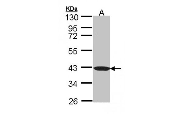
(Western blot analysis of 30 ug whole cell lysate (A:Hep G2) using a 7.5 % SDS PAGE gel and PPP1R16A antibody at a dilution of 1:1000)
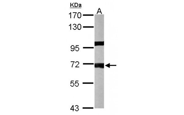
WB (Western Blot)
(Western blot analysis of 30 ug whole cell lysate (A:Hep G2) using a 7.5 % SDS PAGE gel and PPP1R16A antibody at a dilution of 1:1000)
PPP1R16A, Polyclonal Antibody (Cat# AAA107704)
 WB (Western Blot)
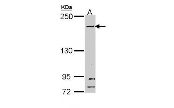
(Western blot analysis of 30 ug of whole cell lysate (A: THP-1; B: HL-60) using a 7.5 % SDS PAGE gel and RED antibody at a dilution of 1:1000)
WB (Western Blot)
(Western blot analysis of 30 ug of whole cell lysate (A: THP-1; B: HL-60) using a 7.5 % SDS PAGE gel and RED antibody at a dilution of 1:1000)
RED, Polyclonal Antibody (Cat# AAA107733)
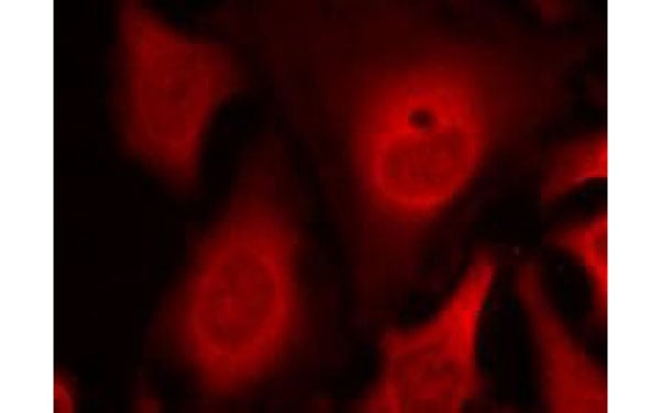
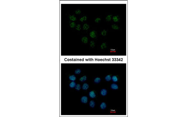
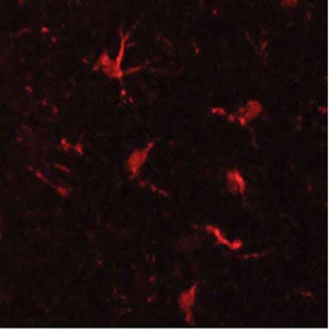 IF (Immunofluorescence)
(Immunofluorescent staining of Integrin alpha M antibodyMac-1 staining of microglial cells in mouse cerebral cortex (red). Photo courtesy of Dr. Felix Eckenstein, University of Vermont.)
IF (Immunofluorescence)
(Immunofluorescent staining of Integrin alpha M antibodyMac-1 staining of microglial cells in mouse cerebral cortex (red). Photo courtesy of Dr. Felix Eckenstein, University of Vermont.)
Integrin alpha M, Polyclonal Antibody (Cat# AAA107771)
 WB (Western Blot)
(Western blot analysis of 30 ug of whole cell lysate (A: A549) using a 7.5 % SDS PAGE gel and DPRP1 antibody at a dilution of 1:1000)
WB (Western Blot)
(Western blot analysis of 30 ug of whole cell lysate (A: A549) using a 7.5 % SDS PAGE gel and DPRP1 antibody at a dilution of 1:1000)
DPRP1, Polyclonal Antibody (Cat# AAA107783)
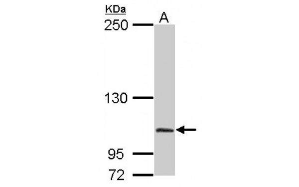
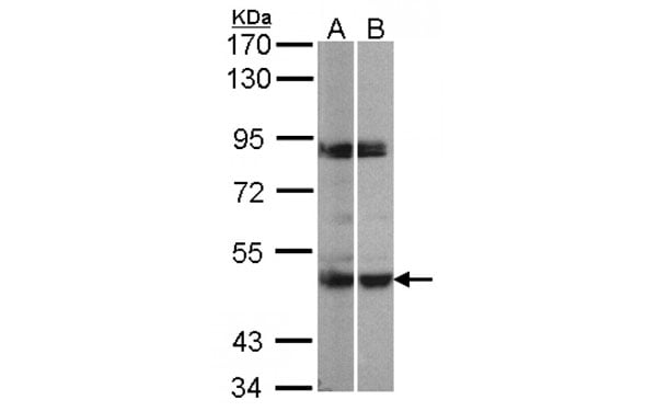
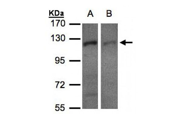 WB (Western Blot)
(Western blot analysis of 30 ug of whole cell lysate (A:H1299; B:HeLa S3) using a 7.5 % SDS PAGE gel and RanBP16 antibody at a dilution of 1:500)
WB (Western Blot)
(Western blot analysis of 30 ug of whole cell lysate (A:H1299; B:HeLa S3) using a 7.5 % SDS PAGE gel and RanBP16 antibody at a dilution of 1:500)
RanBP16, Polyclonal Antibody (Cat# AAA107803)
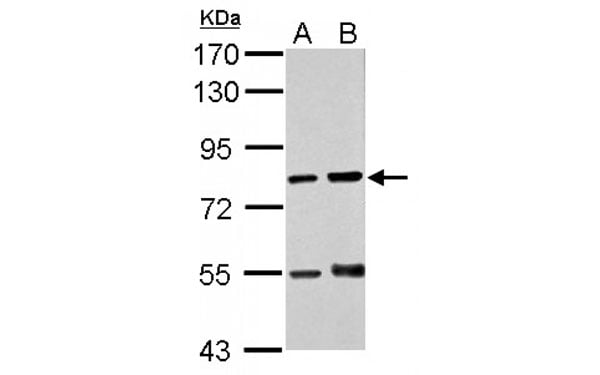
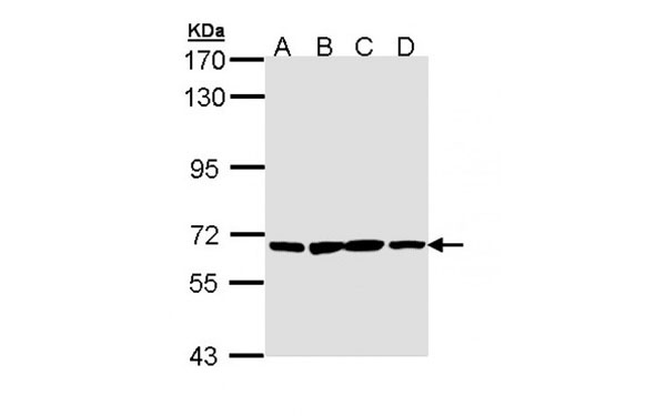 WB (Western Blot)
(Western blot analysis of 30 ug of whole cell lysate (A: H1299; B: Hela; C: Hep G2; D: Molt-4) using a 7.5 % SDS PAGE gel and PAN3 antibody at a dilution of 1:1000)
WB (Western Blot)
(Western blot analysis of 30 ug of whole cell lysate (A: H1299; B: Hela; C: Hep G2; D: Molt-4) using a 7.5 % SDS PAGE gel and PAN3 antibody at a dilution of 1:1000)
PAN3, Polyclonal Antibody (Cat# AAA107805)
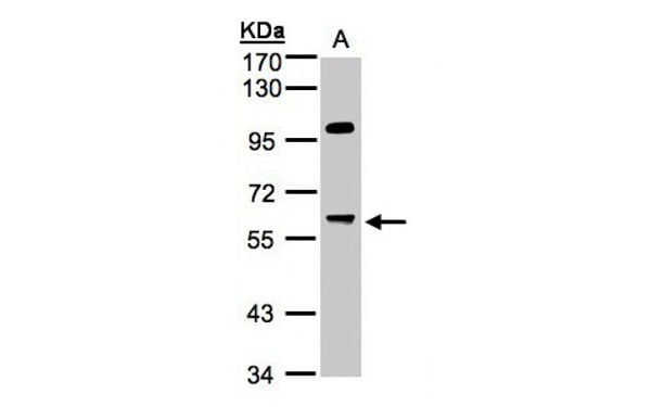
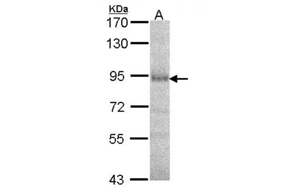
 WB (Western Blot)
(Western blot analysis of 30 ug of whole cell lysate (A:293T; B:H1299) using a 7.5 % SDS PAGE gel and PRAK antibody at a dilution of 1:500)
WB (Western Blot)
(Western blot analysis of 30 ug of whole cell lysate (A:293T; B:H1299) using a 7.5 % SDS PAGE gel and PRAK antibody at a dilution of 1:500)
PRAK, Polyclonal Antibody (Cat# AAA107811)
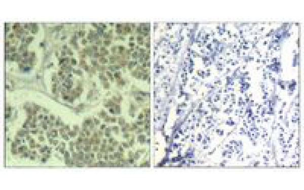 IHC (Immunohiostchemistry)
(immunohistochemical analysis of paraffin-embedded human breast carcinoma tissue using elF2alpha (Phospho-Ser49) antibody (left) or the same antibody preincubated with with blocking peptide (right))
IHC (Immunohiostchemistry)
(immunohistochemical analysis of paraffin-embedded human breast carcinoma tissue using elF2alpha (Phospho-Ser49) antibody (left) or the same antibody preincubated with with blocking peptide (right))
eIF2 alpha, Polyclonal Antibody (Cat# AAA107820)
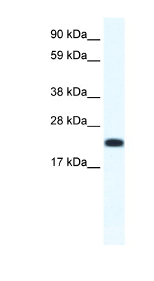
 WB (Western Blot)
(Western blot analysis of 30 ug of whole cell lysate (A: Molt-4) using a 10 % SDS PAGE gel and C1orf165 antibody at a dilution of 1:1000)
WB (Western Blot)
(Western blot analysis of 30 ug of whole cell lysate (A: Molt-4) using a 10 % SDS PAGE gel and C1orf165 antibody at a dilution of 1:1000)
C1orf165, Polyclonal Antibody (Cat# AAA107822)
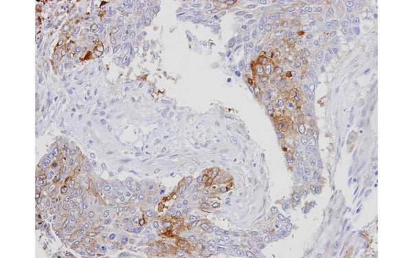
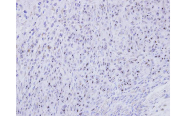 IHC (Immunohistochemisry)
(Immunohistochemical staining of paraffin-embedded SAS xenograft using SMC1B antibody at a dilution of 1:500)
IHC (Immunohistochemisry)
(Immunohistochemical staining of paraffin-embedded SAS xenograft using SMC1B antibody at a dilution of 1:500)
SMC1B, Polyclonal Antibody (Cat# AAA107833)
 WB (Western Blot)
(Western Blot analysis using CITED4 antibodyCITED4 antibody used at 0.2-1 ug/ml to detect target protein.)
WB (Western Blot)
(Western Blot analysis using CITED4 antibodyCITED4 antibody used at 0.2-1 ug/ml to detect target protein.)
CITED4, Polyclonal Antibody (Cat# AAA107071)
ACTH, Polyclonal Antibody (Cat# AAA106665)
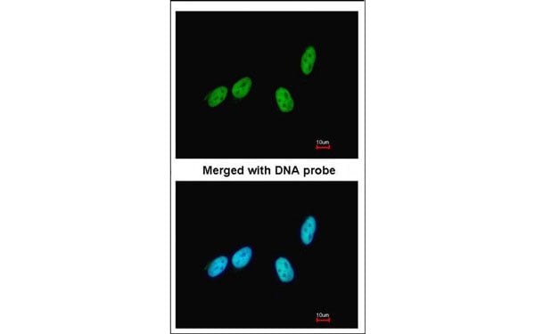
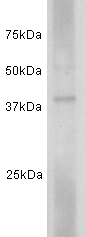 Application Data
(Fig: Western blot analysis on human spleen using anti-CD24 polyclonal antibody)
Application Data
(Fig: Western blot analysis on human spleen using anti-CD24 polyclonal antibody)
CD24, Polyclonal Antibody (Cat# AAA111100)
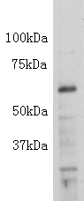 Application Data
Insulin B Chain, Polyclonal Antibody (Cat# AAA111131)
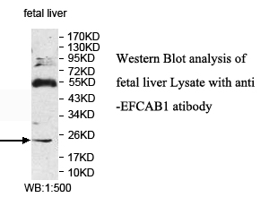
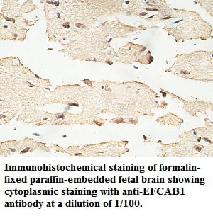 Application Data
EFCAB1, Polyclonal Antibody (Cat# AAA111327)
Predicted: Mouse, Rat
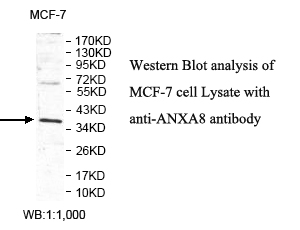
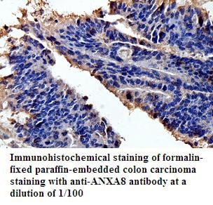 Application Data
ANXA8, Polyclonal Antibody (Cat# AAA111340)
Predicted: Mouse, Rat
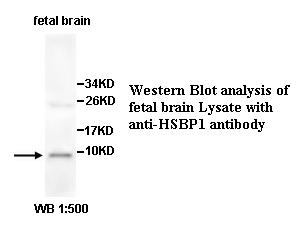
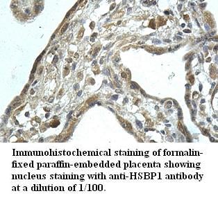 Application Data
HSBP1, Polyclonal Antibody (Cat# AAA111344)
Predicted: Mouse, Rat
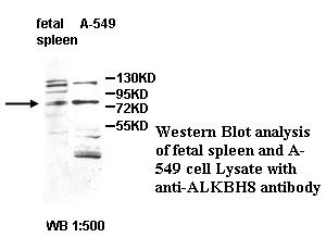
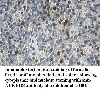 Application Data
ALKBH8, Polyclonal Antibody (Cat# AAA111346)
Predicted: Mouse, Rat
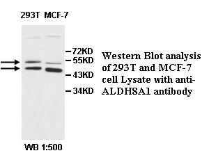
 Application Data
ALDH8A1, Polyclonal Antibody (Cat# AAA111350)
Predicted: Mouse, Rat
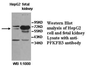
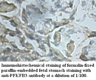 Application Data
PFKFB3, Polyclonal Antibody (Cat# AAA111352)
Predicted: Mouse, Rat
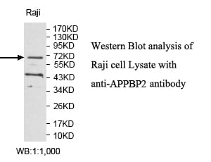
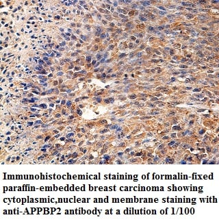 Application Data
APPBP2, Polyclonal Antibody (Cat# AAA111357)
Predicted: Mouse, Rat
 Application Data
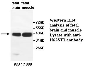
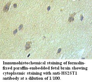
HS2ST1, Polyclonal Antibody (Cat# AAA111362)
Predicted: Mouse, Rat
 Application Data
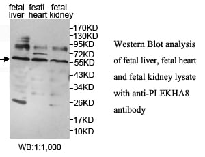
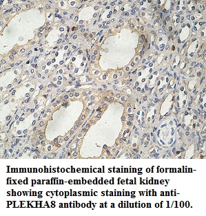
PLEKHA8, Polyclonal Antibody (Cat# AAA111382)
Predicted: Mouse, Rat
 Application Data
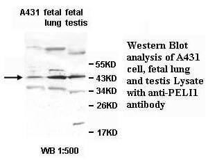
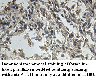
PELI1, Polyclonal Antibody (Cat# AAA111386)
Predicted: Mouse, Rat
 Application Data
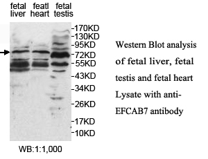
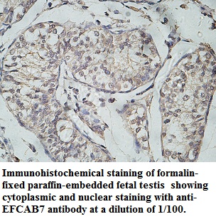
EFCAB7, Polyclonal Antibody (Cat# AAA111394)
Predicted: Mouse, Rat
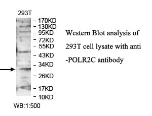
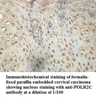 Application Data
POLR2C, Polyclonal Antibody (Cat# AAA111700)
Predicted: Mouse, Rat
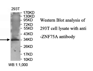
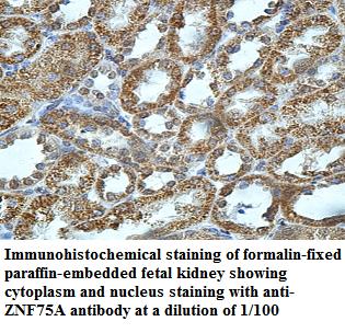 Application Data
ZNF75A, Polyclonal Antibody (Cat# AAA111702)
Predicted: Mouse, Rat
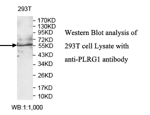
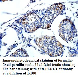 Application Data
PLRG1, Polyclonal Antibody (Cat# AAA111703)
Predicted: Mouse, Rat
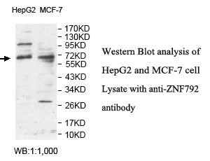
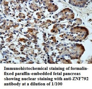 Application Data
ZNF792, Polyclonal Antibody (Cat# AAA111709)
Predicted: Mouse, Rat
 Application Data
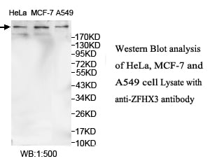
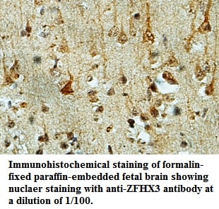
ZFHX3, Polyclonal Antibody (Cat# AAA111713)
Predicted: Mouse, Rat
 Application Data
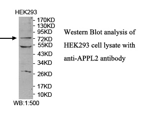
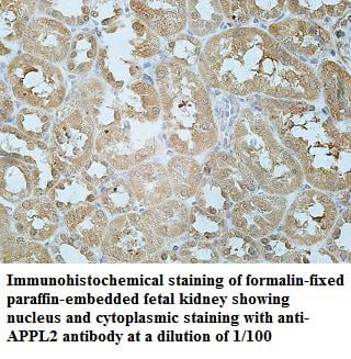
APPL2, Polyclonal Antibody (Cat# AAA111725)
Predicted: Mouse, Rat
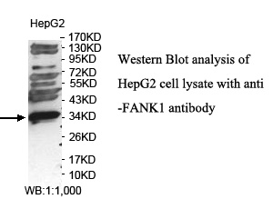
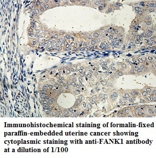 Application Data
FANK1, Polyclonal Antibody (Cat# AAA111742)
Predicted: Mouse, Rat
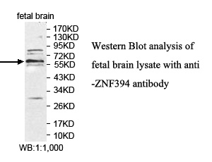
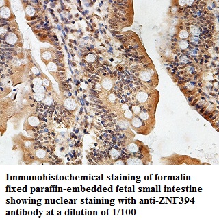 Application Data
ZNF394, Polyclonal Antibody (Cat# AAA111745)
Predicted: Mouse, Rat
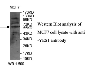
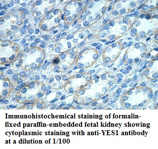 Application Data
YES1, Polyclonal Antibody (Cat# AAA111758)
Predicted: Mouse, Rat
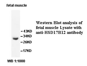
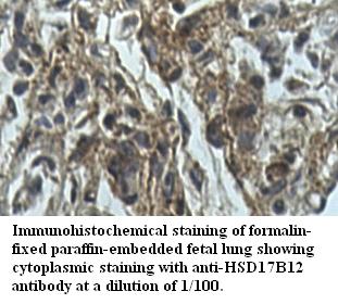 Application Data
HSD17B12, Polyclonal Antibody (Cat# AAA111762)
Predicted: Mouse, Rat
 Application Data
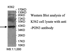
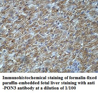
PON3, Polyclonal Antibody (Cat# AAA111764)
Predicted: Mouse, Rat
 Application Data
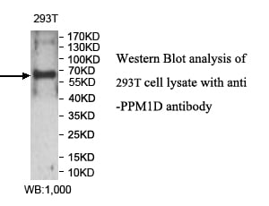
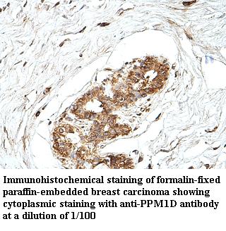
PPM1D, Polyclonal Antibody (Cat# AAA111468)
Predicted: Mouse, Rat
 Application Data
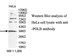
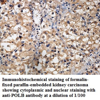
POLB, Polyclonal Antibody (Cat# AAA111473)
Predicted: Mouse, Rat

 Application Data
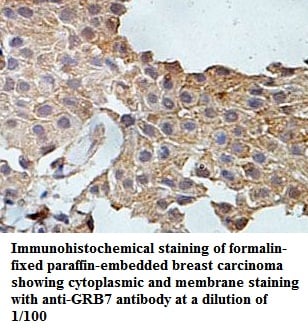
GRB7, Polyclonal Antibody (Cat# AAA111484)
Predicted: Mouse, Rat
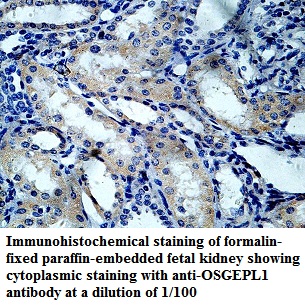 Application Data
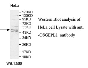
OSGEPL1, Polyclonal Antibody (Cat# AAA111489)
Predicted: Mouse, Rat
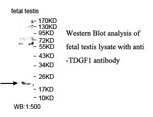
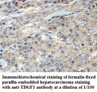 Application Data
TDGF1, Polyclonal Antibody (Cat# AAA111490)
Predicted: Mouse, Rat
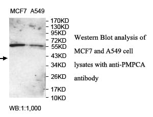
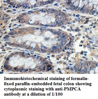 Application Data
PMPCA, Polyclonal Antibody (Cat# AAA111499)
Predicted: Mouse, Rat
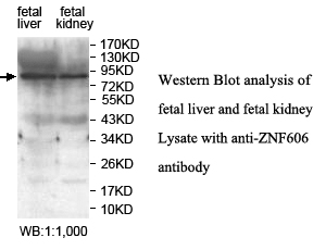
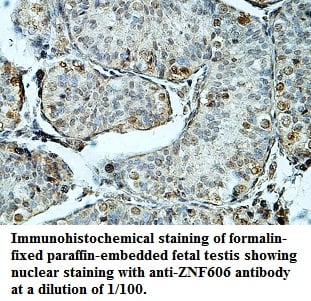 Application Data
ZNF606, Polyclonal Antibody (Cat# AAA111500)
Predicted: Mouse, Rat
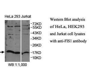
 Application Data
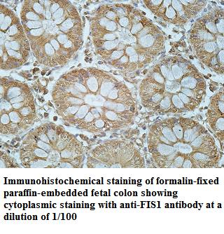
FIS1, Polyclonal Antibody (Cat# AAA111508)
Predicted: Mouse, Rat
 Application Data
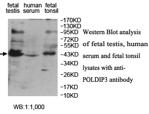
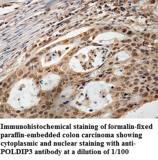
POLDIP3, Polyclonal Antibody (Cat# AAA111515)
Predicted: Mouse, Rat
 Application Data
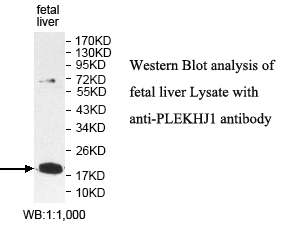
PLEKHJ1, Polyclonal Antibody (Cat# AAA111522)
Predicted: Mouse, Rat
 Application Data
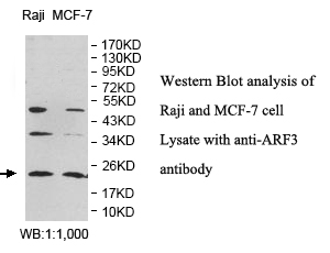
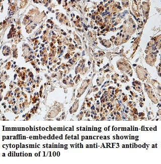
ARF3, Polyclonal Antibody (Cat# AAA111527)
Predicted: Mouse, Rat
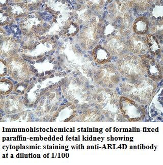 Application Data
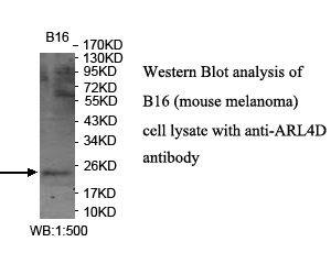
ARL4D, Polyclonal Antibody (Cat# AAA111532)
Predicted: Rat
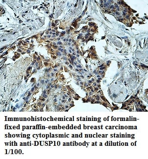 Application Data
DUSP10, Polyclonal Antibody (Cat# AAA111533)
Predicted: Mouse, Rat
 Application Data
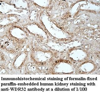
WDR32, Polyclonal Antibody (Cat# AAA111537)
Predicted: Mouse, Rat
What are Polyclonal Antibodies?
Polyclonal antibodies are antibodies that come from multiple B cell clones of a host animal. The typical hosts used for the majority of polyclonal antibody production are rabbits, goats, sheep, and donkeys. These polyclonal antibodies, once having identified their target, will bind to different epitopes located at different regions or sequences on the same protein/antigen. As a result, they are ideal at locating and binding to the target, even if the target is in very low concentrations (due to many different antibodies being able to bind to the same target molecule, which allows for significant amplification of a downstream signal).
Polyclonal antibodies are typically produced by injecting an antigen into a host animal, which causes the animal’s immune system to attack the foreign antigen by mass generating antibodies against it. After a period of time, serum is collected from the animal and purified using physicochemical fractionation, class-specific affinity purification, and/or antigen-affinity purification.
Key Uses of Polyclonal Antibodies
- Western Blotting: This method is used to find specific proteins in biological samples after separating them by size.
- Immunohistochemistry: IHC helps visualize the location of proteins in tissue sections using various staining techniques.
- ELISA: (Enzyme-Linked Immunosorbent Assay) is typically used to identify specific protein quantities in a sample. ELISAs can be either “Quantitative” or “Qualitative”.
- Flow Cytometry: technique that identifies and measures the specific protein on the surface or inside the cells in a fluid suspension.
- Immunoprecipitation: IP isolates and studies a specific protein from a complex mixture using antibodies.
Why Buy Polyclonal Antibodies from AAA Biotech?
1. Ideal for Various Applications
Our antibodies are generally going to be validated for use in multiple types of assays, including ELISA, Western Blotting, Immunohistochemistry, Immunoprecipitation, amongst others. They are ideal for a wide range of research applications.
2. Rigorous Quality Control
All of the antibodies in our catalog undergo strict quality testing to ensure specificity, sensitivity, and consistent performance. We are confident in the ability of our antibodies to provide you with accurate results.
3. Wide Assortment of Antibodies
Antibodies in are catalog can be found for both common and exotic species, and these antibodies are also available in both conjugated and recombinant forms to suit many diverse experimental needs.
4. Highly Purified
Our antibodies are available in purified forms with over 85% purity, as confirmed by SDS-PAGE. They are also available with tags such as His, Flag, GST, or MBP. We cater to customers worldwide.
FAQ
1. How are polyclonal antibodies produced?
Traditionally, polyclonal antibodies are produced by injecting an antigen into a host animal (such as a rabbit or goat), which then triggers an immune response from the host animal. The animal’s B cells produce antibodies that will recognize different parts of the injected antigen. These antibodies are then collected from the animal’s blood and purified for use.
2. How do polyclonal antibodies differ from monoclonal antibodies?
Polyclonal antibodies are a mix of antibodies that bind to different locations (epitopes) of the same antigen, while monoclonal antibodies are identical and bind to just one specific epitope. This makes polyclonal antibodies more versatile and better at detecting proteins that may be present in low quantities or in altered/modified forms.
3. How should I store polyclonal antibodies?
Polyclonal antibodies should be stored at 4°C for short-term use (up to a few weeks) and at -20°C or -80°C for long-term storage. Avoid repeated freeze-thaw cycles by dividing them into small aliquots. Always check the datasheet for specific storage instructions.