Filters
▼Clonality
▼Type
▼Reactivity
▼Gene Name
▼Isotype
▼Host
▼Application
▼Clone
▼Polyclonal Antibodies
At AAA Biotech also known as AAA Bio or AAABio, we provide a broad range of purified polyclonal antibodies (pAbs) that are able to all be browsed online through our website. Due to their high specificity and strong binding affinity, these antibodies are ideal for wide swathes of research and experimental applications.
Our polyclonal antibodies can easily support your work, whether you use them for Western Blotting, Immunocytochemistry (with or without Immunofluorescence used in conjunction), Immunohistochemistry, Immunoprecipitation, and ELISA tests. We highly encourage you to browse our range of pAbs and choose the one that best suits your experimental model.
Viewing 8150-8200 of 96805 product results

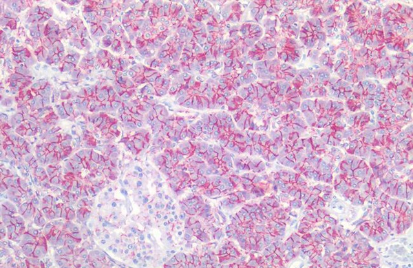 IHC (Immunohistochemistry)
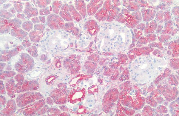
(CLDN3/Claudin 3 Antibody-Human Pancreas: Formalin-Fixed, Paraffin-Embedded (FFPE))
IHC (Immunohistochemistry)
(CLDN3/Claudin 3 Antibody-Human Pancreas: Formalin-Fixed, Paraffin-Embedded (FFPE))
CLDN3/Claudin 3, Polyclonal Antibody (Cat# AAA162862)
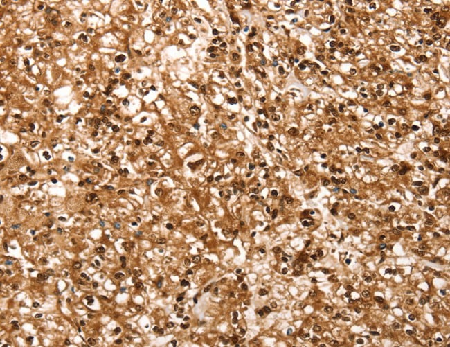
 IHC (Immunohistochemisry)
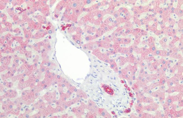
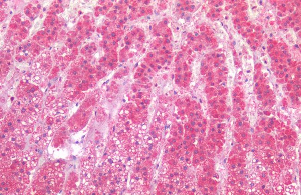
(CLEC4E/MINCLE Antibody-Human Liver: Formalin-Fixed, Paraffin-Embedded (FFPE), at a dilution of 1:100.)
IHC (Immunohistochemisry)
(CLEC4E/MINCLE Antibody-Human Liver: Formalin-Fixed, Paraffin-Embedded (FFPE), at a dilution of 1:100.)
CLEC4E/MINCLE, Polyclonal Antibody (Cat# AAA162867)
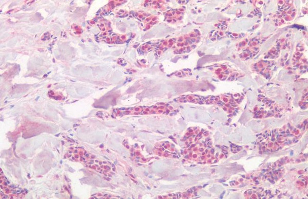
 IHC (Immunohiostchemistry)
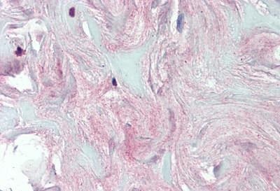
(COL4A5/Collagen IV Alpha5 Antibody-Human Breast, Fibrous Tissue: Formalin-Fixed, Paraffin-Embedded (FFPE))
IHC (Immunohiostchemistry)
(COL4A5/Collagen IV Alpha5 Antibody-Human Breast, Fibrous Tissue: Formalin-Fixed, Paraffin-Embedded (FFPE))
COL4A5/Collagen IV Alpha5, Polyclonal Antibody (Cat# AAA162870)
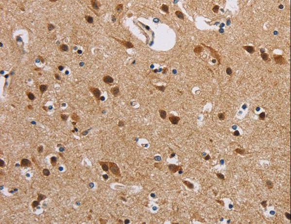
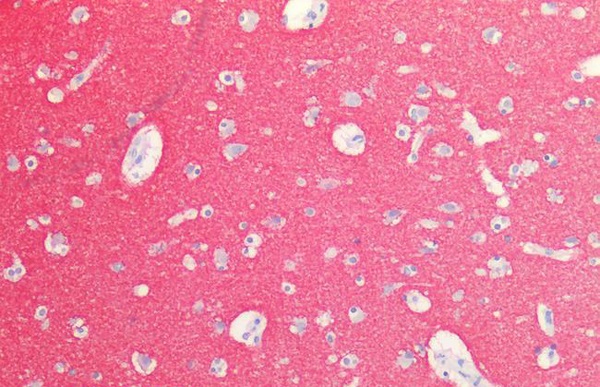
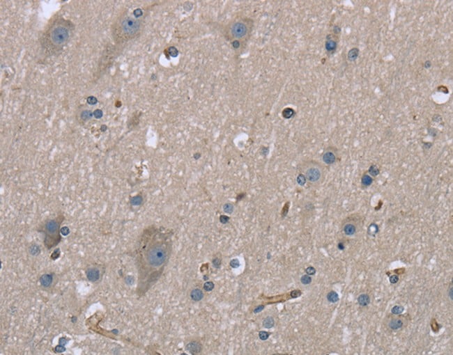
 IHC (Immunohiostchemistry)
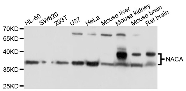
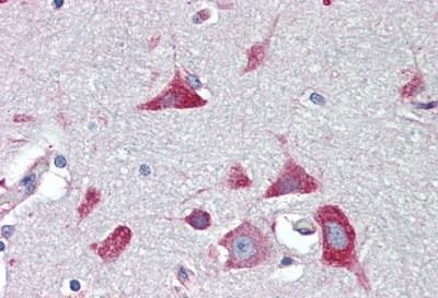
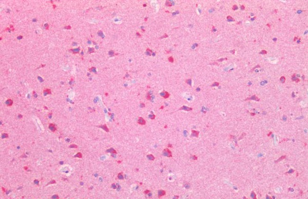
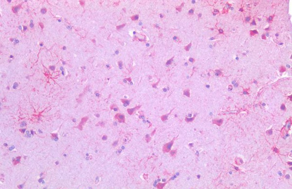
(NACA Antibody-Human Brain, Cortex: Formalin-Fixed, Paraffin-Embedded (FFPE))
IHC (Immunohiostchemistry)
(NACA Antibody-Human Brain, Cortex: Formalin-Fixed, Paraffin-Embedded (FFPE))
NACA, Polyclonal Antibody (Cat# AAA162873)
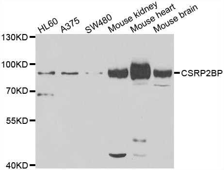
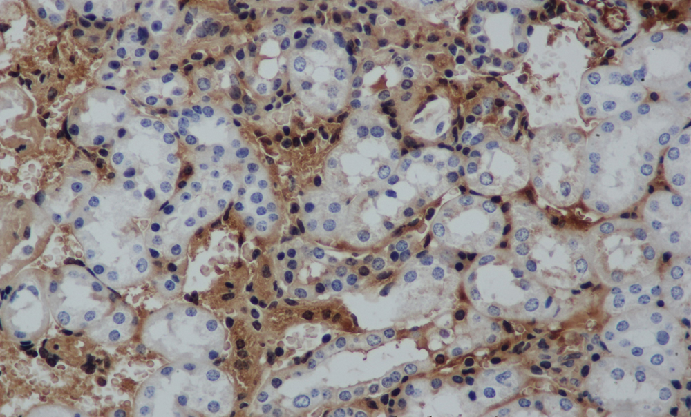
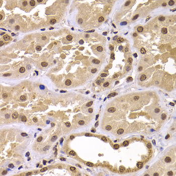 IHC (Immunohistochemistry)
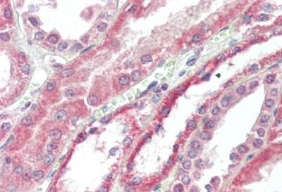
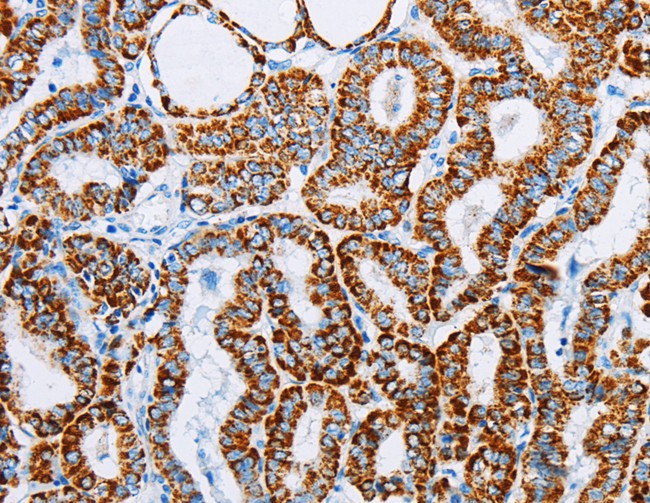
(CSRP2BP Antibody-Immunohistochemistry of paraffin-embedded human kidney tissue.)
IHC (Immunohistochemistry)
(CSRP2BP Antibody-Immunohistochemistry of paraffin-embedded human kidney tissue.)
CSRP2BP, Polyclonal Antibody (Cat# AAA162880)
 IHC (Immunohistochemistry)
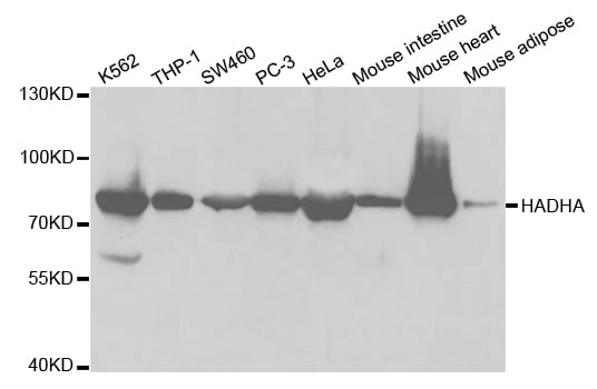
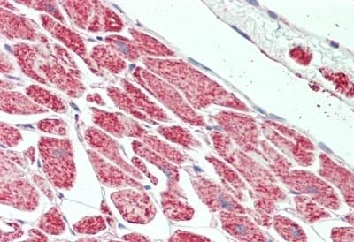
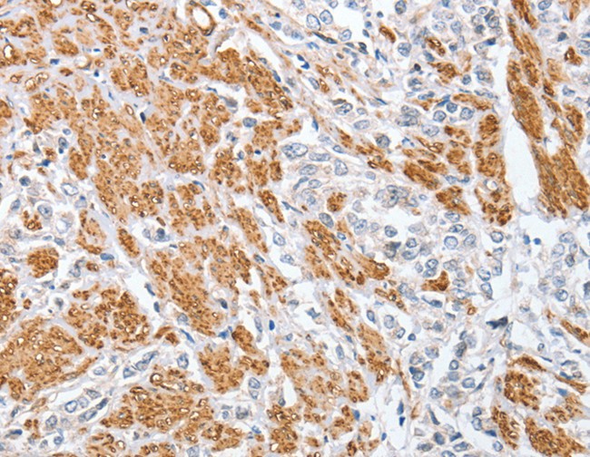
(HADHA Antibody-Human Heart: Formalin-Fixed, Paraffin-Embedded (FFPE))
IHC (Immunohistochemistry)
(HADHA Antibody-Human Heart: Formalin-Fixed, Paraffin-Embedded (FFPE))
HADHA, Polyclonal Antibody (Cat# AAA162894)
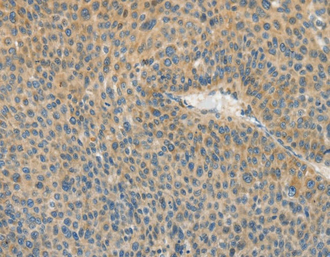
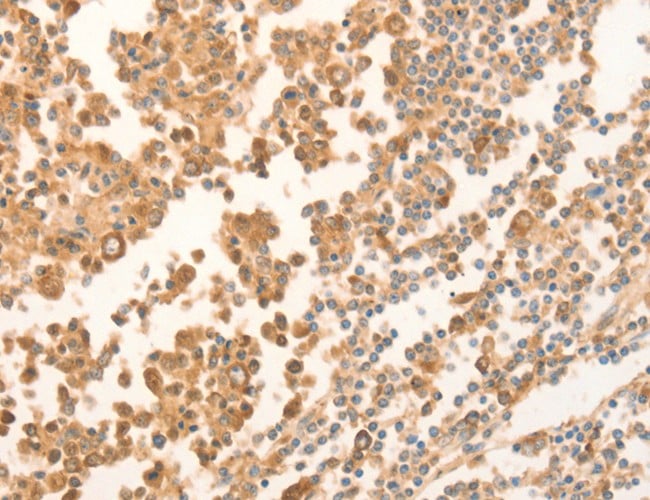
 IHC (Immunohistochemistry)
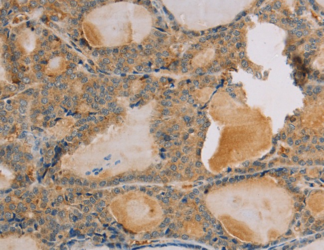
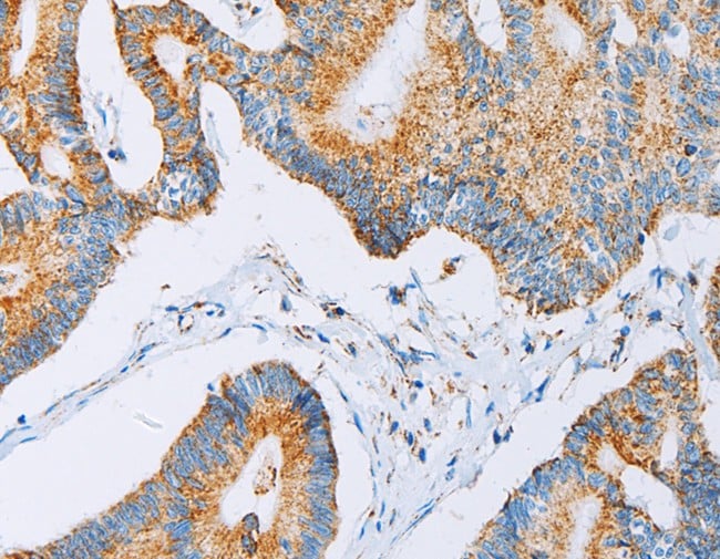
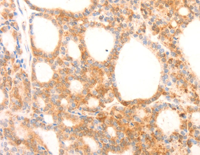
(IL19 Antibody-Immunohistochemistry of paraffin-embedded Human thyroid cancer using IL19 Polyclonal Antibody at dilution of 1:40.)
IHC (Immunohistochemistry)
(IL19 Antibody-Immunohistochemistry of paraffin-embedded Human thyroid cancer using IL19 Polyclonal Antibody at dilution of 1:40.)
IL19, Polyclonal Antibody (Cat# AAA162913)
 IHC (Immunohistochemisry)
(CCN5 Antibody-Human Liver: Formalin-Fixed, Paraffin-Embedded (FFPE))
IHC (Immunohistochemisry)
(CCN5 Antibody-Human Liver: Formalin-Fixed, Paraffin-Embedded (FFPE))
CCN5, Polyclonal Antibody (Cat# AAA162918)
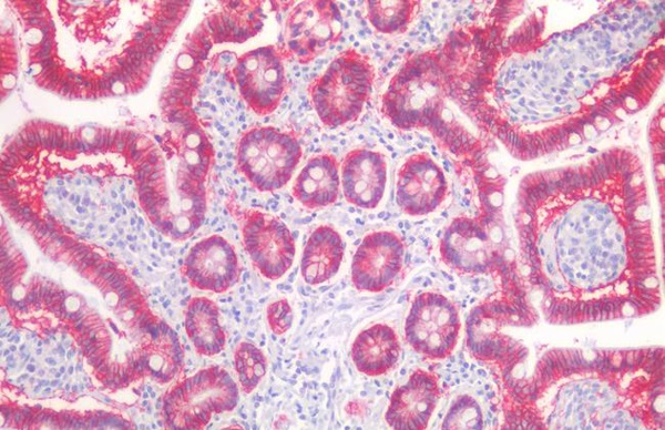
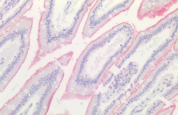
 IHC (Immunohiostchemistry)
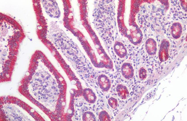
(GUCA2A/Guanylin Antibody-Human Small Intestine: Formalin-Fixed, Paraffin-Embedded (FFPE))
IHC (Immunohiostchemistry)
(GUCA2A/Guanylin Antibody-Human Small Intestine: Formalin-Fixed, Paraffin-Embedded (FFPE))
GUCA2A/Guanylin, Polyclonal Antibody (Cat# AAA162922)
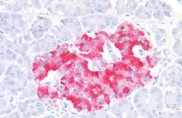
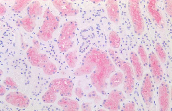
 IHC (Immunohiostchemistry)
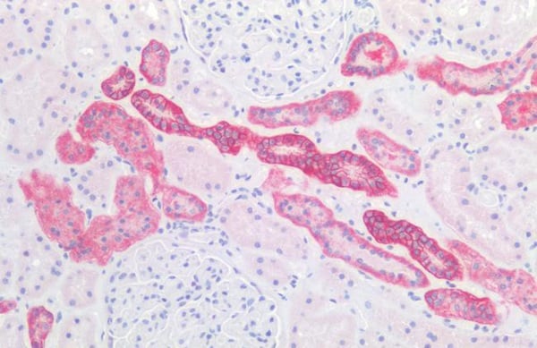
(EPO/Erythropoietin Antibody-Human Kidney: Formalin-Fixed, Paraffin-Embedded (FFPE))
IHC (Immunohiostchemistry)
(EPO/Erythropoietin Antibody-Human Kidney: Formalin-Fixed, Paraffin-Embedded (FFPE))
EPO/Erythropoietin, Polyclonal Antibody (Cat# AAA162924)
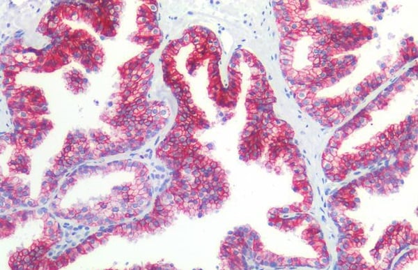
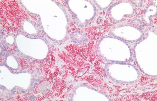
 IHC (Immunohiostchemistry)
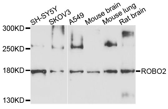
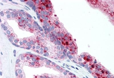
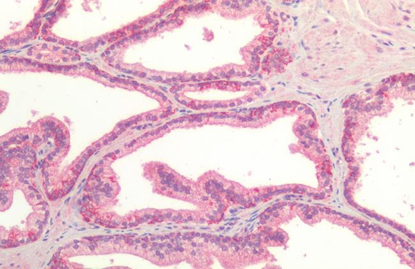
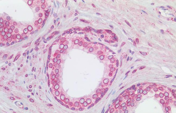
(ROBO2 Antibody-Human Prostate: Formalin-Fixed, Paraffin-Embedded (FFPE))
IHC (Immunohiostchemistry)
(ROBO2 Antibody-Human Prostate: Formalin-Fixed, Paraffin-Embedded (FFPE))
ROBO2, Polyclonal Antibody (Cat# AAA162931)
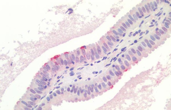
 IHC (Immunohistochemisry)
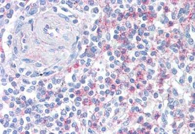
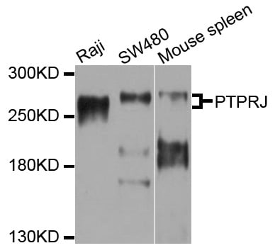
(PTPRJ/CD148 Antibody-Human Kidney: Formalin-Fixed, Paraffin-Embedded (FFPE))
IHC (Immunohistochemisry)
(PTPRJ/CD148 Antibody-Human Kidney: Formalin-Fixed, Paraffin-Embedded (FFPE))
PTPRJ/CD148, Polyclonal Antibody (Cat# AAA162932)
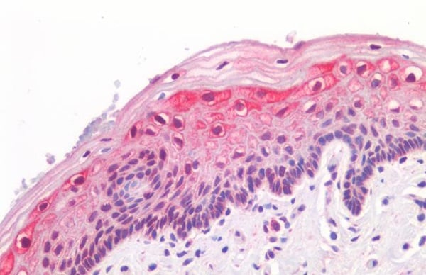
 IHC (Immunohistochemistry)
(PIK3CA/PI3K Alpha Antibody-Human Breast Carcinoma: Formalin-Fixed, Paraffin-Embedded (FFPE))
IHC (Immunohistochemistry)
(PIK3CA/PI3K Alpha Antibody-Human Breast Carcinoma: Formalin-Fixed, Paraffin-Embedded (FFPE))
PIK3CA/PI3K Alpha, Polyclonal Antibody (Cat# AAA162943)
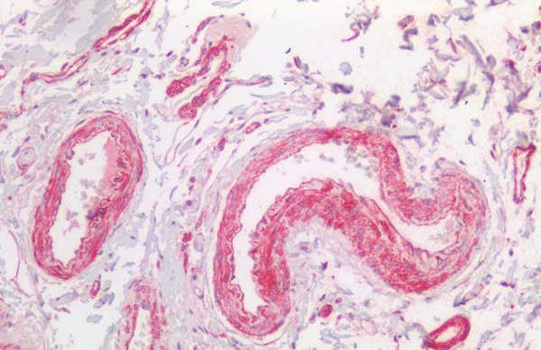
 IHC (Immunohistochemisry)
(HTR1B/5-HT1B Receptor Antibody-Human Liver, Smooth Muscle: Formalin-Fixed, Paraffin-Embedded (FFPE))
IHC (Immunohistochemisry)
(HTR1B/5-HT1B Receptor Antibody-Human Liver, Smooth Muscle: Formalin-Fixed, Paraffin-Embedded (FFPE))
HTR1B/5-HT1B Receptor, Polyclonal Antibody (Cat# AAA162950)
 IHC (Immunohiostchemistry)
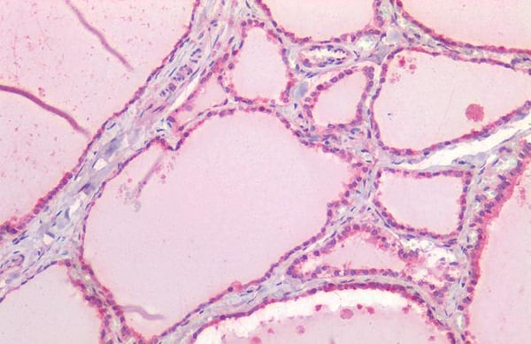
(TSLP Antibody-Human Spleen: Formalin-Fixed, Paraffin-Embedded (FFPE))
IHC (Immunohiostchemistry)
(TSLP Antibody-Human Spleen: Formalin-Fixed, Paraffin-Embedded (FFPE))
TSLP, Polyclonal Antibody (Cat# AAA162970)
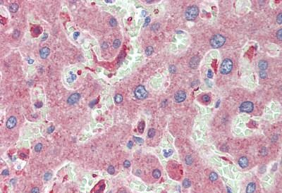
 IHC (Immunohistochemistry)
(KRT18/CK18/Cytokeratin 18 Antibody-Human Liver: Formalin-Fixed, Paraffin-Embedded (FFPE))
IHC (Immunohistochemistry)
(KRT18/CK18/Cytokeratin 18 Antibody-Human Liver: Formalin-Fixed, Paraffin-Embedded (FFPE))
KRT18/CK18/Cytokeratin 18, Polyclonal Antibody (Cat# AAA162973)
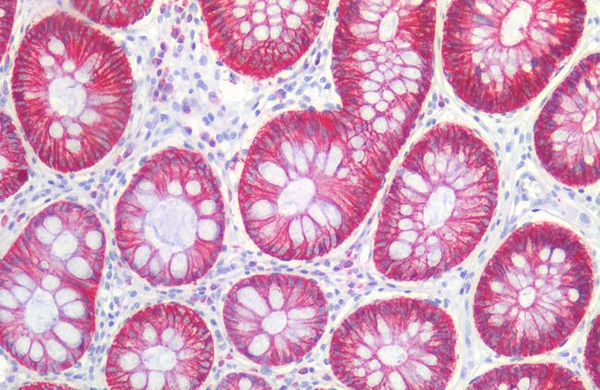
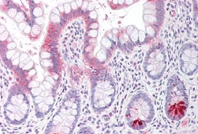
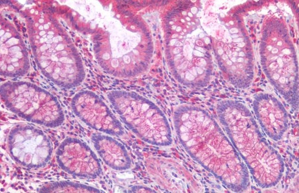
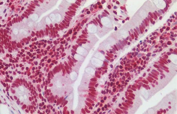
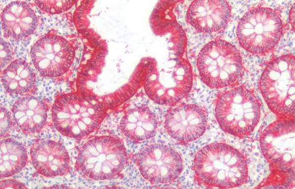 IHC (Immunohistochemisry)
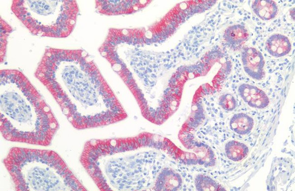
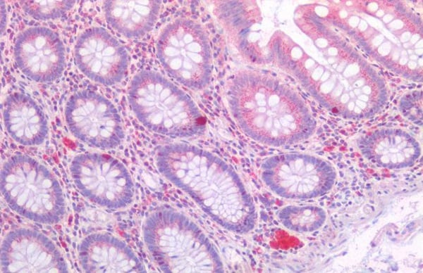
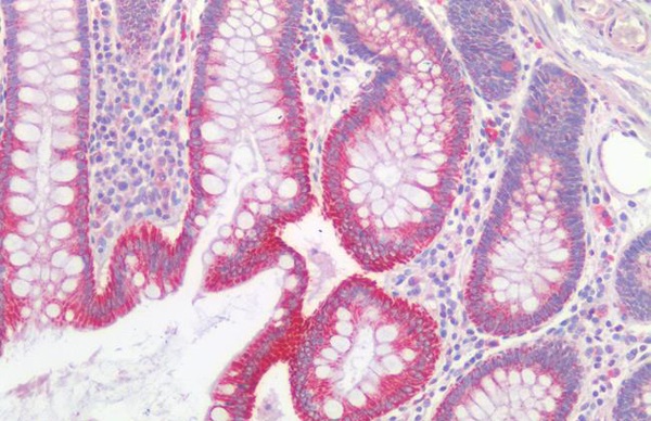
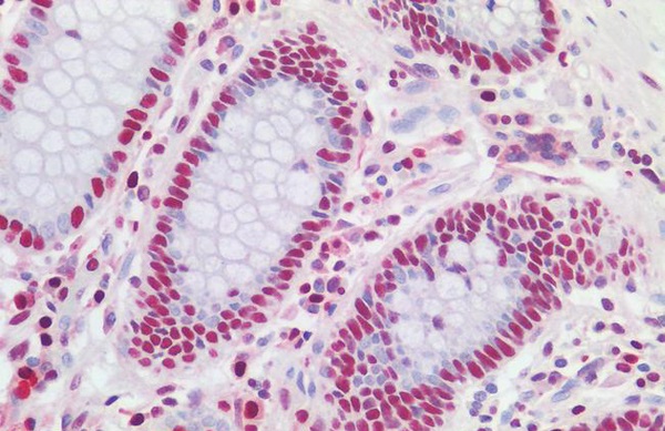
(EPCAM Antibody-Human Colon: Formalin-Fixed, Paraffin-Embedded (FFPE))
IHC (Immunohistochemisry)
(EPCAM Antibody-Human Colon: Formalin-Fixed, Paraffin-Embedded (FFPE))
EPCAM, Polyclonal Antibody (Cat# AAA162977)
 IHC (Immunohiostchemistry)
(CDH1/E Cadherin Antibody-Human Liver: Formalin-Fixed, Paraffin-Embedded (FFPE))
IHC (Immunohiostchemistry)
(CDH1/E Cadherin Antibody-Human Liver: Formalin-Fixed, Paraffin-Embedded (FFPE))
CDH1/E Cadherin, Polyclonal Antibody (Cat# AAA162982)
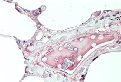
 IHC (Immunohiostchemistry)
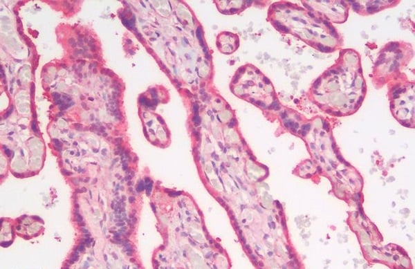
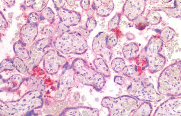
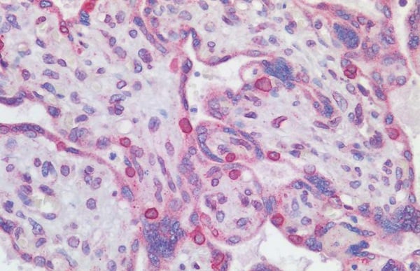
(ANXA1/Annexin A1 Antibody-Human Placenta: Formalin-Fixed, Paraffin-Embedded (FFPE))
IHC (Immunohiostchemistry)
(ANXA1/Annexin A1 Antibody-Human Placenta: Formalin-Fixed, Paraffin-Embedded (FFPE))
ANXA1/Annexin A1, Polyclonal Antibody (Cat# AAA162985)
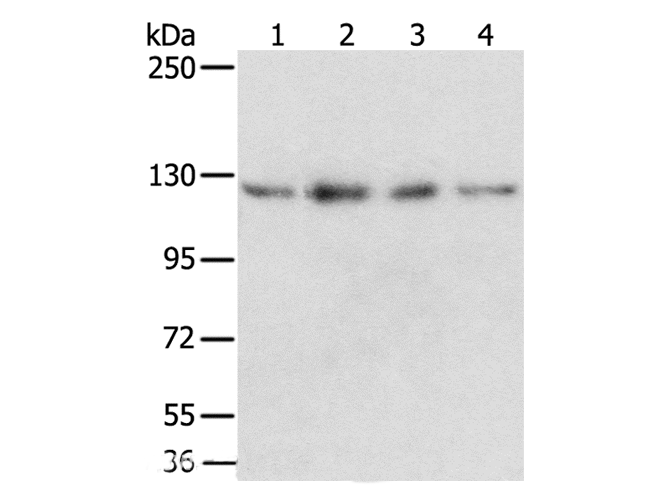
 IHC (Immunohiostchemistry)
(PDGFRA/PDGFR Alpha Antibody-Human Colon: Formalin-Fixed, Paraffin-Embedded (FFPE))
IHC (Immunohiostchemistry)
(PDGFRA/PDGFR Alpha Antibody-Human Colon: Formalin-Fixed, Paraffin-Embedded (FFPE))
PDGFRA/PDGFR Alpha, Polyclonal Antibody (Cat# AAA162994)
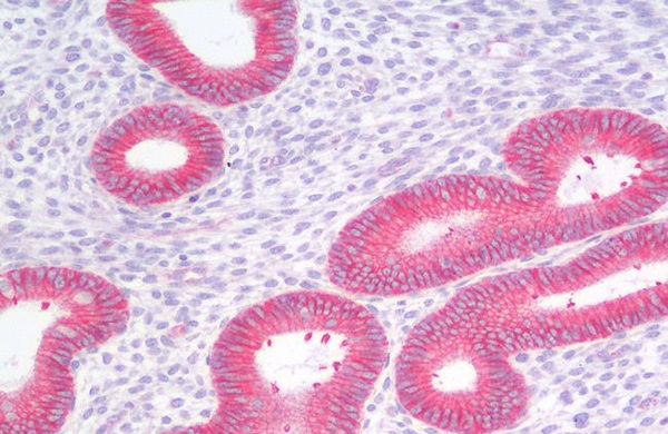
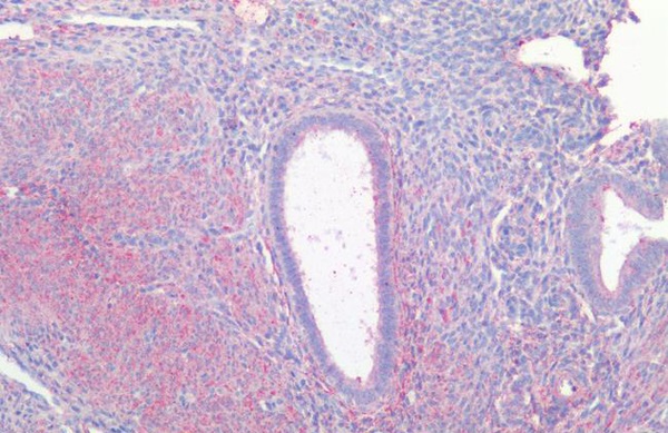
 IHC (Immunohiostchemistry)
(GREM1/Gremlin-1 Antibody-Human Cervix: Formalin-Fixed, Paraffin-Embedded (FFPE))
IHC (Immunohiostchemistry)
(GREM1/Gremlin-1 Antibody-Human Cervix: Formalin-Fixed, Paraffin-Embedded (FFPE))
GREM1/Gremlin-1, Polyclonal Antibody (Cat# AAA162999)
 IHC (Immunohistochemisry)
(HAVCR1/KIM-1 Antibody-Human Adrenal: Formalin-Fixed, Paraffin-Embedded (FFPE))
IHC (Immunohistochemisry)
(HAVCR1/KIM-1 Antibody-Human Adrenal: Formalin-Fixed, Paraffin-Embedded (FFPE))
HAVCR1/KIM-1, Polyclonal Antibody (Cat# AAA163001)
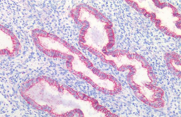
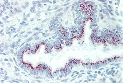
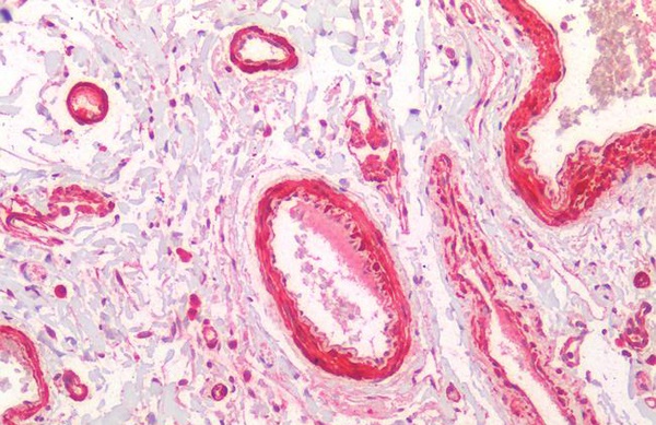
 IHC (Immunohistochemistry)
(TJP1/ZO-1 Antibody-Human Prostate: Formalin-Fixed, Paraffin-Embedded (FFPE))
IHC (Immunohistochemistry)
(TJP1/ZO-1 Antibody-Human Prostate: Formalin-Fixed, Paraffin-Embedded (FFPE))
TJP1/ZO-1, Polyclonal Antibody (Cat# AAA163012)
 IHC (Immunohistochemistry)
(USP19 Antibody-Human Placenta: Formalin-Fixed, Paraffin-Embedded (FFPE))
IHC (Immunohistochemistry)
(USP19 Antibody-Human Placenta: Formalin-Fixed, Paraffin-Embedded (FFPE))
USP19, Polyclonal Antibody (Cat# AAA163017)
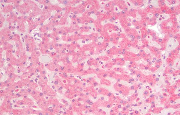
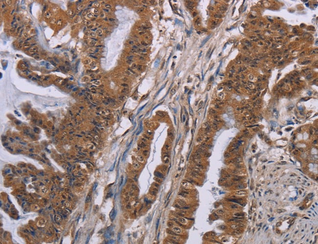
 IHC (Immunohistochemistry)
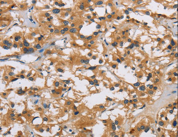
(RARRES2/Chemerin Antibody-Human Liver: Formalin-Fixed, Paraffin-Embedded (FFPE))
IHC (Immunohistochemistry)
(RARRES2/Chemerin Antibody-Human Liver: Formalin-Fixed, Paraffin-Embedded (FFPE))
RARRES2/Chemerin, Polyclonal Antibody (Cat# AAA163352)
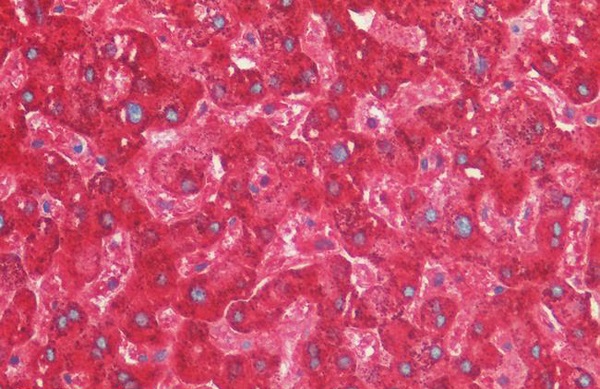
 IHC (Immunohiostchemistry)
(LRRC8A/LRRC8 Antibody-Human Brain, Cortex: Formalin-Fixed, Paraffin-Embedded (FFPE))
IHC (Immunohiostchemistry)
(LRRC8A/LRRC8 Antibody-Human Brain, Cortex: Formalin-Fixed, Paraffin-Embedded (FFPE))
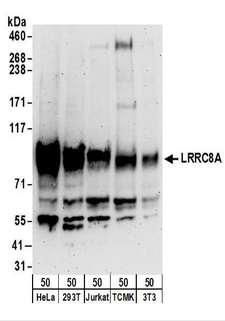
LRRC8A/LRRC8, Polyclonal Antibody (Cat# AAA163353)
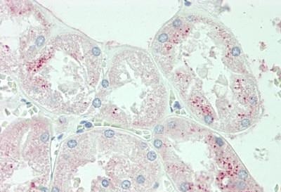
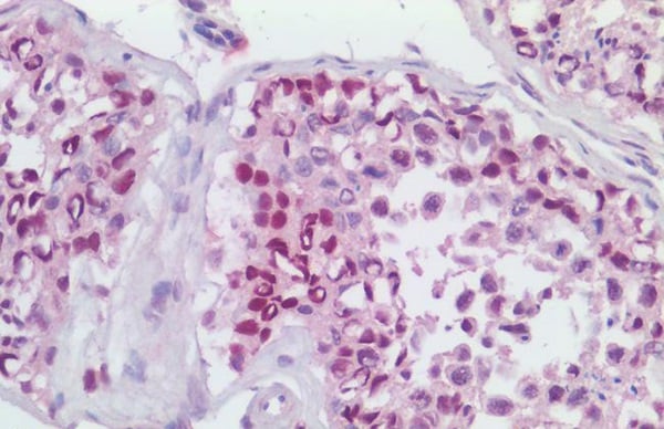 IHC (Immunohiostchemistry)
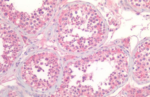
(MCM4 Antibody-Human Testis: Formalin-Fixed, Paraffin-Embedded (FFPE))
IHC (Immunohiostchemistry)
(MCM4 Antibody-Human Testis: Formalin-Fixed, Paraffin-Embedded (FFPE))
MCM4, Polyclonal Antibody (Cat# AAA163366)
 IHC (Immunohistochemisry)
(SUB1 Antibody-Human Colon: Formalin-Fixed, Paraffin-Embedded (FFPE))
IHC (Immunohistochemisry)
(SUB1 Antibody-Human Colon: Formalin-Fixed, Paraffin-Embedded (FFPE))
SUB1, Polyclonal Antibody (Cat# AAA163368)
 IHC (Immunohiostchemistry)
(RANGAP1 Antibody-Human Placenta: Formalin-Fixed, Paraffin-Embedded (FFPE))
IHC (Immunohiostchemistry)
(RANGAP1 Antibody-Human Placenta: Formalin-Fixed, Paraffin-Embedded (FFPE))
RANGAP1, Polyclonal Antibody (Cat# AAA163373)

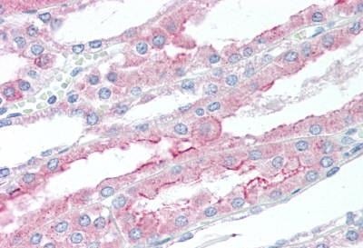
 IHC (Immunohistochemisry)
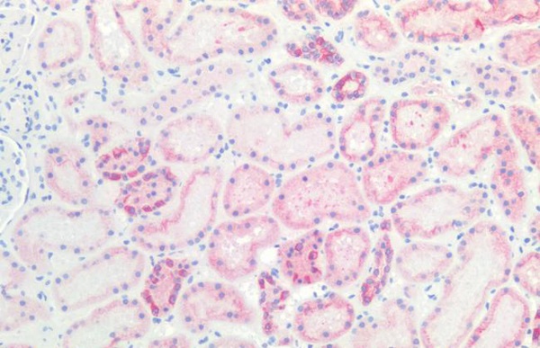
(SLC6A19 Antibody-Human Kidney: Formalin-Fixed, Paraffin-Embedded (FFPE))
IHC (Immunohistochemisry)
(SLC6A19 Antibody-Human Kidney: Formalin-Fixed, Paraffin-Embedded (FFPE))
SLC6A19, Polyclonal Antibody (Cat# AAA163375)

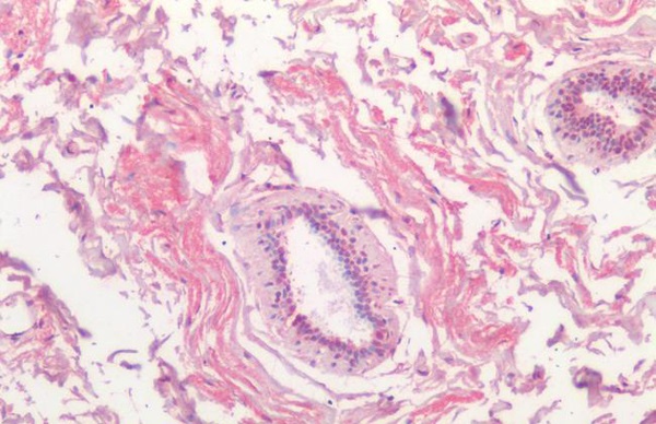
 IHC (Immunohiostchemistry)
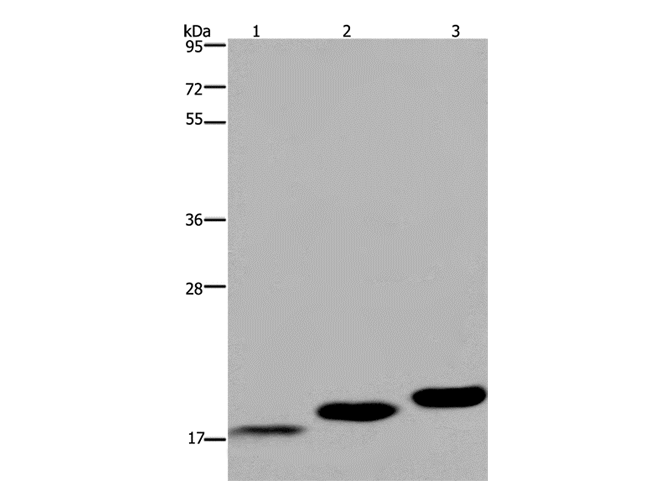
IL11, Polyclonal Antibody (Cat# AAA163383)
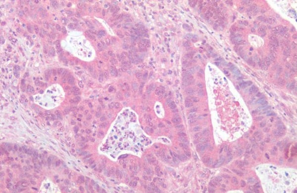
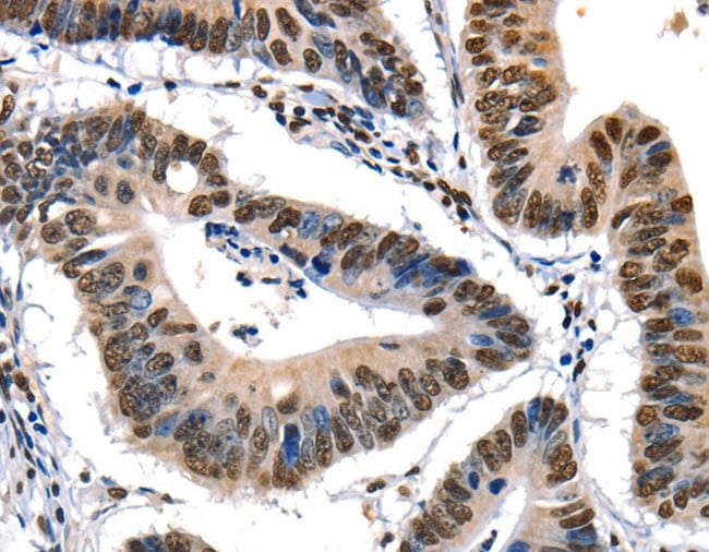 IHC (Immunohiostchemistry)
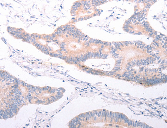
(Immunohistochemistry of paraffin-embedded Human colon cancer tissue using XRCC6 Polyclonal Antibody at dilution of 1:10)
IHC (Immunohiostchemistry)
(Immunohistochemistry of paraffin-embedded Human colon cancer tissue using XRCC6 Polyclonal Antibody at dilution of 1:10)
XRCC6, Polyclonal Antibody (Cat# AAA166108)
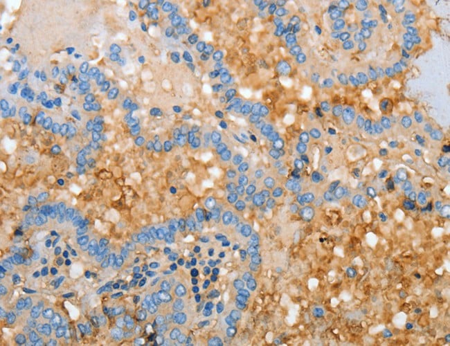
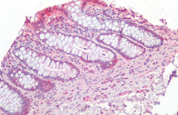
 IHC (Immunohistochemisry)
(Immunohistochemistry of paraffin-embedded Human esophagus cancer using APOH Polyclonal Antibody at dilution of 1:30)
IHC (Immunohistochemisry)
(Immunohistochemistry of paraffin-embedded Human esophagus cancer using APOH Polyclonal Antibody at dilution of 1:30)
APOH, Polyclonal Antibody (Cat# AAA166111)
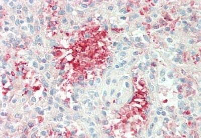
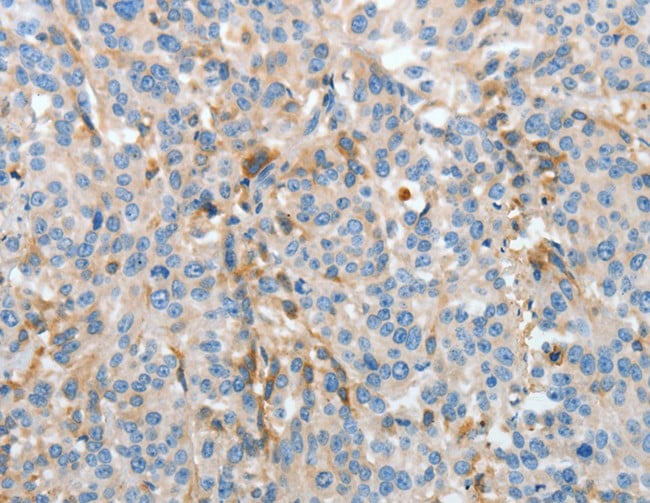
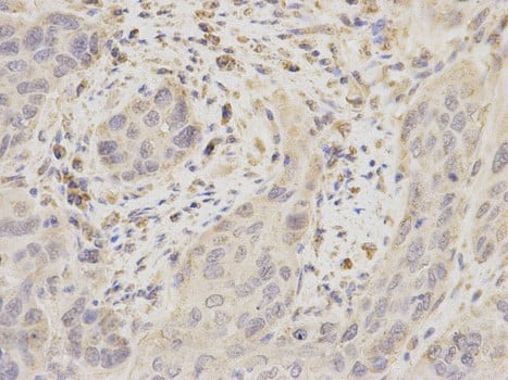
 IHC (Immunohiostchemistry)
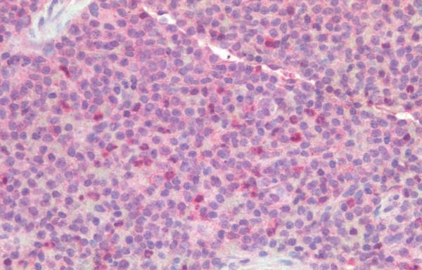
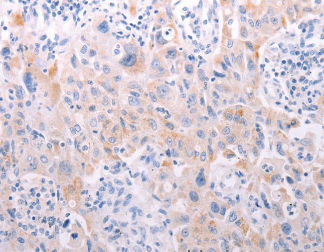
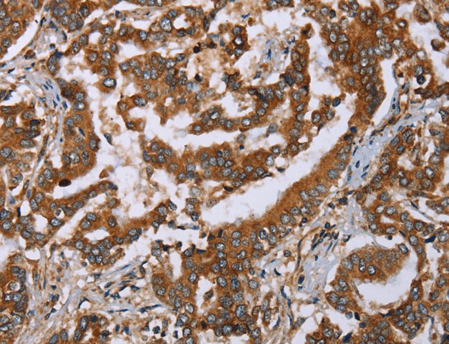
(Immunohistochemistry of paraffin-embedded Human thyroid cancer tissue using PARP3 Polyclonal Antibody at dilution 1:25)
IHC (Immunohiostchemistry)
(Immunohistochemistry of paraffin-embedded Human thyroid cancer tissue using PARP3 Polyclonal Antibody at dilution 1:25)
PARP3, Polyclonal Antibody (Cat# AAA166112)
 IHC (Immunohiostchemistry)
(Immunohistochemistry of paraffin-embedded Human lung cancer tissue using ANKRA2 Polyclonal Antibody at dilution 1:25)
IHC (Immunohiostchemistry)
(Immunohistochemistry of paraffin-embedded Human lung cancer tissue using ANKRA2 Polyclonal Antibody at dilution 1:25)
ANKRA2, Polyclonal Antibody (Cat# AAA166117)
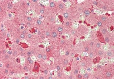
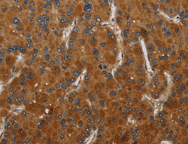
 IHC (Immunohiostchemistry)
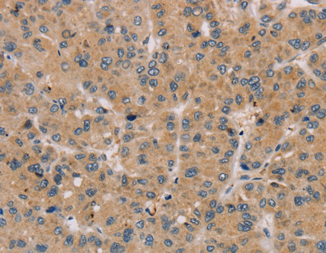
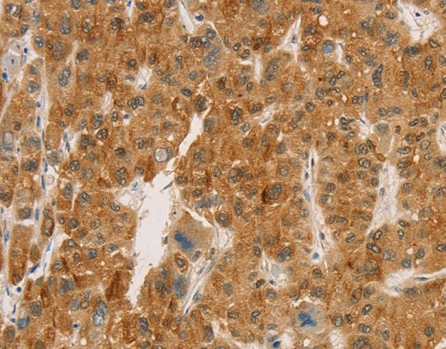
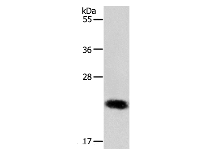
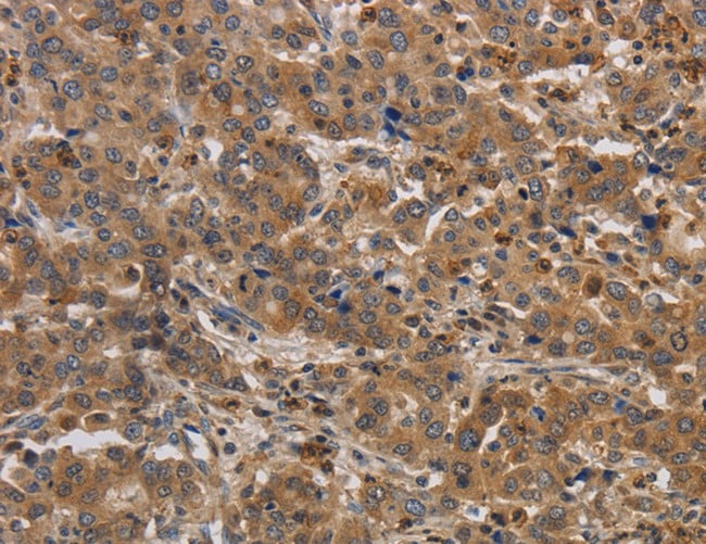
(Immunohistochemistry of paraffin-embedded Human liver cancer tissue using TIMP1 Polyclonal Antibody at dilution 1:15)
IHC (Immunohiostchemistry)
(Immunohistochemistry of paraffin-embedded Human liver cancer tissue using TIMP1 Polyclonal Antibody at dilution 1:15)
TIMP1, Polyclonal Antibody (Cat# AAA166206)
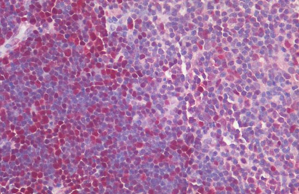
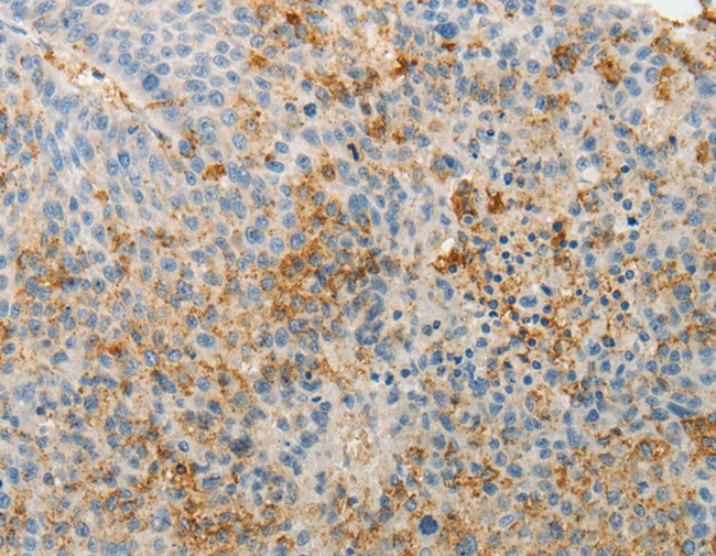
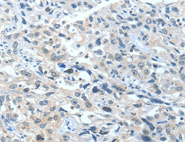
 IHC (Immunohistochemisry)
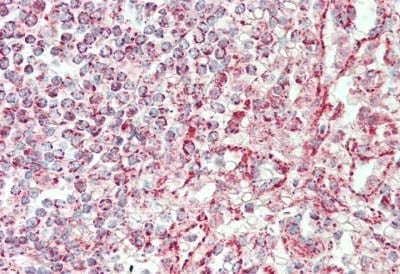
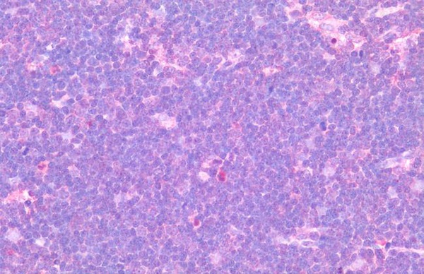
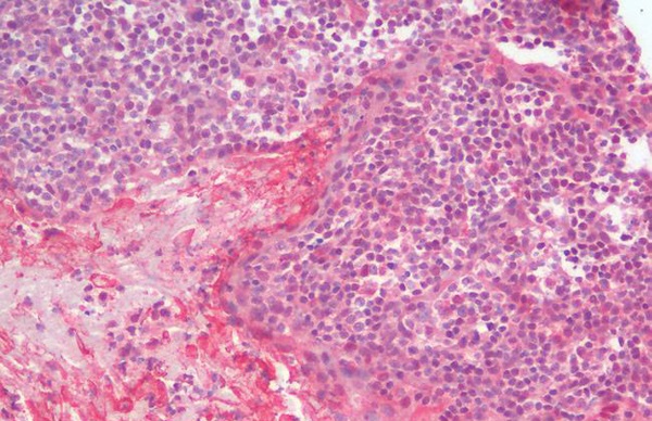
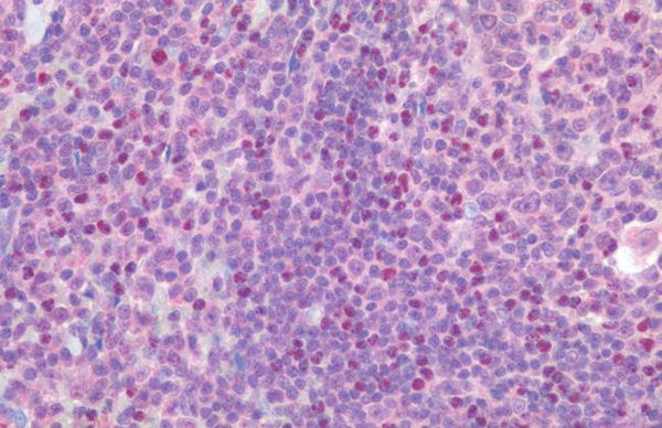
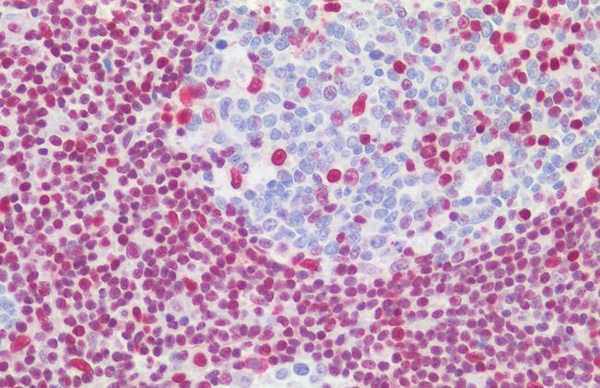
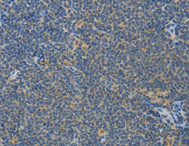
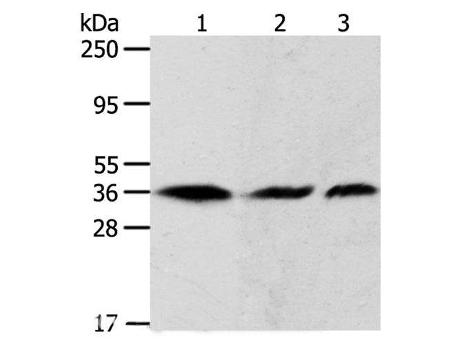
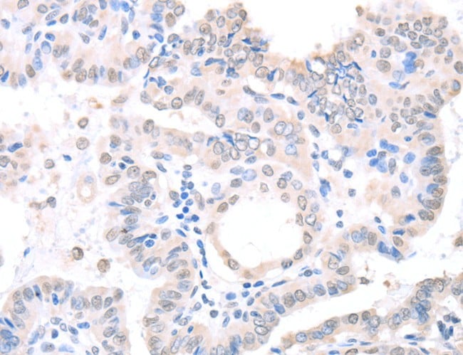
(Immunohistochemistry of paraffin-embedded Human lymphoma using ASPA Polyclonal Antibody at dilution of 1:80)
IHC (Immunohistochemisry)
(Immunohistochemistry of paraffin-embedded Human lymphoma using ASPA Polyclonal Antibody at dilution of 1:80)
ASPA, Polyclonal Antibody (Cat# AAA166208)
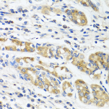
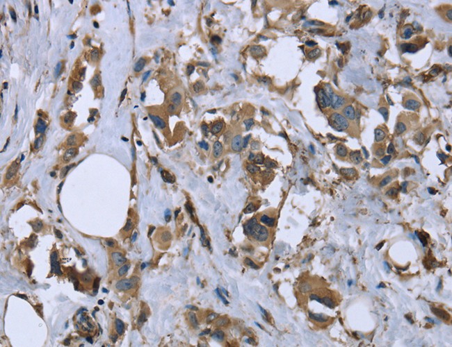
 IHC (Immunohistochemisry)
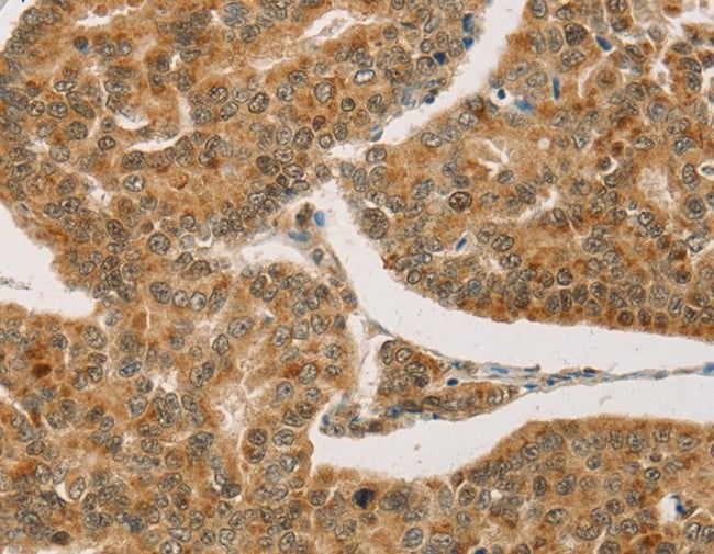
(Immunohistochemistry of paraffin-embedded Human breast cancer using CPA2 Polyclonal Antibody at dilution of 1:20)
IHC (Immunohistochemisry)
(Immunohistochemistry of paraffin-embedded Human breast cancer using CPA2 Polyclonal Antibody at dilution of 1:20)
CPA2, Polyclonal Antibody (Cat# AAA166211)
 IHC (Immunohistochemisry)
(Immunohistochemistry of paraffin-embedded Human breast cancer using MYL12B Polyclonal Antibody at dilution of 1:20)
IHC (Immunohistochemisry)
(Immunohistochemistry of paraffin-embedded Human breast cancer using MYL12B Polyclonal Antibody at dilution of 1:20)
MYL12B, Polyclonal Antibody (Cat# AAA166218)
 IHC (Immunohiostchemistry)
(Immunohistochemistry of paraffin-embedded Human esophagus cancer tissue using GRK1 Polyclonal Antibody at dilution 1:40)
IHC (Immunohiostchemistry)
(Immunohistochemistry of paraffin-embedded Human esophagus cancer tissue using GRK1 Polyclonal Antibody at dilution 1:40)
GRK1, Polyclonal Antibody (Cat# AAA166224)
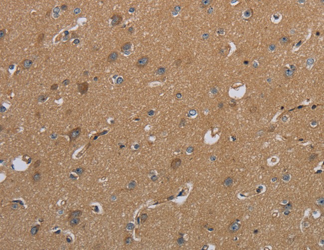 IHC (Immunohistochemisry)
(Immunohistochemistry of paraffin-embedded Human brain using CSH1 Polyclonal Antibody at dilution of 1:50)
IHC (Immunohistochemisry)
(Immunohistochemistry of paraffin-embedded Human brain using CSH1 Polyclonal Antibody at dilution of 1:50)
CSH1, Polyclonal Antibody (Cat# AAA166225)
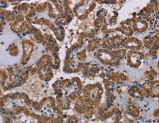
 IHC (Immunohistochemisry)
(Immunohistochemistry of paraffin-embedded Human thyroid cancer using ACAA2 Polyclonal Antibody at dilution of 1:30)
IHC (Immunohistochemisry)
(Immunohistochemistry of paraffin-embedded Human thyroid cancer using ACAA2 Polyclonal Antibody at dilution of 1:30)
ACAA2, Polyclonal Antibody (Cat# AAA166227)
 IHC (Immunohiostchemistry)
(Immunohistochemistry of paraffin-embedded Human esophagus cancer tissue using CENPC Polyclonal Antibody at dilution 1:70)
IHC (Immunohiostchemistry)
(Immunohistochemistry of paraffin-embedded Human esophagus cancer tissue using CENPC Polyclonal Antibody at dilution 1:70)
CENPC, Polyclonal Antibody (Cat# AAA166233)
 IHC (Immunohiostchemistry)
(Immunohistochemistry of paraffin-embedded Human liver cancer using RAB22A Polyclonal Antibody at dilution of 1:20)
IHC (Immunohiostchemistry)
(Immunohistochemistry of paraffin-embedded Human liver cancer using RAB22A Polyclonal Antibody at dilution of 1:20)
RAB22A, Polyclonal Antibody (Cat# AAA166235)
 IHC (Immunohiostchemistry)
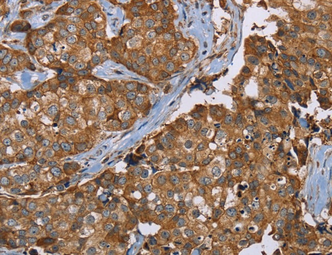
(Immunohistochemistry of paraffin-embedded Human thyroid cancer using APEX1 Polyclonal Antibody at dilution of 1:20)
IHC (Immunohiostchemistry)
(Immunohistochemistry of paraffin-embedded Human thyroid cancer using APEX1 Polyclonal Antibody at dilution of 1:20)
APEX1, Polyclonal Antibody (Cat# AAA166250)
 IHC (Immunohiostchemistry)
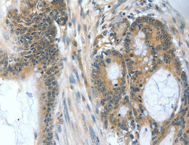
(Immunohistochemistry of paraffin-embedded Human prostate cancer using RBM5 Polyclonal Antibody at dilution of 1:30)
IHC (Immunohiostchemistry)
(Immunohistochemistry of paraffin-embedded Human prostate cancer using RBM5 Polyclonal Antibody at dilution of 1:30)
RBM5, Polyclonal Antibody (Cat# AAA166258)
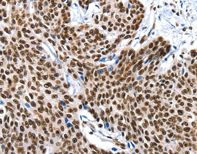
 IHC (Immunohistochemisry)
(Immunohistochemistry of paraffin-embedded Human thyroid cancer using NAP1L1 Polyclonal Antibody at dilution of 1:70)
IHC (Immunohistochemisry)
(Immunohistochemistry of paraffin-embedded Human thyroid cancer using NAP1L1 Polyclonal Antibody at dilution of 1:70)
NAP1L1, Polyclonal Antibody (Cat# AAA166261)
 IHC (Immunohiostchemistry)
(Immunohistochemistry of paraffin-embedded Human breast cancer tissue using PLXNB1 Polyclonal Antibody at dilution 1:50)
IHC (Immunohiostchemistry)
(Immunohistochemistry of paraffin-embedded Human breast cancer tissue using PLXNB1 Polyclonal Antibody at dilution 1:50)
PLXNB1, Polyclonal Antibody (Cat# AAA166279)
 IHC (Immunohiostchemistry)
(Immunohistochemistry of paraffin-embedded Human esophagus cancer tissue using BAD Polyclonal Antibody at dilution 1:60)
IHC (Immunohiostchemistry)
(Immunohistochemistry of paraffin-embedded Human esophagus cancer tissue using BAD Polyclonal Antibody at dilution 1:60)
BAD, Polyclonal Antibody (Cat# AAA166280)

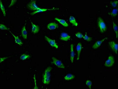
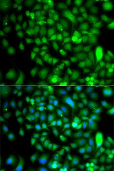
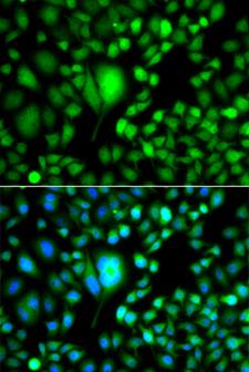
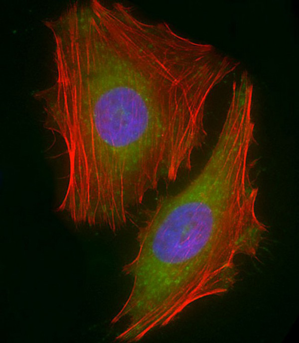
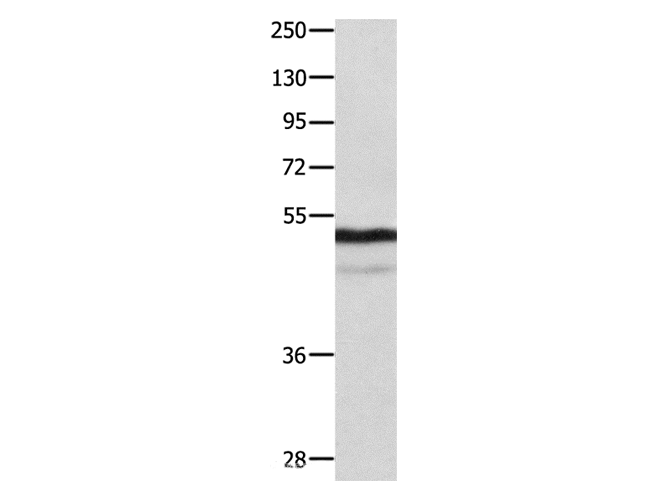
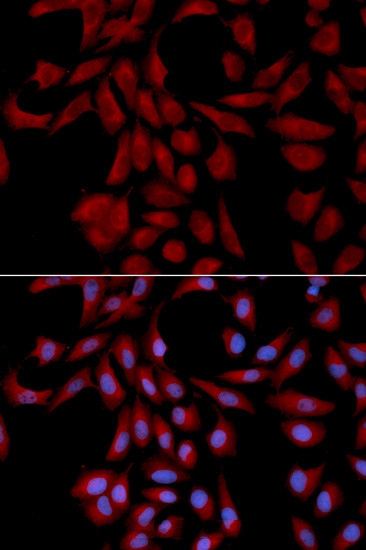 IF (Immunofluorescence)
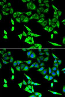
(Immunofluorescence analysis of U2OS cell using UBE2I antibody. Blue: DAPI for nuclear staining.)
IF (Immunofluorescence)
(Immunofluorescence analysis of U2OS cell using UBE2I antibody. Blue: DAPI for nuclear staining.)
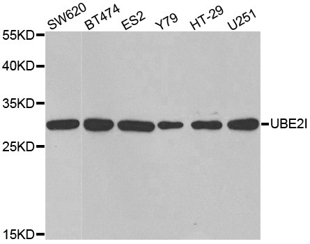
UBE2I, Polyclonal Antibody (Cat# AAA166284)
What are Polyclonal Antibodies?
Polyclonal antibodies are antibodies that come from multiple B cell clones of a host animal. The typical hosts used for the majority of polyclonal antibody production are rabbits, goats, sheep, and donkeys. These polyclonal antibodies, once having identified their target, will bind to different epitopes located at different regions or sequences on the same protein/antigen. As a result, they are ideal at locating and binding to the target, even if the target is in very low concentrations (due to many different antibodies being able to bind to the same target molecule, which allows for significant amplification of a downstream signal).
Polyclonal antibodies are typically produced by injecting an antigen into a host animal, which causes the animal’s immune system to attack the foreign antigen by mass generating antibodies against it. After a period of time, serum is collected from the animal and purified using physicochemical fractionation, class-specific affinity purification, and/or antigen-affinity purification.
Key Uses of Polyclonal Antibodies
- Western Blotting: This method is used to find specific proteins in biological samples after separating them by size.
- Immunohistochemistry: IHC helps visualize the location of proteins in tissue sections using various staining techniques.
- ELISA: (Enzyme-Linked Immunosorbent Assay) is typically used to identify specific protein quantities in a sample. ELISAs can be either “Quantitative” or “Qualitative”.
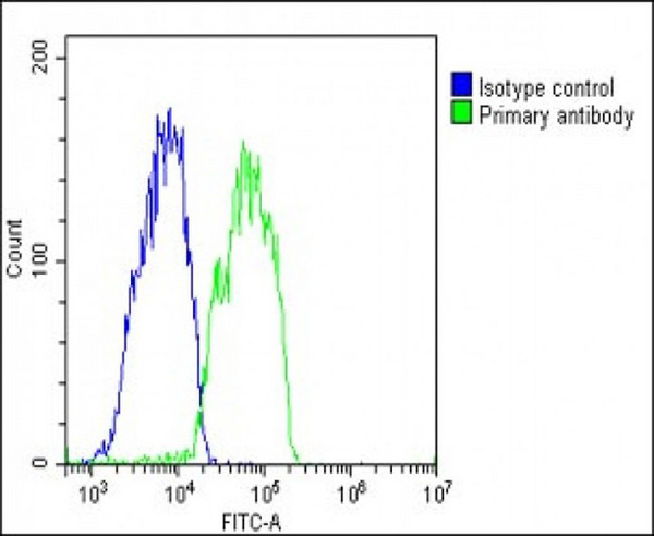
- Flow Cytometry: technique that identifies and measures the specific protein on the surface or inside the cells in a fluid suspension.
- Immunoprecipitation: IP isolates and studies a specific protein from a complex mixture using antibodies.
Why Buy Polyclonal Antibodies from AAA Biotech?
1. Ideal for Various Applications
Our antibodies are generally going to be validated for use in multiple types of assays, including ELISA, Western Blotting, Immunohistochemistry, Immunoprecipitation, amongst others. They are ideal for a wide range of research applications.
2. Rigorous Quality Control
All of the antibodies in our catalog undergo strict quality testing to ensure specificity, sensitivity, and consistent performance. We are confident in the ability of our antibodies to provide you with accurate results.
3. Wide Assortment of Antibodies
Antibodies in are catalog can be found for both common and exotic species, and these antibodies are also available in both conjugated and recombinant forms to suit many diverse experimental needs.
4. Highly Purified
Our antibodies are available in purified forms with over 85% purity, as confirmed by SDS-PAGE. They are also available with tags such as His, Flag, GST, or MBP. We cater to customers worldwide.
FAQ
1. How are polyclonal antibodies produced?
Traditionally, polyclonal antibodies are produced by injecting an antigen into a host animal (such as a rabbit or goat), which then triggers an immune response from the host animal. The animal’s B cells produce antibodies that will recognize different parts of the injected antigen. These antibodies are then collected from the animal’s blood and purified for use.
2. How do polyclonal antibodies differ from monoclonal antibodies?
Polyclonal antibodies are a mix of antibodies that bind to different locations (epitopes) of the same antigen, while monoclonal antibodies are identical and bind to just one specific epitope. This makes polyclonal antibodies more versatile and better at detecting proteins that may be present in low quantities or in altered/modified forms.
3. How should I store polyclonal antibodies?
Polyclonal antibodies should be stored at 4°C for short-term use (up to a few weeks) and at -20°C or -80°C for long-term storage. Avoid repeated freeze-thaw cycles by dividing them into small aliquots. Always check the datasheet for specific storage instructions.