Filters
▼Clonality
▼Type
▼Reactivity
▼Gene Name
▼Isotype
▼Host
▼Application
▼Clone
▼Polyclonal Antibodies
At AAA Biotech also known as AAA Bio or AAABio, we provide a broad range of purified polyclonal antibodies (pAbs) that are able to all be browsed online through our website. Due to their high specificity and strong binding affinity, these antibodies are ideal for wide swathes of research and experimental applications.
Our polyclonal antibodies can easily support your work, whether you use them for Western Blotting, Immunocytochemistry (with or without Immunofluorescence used in conjunction), Immunohistochemistry, Immunoprecipitation, and ELISA tests. We highly encourage you to browse our range of pAbs and choose the one that best suits your experimental model.
Viewing 7950-8000 of 96805 product results
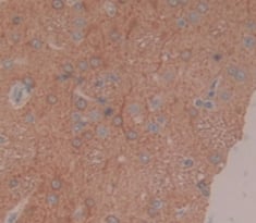
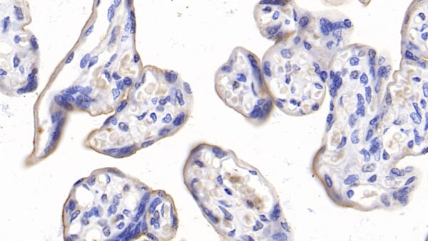
 IHC (Immunohistochemisry)
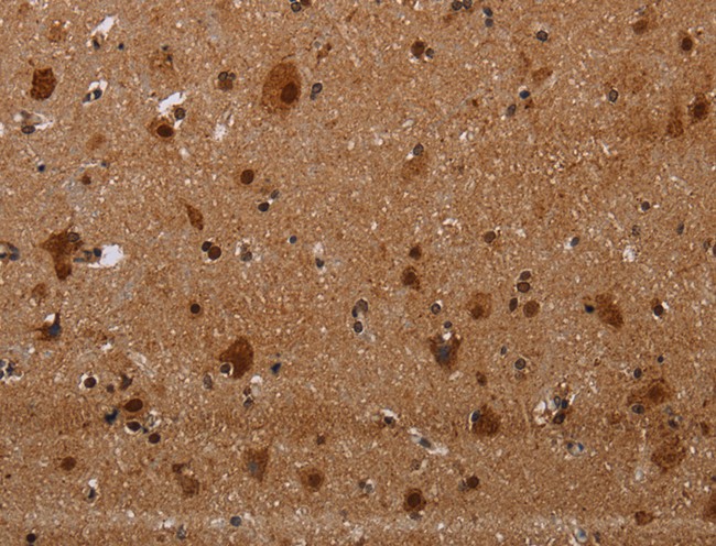
(Immunohistochemistry of paraffin-embedded Human brain using MAGEE1 Polyclonal Antibody at dilution of 1:30)
IHC (Immunohistochemisry)
(Immunohistochemistry of paraffin-embedded Human brain using MAGEE1 Polyclonal Antibody at dilution of 1:30)
MAGEE1, Polyclonal Antibody (Cat# AAA166811)
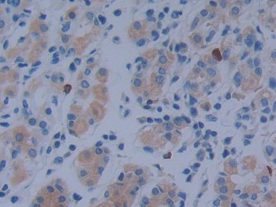
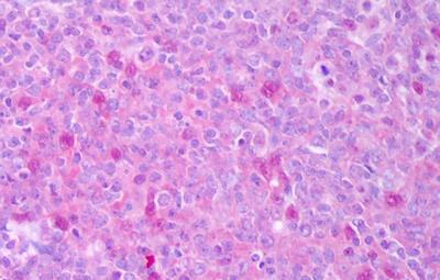
 IHC (Immunohiostchemistry)
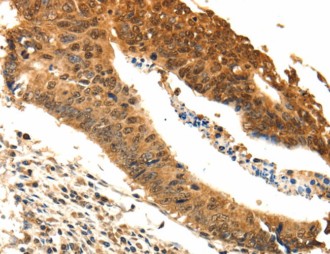
(Immunohistochemistry of paraffin-embedded Human thyroid cancer tissue using ODC1 Polyclonal Antibody at dilution 1:25)
IHC (Immunohiostchemistry)
(Immunohistochemistry of paraffin-embedded Human thyroid cancer tissue using ODC1 Polyclonal Antibody at dilution 1:25)
ODC1, Polyclonal Antibody (Cat# AAA166702)
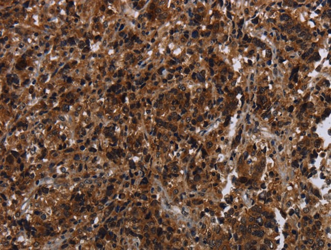
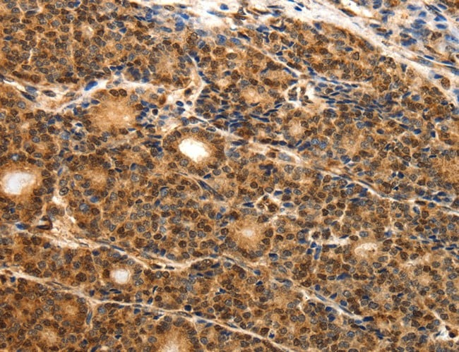
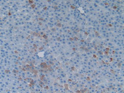
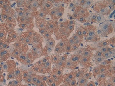
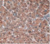
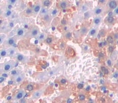
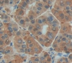
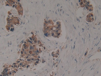
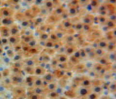
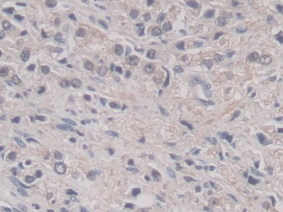
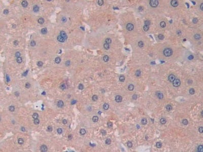
 IHC (Immunohiostchemistry)
(Immunohistochemistry of paraffin-embedded Human liver cancer tissue using E2F4 Polyclonal Antibody at dilution 1:40)
IHC (Immunohiostchemistry)
(Immunohistochemistry of paraffin-embedded Human liver cancer tissue using E2F4 Polyclonal Antibody at dilution 1:40)
E2F4, Polyclonal Antibody (Cat# AAA166703)
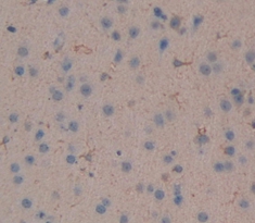
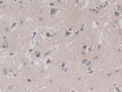
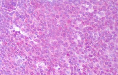
 IHC (Immunohiostchemistry)
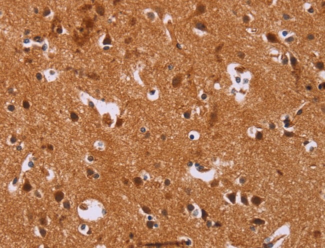
(Immunohistochemistry of paraffin-embedded Human brain tissue using CALCA Polyclonal Antibody at dilution 1:25)
IHC (Immunohiostchemistry)
(Immunohistochemistry of paraffin-embedded Human brain tissue using CALCA Polyclonal Antibody at dilution 1:25)
CALCA, Polyclonal Antibody (Cat# AAA166704)
 IHC (Immunohiostchemistry)
(DAB staining on fromalin fixed paraffin-embedded Liver tissue))
IHC (Immunohiostchemistry)
(DAB staining on fromalin fixed paraffin-embedded Liver tissue))
Motilin (MTL), Polyclonal Antibody (Cat# AAA130725)


 IHC (Immunohistochemistry)
(DAB staining on IHC-P; Samples: Rat Liver Tissue)
IHC (Immunohistochemistry)
(DAB staining on IHC-P; Samples: Rat Liver Tissue)
Heat Shock 70kDa Binding Protein 1 (HSPBP1), Polyclonal Antibody (Cat# AAA130727)
 IHC (Immunohiostchemistry)
(DABstainingonIHC-P.Samples:RatTissue))
IHC (Immunohiostchemistry)
(DABstainingonIHC-P.Samples:RatTissue))
Coagulation Factor IX (F9), Polyclonal Antibody (Cat# AAA130754)
 IHC (Immunohiostchemistry)
(DABstainingonIHC-P.Samples:RatTissue))
IHC (Immunohiostchemistry)
(DABstainingonIHC-P.Samples:RatTissue))
Ephrin A4 (EFNA4), Polyclonal Antibody (Cat# AAA131152)
Ectonucleotide Pyrophosphatase/Phosphodiesterase 1 (ENPP1), Polyclonal Antibody (Cat# AAA131154)
 IHC (Immunohistochemisry)
(DAB staining on IHC-P.Samples: Human Tissue)
IHC (Immunohistochemisry)
(DAB staining on IHC-P.Samples: Human Tissue)
V-Myc Myelocytomatosis Viral Oncogene Homolog 1, Lung Carcinoma Derived (MYCL1), Polyclonal Antibody (Cat# AAA131160)
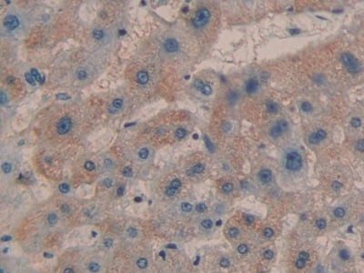
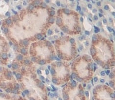
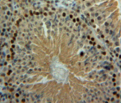
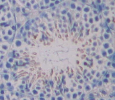
 IHC (Immunohistochemisry)
(DAB staining on IHC-P; Samples: Human Liver Tissue))
IHC (Immunohistochemisry)
(DAB staining on IHC-P; Samples: Human Liver Tissue))
Toll Interacting Protein (TOLLIP), Polyclonal Antibody (Cat# AAA131184)
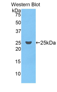
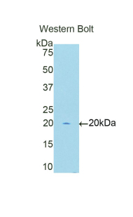
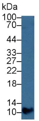
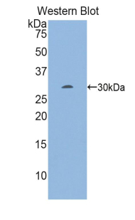
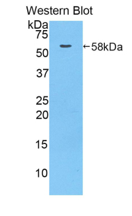
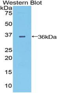
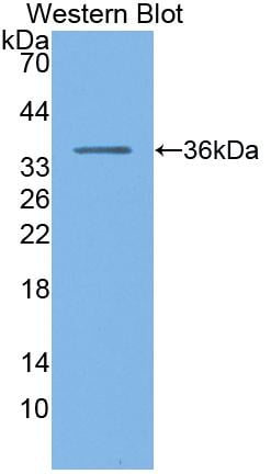
 WB (Western Blot)
(Western Blot;Sample: Recombinant MT1E, Human.)
WB (Western Blot)
(Western Blot;Sample: Recombinant MT1E, Human.)
Metallothionein 1E (MT1E), Polyclonal Antibody (Cat# AAA131341)
 IHC (Immunohiostchemistry)
(DABstainingonIHC-P.Samples:MouseTissue))
IHC (Immunohiostchemistry)
(DABstainingonIHC-P.Samples:MouseTissue))
Angiopoietin Like Protein 7 (ANGPTL7), Polyclonal Antibody (Cat# AAA131027)
 IHC (Immunohistochemistry)
(DAB staining on IHC-P. Samples: Human Tissue))
IHC (Immunohistochemistry)
(DAB staining on IHC-P. Samples: Human Tissue))
Oncomodulin (OCM), Polyclonal Antibody (Cat# AAA130958)
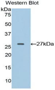
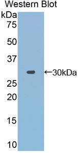
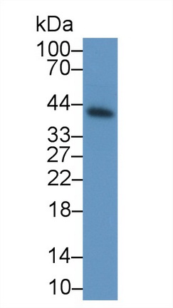
 WB (Western Blot)
(Western Blot: Sample: Recombinant HBb, Gallus.)
WB (Western Blot)
(Western Blot: Sample: Recombinant HBb, Gallus.)
Hemoglobin Beta (HBb), Polyclonal Antibody (Cat# AAA130995)

 IHC (Immunohistochemisry)
(DAB staining on IHC-P; Samples: Mouse Kidney Tissue))
IHC (Immunohistochemisry)
(DAB staining on IHC-P; Samples: Mouse Kidney Tissue))
B-Cell CLL/Lymphoma 2 Like Protein (Bcl2L), Polyclonal Antibody (Cat# AAA130888)
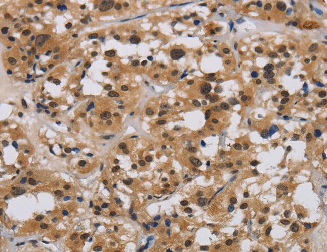
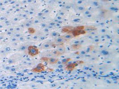
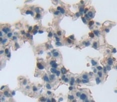
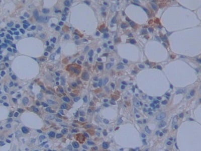
 IHC (Immunohistochemistry)
(DAB staining on IHC-P; Samples: Human Liver cancer Tissue; Primary Ab: 10ug/ml Rabbit Anti-Human NPNT Antibody Second Ab: 2ug/mL HRP-Linked Caprine Anti-Rabbit IgG Polyclonal Antibody)
IHC (Immunohistochemistry)
(DAB staining on IHC-P; Samples: Human Liver cancer Tissue; Primary Ab: 10ug/ml Rabbit Anti-Human NPNT Antibody Second Ab: 2ug/mL HRP-Linked Caprine Anti-Rabbit IgG Polyclonal Antibody)
Nephronectin (NPNT), Polyclonal Antibody (Cat# AAA131276)
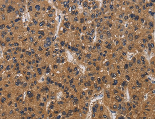
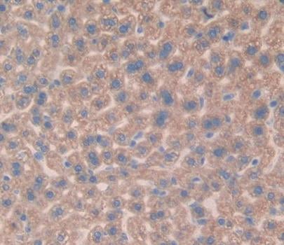
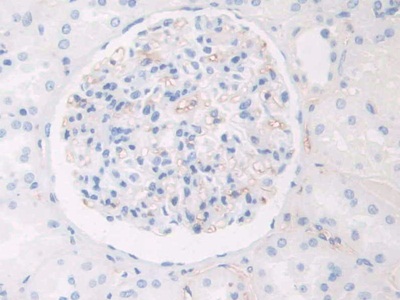
 IHC (Immunohistochemistry)
(DAB staining on IHC-P; Samples: Human Liver Tissue.)
IHC (Immunohistochemistry)
(DAB staining on IHC-P; Samples: Human Liver Tissue.)
Relaxin 2 (RLN2), Polyclonal Antibody (Cat# AAA131402)
 IHC (Immunohistochemisry)
(DAB staining on IHC-P. Samples: Mouse Tissue))
IHC (Immunohistochemisry)
(DAB staining on IHC-P. Samples: Mouse Tissue))
Vitamin D Binding Protein (DBP), Polyclonal Antibody (Cat# AAA131930)
 IHC (Immunohiostchemistry)
(DABstainingonIHC-P.Samples:MouseTissue))
IHC (Immunohiostchemistry)
(DABstainingonIHC-P.Samples:MouseTissue))
Early Growth Response Protein 2 (EGR2), Polyclonal Antibody (Cat# AAA131947)
 IHC (Immunohistochemisry)
(DAB staining on IHC-P. Samples: Mouse Tissue))
IHC (Immunohistochemisry)
(DAB staining on IHC-P. Samples: Mouse Tissue))
Interferon Alpha 4 (IFNa4), Polyclonal Antibody (Cat# AAA131677)

 IHC (Immunohiostchemistry)
(DABstainingonIHC-P.Samples:RatTissue))
IHC (Immunohiostchemistry)
(DABstainingonIHC-P.Samples:RatTissue))
Sulfite Oxidase (SUOX), Polyclonal Antibody (Cat# AAA131682)
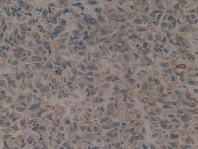
 IHC (Immunohistochemistry)
(DAB staining on IHC-P; Samples: Human Breast cancer Tissue))
IHC (Immunohistochemistry)
(DAB staining on IHC-P; Samples: Human Breast cancer Tissue))
Complement Factor B (CFB), Polyclonal Antibody (Cat# AAA131593)
 IHC (Immunohiostchemistry)
(DABstainingonIHC-P.Samples:HumanTissue))
IHC (Immunohiostchemistry)
(DABstainingonIHC-P.Samples:HumanTissue))
Aspartate Aminotransferase 2 (AST2), Polyclonal Antibody (Cat# AAA131610)
 IHC (Immunohistochemisry)
(DAB staining on IHC-P; Samples: Human Stomach Tissue))
IHC (Immunohistochemisry)
(DAB staining on IHC-P; Samples: Human Stomach Tissue))
Metastasis Associated In Colon Cancer 1 (MACC1), Polyclonal Antibody (Cat# AAA131696)
 IHC (Immunohistochemistry)
(DAB staining on IHC-P; Samples: Human Liver cancer Tissue))
IHC (Immunohistochemistry)
(DAB staining on IHC-P; Samples: Human Liver cancer Tissue))
Keratin 16 (KRT16), Polyclonal Antibody (Cat# AAA131720)

 IHC (Immunohiostchemistry)
(DAB staining on fromalin fixed paraffin-embedded testis tissue))
IHC (Immunohiostchemistry)
(DAB staining on fromalin fixed paraffin-embedded testis tissue))
Left/Right Determination Factor 2 (LEFTY2), Polyclonal Antibody (Cat# AAA131731)

 IHC (Immunohistochemisry)
(DAB staining on IHC-P; Samples: Human Breast Cancer Tissue.)
IHC (Immunohistochemisry)
(DAB staining on IHC-P; Samples: Human Breast Cancer Tissue.)
S100 Calcium Binding Protein A11 (S100A11), Polyclonal Antibody (Cat# AAA131856)


 IHC (Immunohistochemisry)
(DAB staining on IHC-P;Samples: Human Stomach Tissue;Primary Ab: 20ug/ml Rabbit Anti-Human IL1RA AntibodySecond Ab: 2ug/mL HRP-Linked Caprine Anti-Rabbit IgG Polyclonal Antibody)
IHC (Immunohistochemisry)
(DAB staining on IHC-P;Samples: Human Stomach Tissue;Primary Ab: 20ug/ml Rabbit Anti-Human IL1RA AntibodySecond Ab: 2ug/mL HRP-Linked Caprine Anti-Rabbit IgG Polyclonal Antibody)
Interleukin 1 Receptor Antagonist (IL1RA), Polyclonal Antibody (Cat# AAA131549)
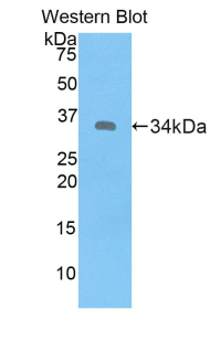
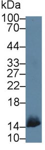
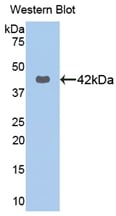
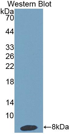
 WB (Western Blot)
(Western Blot: Sample: Recombinant protein.)
WB (Western Blot)
(Western Blot: Sample: Recombinant protein.)
Diamine Oxidase (DAO), Polyclonal Antibody (Cat# AAA132024)
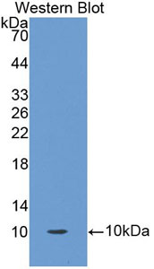
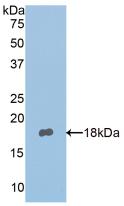
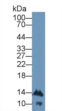
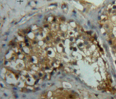
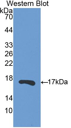
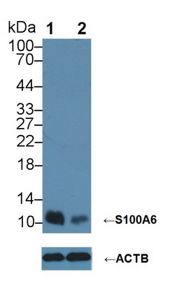 Knockout Validation
(Knockout Validation: Lane 1: Wild-type Hela cell lysate;;Lane 2: S100A6 knockout Hela cell lysate;;Predicted MW: 10kDa ;Observed MW: 10kDa;Primary Ab: 4ug/ml Rabbit Anti-Human S100A6 Antibody;Second Ab: 0.2ug/mL HRP-Linked Caprine Anti-Rabbit IgG Polyclonal Antibody;)
Knockout Validation
(Knockout Validation: Lane 1: Wild-type Hela cell lysate;;Lane 2: S100A6 knockout Hela cell lysate;;Predicted MW: 10kDa ;Observed MW: 10kDa;Primary Ab: 4ug/ml Rabbit Anti-Human S100A6 Antibody;Second Ab: 0.2ug/mL HRP-Linked Caprine Anti-Rabbit IgG Polyclonal Antibody;)
S100 Calcium Binding Protein A6 (S100A6), Polyclonal Antibody (Cat# AAA131799)
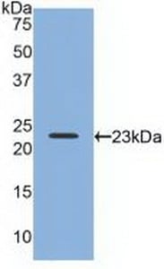
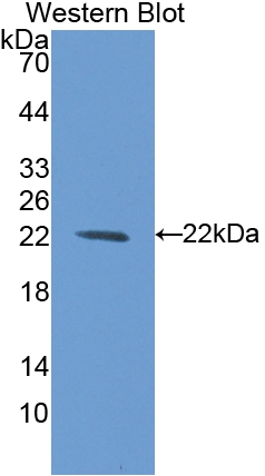
 WB (Western Blot)
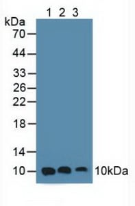
(Sample:Lane 1: Porcine Cerebrum lysate;Lane 2: Mouse Cerebrum lysate;Lane 3: Hela cell lysatePrimary Ab: 1ug/ml Rabbit Anti-Porcine CYPA AntibodySecond Ab: 0.2ug/mL HRP-Linked Caprine Anti-Rabbit IgG Polyclonal Antibody)
WB (Western Blot)
(Sample:Lane 1: Porcine Cerebrum lysate;Lane 2: Mouse Cerebrum lysate;Lane 3: Hela cell lysatePrimary Ab: 1ug/ml Rabbit Anti-Porcine CYPA AntibodySecond Ab: 0.2ug/mL HRP-Linked Caprine Anti-Rabbit IgG Polyclonal Antibody)
Cyclophilin A (CYPA), Polyclonal Antibody (Cat# AAA131840)
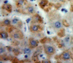
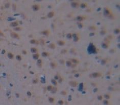
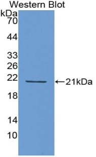
 IHC (Immunohistochemisry)
(DAB staining on IHC-P; Samples: Human Cerebrum Tissue))
IHC (Immunohistochemisry)
(DAB staining on IHC-P; Samples: Human Cerebrum Tissue))
Haptoglobin Related Protein (HPR), Polyclonal Antibody (Cat# AAA131841)
Fatty Acid Binding Protein 1, Liver (FABP1), Polyclonal Antibody (Cat# AAA132198)
 IHC (Immunohiostchemistry)
(DAB staining on fromalin fixed paraffin-embedded testis tissue))
IHC (Immunohiostchemistry)
(DAB staining on fromalin fixed paraffin-embedded testis tissue))
Bleomycin Hydrolase (BLMH), Polyclonal Antibody (Cat# AAA132084)
 IHC (Immunohiostchemistry)
(DAB staining on IHC-P. Samples: Mouse Tissue))
IHC (Immunohiostchemistry)
(DAB staining on IHC-P. Samples: Mouse Tissue))
Dispatched Homolog 1 (DISP1), Polyclonal Antibody (Cat# AAA132307)
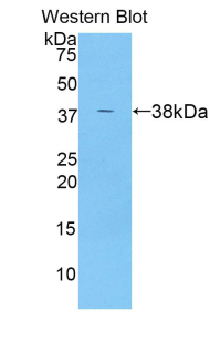
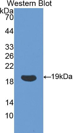
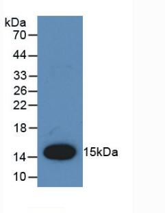
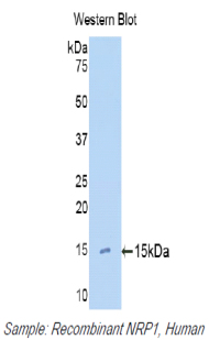 WB (Western Blot)
(Western Blot: Sample: Recombinant protein.)
WB (Western Blot)
(Western Blot: Sample: Recombinant protein.)
Neuropilin 1 (NRP1), Polyclonal Antibody (Cat# AAA132475)
Mannose Binding Lectin (MBL), Polyclonal Antibody (Cat# AAA132487)
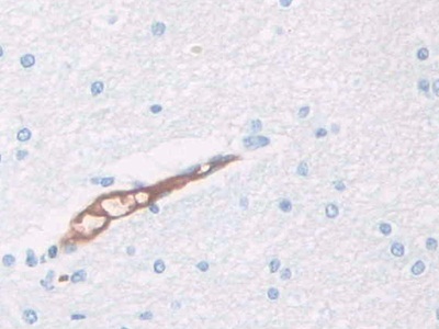
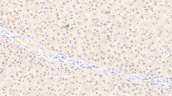
 IHC (Immunohiostchemistry)
(DAB staining on fromalin fixed paraffin-embedded liver tissue))
IHC (Immunohiostchemistry)
(DAB staining on fromalin fixed paraffin-embedded liver tissue))
Monocyte Chemotactic Protein 1 (MCP1), Polyclonal Antibody (Cat# AAA132332)
 IHC (Immunohiostchemistry)
(DAB staining on fromalin fixed paraffin-embedded Kidney tissue))
IHC (Immunohiostchemistry)
(DAB staining on fromalin fixed paraffin-embedded Kidney tissue))
Protectin (CD59), Polyclonal Antibody (Cat# AAA132563)
 IHC (Immunohistochemistry)
(DAB staining on IHC-P; Samples: Human Prostate cancer Tissue))
IHC (Immunohistochemistry)
(DAB staining on IHC-P; Samples: Human Prostate cancer Tissue))
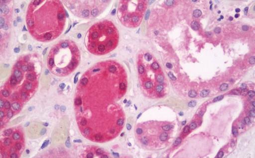
Pepsinogen C (PGC), Polyclonal Antibody (Cat# AAA132583)
 IHC (Immunohiostchemistry)
(DABstainingonIHC-P.Samples:RatTissue))
IHC (Immunohiostchemistry)
(DABstainingonIHC-P.Samples:RatTissue))
Matrix Metalloproteinase 13 (MMP13), Polyclonal Antibody (Cat# AAA132584)
 IHC (Immunohistochemisry)
(DAB staining on IHC-P. Samples: Human Tissue))
IHC (Immunohistochemisry)
(DAB staining on IHC-P. Samples: Human Tissue))
Adiponectin Receptor 2 (ADIPOR2), Polyclonal Antibody (Cat# AAA132606)
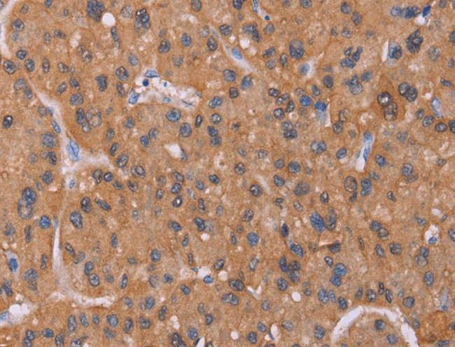
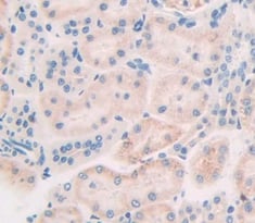
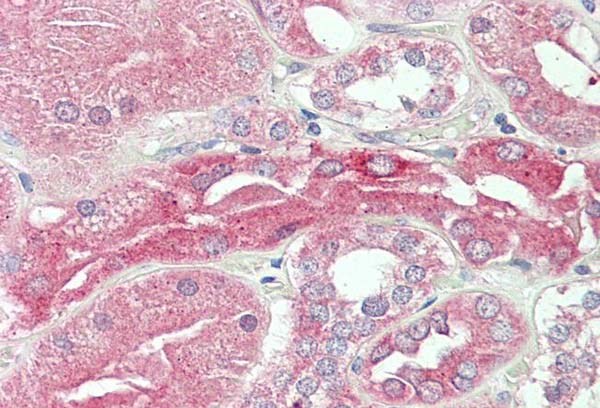
 IHC (Immunohistochemisry)
(DAB staining on IHC-P; Samples: Human Liver Tissue))
IHC (Immunohistochemisry)
(DAB staining on IHC-P; Samples: Human Liver Tissue))
Ribonuclease A13 (RNASE13), Polyclonal Antibody (Cat# AAA132184)
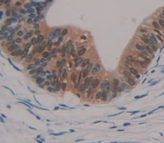
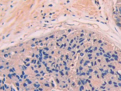
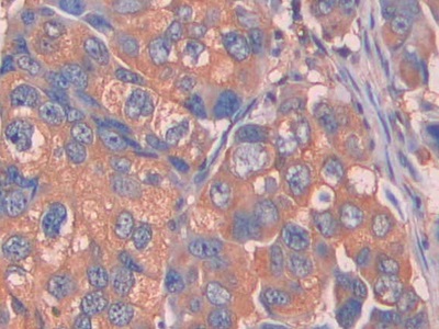
 IHC (Immunohistochemistry)
(Anti-CD40LG/CD54 antibody IHC of human tonsil. Immunohistochemistry of formalin-fixed, paraffin-embedded tissue after heat-induced antigen retrieval. Antibody concentration 5 ug/ml.)
IHC (Immunohistochemistry)
(Anti-CD40LG/CD54 antibody IHC of human tonsil. Immunohistochemistry of formalin-fixed, paraffin-embedded tissue after heat-induced antigen retrieval. Antibody concentration 5 ug/ml.)
CD40L, Polyclonal Antibody (Cat# AAA162246)
 IHC (Immunohistochemistry)
(Anti-IIL-22 antibody IHC of human tonsil. Immunohistochemistry of formalin-fixed, paraffin-embedded tissue after heat-induced antigen retrieval. Antibody concentration 5 ug/ml.)
IHC (Immunohistochemistry)
(Anti-IIL-22 antibody IHC of human tonsil. Immunohistochemistry of formalin-fixed, paraffin-embedded tissue after heat-induced antigen retrieval. Antibody concentration 5 ug/ml.)
IL22, Polyclonal Antibody (Cat# AAA162252)
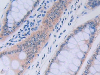
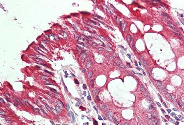
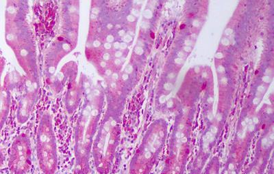 IHC (Immunohistochemistry)
(Anti-IL28A antibody IHC of human small intestine. Immunohistochemistry of formalin-fixed, paraffin-embedded tissue after heat-induced antigen retrieval. Antibody concentration 5 ug/ml.)
IHC (Immunohistochemistry)
(Anti-IL28A antibody IHC of human small intestine. Immunohistochemistry of formalin-fixed, paraffin-embedded tissue after heat-induced antigen retrieval. Antibody concentration 5 ug/ml.)
IFNL2/IL28A, Polyclonal Antibody (Cat# AAA162255)
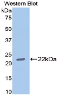
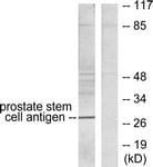 WB (Western Blot)
(Western blot of extracts from HepG2 cells, using Prostate Stem Cell Antigen Antibody. The lane on the right is treated with the synthesized peptide.)
WB (Western Blot)
(Western blot of extracts from HepG2 cells, using Prostate Stem Cell Antigen Antibody. The lane on the right is treated with the synthesized peptide.)
PSCA, Polyclonal Antibody (Cat# AAA162287)
Predicted: Mouse
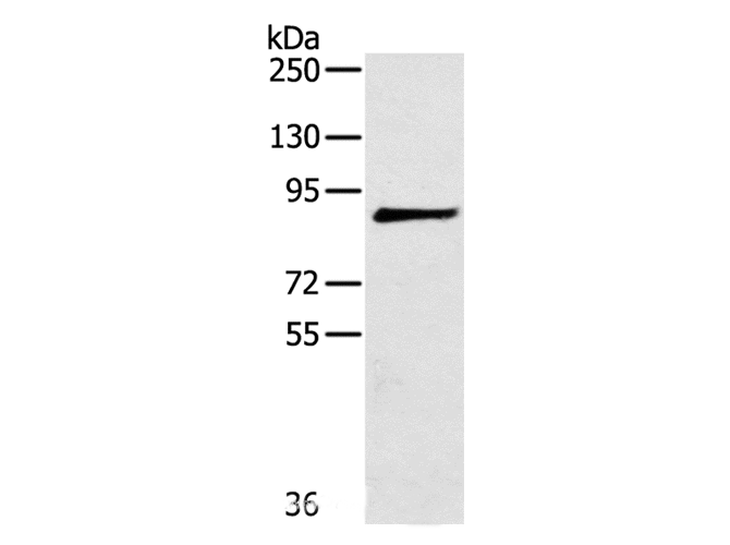
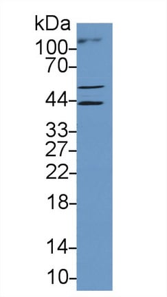
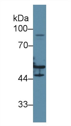
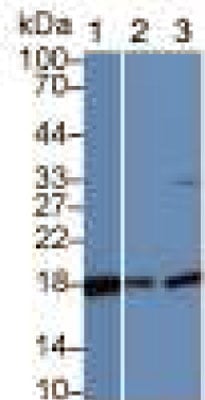
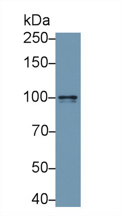
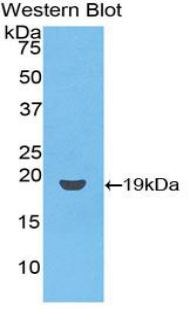
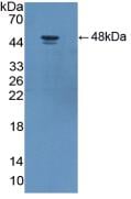
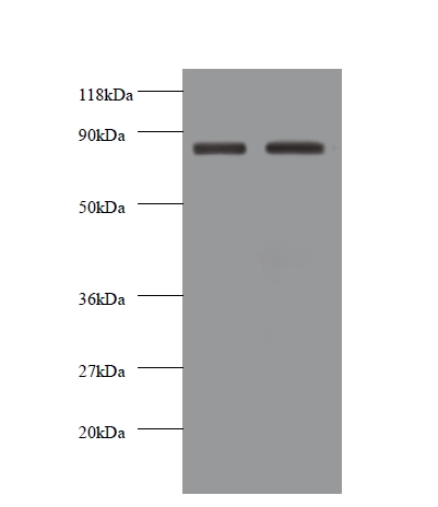 WB (Western Blot)
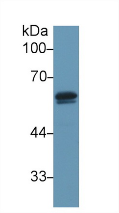
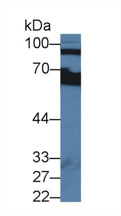
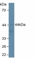
(Western blot of Cadherin-12 antibody at 2 ug/ml. Lane 1: EC109 whole cell lysate. Lane 2: 293T whole cell lysate. Secondary: Goat polyclonal to rabbit IgG at 1:10000 dilution. Predicted band size: 87 kDa. Observed band size: 87 kDa. This image was taken for the unconjugated form of this product. Other forms have not been tested.)
WB (Western Blot)
(Western blot of Cadherin-12 antibody at 2 ug/ml. Lane 1: EC109 whole cell lysate. Lane 2: 293T whole cell lysate. Secondary: Goat polyclonal to rabbit IgG at 1:10000 dilution. Predicted band size: 87 kDa. Observed band size: 87 kDa. This image was taken for the unconjugated form of this product. Other forms have not been tested.)
CDH12/Cadherin 12, Polyclonal Antibody (Cat# AAA162293)
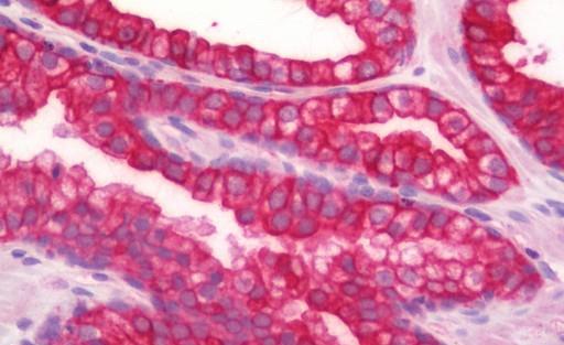
 IHC (Immunohiostchemistry)
(Human Colon: Formalin-Fixed, Paraffin-Embedded (FFPE))
IHC (Immunohiostchemistry)
(Human Colon: Formalin-Fixed, Paraffin-Embedded (FFPE))
ACHE/Acetylcholinesterase, Polyclonal Antibody (Cat# AAA162304)
Predicted: Mouse, Rat
What are Polyclonal Antibodies?
Polyclonal antibodies are antibodies that come from multiple B cell clones of a host animal. The typical hosts used for the majority of polyclonal antibody production are rabbits, goats, sheep, and donkeys. These polyclonal antibodies, once having identified their target, will bind to different epitopes located at different regions or sequences on the same protein/antigen. As a result, they are ideal at locating and binding to the target, even if the target is in very low concentrations (due to many different antibodies being able to bind to the same target molecule, which allows for significant amplification of a downstream signal).
Polyclonal antibodies are typically produced by injecting an antigen into a host animal, which causes the animal’s immune system to attack the foreign antigen by mass generating antibodies against it. After a period of time, serum is collected from the animal and purified using physicochemical fractionation, class-specific affinity purification, and/or antigen-affinity purification.
Key Uses of Polyclonal Antibodies
- Western Blotting: This method is used to find specific proteins in biological samples after separating them by size.
- Immunohistochemistry: IHC helps visualize the location of proteins in tissue sections using various staining techniques.
- ELISA: (Enzyme-Linked Immunosorbent Assay) is typically used to identify specific protein quantities in a sample. ELISAs can be either “Quantitative” or “Qualitative”.
- Flow Cytometry: technique that identifies and measures the specific protein on the surface or inside the cells in a fluid suspension.
- Immunoprecipitation: IP isolates and studies a specific protein from a complex mixture using antibodies.
Why Buy Polyclonal Antibodies from AAA Biotech?
1. Ideal for Various Applications
Our antibodies are generally going to be validated for use in multiple types of assays, including ELISA, Western Blotting, Immunohistochemistry, Immunoprecipitation, amongst others. They are ideal for a wide range of research applications.
2. Rigorous Quality Control
All of the antibodies in our catalog undergo strict quality testing to ensure specificity, sensitivity, and consistent performance. We are confident in the ability of our antibodies to provide you with accurate results.
3. Wide Assortment of Antibodies
Antibodies in are catalog can be found for both common and exotic species, and these antibodies are also available in both conjugated and recombinant forms to suit many diverse experimental needs.
4. Highly Purified
Our antibodies are available in purified forms with over 85% purity, as confirmed by SDS-PAGE. They are also available with tags such as His, Flag, GST, or MBP. We cater to customers worldwide.
FAQ
1. How are polyclonal antibodies produced?
Traditionally, polyclonal antibodies are produced by injecting an antigen into a host animal (such as a rabbit or goat), which then triggers an immune response from the host animal. The animal’s B cells produce antibodies that will recognize different parts of the injected antigen. These antibodies are then collected from the animal’s blood and purified for use.
2. How do polyclonal antibodies differ from monoclonal antibodies?
Polyclonal antibodies are a mix of antibodies that bind to different locations (epitopes) of the same antigen, while monoclonal antibodies are identical and bind to just one specific epitope. This makes polyclonal antibodies more versatile and better at detecting proteins that may be present in low quantities or in altered/modified forms.
3. How should I store polyclonal antibodies?
Polyclonal antibodies should be stored at 4°C for short-term use (up to a few weeks) and at -20°C or -80°C for long-term storage. Avoid repeated freeze-thaw cycles by dividing them into small aliquots. Always check the datasheet for specific storage instructions.