Filters
▼Clonality
▼Type
▼Reactivity
▼Gene Name
▼Isotype
▼Host
▼Application
▼Clone
▼Polyclonal Antibodies
At AAA Biotech also known as AAA Bio or AAABio, we provide a broad range of purified polyclonal antibodies (pAbs) that are able to all be browsed online through our website. Due to their high specificity and strong binding affinity, these antibodies are ideal for wide swathes of research and experimental applications.
Our polyclonal antibodies can easily support your work, whether you use them for Western Blotting, Immunocytochemistry (with or without Immunofluorescence used in conjunction), Immunohistochemistry, Immunoprecipitation, and ELISA tests. We highly encourage you to browse our range of pAbs and choose the one that best suits your experimental model.
Viewing 7750-7800 of 96805 product results
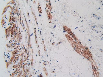
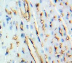 IHC (Immunohiostchemistry)
(DAB staining on fromalin fixed paraffin-embedded Heart tissue))
IHC (Immunohiostchemistry)
(DAB staining on fromalin fixed paraffin-embedded Heart tissue))
Brain Natriuretic Peptide (BNP), Polyclonal Antibody (Cat# AAA133499)
 IHC (Immunohiostchemistry)
(DABstainingonIHC-P.Samples:RatTissue))
IHC (Immunohiostchemistry)
(DABstainingonIHC-P.Samples:RatTissue))
Keratin 10 (KRT10), Polyclonal Antibody (Cat# AAA133759)
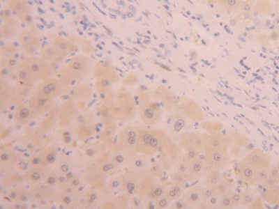
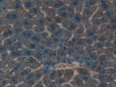
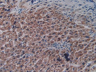
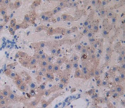
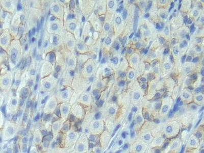
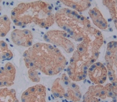
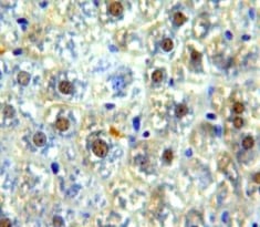
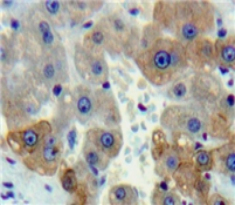
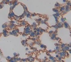
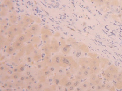 IHC (Immunohistochemistry)
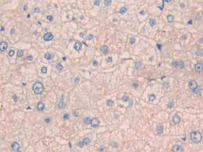
(DAB staining on IHC-P; Samples: Human Liver Tissue))
IHC (Immunohistochemistry)
(DAB staining on IHC-P; Samples: Human Liver Tissue))
Alanine Aminotransferase (ALT), Polyclonal Antibody (Cat# AAA133768)
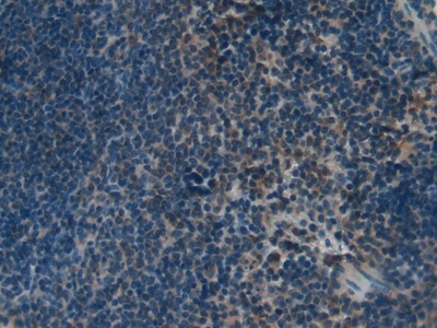
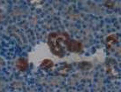
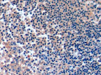
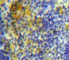
 IHC (Immunohiostchemistry)
(DAB staining on fromalin fixed paraffin-embedded spleen tissue))
IHC (Immunohiostchemistry)
(DAB staining on fromalin fixed paraffin-embedded spleen tissue))
Perforin 1 (PRF1), Polyclonal Antibody (Cat# AAA133771)
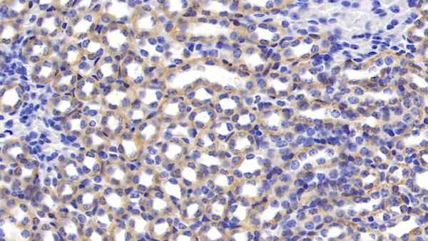
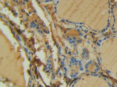
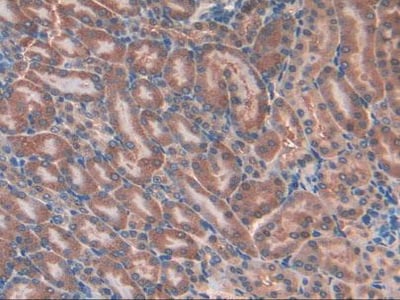
 IHC (Immunohistochemistry)
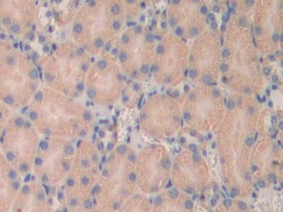
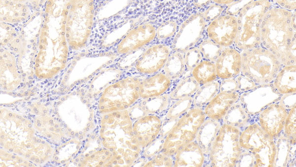
(DAB staining on IHC-P;Samples: Mouse Kidney Tissue;Primary Ab: 10ug/ml Rabbit Anti-Mouse KLK4 AntibodySecond Ab: 2ug/mL HRP-Linked Caprine Anti-Rabbit IgG Polyclonal Antibody)
IHC (Immunohistochemistry)
(DAB staining on IHC-P;Samples: Mouse Kidney Tissue;Primary Ab: 10ug/ml Rabbit Anti-Mouse KLK4 AntibodySecond Ab: 2ug/mL HRP-Linked Caprine Anti-Rabbit IgG Polyclonal Antibody)
Kallikrein 4 (KLK4), Polyclonal Antibody (Cat# AAA133772)
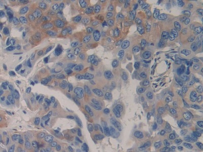
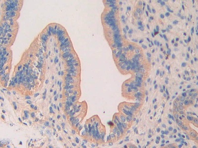
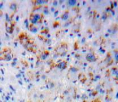
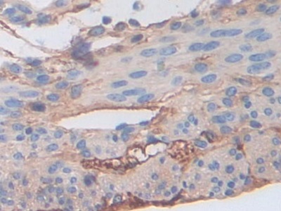
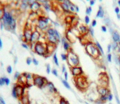
 IHC (Immunohistochemistry)
(DAB staining on IHC-P; Samples: Human Prostate Gland Cancer Tissue.)
IHC (Immunohistochemistry)
(DAB staining on IHC-P; Samples: Human Prostate Gland Cancer Tissue.)
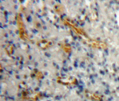
Vascular Endothelial Growth Factor Receptor 2 (VEGFR2), Polyclonal Antibody (Cat# AAA133774)
 IHC (Immunohistochemistry)
(DAB staining on IHC-P; Samples: Mouse Vas Deferens Tissue)
IHC (Immunohistochemistry)
(DAB staining on IHC-P; Samples: Mouse Vas Deferens Tissue)
Clusterin (CLU), Polyclonal Antibody (Cat# AAA133775)
 IHC (Immunohiostchemistry)
(DAB staining on fromalin fixed paraffin-embedded kidney tissue))
IHC (Immunohiostchemistry)
(DAB staining on fromalin fixed paraffin-embedded kidney tissue))
Anterior Gradient Protein 2 (AGR2), Polyclonal Antibody (Cat# AAA133777)
 IHC (Immunohiostchemistry)
(DABstainingonIHC-P.Samples:RatTissue))
IHC (Immunohiostchemistry)
(DABstainingonIHC-P.Samples:RatTissue))
A Disintegrin And Metalloproteinase With Thrombospondin 7 (ADAMTS7), Polyclonal Antibody (Cat# AAA133785)
 IHC (Immunohiostchemistry)
(DABstainingonIHC-P.Samples:MouseTissue))
IHC (Immunohiostchemistry)
(DABstainingonIHC-P.Samples:MouseTissue))
Tubulin Beta 1 (TUBb1), Polyclonal Antibody (Cat# AAA133790)
 IHC (Immunohiostchemistry)
(DABstainingonIHC-P.Samples:MouseTissue))
IHC (Immunohiostchemistry)
(DABstainingonIHC-P.Samples:MouseTissue))
Neuropilin 1 (NRP1), Polyclonal Antibody (Cat# AAA133792)
Interleukin 8 Receptor Beta (IL8Rb), Polyclonal Antibody (Cat# AAA133797)
 IHC (Immunohistochemistry)
(DAB staining on IHC-P; Samples: Mouse Kidney Tissue))
IHC (Immunohistochemistry)
(DAB staining on IHC-P; Samples: Mouse Kidney Tissue))
Laminin Alpha 4 (LAMa4), Polyclonal Antibody (Cat# AAA133798)

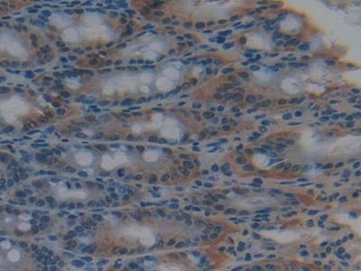
 IHC (Immunohistochemisry)
(DAB staining on IHC-P; Samples: Rat Stomach Tissue))
IHC (Immunohistochemisry)
(DAB staining on IHC-P; Samples: Rat Stomach Tissue))
Cadherin, Epithelial (CDHE), Polyclonal Antibody (Cat# AAA133799)

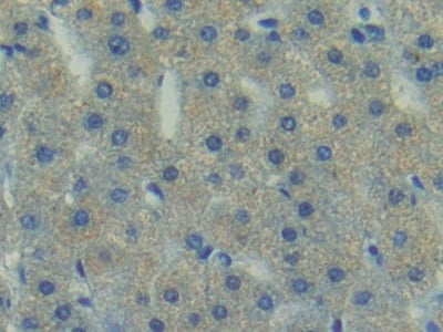
 IHC (Immunohiostchemistry)
(DAB staining on fromalin fixed paraffin-embedded Liver tissue))
IHC (Immunohiostchemistry)
(DAB staining on fromalin fixed paraffin-embedded Liver tissue))
Interleukin 7 Receptor (IL7R), Polyclonal Antibody (Cat# AAA133803)
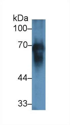
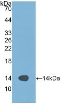
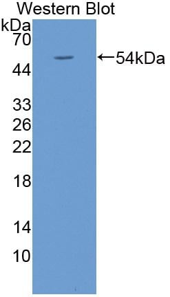
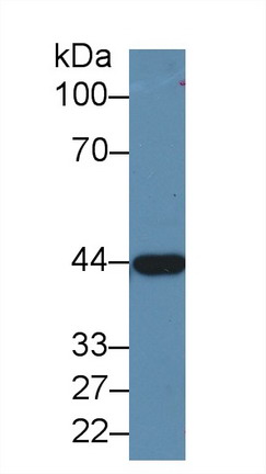
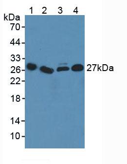
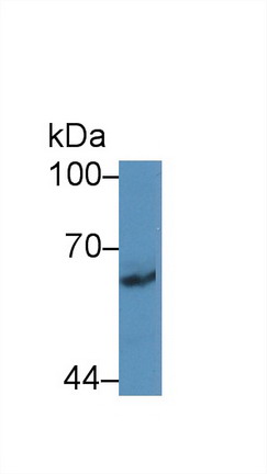
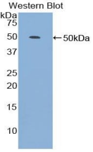
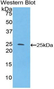
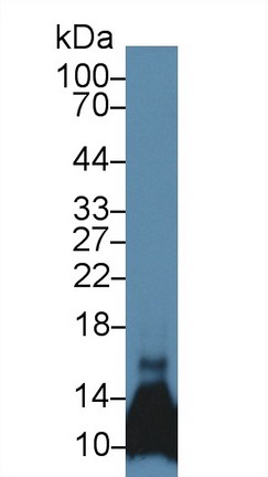
 WB (Western Blot)
(Western Blot: Sample: Equine Plasma Primary Ab: 1ug/ml Rabbit Anti-Equine)
WB (Western Blot)
(Western Blot: Sample: Equine Plasma Primary Ab: 1ug/ml Rabbit Anti-Equine)
Albumin (ALB), Polyclonal Antibody (Cat# AAA133804)
 IHC (Immunohiostchemistry)
(DABstainingonIHC-P.Samples:HumanTissue))
IHC (Immunohiostchemistry)
(DABstainingonIHC-P.Samples:HumanTissue))
Urocortin 3 (UCN3), Polyclonal Antibody (Cat# AAA133809)
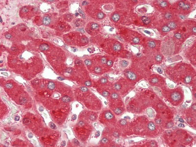 IHC (Immunohistochemisry)
(HE staining on IHC-P; Samples: Human Liver Tissue.)
IHC (Immunohistochemisry)
(HE staining on IHC-P; Samples: Human Liver Tissue.)
Ceruloplasmin (CP), Polyclonal Antibody (Cat# AAA133816)
 IHC (Immunohiostchemistry)
(DABstainingonIHC-P.Samples:MouseTissue))
IHC (Immunohiostchemistry)
(DABstainingonIHC-P.Samples:MouseTissue))
Hyaluronan Binding Protein 1 (HABP1), Polyclonal Antibody (Cat# AAA133817)
 IHC (Immunohistochemisry)
(DAB staining on fromalin fixed paraffin- embedded liver tissue))
IHC (Immunohistochemisry)
(DAB staining on fromalin fixed paraffin- embedded liver tissue))
Carboxypeptidase N1 (CPN1), Polyclonal Antibody (Cat# AAA133824)
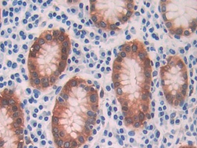
 IHC (Immunohistochemisry)
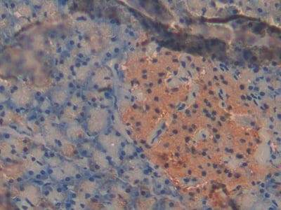
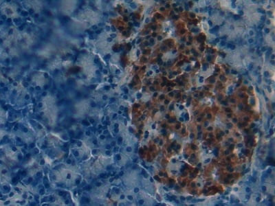
(DAB staining on IHC-P; Samples: Mouse Pancreas Tissue))
IHC (Immunohistochemisry)
(DAB staining on IHC-P; Samples: Mouse Pancreas Tissue))
Pyruvate Dehydrogenase Kinase Isozyme 2 (PDK2), Polyclonal Antibody (Cat# AAA133827)
 IHC (Immunohistochemisry)
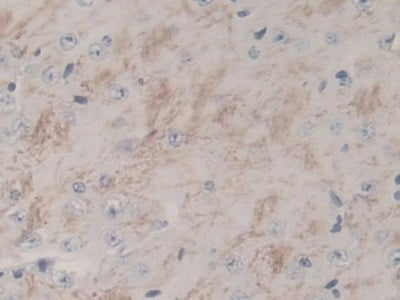
(DAB staining on IHC-P. Samples: Rat Tissue))
IHC (Immunohistochemisry)
(DAB staining on IHC-P. Samples: Rat Tissue))
Nitric Oxide Synthase 1, Neuronal (NOS1), Polyclonal Antibody (Cat# AAA133829)
 IHC (Immunohiostchemistry)
(DABstainingonIHC-P.Samples:RatTissue))
IHC (Immunohiostchemistry)
(DABstainingonIHC-P.Samples:RatTissue))
Neuropilin 1 (NRP1), Polyclonal Antibody (Cat# AAA133830)
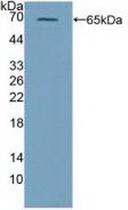
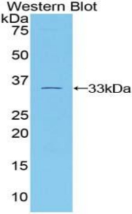
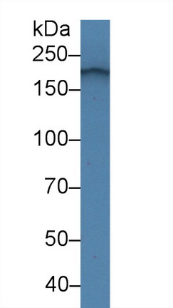
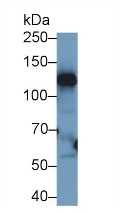
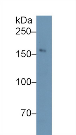
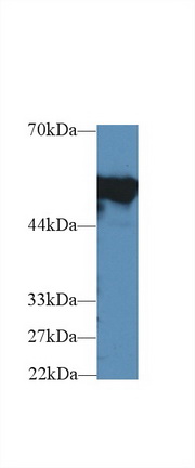
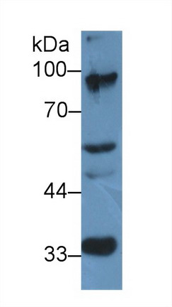
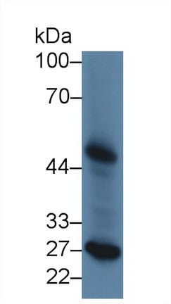
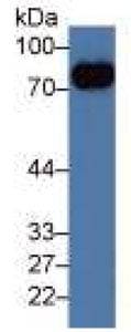
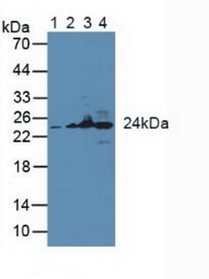
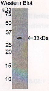
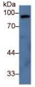 WB (Western Blot)
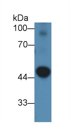
(Western Blot;Sample: Porcine Liver lysatePrimary Ab: 2ug/ml Rabbit Anti-Porcine C7 AntibodySecond Ab: 0.2ug/ml HRP-Linked Caprine Anti-Rabbit IgG Polyclonal Antibody)
WB (Western Blot)
(Western Blot;Sample: Porcine Liver lysatePrimary Ab: 2ug/ml Rabbit Anti-Porcine C7 AntibodySecond Ab: 0.2ug/ml HRP-Linked Caprine Anti-Rabbit IgG Polyclonal Antibody)
Complement Component 7 (C7), Polyclonal Antibody (Cat# AAA133831)
 IHC (Immunohiostchemistry)
(DABstainingonIHC-P.Samples:HumanTissue))
IHC (Immunohiostchemistry)
(DABstainingonIHC-P.Samples:HumanTissue))
Platelet Derived Growth Factor C (PDGFC), Polyclonal Antibody (Cat# AAA133389)
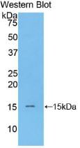
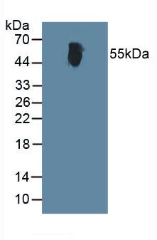
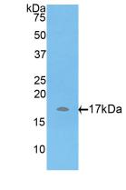
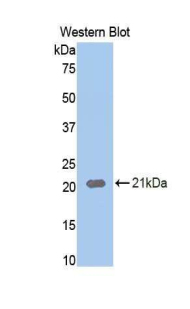
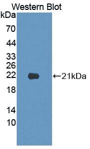
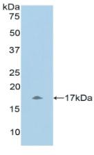
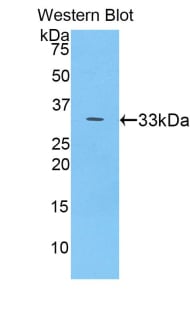
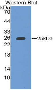
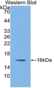
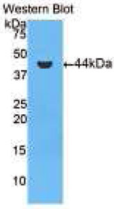
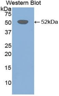
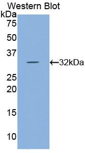
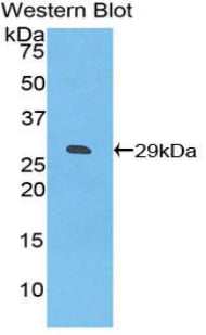
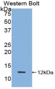
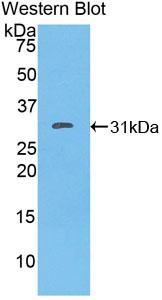
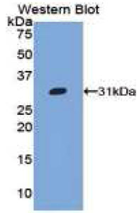
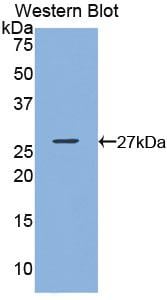
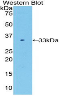
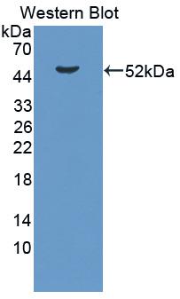 WB (Western Blot)
((Figure. Western Blot; Sample: Recombinant protein.))
WB (Western Blot)
((Figure. Western Blot; Sample: Recombinant protein.))
Lipopolysaccharide Binding Protein (LBP), Polyclonal Antibody (Cat# AAA133404)
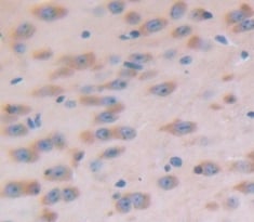
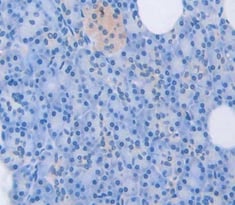
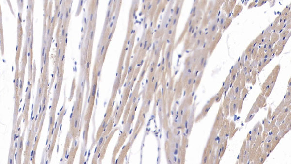
 IHC (Immunohiostchemistry)
(DABstainingonIHC-P.Samples:HumanTissue))
IHC (Immunohiostchemistry)
(DABstainingonIHC-P.Samples:HumanTissue))
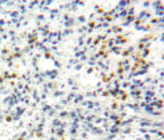
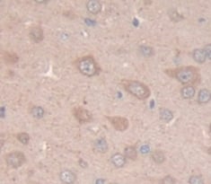
Paraneoplastic Antigen MA2 (PNMA2), Polyclonal Antibody (Cat# AAA133375)
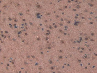
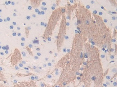 IHC (Immunohistochemistry)
(DAB staining on IHC-P; Samples: Mouse Cerebrum Tissue))
IHC (Immunohistochemistry)
(DAB staining on IHC-P; Samples: Mouse Cerebrum Tissue))
Fibrinogen Beta (FGb), Polyclonal Antibody (Cat# AAA134946)

 IHC (Immunohistochemistry)
(DAB staining on IHC-P; Samples: Human Stomach Tissue))
IHC (Immunohistochemistry)
(DAB staining on IHC-P; Samples: Human Stomach Tissue))
Alcohol Dehydrogenase 1 (ADH1), Polyclonal Antibody (Cat# AAA134947)
 IHC (Immunohistochemisry)
(DAB staining on fromalin fixed paraffin-embedded Liver tissue))
IHC (Immunohistochemisry)
(DAB staining on fromalin fixed paraffin-embedded Liver tissue))
Nucleoporin 98kDa (NUP98), Polyclonal Antibody (Cat# AAA134948)
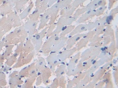
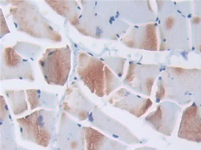 IHC (Immunohistochemistry)
(DAB staining on IHC-P; Samples: Rat Skeletal muscle Tissue))
IHC (Immunohistochemistry)
(DAB staining on IHC-P; Samples: Rat Skeletal muscle Tissue))
Elongin A (ELOA), Polyclonal Antibody (Cat# AAA134949)
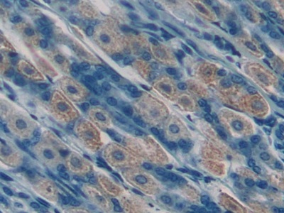
 IHC (Immunohistochemistry)
(DAB staining on IHC-P; Samples: Mouse Stomach Tissue.))
IHC (Immunohistochemistry)
(DAB staining on IHC-P; Samples: Mouse Stomach Tissue.))
Phospholipase A2, Calcium Independent (iPLA2), Polyclonal Antibody (Cat# AAA134952)

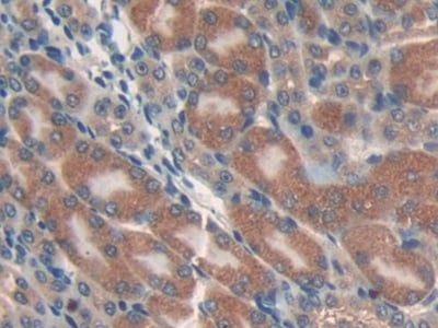
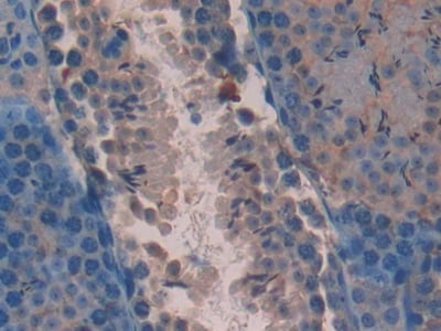 IHC (Immunohistochemisry)
(DAB staining on IHC-P; Samples: Mouse Testis Tissue))
IHC (Immunohistochemisry)
(DAB staining on IHC-P; Samples: Mouse Testis Tissue))
Apoptosis Associated Tyrosine Kinase (AATK), Polyclonal Antibody (Cat# AAA134961)
 IHC (Immunohiostchemistry)
(DAB staining on fromalin fixed paraffin-embedded Stomach tissue))
IHC (Immunohiostchemistry)
(DAB staining on fromalin fixed paraffin-embedded Stomach tissue))
Killer Cell Lectin Like Receptor Subfamily C, Member 2 (KLRC2), Polyclonal Antibody (Cat# AAA134966)

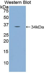
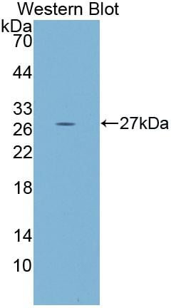
 Application Data
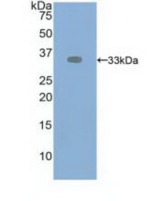
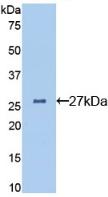
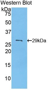
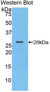
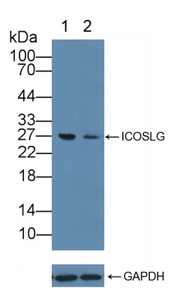
(Knockout Validation: Lane 1: Wild-type Jurkat cell lysate; Lane 2: ICOSLG knockout Jurkat cell lysate; Predicted MW: 33kd Observed MW: 27kd Primary Ab: 2ug/ml Rabbit Anti-Human ICOSLG Antibody Second Ab: 0.2ug/mL HRP-Linked Caprine Anti-Rabbit IgG Polyclonal Antibody)
Application Data
(Knockout Validation: Lane 1: Wild-type Jurkat cell lysate; Lane 2: ICOSLG knockout Jurkat cell lysate; Predicted MW: 33kd Observed MW: 27kd Primary Ab: 2ug/ml Rabbit Anti-Human ICOSLG Antibody Second Ab: 0.2ug/mL HRP-Linked Caprine Anti-Rabbit IgG Polyclonal Antibody)
Inducible T-Cell Co Stimulator Ligand (ICOSLG), Polyclonal Antibody (Cat# AAA134970)
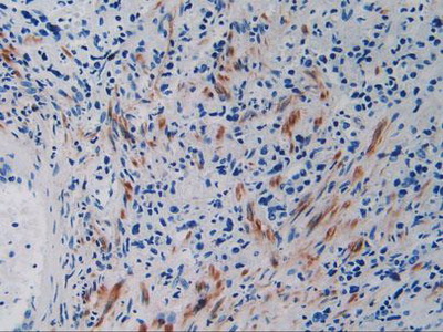
 IHC (Immunohistochemisry)
(DAB staining on IHC-P. Samples: Human Tissue))
IHC (Immunohistochemisry)
(DAB staining on IHC-P. Samples: Human Tissue))
Neurofilament 3 (NEF3), Polyclonal Antibody (Cat# AAA134971)
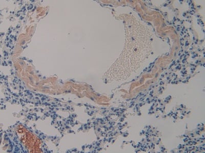
 IHC (Immunohiostchemistry)
(DAB staining on fromalin fixed paraffin-embedded Liver tissue))
IHC (Immunohiostchemistry)
(DAB staining on fromalin fixed paraffin-embedded Liver tissue))
Peptide YY (PYY), Polyclonal Antibody (Cat# AAA134973)
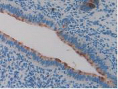
 IHC (Immunohistochemisry)
(DAB staining on fromalin fixed paraffin-embedded Liver tissue))
IHC (Immunohistochemisry)
(DAB staining on fromalin fixed paraffin-embedded Liver tissue))
Protocadherin Beta 2 (PCDHb2), Polyclonal Antibody (Cat# AAA134976)
Periostin (POSTN), Polyclonal Antibody (Cat# AAA134981)
 IHC (Immunohistochemisry)
(DAB staining on fromalin fixed paraffin-embedded Spleen tissue))
IHC (Immunohistochemisry)
(DAB staining on fromalin fixed paraffin-embedded Spleen tissue))
Phospholipase A2, Lipoprotein Associated (LpPLA2), Polyclonal Antibody (Cat# AAA134983)
 IHC (Immunohiostchemistry)
(DABstainingonIHC-P.Samples:MouseTissue))
IHC (Immunohiostchemistry)
(DABstainingonIHC-P.Samples:MouseTissue))
Sprouty Homolog 3 (SPRY3), Polyclonal Antibody (Cat# AAA134984)
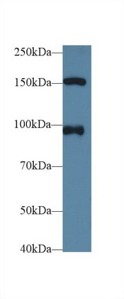
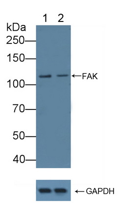 Application Data
(Knockout Validation: Lane 1: Wild-type Jurkat cell lysate; Lane 2: FAK knockout Jurkat cell lysate; Predicted MW: 39,48,63,100,120kd Observed MW: 120kd Primary Ab: 1ug/ml Rabbit Anti-Human FAK Antibody Second Ab: 0.2ug/mL HRP-Linked Caprine Anti-Rabbit IgG Polyclonal Antibody)
Application Data
(Knockout Validation: Lane 1: Wild-type Jurkat cell lysate; Lane 2: FAK knockout Jurkat cell lysate; Predicted MW: 39,48,63,100,120kd Observed MW: 120kd Primary Ab: 1ug/ml Rabbit Anti-Human FAK Antibody Second Ab: 0.2ug/mL HRP-Linked Caprine Anti-Rabbit IgG Polyclonal Antibody)
Focal Adhesion Kinase (FAK), Polyclonal Antibody (Cat# AAA134986)
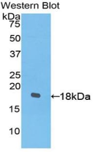
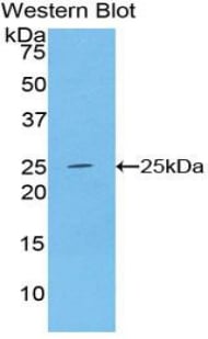
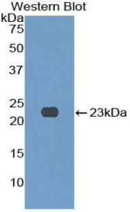
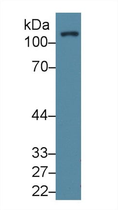
 WB (Western Blot)
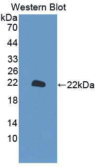
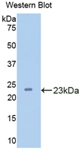
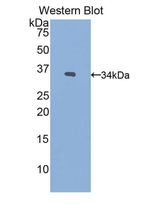
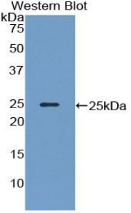
((Figure. Western Blot; Sample: Recombinant protein.))
WB (Western Blot)
((Figure. Western Blot; Sample: Recombinant protein.))
Protein Tyrosine Phosphatase Receptor Type B (PTPRB), Polyclonal Antibody (Cat# AAA134990)
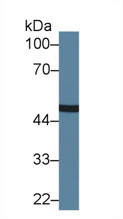
 WB (Western Blot)
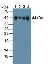
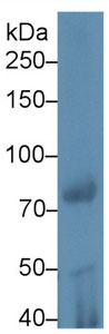
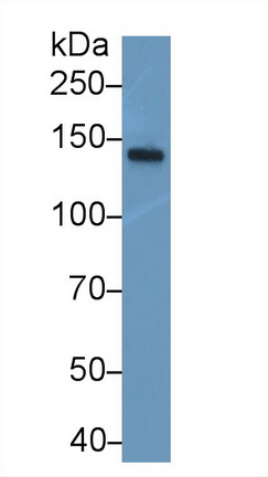
(Sample: Mouse Lymphocyte lysate; Primary Ab: 2ug/ml Rabbit Anti-Mouse GP5 Antibody Second Ab: 0.2ug/mL HRP-Linked Caprine Anti-Rabbit IgG Polyclonal Antibody)
WB (Western Blot)
(Sample: Mouse Lymphocyte lysate; Primary Ab: 2ug/ml Rabbit Anti-Mouse GP5 Antibody Second Ab: 0.2ug/mL HRP-Linked Caprine Anti-Rabbit IgG Polyclonal Antibody)
Glycoprotein V, Platelet (GP5), Polyclonal Antibody (Cat# AAA134998)
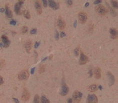
 IHC (Immunohistochemistry)
(DAB staining on IHC-P; Samples: Mouse Brain Tissue)
IHC (Immunohistochemistry)
(DAB staining on IHC-P; Samples: Mouse Brain Tissue)
Glutathione S Transferase Kappa 1 (GSTk1), Polyclonal Antibody (Cat# AAA135000)

 IHC (Immunohistochemistry)
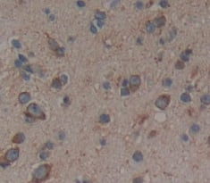
(DAB staining on IHC-P; Samples: Mouse Brain Tissue))
IHC (Immunohistochemistry)
(DAB staining on IHC-P; Samples: Mouse Brain Tissue))
Macrophage Inflammatory Protein Related Protein 1 (MRP1), Polyclonal Antibody (Cat# AAA134883)
Fibrinogen Like Protein 1 (FGL1), Polyclonal Antibody (Cat# AAA134884)
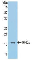
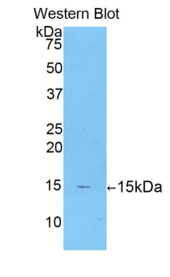
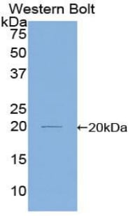
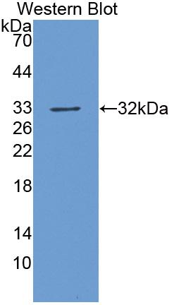
 WB (Western Blot)
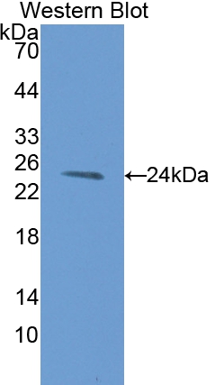
(Used in Western Blot, Sample: Recombinant IFNa/bR1, Rat)
WB (Western Blot)
(Used in Western Blot, Sample: Recombinant IFNa/bR1, Rat)
Interferon Alpha/Beta Receptor 1 (IFNa/bR1), Polyclonal Antibody (Cat# AAA134885)
 IHC (Immunohiostchemistry)
(DAB staining on fromalin fixed paraffin-embedded Liver tissue))
IHC (Immunohiostchemistry)
(DAB staining on fromalin fixed paraffin-embedded Liver tissue))
Fucosidase Alpha L1, Tissue (FUCa1), Polyclonal Antibody (Cat# AAA134889)
Runt Related Transcription Factor 2 (RUNX2), Polyclonal Antibody (Cat# AAA134890)
What are Polyclonal Antibodies?
Polyclonal antibodies are antibodies that come from multiple B cell clones of a host animal. The typical hosts used for the majority of polyclonal antibody production are rabbits, goats, sheep, and donkeys. These polyclonal antibodies, once having identified their target, will bind to different epitopes located at different regions or sequences on the same protein/antigen. As a result, they are ideal at locating and binding to the target, even if the target is in very low concentrations (due to many different antibodies being able to bind to the same target molecule, which allows for significant amplification of a downstream signal).
Polyclonal antibodies are typically produced by injecting an antigen into a host animal, which causes the animal’s immune system to attack the foreign antigen by mass generating antibodies against it. After a period of time, serum is collected from the animal and purified using physicochemical fractionation, class-specific affinity purification, and/or antigen-affinity purification.
Key Uses of Polyclonal Antibodies
- Western Blotting: This method is used to find specific proteins in biological samples after separating them by size.
- Immunohistochemistry: IHC helps visualize the location of proteins in tissue sections using various staining techniques.
- ELISA: (Enzyme-Linked Immunosorbent Assay) is typically used to identify specific protein quantities in a sample. ELISAs can be either “Quantitative” or “Qualitative”.
- Flow Cytometry: technique that identifies and measures the specific protein on the surface or inside the cells in a fluid suspension.
- Immunoprecipitation: IP isolates and studies a specific protein from a complex mixture using antibodies.
Why Buy Polyclonal Antibodies from AAA Biotech?
1. Ideal for Various Applications
Our antibodies are generally going to be validated for use in multiple types of assays, including ELISA, Western Blotting, Immunohistochemistry, Immunoprecipitation, amongst others. They are ideal for a wide range of research applications.
2. Rigorous Quality Control
All of the antibodies in our catalog undergo strict quality testing to ensure specificity, sensitivity, and consistent performance. We are confident in the ability of our antibodies to provide you with accurate results.
3. Wide Assortment of Antibodies
Antibodies in are catalog can be found for both common and exotic species, and these antibodies are also available in both conjugated and recombinant forms to suit many diverse experimental needs.
4. Highly Purified
Our antibodies are available in purified forms with over 85% purity, as confirmed by SDS-PAGE. They are also available with tags such as His, Flag, GST, or MBP. We cater to customers worldwide.
FAQ
1. How are polyclonal antibodies produced?
Traditionally, polyclonal antibodies are produced by injecting an antigen into a host animal (such as a rabbit or goat), which then triggers an immune response from the host animal. The animal’s B cells produce antibodies that will recognize different parts of the injected antigen. These antibodies are then collected from the animal’s blood and purified for use.
2. How do polyclonal antibodies differ from monoclonal antibodies?
Polyclonal antibodies are a mix of antibodies that bind to different locations (epitopes) of the same antigen, while monoclonal antibodies are identical and bind to just one specific epitope. This makes polyclonal antibodies more versatile and better at detecting proteins that may be present in low quantities or in altered/modified forms.
3. How should I store polyclonal antibodies?
Polyclonal antibodies should be stored at 4°C for short-term use (up to a few weeks) and at -20°C or -80°C for long-term storage. Avoid repeated freeze-thaw cycles by dividing them into small aliquots. Always check the datasheet for specific storage instructions.