Filters
▼Clonality
▼Type
▼Reactivity
▼Gene Name
▼Isotype
▼Host
▼Application
▼Clone
▼Polyclonal Antibodies
At AAA Biotech also known as AAA Bio or AAABio, we provide a broad range of purified polyclonal antibodies (pAbs) that are able to all be browsed online through our website. Due to their high specificity and strong binding affinity, these antibodies are ideal for wide swathes of research and experimental applications.
Our polyclonal antibodies can easily support your work, whether you use them for Western Blotting, Immunocytochemistry (with or without Immunofluorescence used in conjunction), Immunohistochemistry, Immunoprecipitation, and ELISA tests. We highly encourage you to browse our range of pAbs and choose the one that best suits your experimental model.
Viewing 7650-7700 of 96805 product results
 FCM/FACS (Flow Cytometry)
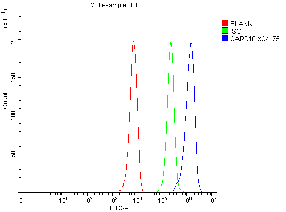
(Figure 5. Flow Cytometry analysis of HepG2 cells using anti-CARD10 antibody (AAA128228).Overlay histogram showing HepG2 cells stained with AAA128228 (Blue line). To facilitate intracellular staining, cells were fixed with 4% paraformaldehyde and permeabilized with permeabilization buffer. The cells were blocked with 10% normal goat serum. And then incubated with rabbit anti-CARD10 Antibody (AAA128228, 1ug/1x106 cells) for 30 min at 20 degree C. DyLight®488 conjugated goat anti-rabbit IgG was used as secondary antibody for 30 minutes at 20 degree C. Isotype control antibody (Green line) was rabbit IgG (1ug/1x106) used under the same conditions. Unlabelled sample without incubation with primary antibody and secondary antibody (Red line) was used as a blank control.)
FCM/FACS (Flow Cytometry)
(Figure 5. Flow Cytometry analysis of HepG2 cells using anti-CARD10 antibody (AAA128228).Overlay histogram showing HepG2 cells stained with AAA128228 (Blue line). To facilitate intracellular staining, cells were fixed with 4% paraformaldehyde and permeabilized with permeabilization buffer. The cells were blocked with 10% normal goat serum. And then incubated with rabbit anti-CARD10 Antibody (AAA128228, 1ug/1x106 cells) for 30 min at 20 degree C. DyLight®488 conjugated goat anti-rabbit IgG was used as secondary antibody for 30 minutes at 20 degree C. Isotype control antibody (Green line) was rabbit IgG (1ug/1x106) used under the same conditions. Unlabelled sample without incubation with primary antibody and secondary antibody (Red line) was used as a blank control.)
CARD10, Polyclonal Antibody (Cat# AAA128228)
 FCM/FACS (Flow Cytometry)
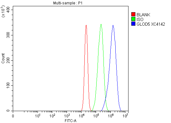
(Figure 4. Flow Cytometry analysis of RT4 cells using anti-GLOD5 antibody (AAA128230).Overlay histogram showing RT4 cells stained with AAA128230 (Blue line). To facilitate intracellular staining, cells were fixed with 4% paraformaldehyde and permeabilized with permeabilization buffer. The cells were blocked with 10% normal goat serum. And then incubated with rabbit anti-GLOD5 Antibody (AAA128230, 1ug/1x106 cells) for 30 min at 20 degree C. DyLight®488 conjugated goat anti-rabbit IgG was used as secondary antibody for 30 minutes at 20 degree C. Isotype control antibody (Green line) was rabbit IgG (1ug/1x106) used under the same conditions. Unlabelled sample without incubation with primary antibody and secondary antibody (Red line) was used as a blank control.)
FCM/FACS (Flow Cytometry)
(Figure 4. Flow Cytometry analysis of RT4 cells using anti-GLOD5 antibody (AAA128230).Overlay histogram showing RT4 cells stained with AAA128230 (Blue line). To facilitate intracellular staining, cells were fixed with 4% paraformaldehyde and permeabilized with permeabilization buffer. The cells were blocked with 10% normal goat serum. And then incubated with rabbit anti-GLOD5 Antibody (AAA128230, 1ug/1x106 cells) for 30 min at 20 degree C. DyLight®488 conjugated goat anti-rabbit IgG was used as secondary antibody for 30 minutes at 20 degree C. Isotype control antibody (Green line) was rabbit IgG (1ug/1x106) used under the same conditions. Unlabelled sample without incubation with primary antibody and secondary antibody (Red line) was used as a blank control.)
GLOD5, Polyclonal Antibody (Cat# AAA128230)
 FCM/FACS (Flow Cytometry)
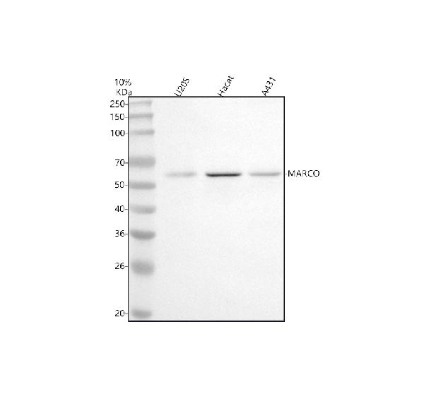
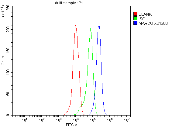
(Figure 2. Flow Cytometry analysis of THP-1 cells using anti-MARCO antibody (AAA128237).Overlay histogram showing THP-1 cells stained with AAA128237 (Blue line). The cells were fixed with 4% paraformaldehyde and blocked with 10% normal goat serum. And then incubated with rabbit anti-MARCO Antibody (AAA128237, 1ug/1x106 cells) for 30 min at 20 degree C. DyLight®488 conjugated goat anti-rabbit IgG was used as secondary antibody for 30 minutes at 20 degree C. Isotype control antibody (Green line) was rabbit IgG (1ug/1x106) used under the same conditions. Unlabelled sample without incubation with primary antibody and secondary antibody (Red line) was used as a blank control.)
FCM/FACS (Flow Cytometry)
(Figure 2. Flow Cytometry analysis of THP-1 cells using anti-MARCO antibody (AAA128237).Overlay histogram showing THP-1 cells stained with AAA128237 (Blue line). The cells were fixed with 4% paraformaldehyde and blocked with 10% normal goat serum. And then incubated with rabbit anti-MARCO Antibody (AAA128237, 1ug/1x106 cells) for 30 min at 20 degree C. DyLight®488 conjugated goat anti-rabbit IgG was used as secondary antibody for 30 minutes at 20 degree C. Isotype control antibody (Green line) was rabbit IgG (1ug/1x106) used under the same conditions. Unlabelled sample without incubation with primary antibody and secondary antibody (Red line) was used as a blank control.)
MARCO, Polyclonal Antibody (Cat# AAA128237)
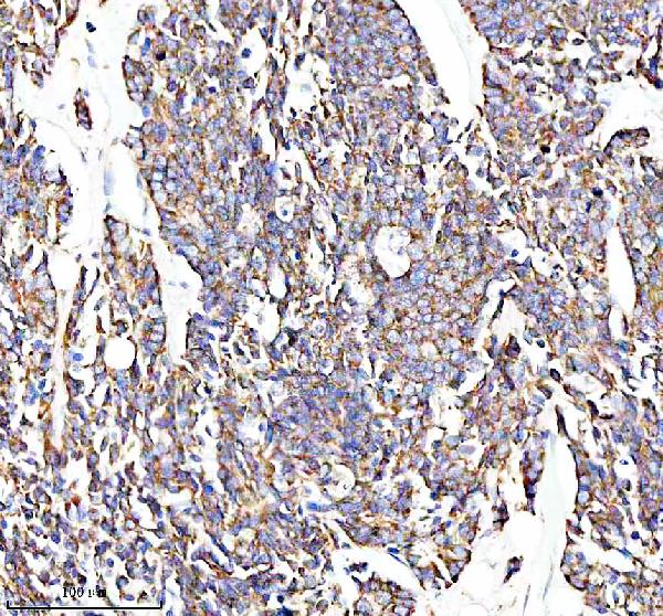
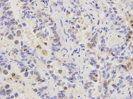
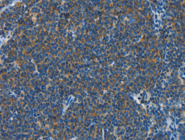
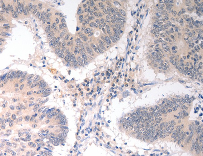
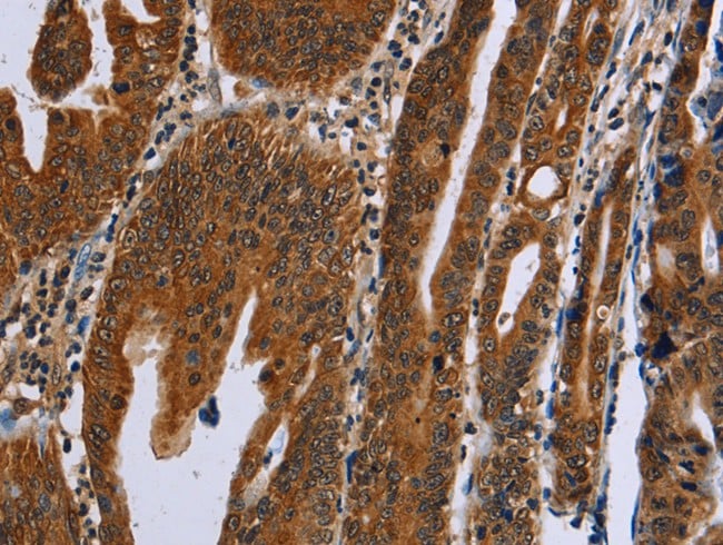
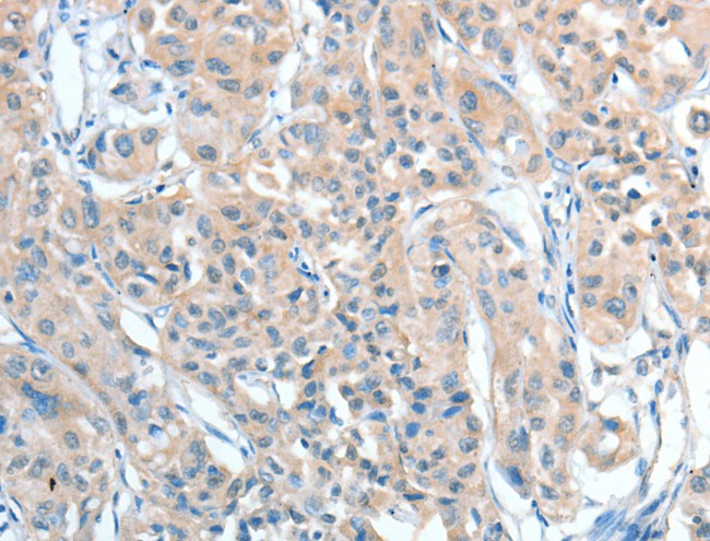
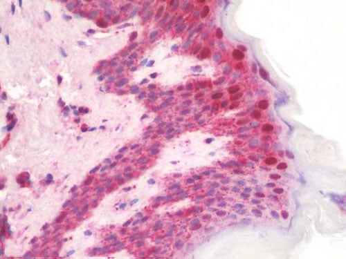
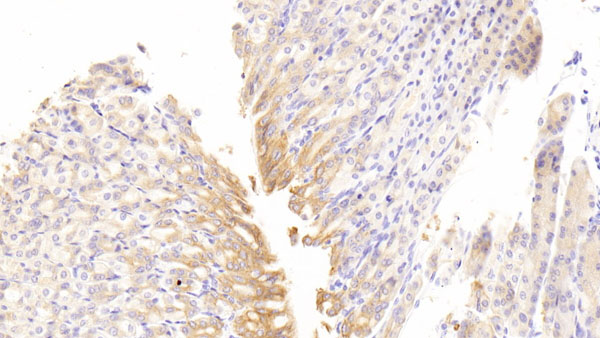
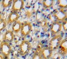
 IHC (Immunohiostchemistry)
(Immunohistochemistry of paraffin-embedded Human colon cancer tissue using FHL3 Polyclonal Antibody at dilution 1:40)
IHC (Immunohiostchemistry)
(Immunohistochemistry of paraffin-embedded Human colon cancer tissue using FHL3 Polyclonal Antibody at dilution 1:40)
FHL3, Polyclonal Antibody (Cat# AAA167154)
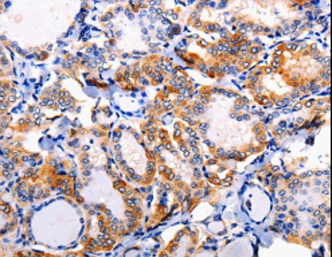
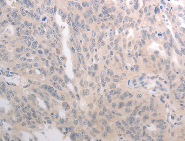
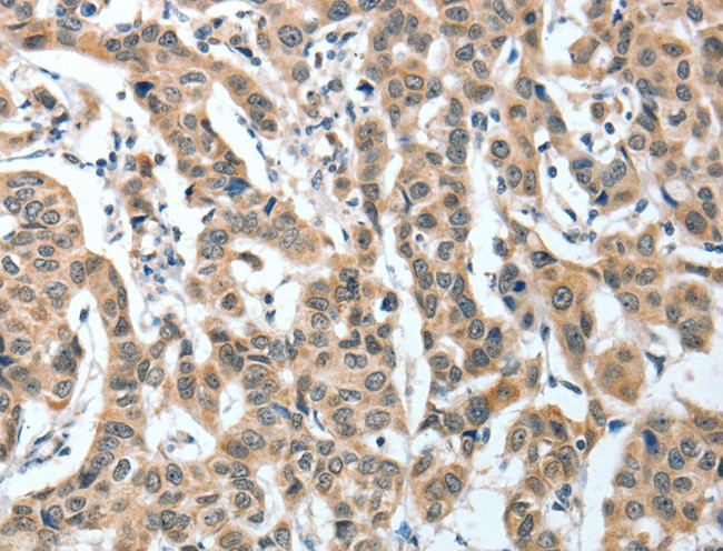
 IHC (Immunohistochemisry)
(Immunohistochemistry of paraffin-embedded Human thyroid cancer using YARS2 Polyclonal Antibody at dilution of 1:20)
IHC (Immunohistochemisry)
(Immunohistochemistry of paraffin-embedded Human thyroid cancer using YARS2 Polyclonal Antibody at dilution of 1:20)
YARS2, Polyclonal Antibody (Cat# AAA167156)
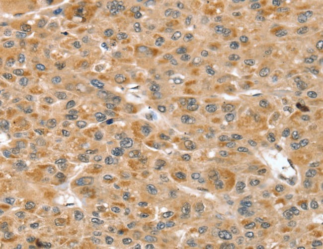
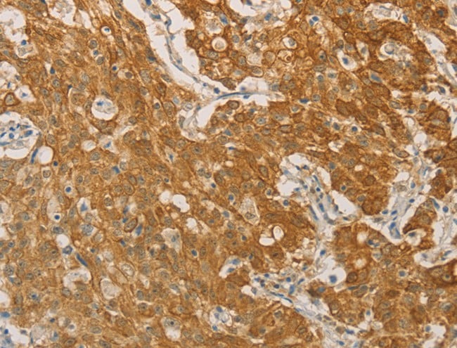
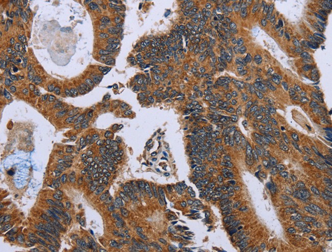
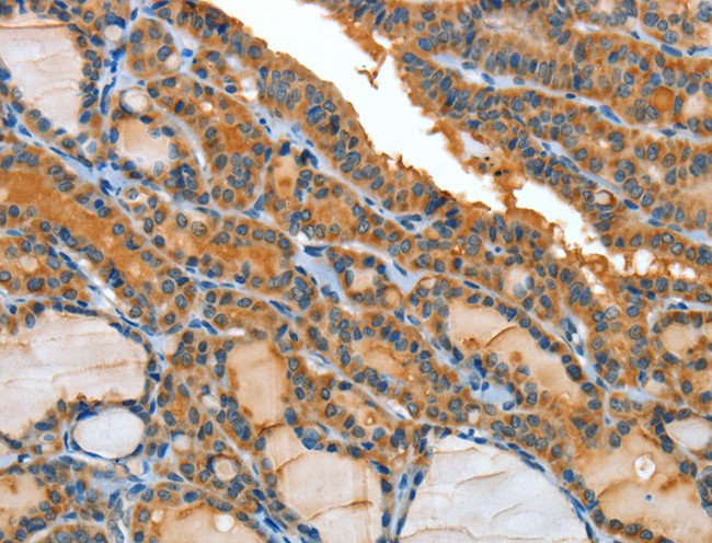
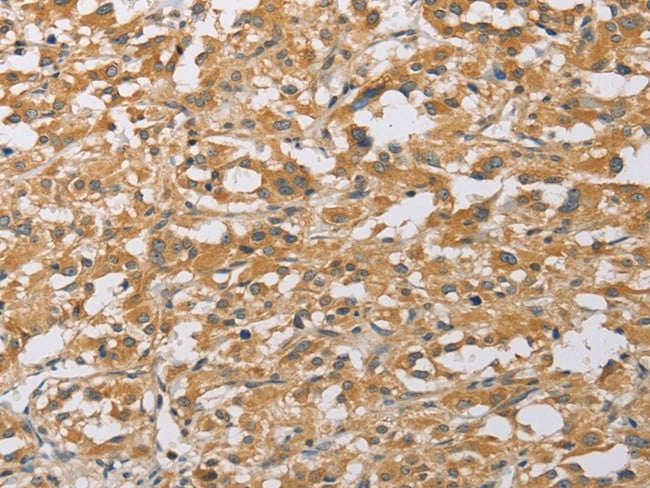
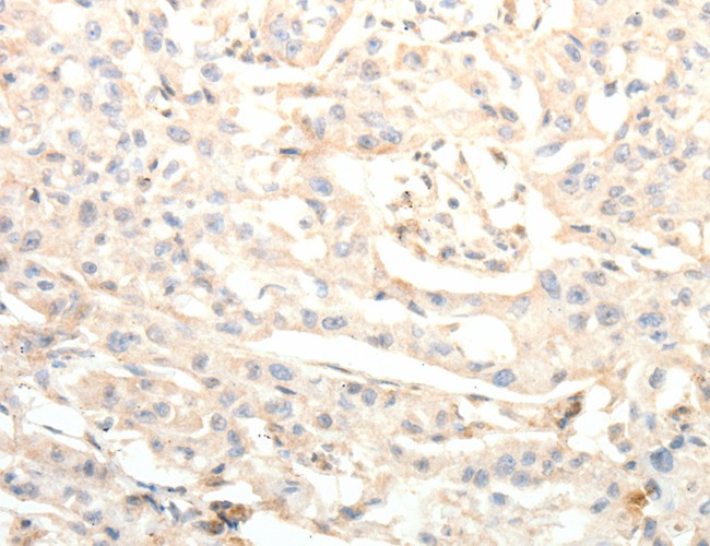
 IHC (Immunohiostchemistry)
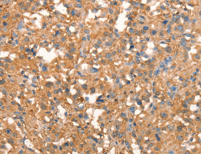
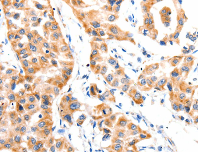
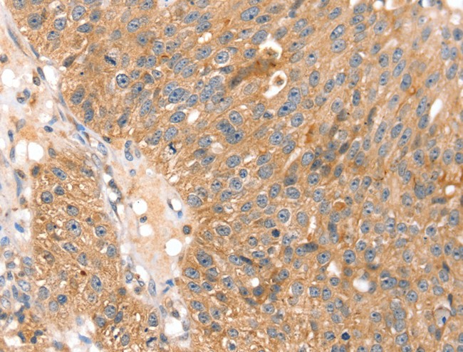
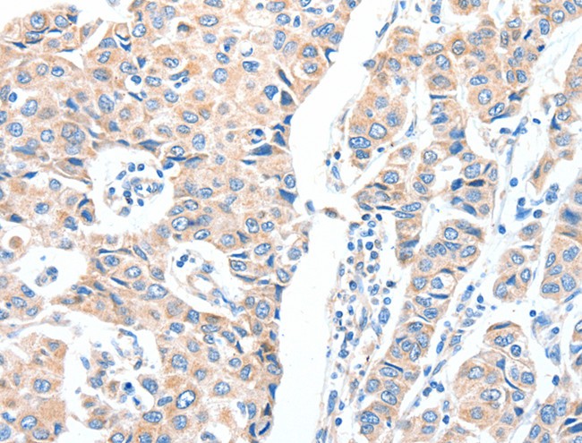
(Immunohistochemistry of paraffin-embedded Human liver cancer tissue using AP1B1 Polyclonal Antibody at dilution 1:100)
IHC (Immunohiostchemistry)
(Immunohistochemistry of paraffin-embedded Human liver cancer tissue using AP1B1 Polyclonal Antibody at dilution 1:100)
AP1B1, Polyclonal Antibody (Cat# AAA167158)
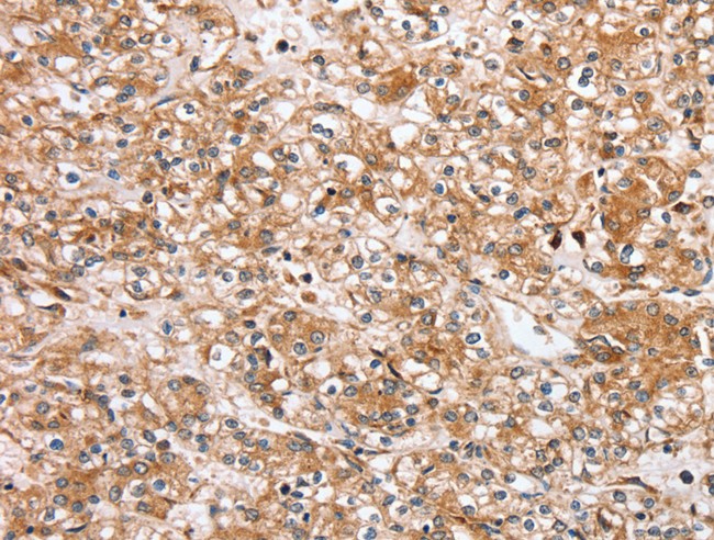
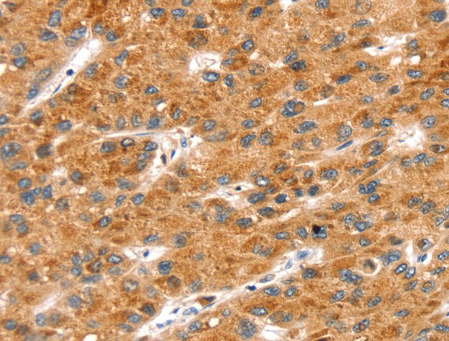
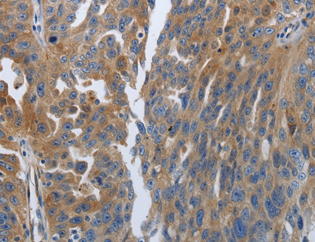
 IHC (Immunohiostchemistry)
(Immunohistochemistry of paraffin-embedded Human sarcoma tissue using KIF1C Polyclonal Antibody at dilution 1:60)
IHC (Immunohiostchemistry)
(Immunohistochemistry of paraffin-embedded Human sarcoma tissue using KIF1C Polyclonal Antibody at dilution 1:60)
KIF1C, Polyclonal Antibody (Cat# AAA167160)
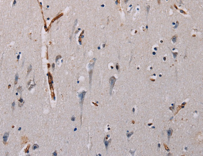
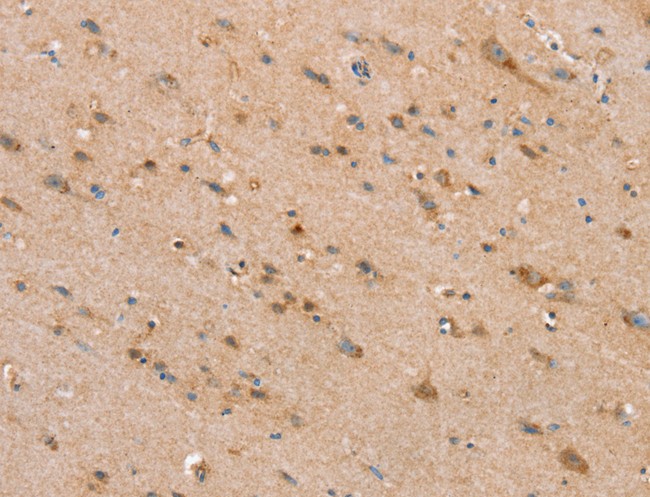
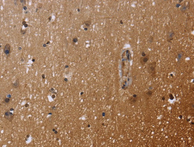
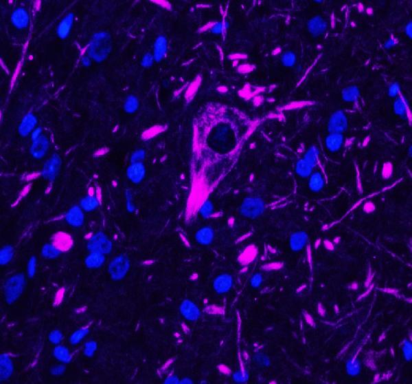
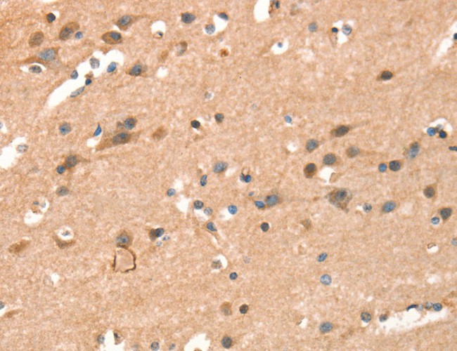 IHC (Immunohistochemisry)
(Immunohistochemistry of paraffin-embedded Human brain using CRELD1 Polyclonal Antibody at dilution of 1:40)
IHC (Immunohistochemisry)
(Immunohistochemistry of paraffin-embedded Human brain using CRELD1 Polyclonal Antibody at dilution of 1:40)
CRELD1, Polyclonal Antibody (Cat# AAA167173)
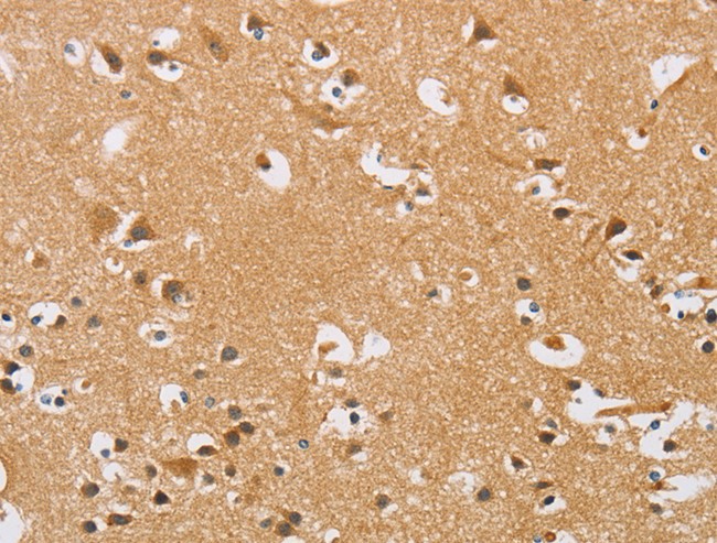
 IHC (Immunohistochemisry)
(Immunohistochemistry of paraffin-embedded Human brain using PLCB3 Polyclonal Antibody at dilution of 1:40)
IHC (Immunohistochemisry)
(Immunohistochemistry of paraffin-embedded Human brain using PLCB3 Polyclonal Antibody at dilution of 1:40)
PLCB3, Polyclonal Antibody (Cat# AAA167192)
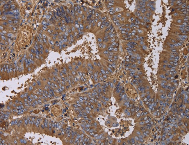
 IHC (Immunohistochemisry)
(Immunohistochemistry of paraffin-embedded Human lung cancer using JUP Polyclonal Antibody at dilution of 1:50)
IHC (Immunohistochemisry)
(Immunohistochemistry of paraffin-embedded Human lung cancer using JUP Polyclonal Antibody at dilution of 1:50)
JUP, Polyclonal Antibody (Cat# AAA167194)
 IHC (Immunohiostchemistry)
(Immunohistochemistry of paraffin-embedded Human brain tissue using CCND3 Polyclonal Antibody at dilution 1:40)
IHC (Immunohiostchemistry)
(Immunohistochemistry of paraffin-embedded Human brain tissue using CCND3 Polyclonal Antibody at dilution 1:40)
CCND3, Polyclonal Antibody (Cat# AAA167213)
 IHC (Immunohiostchemistry)
(Immunohistochemistry of paraffin-embedded Human lung cancer tissue using pan CDH Polyclonal Antibody at dilution 1:20)
IHC (Immunohiostchemistry)
(Immunohistochemistry of paraffin-embedded Human lung cancer tissue using pan CDH Polyclonal Antibody at dilution 1:20)
CDH, Polyclonal Antibody (Cat# AAA167223)
 IHC (Immunohistochemisry)
(Immunohistochemistry of paraffin-embedded Human ovarian cancer using RNH1 Polyclonal Antibody at dilution of 1:30)
IHC (Immunohistochemisry)
(Immunohistochemistry of paraffin-embedded Human ovarian cancer using RNH1 Polyclonal Antibody at dilution of 1:30)
RNH1, Polyclonal Antibody (Cat# AAA167230)
 FCM/FACS (Flow Cytometry)
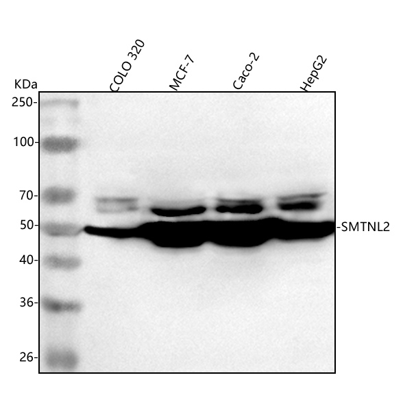
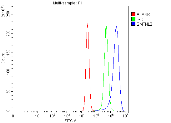
(Figure 3. Flow Cytometry analysis of MCF-7 cells using anti-SMTNL2 antibody (AAA127919).Overlay histogram showing MCF-7 cells stained with AAA127919 (Blue line). To facilitate intracellular staining, cells were fixed with 4% paraformaldehyde and permeabilized with permeabilization buffer. The cells were blocked with 10% normal goat serum. And then incubated with rabbit anti-SMTNL2 Antibody (AAA127919, 1ug/1x106 cells) for 30 min at 20 degree C. DyLight488 conjugated goat anti-rabbit IgG was used as secondary antibody for 30 minutes at 20 degree C. Isotype control antibody (Green line) was rabbit IgG (1ug/1x106) used under the same conditions. Unlabelled sample (Red line) was also used as a control.)
FCM/FACS (Flow Cytometry)
(Figure 3. Flow Cytometry analysis of MCF-7 cells using anti-SMTNL2 antibody (AAA127919).Overlay histogram showing MCF-7 cells stained with AAA127919 (Blue line). To facilitate intracellular staining, cells were fixed with 4% paraformaldehyde and permeabilized with permeabilization buffer. The cells were blocked with 10% normal goat serum. And then incubated with rabbit anti-SMTNL2 Antibody (AAA127919, 1ug/1x106 cells) for 30 min at 20 degree C. DyLight488 conjugated goat anti-rabbit IgG was used as secondary antibody for 30 minutes at 20 degree C. Isotype control antibody (Green line) was rabbit IgG (1ug/1x106) used under the same conditions. Unlabelled sample (Red line) was also used as a control.)
SMTNL2, Polyclonal Antibody (Cat# AAA127919)

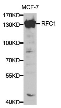
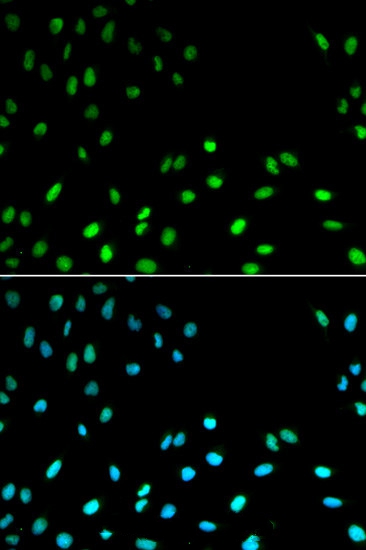 IF (Immunofluorescence)
(Immunofluorescence analysis of MCF7 cell using RFC1 antibody. Blue: DAPI for nuclear staining.)
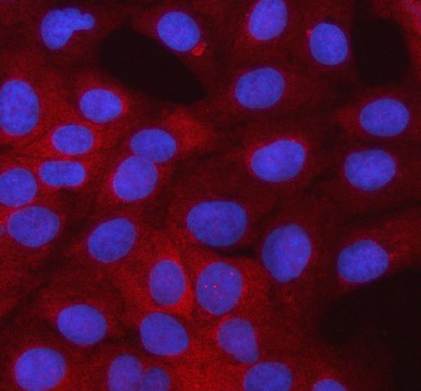
IF (Immunofluorescence)
(Immunofluorescence analysis of MCF7 cell using RFC1 antibody. Blue: DAPI for nuclear staining.)
RFC1, Polyclonal Antibody (Cat# AAA166903)
 IHC (Immunohiostchemistry)
(Immunohistochemistry of paraffin-embedded Human lung cancer tissue using CNTNAP3 Polyclonal Antibody at dilution 1:40)
IHC (Immunohiostchemistry)
(Immunohistochemistry of paraffin-embedded Human lung cancer tissue using CNTNAP3 Polyclonal Antibody at dilution 1:40)
CNTNAP3, Polyclonal Antibody (Cat# AAA166939)
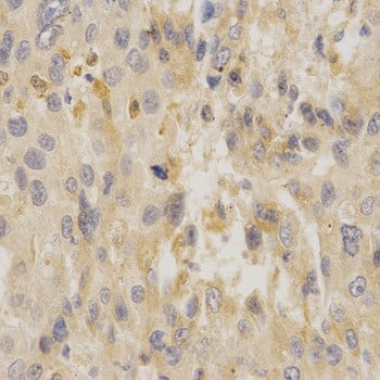
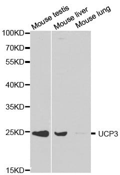
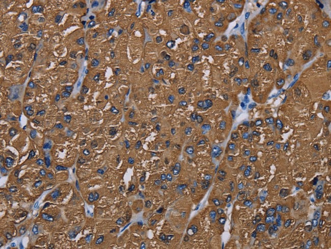
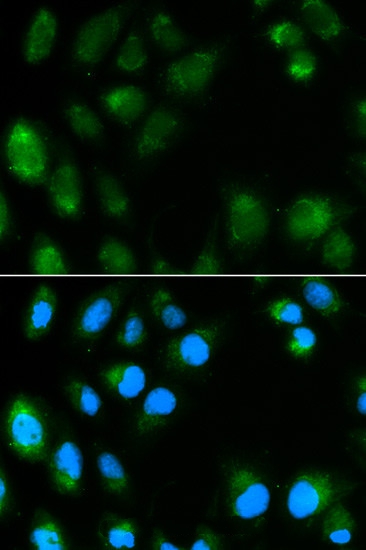 IF (Immunofluorescence)
(Immunofluorescence analysis of MCF7 cell using UCP3 antibody. Blue: DAPI for nuclear staining.)
IF (Immunofluorescence)
(Immunofluorescence analysis of MCF7 cell using UCP3 antibody. Blue: DAPI for nuclear staining.)
UCP3, Polyclonal Antibody (Cat# AAA166948)
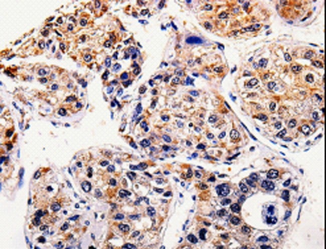
 IHC (Immunohistochemisry)
(Immunohistochemistry of paraffin-embedded Human thyroid cancer using BMP3 Polyclonal Antibody at dilution of 1:20)
IHC (Immunohistochemisry)
(Immunohistochemistry of paraffin-embedded Human thyroid cancer using BMP3 Polyclonal Antibody at dilution of 1:20)
BMP3, Polyclonal Antibody (Cat# AAA166949)
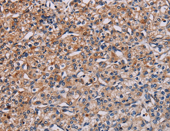
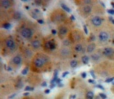
 IHC (Immunohiostchemistry)
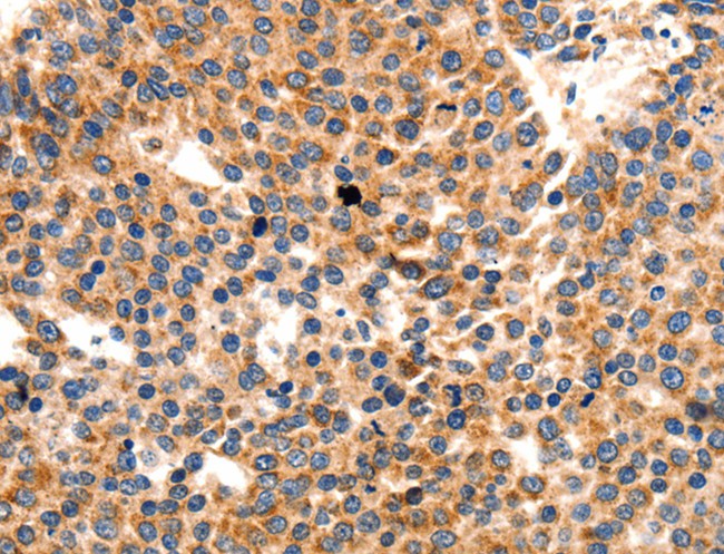
(Immunohistochemistry of paraffin-embedded Human liver cancer tissue using CD5L Polyclonal Antibody at dilution 1:40)
IHC (Immunohiostchemistry)
(Immunohistochemistry of paraffin-embedded Human liver cancer tissue using CD5L Polyclonal Antibody at dilution 1:40)
CD5L, Polyclonal Antibody (Cat# AAA166955)
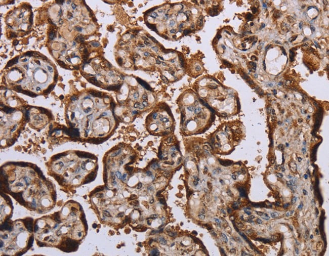
 IHC (Immunohistochemisry)
(Immunohistochemistry of paraffin-embedded Human gastric cancer using PSMB7 Polyclonal Antibody at dilution of 1:25)
IHC (Immunohistochemisry)
(Immunohistochemistry of paraffin-embedded Human gastric cancer using PSMB7 Polyclonal Antibody at dilution of 1:25)
PSMB7, Polyclonal Antibody (Cat# AAA166957)
 IHC (Immunohistochemisry)
(Immunohistochemistry of paraffin-embedded Human thyroid cancer using FADS1 Polyclonal Antibody at dilution of 1:60)
IHC (Immunohistochemisry)
(Immunohistochemistry of paraffin-embedded Human thyroid cancer using FADS1 Polyclonal Antibody at dilution of 1:60)
FADS1, Polyclonal Antibody (Cat# AAA166959)
 IHC (Immunohiostchemistry)
(Immunohistochemistry of paraffin-embedded Human esophagus cancer tissue using SCN1B Polyclonal Antibody at dilution 1:35)
IHC (Immunohiostchemistry)
(Immunohistochemistry of paraffin-embedded Human esophagus cancer tissue using SCN1B Polyclonal Antibody at dilution 1:35)
SCN1B, Polyclonal Antibody (Cat# AAA166961)
 IHC (Immunohiostchemistry)
(Immunohistochemistry of paraffin-embedded Human ovarian cancer tissue using ICOSLG Polyclonal Antibody at dilution 1:30)
IHC (Immunohiostchemistry)
(Immunohistochemistry of paraffin-embedded Human ovarian cancer tissue using ICOSLG Polyclonal Antibody at dilution 1:30)
ICOSLG, Polyclonal Antibody (Cat# AAA166968)
 IHC (Immunohistochemisry)
(Immunohistochemistry of paraffin-embedded Human gastric cancer using GH2 Polyclonal Antibody at dilution of 1:60)
IHC (Immunohistochemisry)
(Immunohistochemistry of paraffin-embedded Human gastric cancer using GH2 Polyclonal Antibody at dilution of 1:60)
GH2, Polyclonal Antibody (Cat# AAA167067)
 IHC (Immunohiostchemistry)
(Immunohistochemistry of paraffin-embedded Human thyroid cancer tissue using IL1R1 Polyclonal Antibody at dilution 1:25)
IHC (Immunohiostchemistry)
(Immunohistochemistry of paraffin-embedded Human thyroid cancer tissue using IL1R1 Polyclonal Antibody at dilution 1:25)
IL1R1, Polyclonal Antibody (Cat# AAA167113)
 IHC (Immunohiostchemistry)
(Immunohistochemistry of paraffin-embedded Human brain tissue using SLC25A20 Polyclonal Antibody at dilution 1:30)
IHC (Immunohiostchemistry)
(Immunohistochemistry of paraffin-embedded Human brain tissue using SLC25A20 Polyclonal Antibody at dilution 1:30)
SLC25A20, Polyclonal Antibody (Cat# AAA167120)

 IHC (Immunohiostchemistry)
(Immunohistochemistry of paraffin-embedded Human lung cancer tissue using AGBL2 Polyclonal Antibody at dilution 1:25)
IHC (Immunohiostchemistry)
(Immunohistochemistry of paraffin-embedded Human lung cancer tissue using AGBL2 Polyclonal Antibody at dilution 1:25)
AGBL2, Polyclonal Antibody (Cat# AAA167125)
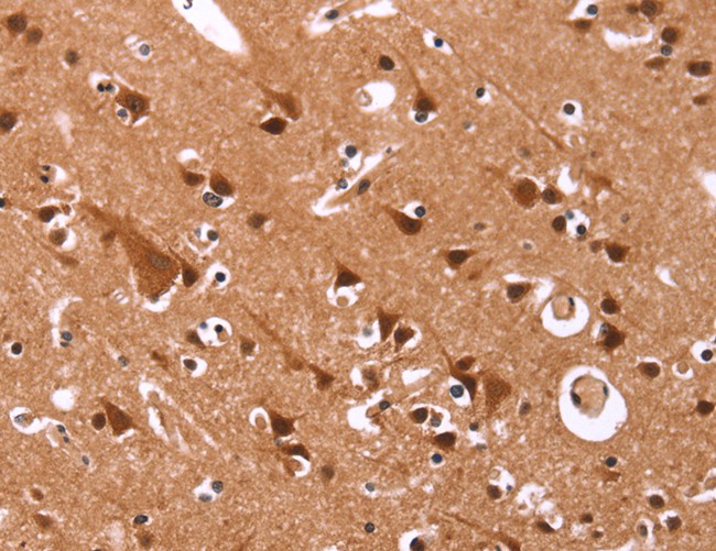
 IHC (Immunohiostchemistry)
(Immunohistochemistry of paraffin-embedded Human brain tissue using ARHGEF1 Polyclonal Antibody at dilution 1:50)
IHC (Immunohiostchemistry)
(Immunohistochemistry of paraffin-embedded Human brain tissue using ARHGEF1 Polyclonal Antibody at dilution 1:50)
ARHGEF1, Polyclonal Antibody (Cat# AAA167126)
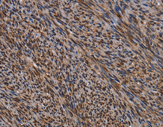
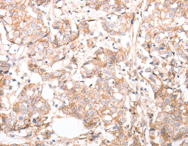
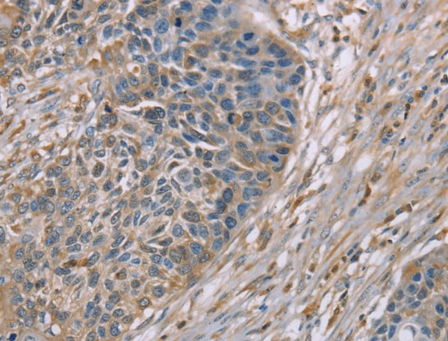
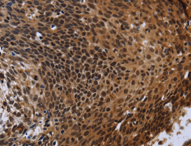
 IHC (Immunohistochemisry)
(Immunohistochemistry of paraffin-embedded Human lung cancer using BIRC2 Polyclonal Antibody at dilution of 1:20)
IHC (Immunohistochemisry)
(Immunohistochemistry of paraffin-embedded Human lung cancer using BIRC2 Polyclonal Antibody at dilution of 1:20)
BIRC2, Polyclonal Antibody (Cat# AAA167140)
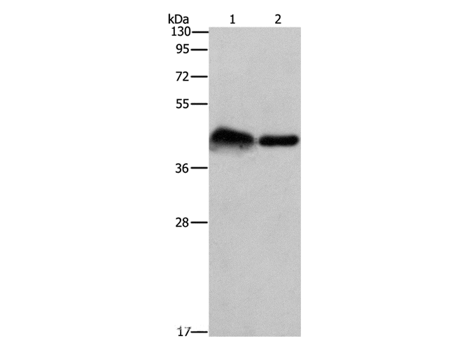
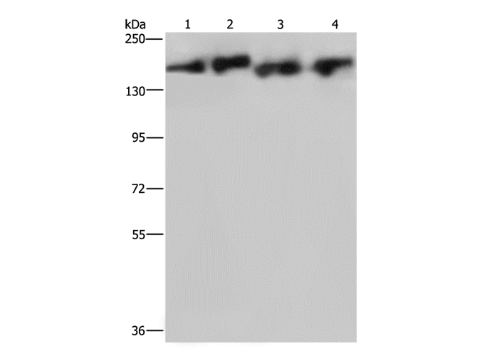
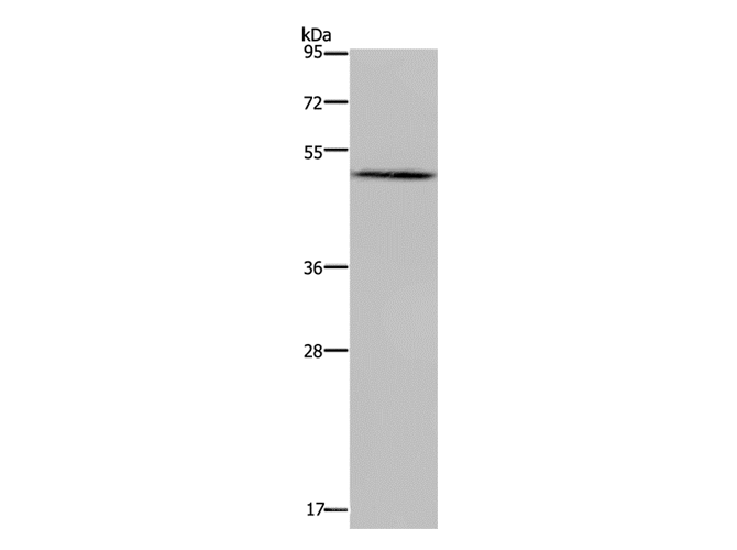
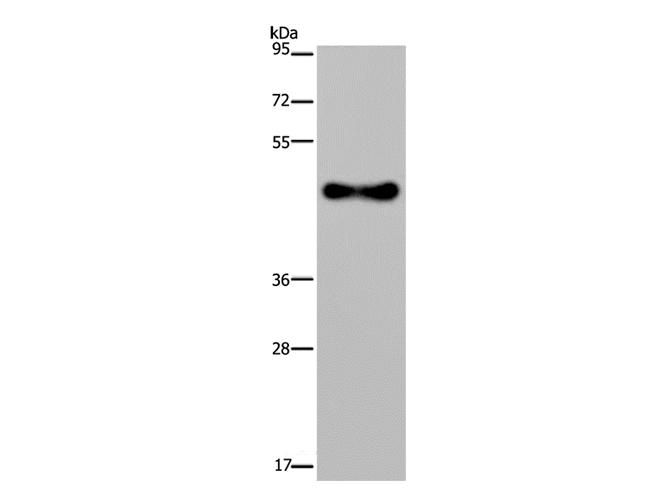
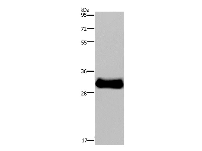
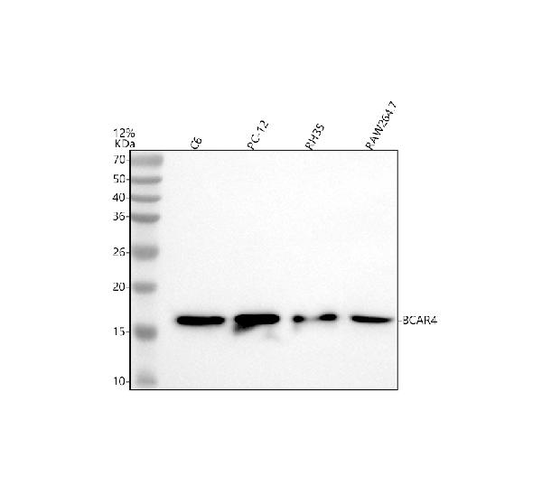
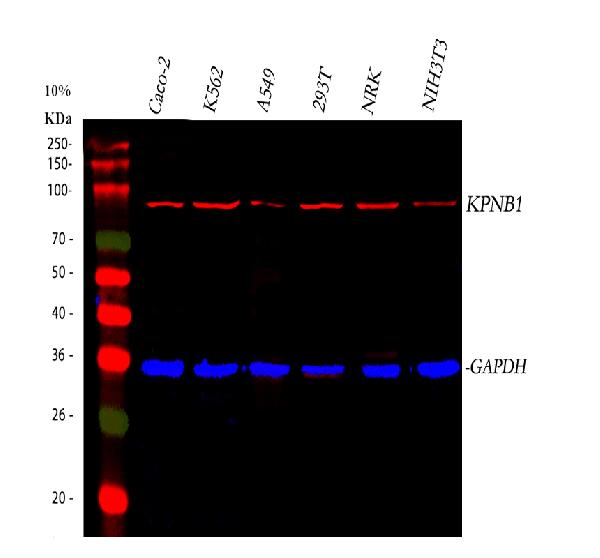
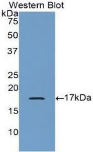
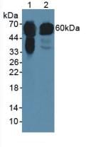
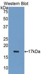
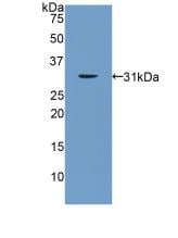
 WB (Western Blot)
(Western Blot analysis of Human testis tissue, Jurkat cell and Mouse liver tissue using KIRREL2 Polyclonal Antibody at dilution of 1:500)
WB (Western Blot)
(Western Blot analysis of Human testis tissue, Jurkat cell and Mouse liver tissue using KIRREL2 Polyclonal Antibody at dilution of 1:500)
KIRREL2, Polyclonal Antibody (Cat# AAA167144)
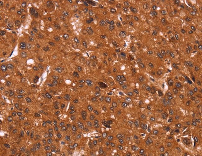
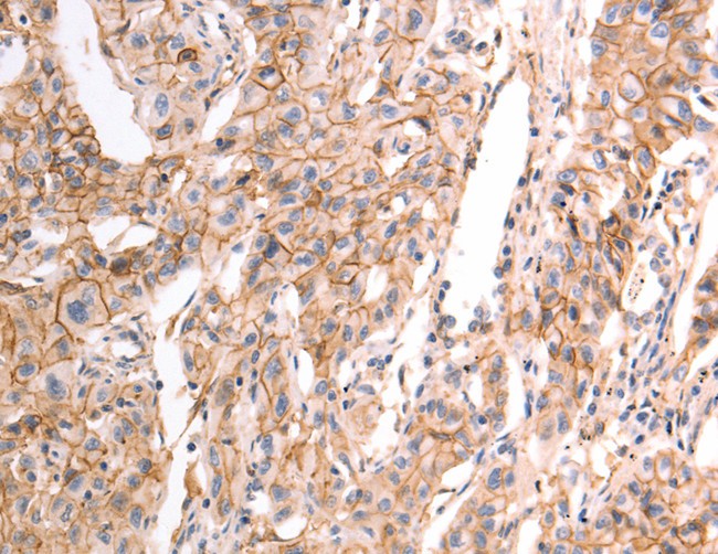
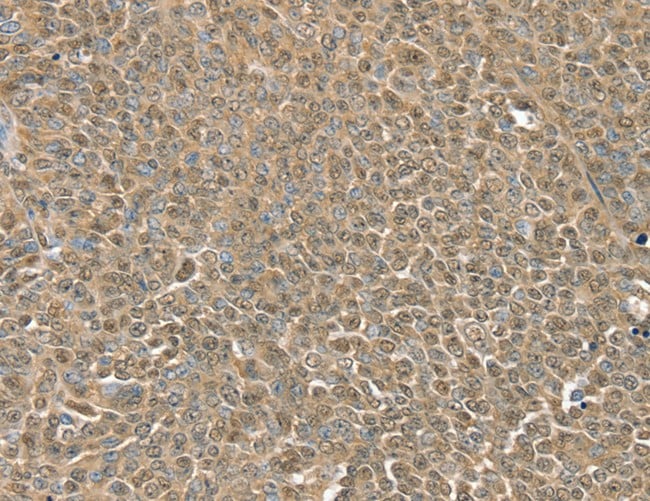
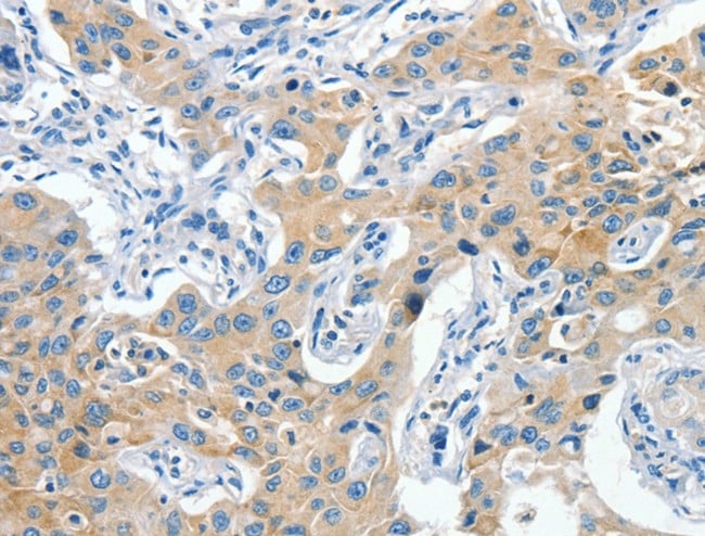
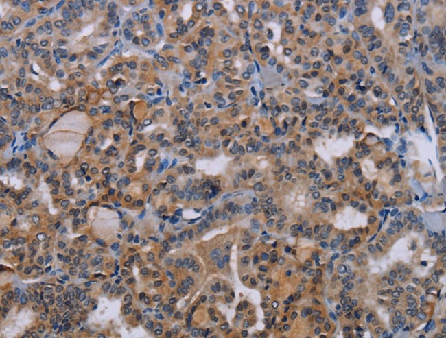
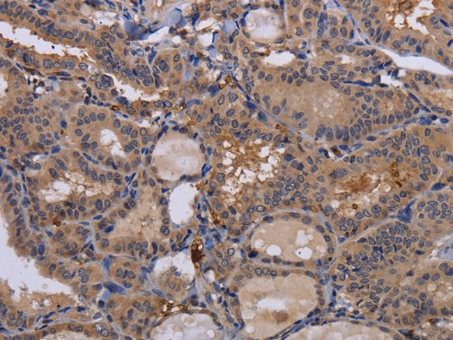
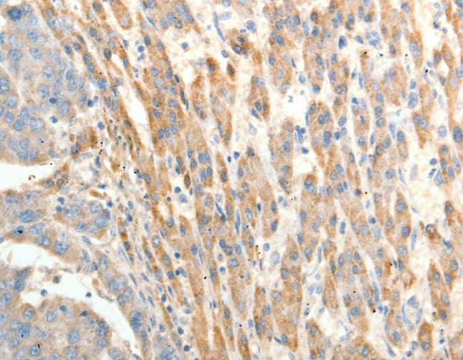
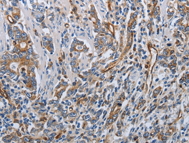
 IHC (Immunohiostchemistry)
(Immunohistochemistry of paraffin-embedded Human liver cancer tissue using PAFAH1B1 Polyclonal Antibody at dilution 1:25)
IHC (Immunohiostchemistry)
(Immunohistochemistry of paraffin-embedded Human liver cancer tissue using PAFAH1B1 Polyclonal Antibody at dilution 1:25)
PAFAH1B1, Polyclonal Antibody (Cat# AAA166991)
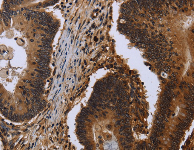
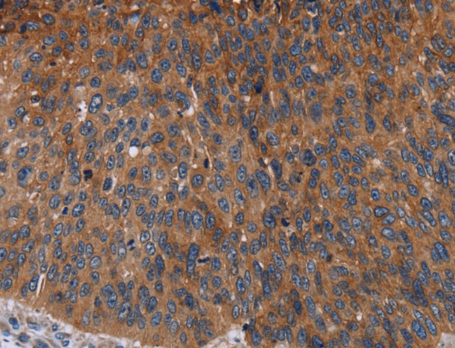
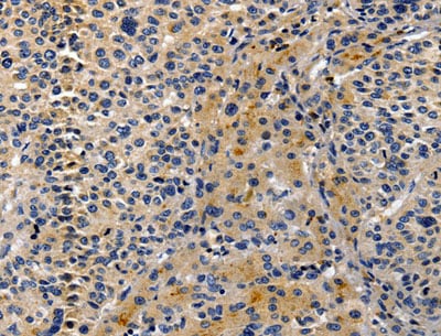
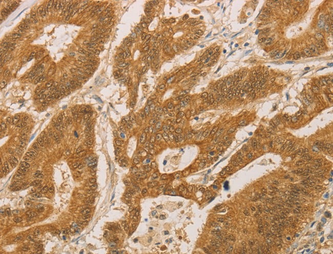
 IHC (Immunohistochemisry)
(Immunohistochemistry of paraffin-embedded Human brain using FHL3 Polyclonal Antibody at dilution of 1:40)
IHC (Immunohistochemisry)
(Immunohistochemistry of paraffin-embedded Human brain using FHL3 Polyclonal Antibody at dilution of 1:40)
FHL3, Polyclonal Antibody (Cat# AAA166996)
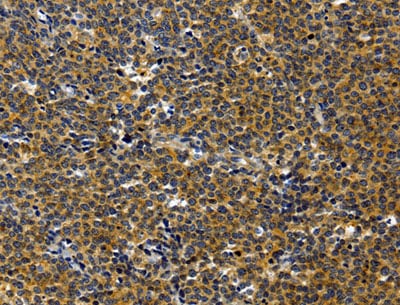
 IHC (Immunohiostchemistry)
(Immunohistochemistry of paraffin-embedded Human thyroid cancer using CWC27 Polyclonal Antibody at dilution of 1:40)
IHC (Immunohiostchemistry)
(Immunohistochemistry of paraffin-embedded Human thyroid cancer using CWC27 Polyclonal Antibody at dilution of 1:40)
CWC27, Polyclonal Antibody (Cat# AAA167001)
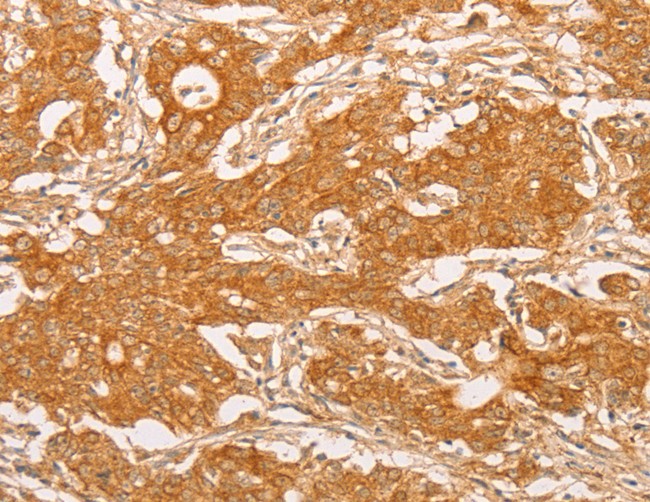
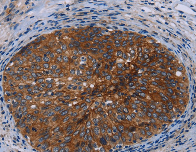
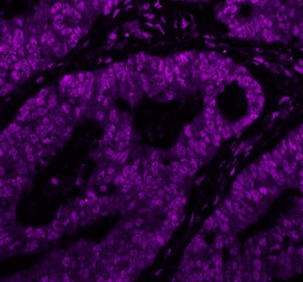
 IHC (Immunohistochemisry)
(Immunohistochemistry of paraffin-embedded Human breast cancer using SOX11 Polyclonal Antibody at dilution of 1:20)
IHC (Immunohistochemisry)
(Immunohistochemistry of paraffin-embedded Human breast cancer using SOX11 Polyclonal Antibody at dilution of 1:20)
SOX11, Polyclonal Antibody (Cat# AAA167015)
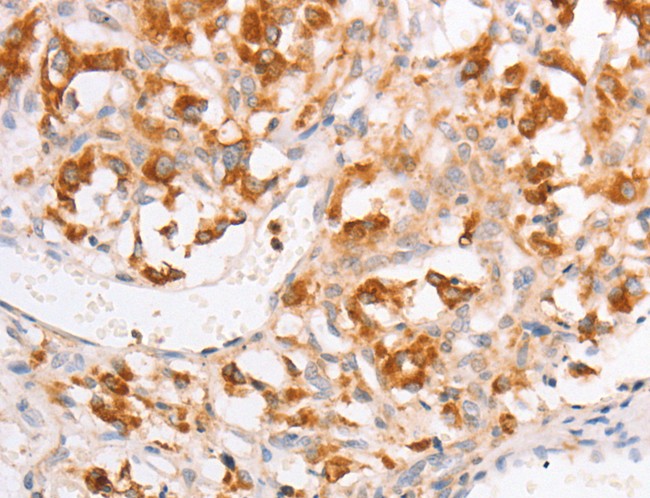
 IHC (Immunohistochemisry)
(Immunohistochemistry of paraffin-embedded Human gastric cancer using SOCS1 Polyclonal Antibody at dilution of 1:70)
IHC (Immunohistochemisry)
(Immunohistochemistry of paraffin-embedded Human gastric cancer using SOCS1 Polyclonal Antibody at dilution of 1:70)
SOCS1, Polyclonal Antibody (Cat# AAA167032)
 IHC (Immunohiostchemistry)
(Immunohistochemistry of paraffin-embedded Human thyroid cancer tissue using ADCK1 Polyclonal Antibody at dilution 1:40)
IHC (Immunohiostchemistry)
(Immunohistochemistry of paraffin-embedded Human thyroid cancer tissue using ADCK1 Polyclonal Antibody at dilution 1:40)
ADCK1, Polyclonal Antibody (Cat# AAA167062)
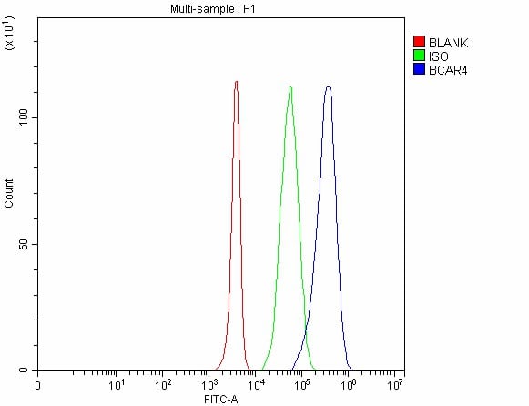 FCM/FACS (Flow Cytometry)
(Figure 2. Flow Cytometry analysis of 293T cells using anti-BCAR4 antibody (AAA127947).Overlay histogram showing 293T cells stained with AAA127947 (Blue line). The cells were fixed with 4% paraformaldehyde and blocked with 10% normal goat serum. And then incubated with rabbit anti-BCAR4 Antibody (AAA127947, 1ug/1x106 cells) for 30 min at 20 degree C. DyLight488 conjugated goat anti-rabbit IgG was used as secondary antibody for 30 minutes at 20 degree C. Isotype control antibody (Green line) was rabbit IgG (1ug/1x106) used under the same conditions. Unlabelled sample without incubation with primary antibody and secondary antibody (Red line) was used as a blank control.)
FCM/FACS (Flow Cytometry)
(Figure 2. Flow Cytometry analysis of 293T cells using anti-BCAR4 antibody (AAA127947).Overlay histogram showing 293T cells stained with AAA127947 (Blue line). The cells were fixed with 4% paraformaldehyde and blocked with 10% normal goat serum. And then incubated with rabbit anti-BCAR4 Antibody (AAA127947, 1ug/1x106 cells) for 30 min at 20 degree C. DyLight488 conjugated goat anti-rabbit IgG was used as secondary antibody for 30 minutes at 20 degree C. Isotype control antibody (Green line) was rabbit IgG (1ug/1x106) used under the same conditions. Unlabelled sample without incubation with primary antibody and secondary antibody (Red line) was used as a blank control.)
BCAR4, Polyclonal Antibody (Cat# AAA127947)
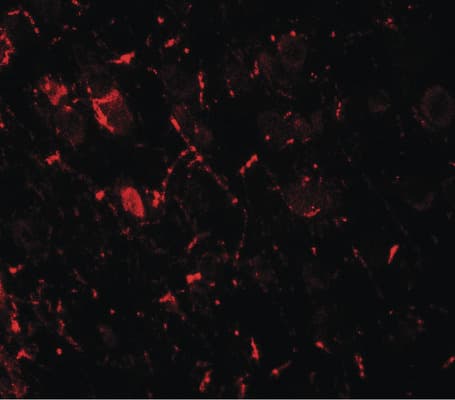
 IF (Immunofluorescence)
(Figure 3. IF analysis of NEFH using anti-NEFH antibody (MA1071).NEFH was detected in a paraffin-embedded section of human colon cancer tissue. Heat mediated antigen retrieval was performed in EDTA buffer (pH 8.0, epitope retrieval solution). The tissue section was blocked with 10% goat serum. The tissue section was then incubated with 5ug/mL mouse anti-NEFH Antibody (MA1071) overnight at 4 degree C. DyLight 647 Conjugated Goat Anti-Mouse IgG (AAA127949) was used as secondary antibody at 1:500 dilution and incubated for 30 minutes at 37 degree C. The section was counterstained with DAPI. Visualize using a fluorescence microscope and filter sets appropriate for the label used.)
IF (Immunofluorescence)
(Figure 3. IF analysis of NEFH using anti-NEFH antibody (MA1071).NEFH was detected in a paraffin-embedded section of human colon cancer tissue. Heat mediated antigen retrieval was performed in EDTA buffer (pH 8.0, epitope retrieval solution). The tissue section was blocked with 10% goat serum. The tissue section was then incubated with 5ug/mL mouse anti-NEFH Antibody (MA1071) overnight at 4 degree C. DyLight 647 Conjugated Goat Anti-Mouse IgG (AAA127949) was used as secondary antibody at 1:500 dilution and incubated for 30 minutes at 37 degree C. The section was counterstained with DAPI. Visualize using a fluorescence microscope and filter sets appropriate for the label used.)
IgG, Polyclonal Secondary Antibody (Cat# AAA127949)
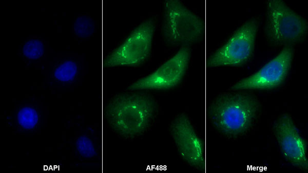
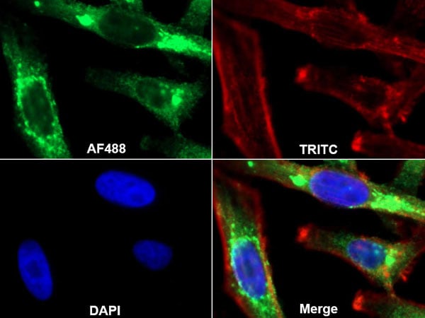 IF (Immunofluorescence)
(AF488 staining on IF;Sample: HepG2 cellTRITC Phalloidin at 100 nM (shown in pseudo color red)Primary Ab: 20ug/ml Rabbit Anti-Human GOLPH2 AntibodySecond Ab: 2ug/ml AF488-Linked Caprine Anti-Rabbit IgG Polyclonal Antibody)
IF (Immunofluorescence)
(AF488 staining on IF;Sample: HepG2 cellTRITC Phalloidin at 100 nM (shown in pseudo color red)Primary Ab: 20ug/ml Rabbit Anti-Human GOLPH2 AntibodySecond Ab: 2ug/ml AF488-Linked Caprine Anti-Rabbit IgG Polyclonal Antibody)
Golgi Phosphoprotein 2 (GOLPH2), Polyclonal Antibody (Cat# AAA162082)
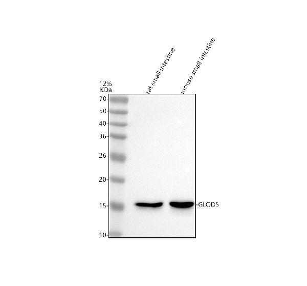
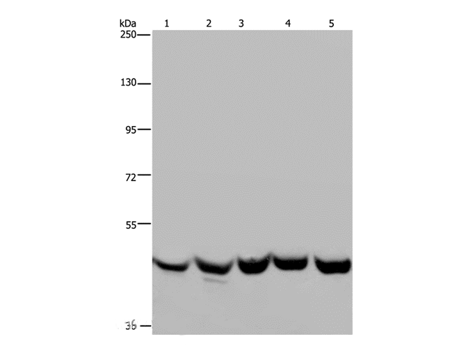
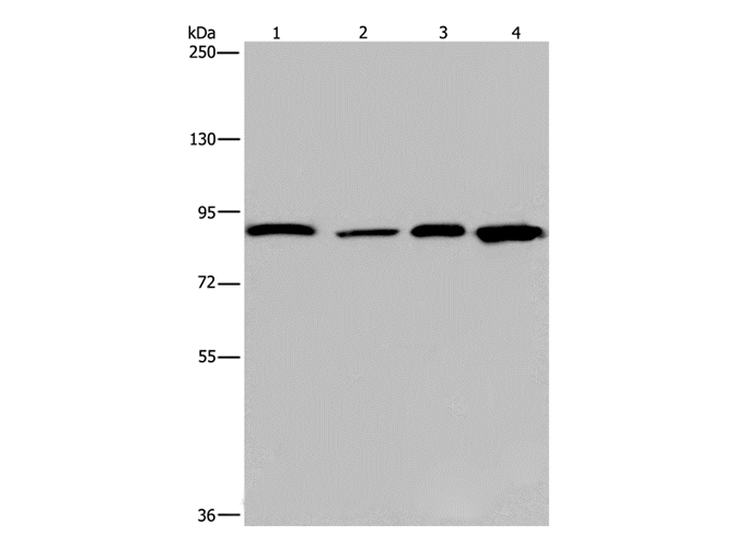
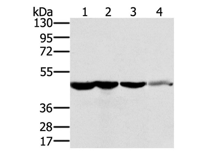
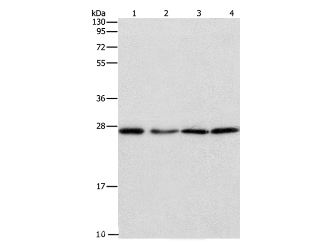
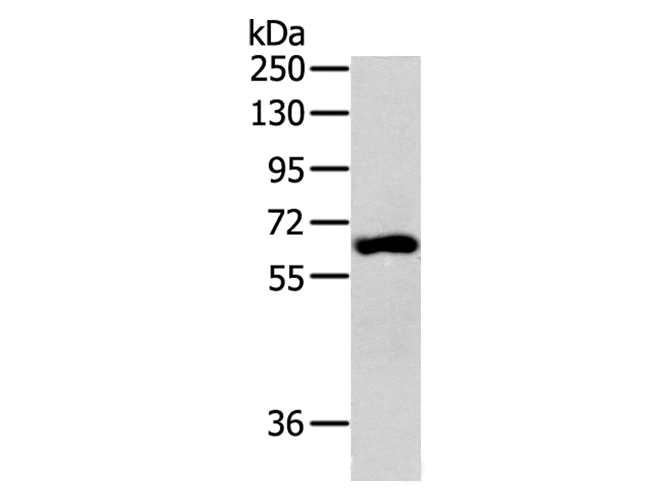
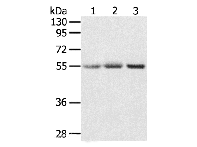
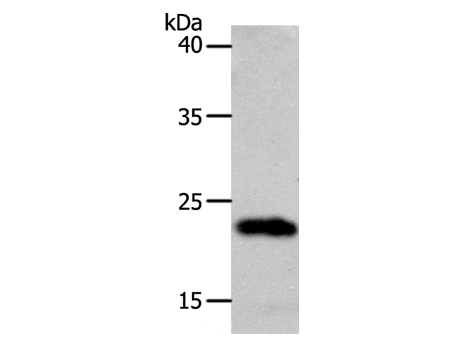
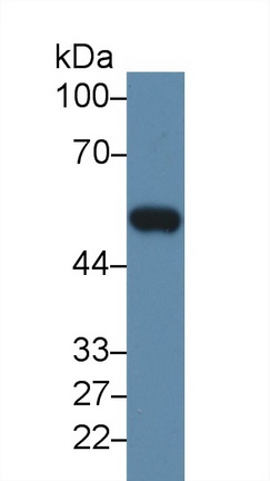
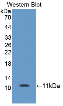
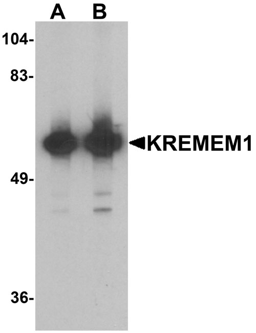 WB (Western Blot)
(Western blot analysis of KREMEN1 in rat small intestine tissue lysate with KREMEN1 antibody at (A) 0.5 and (B) 1 ug/ml)
WB (Western Blot)
(Western blot analysis of KREMEN1 in rat small intestine tissue lysate with KREMEN1 antibody at (A) 0.5 and (B) 1 ug/ml)
KREMEN1 / KREMEN-1, Polyclonal Antibody (Cat# AAA162142)
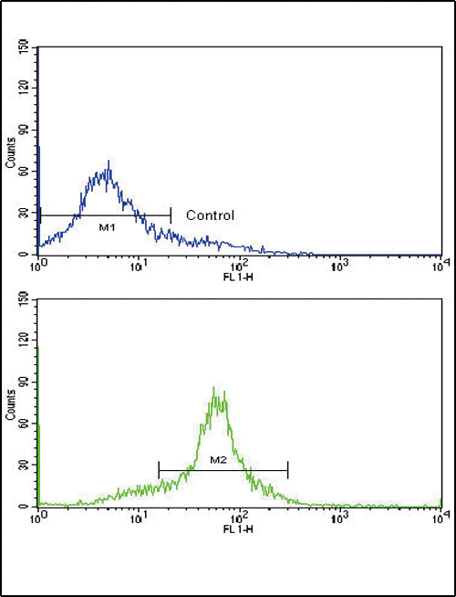 FCM/FACS (Flow Cytometry)
(Flow cytometric of WiDr cells using Glucagon Antibody (bottom histogram) compared to a negative control cell (top histogram). FITC-conjugated goat-anti-rabbit secondary antibodies were used for the analysis.)
FCM/FACS (Flow Cytometry)
(Flow cytometric of WiDr cells using Glucagon Antibody (bottom histogram) compared to a negative control cell (top histogram). FITC-conjugated goat-anti-rabbit secondary antibodies were used for the analysis.)
GCG / Glucagon, Polyclonal Antibody (Cat# AAA162143)
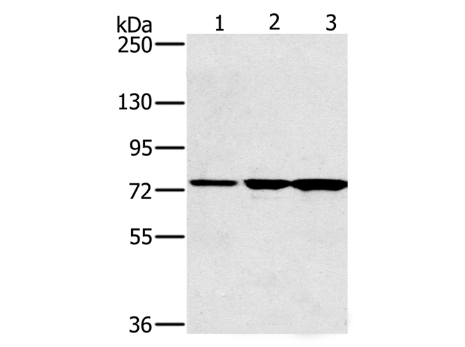
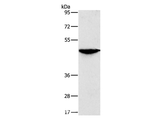
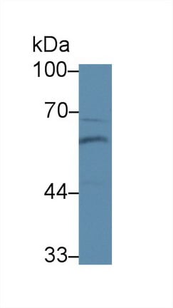
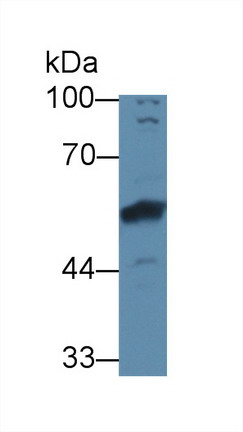
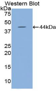
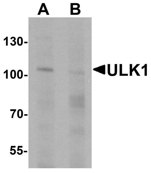 WB (Western Blot)
(Western blot analysis of ULK1 in rat brain tissue lysate with ULK1 antibody at 1 ug/ml in (A) the absence and (B) the presence of blocking peptide.)
WB (Western Blot)
(Western blot analysis of ULK1 in rat brain tissue lysate with ULK1 antibody at 1 ug/ml in (A) the absence and (B) the presence of blocking peptide.)
ULK1, Polyclonal Antibody (Cat# AAA162155)
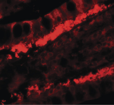
 IHC (Immunohistochemisry)
(DAB staining on IHC-P; Samples: Human Glioma Tissue))
IHC (Immunohistochemisry)
(DAB staining on IHC-P; Samples: Human Glioma Tissue))
Aquaporin 1, Colton Blood Group (AQP1), Polyclonal Antibody (Cat# AAA133060)
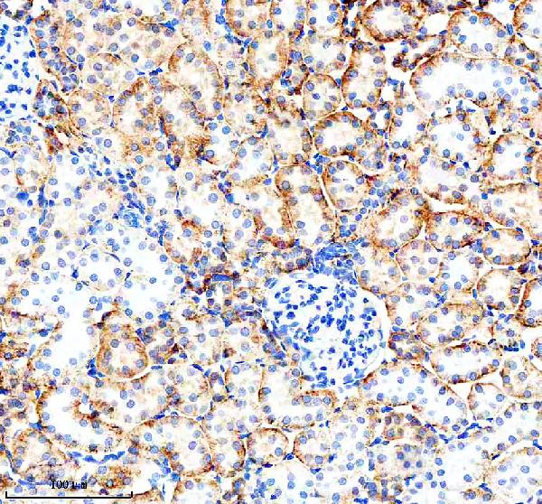
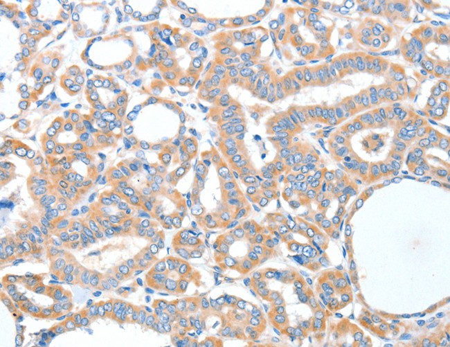
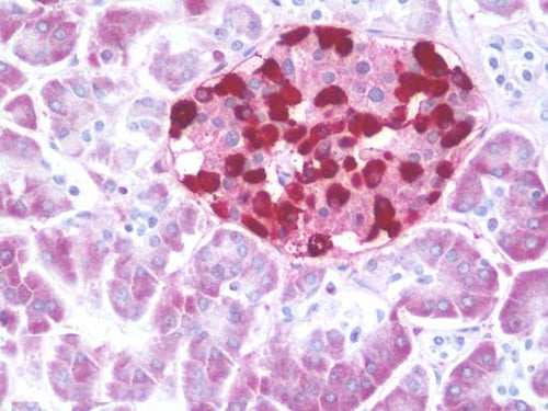
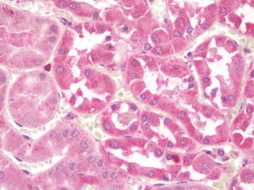
 IHC (Immunohistochemistry)
(DAB staining on fromalin fixed paraffin-embedded Kidney tissue))
IHC (Immunohistochemistry)
(DAB staining on fromalin fixed paraffin-embedded Kidney tissue))
Bone Morphogenetic Protein 6 (BMP6), Polyclonal Antibody (Cat# AAA132793)
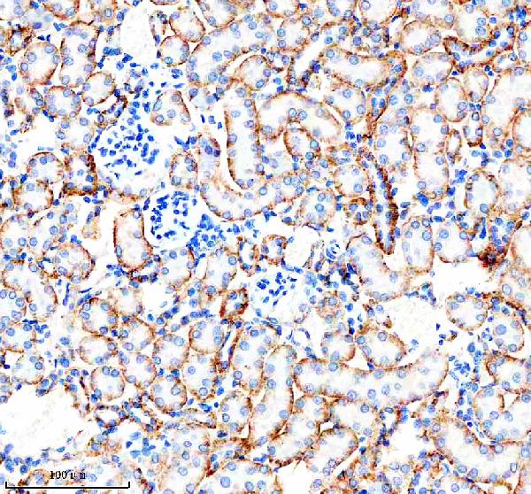
 IHC (Immunohistochemisry)
(DAB staining on fromalin fixed paraffin-embedded Kidney tissue))
IHC (Immunohistochemisry)
(DAB staining on fromalin fixed paraffin-embedded Kidney tissue))
Keratin 5 (KRT5), Polyclonal Antibody (Cat# AAA132840)
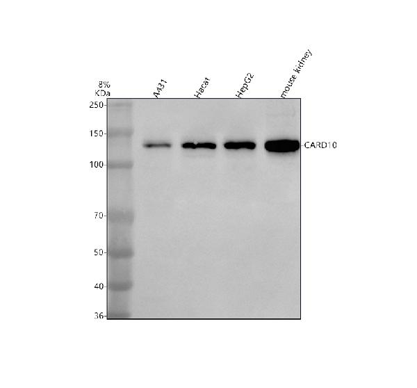
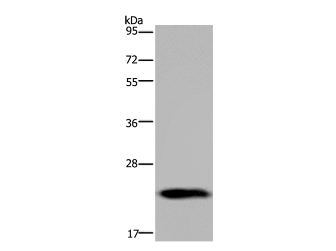
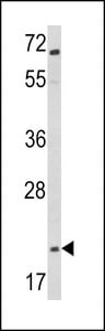
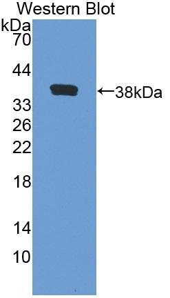
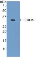
 WB (Western Blot)
(Western Blot: Sample: Recombinant protein.)
WB (Western Blot)
(Western Blot: Sample: Recombinant protein.)
Matrix Metalloproteinase 12 (MMP12), Polyclonal Antibody (Cat# AAA132656)
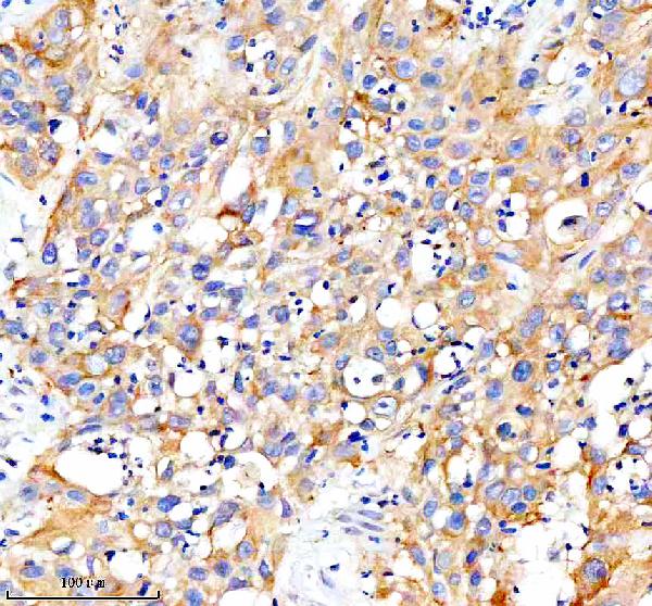
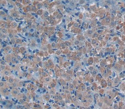
 IHC (Immunohiostchemistry)
(DAB staining on fromalin fixed paraffin-embedded Kidney tissue))
IHC (Immunohiostchemistry)
(DAB staining on fromalin fixed paraffin-embedded Kidney tissue))
A Disintegrin And Metalloproteinase With Thrombospondin 1 (ADAMTS1), Polyclonal Antibody (Cat# AAA132658)
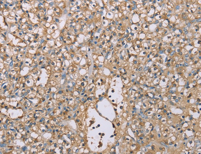
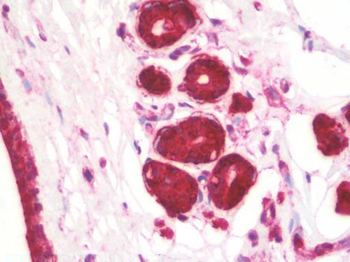
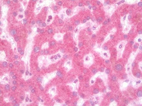
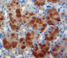
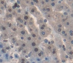
 IHC (Immunohiostchemistry)
(DAB staining on fromalin fixed paraffin-embedded Liver tissue))
IHC (Immunohiostchemistry)
(DAB staining on fromalin fixed paraffin-embedded Liver tissue))
Hyaluronoglucosaminidase 2 (HYAL2), Polyclonal Antibody (Cat# AAA132665)

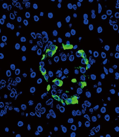
 IHC (Immunohistochemisry)
(DAB staining on IHC-P. (Samples: Rat Tissue))
IHC (Immunohistochemisry)
(DAB staining on IHC-P. (Samples: Rat Tissue))
Paraneoplastic Antigen MA2 (PNMA2), Polyclonal Antibody (Cat# AAA132708)
 IHC (Immunohiostchemistry)
(DABstainingonIHC-P.Samples:RatTissue))
IHC (Immunohiostchemistry)
(DABstainingonIHC-P.Samples:RatTissue))
Myeloid Progenitor Inhibitory Factor 2 (MPIF2), Polyclonal Antibody (Cat# AAA132729)
What are Polyclonal Antibodies?
Polyclonal antibodies are antibodies that come from multiple B cell clones of a host animal. The typical hosts used for the majority of polyclonal antibody production are rabbits, goats, sheep, and donkeys. These polyclonal antibodies, once having identified their target, will bind to different epitopes located at different regions or sequences on the same protein/antigen. As a result, they are ideal at locating and binding to the target, even if the target is in very low concentrations (due to many different antibodies being able to bind to the same target molecule, which allows for significant amplification of a downstream signal).
Polyclonal antibodies are typically produced by injecting an antigen into a host animal, which causes the animal’s immune system to attack the foreign antigen by mass generating antibodies against it. After a period of time, serum is collected from the animal and purified using physicochemical fractionation, class-specific affinity purification, and/or antigen-affinity purification.
Key Uses of Polyclonal Antibodies
- Western Blotting: This method is used to find specific proteins in biological samples after separating them by size.
- Immunohistochemistry: IHC helps visualize the location of proteins in tissue sections using various staining techniques.
- ELISA: (Enzyme-Linked Immunosorbent Assay) is typically used to identify specific protein quantities in a sample. ELISAs can be either “Quantitative” or “Qualitative”.
- Flow Cytometry: technique that identifies and measures the specific protein on the surface or inside the cells in a fluid suspension.
- Immunoprecipitation: IP isolates and studies a specific protein from a complex mixture using antibodies.
Why Buy Polyclonal Antibodies from AAA Biotech?
1. Ideal for Various Applications
Our antibodies are generally going to be validated for use in multiple types of assays, including ELISA, Western Blotting, Immunohistochemistry, Immunoprecipitation, amongst others. They are ideal for a wide range of research applications.
2. Rigorous Quality Control
All of the antibodies in our catalog undergo strict quality testing to ensure specificity, sensitivity, and consistent performance. We are confident in the ability of our antibodies to provide you with accurate results.
3. Wide Assortment of Antibodies
Antibodies in are catalog can be found for both common and exotic species, and these antibodies are also available in both conjugated and recombinant forms to suit many diverse experimental needs.
4. Highly Purified
Our antibodies are available in purified forms with over 85% purity, as confirmed by SDS-PAGE. They are also available with tags such as His, Flag, GST, or MBP. We cater to customers worldwide.
FAQ
1. How are polyclonal antibodies produced?
Traditionally, polyclonal antibodies are produced by injecting an antigen into a host animal (such as a rabbit or goat), which then triggers an immune response from the host animal. The animal’s B cells produce antibodies that will recognize different parts of the injected antigen. These antibodies are then collected from the animal’s blood and purified for use.
2. How do polyclonal antibodies differ from monoclonal antibodies?
Polyclonal antibodies are a mix of antibodies that bind to different locations (epitopes) of the same antigen, while monoclonal antibodies are identical and bind to just one specific epitope. This makes polyclonal antibodies more versatile and better at detecting proteins that may be present in low quantities or in altered/modified forms.
3. How should I store polyclonal antibodies?
Polyclonal antibodies should be stored at 4°C for short-term use (up to a few weeks) and at -20°C or -80°C for long-term storage. Avoid repeated freeze-thaw cycles by dividing them into small aliquots. Always check the datasheet for specific storage instructions.