Filters
▼Clonality
▼Type
▼Reactivity
▼Gene Name
▼Isotype
▼Host
▼Application
▼Clone
▼Polyclonal Antibodies
At AAA Biotech also known as AAA Bio or AAABio, we provide a broad range of purified polyclonal antibodies (pAbs) that are able to all be browsed online through our website. Due to their high specificity and strong binding affinity, these antibodies are ideal for wide swathes of research and experimental applications.
Our polyclonal antibodies can easily support your work, whether you use them for Western Blotting, Immunocytochemistry (with or without Immunofluorescence used in conjunction), Immunohistochemistry, Immunoprecipitation, and ELISA tests. We highly encourage you to browse our range of pAbs and choose the one that best suits your experimental model.
Viewing 7450-7500 of 96805 product results
 IHC (Immunohistochemisry)
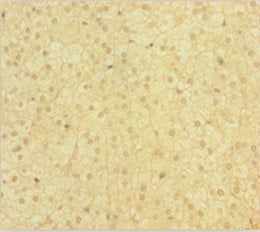
(Immunofluorescence staining of HepG2 cells with AAA118241 at 1:133,counter-stained with DAPI. The cells were fixed in 4% formaldehyde, permeabilized using 0.2% Triton X-100 and blocked in 10% normal Goat Serum. The cells were then incubated with the antibody overnight at 4 degree C.The secondary antibody was Alexa Fluor 488-congugated AffiniPure Goat Anti-Rabbit IgG (H+L).)
IHC (Immunohistochemisry)
(Immunofluorescence staining of HepG2 cells with AAA118241 at 1:133,counter-stained with DAPI. The cells were fixed in 4% formaldehyde, permeabilized using 0.2% Triton X-100 and blocked in 10% normal Goat Serum. The cells were then incubated with the antibody overnight at 4 degree C.The secondary antibody was Alexa Fluor 488-congugated AffiniPure Goat Anti-Rabbit IgG (H+L).)
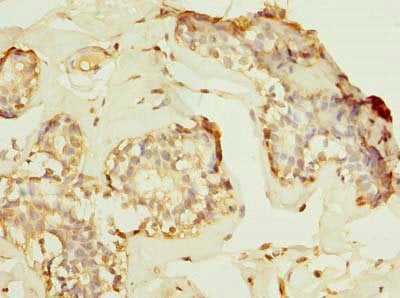
Eukaryotic translation initiation factor 3 subunit G, Polyclonal Antibody (Cat# AAA118241)
 IHC (Immunohiostchemistry)
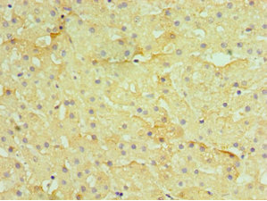
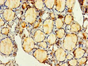
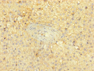
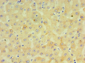
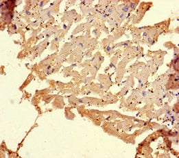
(Immunohistochemistry of paraffin-embedded human liver using AAA118242 at dilution 1:100)
IHC (Immunohiostchemistry)
(Immunohistochemistry of paraffin-embedded human liver using AAA118242 at dilution 1:100)
Histone H2A.Z, Polyclonal Antibody (Cat# AAA118242)
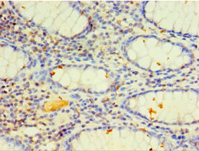
 IHC (Immunohistochemisry)
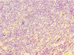
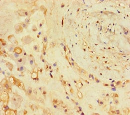
(Immunohistochemistry of paraffin-embedded human rectum tissue using AAA118420 at dilution 1:100)
IHC (Immunohistochemisry)
(Immunohistochemistry of paraffin-embedded human rectum tissue using AAA118420 at dilution 1:100)
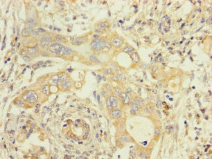
CHI3L2, Polyclonal Antibody (Cat# AAA118420)
Ornithine carbamoyltransferase, Polyclonal Antibody (Cat# AAA118421)
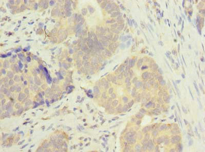
 IHC (Immunohiostchemistry)
(Immunohistochemistry of paraffin-embedded human breast cancer using AAA118428 at dilution of 1:100)
IHC (Immunohiostchemistry)
(Immunohistochemistry of paraffin-embedded human breast cancer using AAA118428 at dilution of 1:100)
FAM53B, Polyclonal Antibody (Cat# AAA118428)
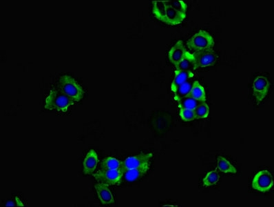
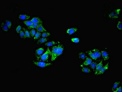
 IF (Immunofluorescence)
(Immunofluorescent analysis of 293 cells using AAA118436 at a dilution of 1:100 and Alexa Fluor 488-congugated AffiniPure Goat Anti-Rabbit IgG(H+L))
IF (Immunofluorescence)
(Immunofluorescent analysis of 293 cells using AAA118436 at a dilution of 1:100 and Alexa Fluor 488-congugated AffiniPure Goat Anti-Rabbit IgG(H+L))
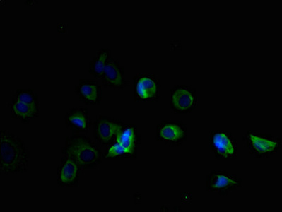
Squalene synthase, Polyclonal Antibody (Cat# AAA118436)
ELAVL4, Polyclonal Antibody (Cat# AAA118438)
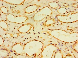
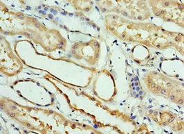
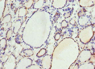
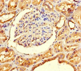
 IHC (Immunohistochemisry)
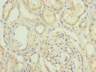
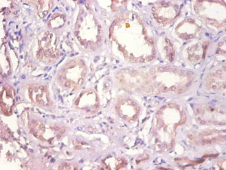
(Immunohistochemistry of paraffin-embedded human kidney using AAA118445 at dilution 1:100)
IHC (Immunohistochemisry)
(Immunohistochemistry of paraffin-embedded human kidney using AAA118445 at dilution 1:100)
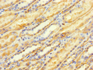
Interferon regulatory factor 2, Polyclonal Antibody (Cat# AAA118445)
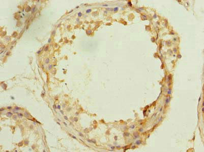
 IHC (Immunohistochemisry)
(Immunohistochemistry of paraffin-embedded human kidney tissue using AAA118456 at dilution of 1:100)
IHC (Immunohistochemisry)
(Immunohistochemistry of paraffin-embedded human kidney tissue using AAA118456 at dilution of 1:100)
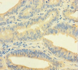
TMEM184B, Polyclonal Antibody (Cat# AAA118456)
 IHC (Immunohiostchemistry)
(Immunohistochemistry of paraffin-embedded human adrenal gland tissue using AAA118457 at dilution 1:100)
IHC (Immunohiostchemistry)
(Immunohistochemistry of paraffin-embedded human adrenal gland tissue using AAA118457 at dilution 1:100)
Crossover junction endonuclease EME1, Polyclonal Antibody (Cat# AAA118457)
 IHC (Immunohiostchemistry)
(Immunohistochemistry of paraffin-embedded human adrenal gland tissue using AAA118459 at dilution of 1:100)
IHC (Immunohiostchemistry)
(Immunohistochemistry of paraffin-embedded human adrenal gland tissue using AAA118459 at dilution of 1:100)
PITPNC1, Polyclonal Antibody (Cat# AAA118459)
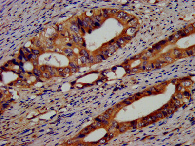
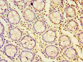
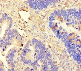
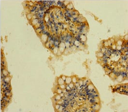
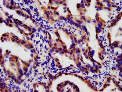
 IHC (Immunohistochemisry)
(Immunohistochemistry of paraffin-embedded human colon cancer using AAA118463 at dilution 1:100)
IHC (Immunohistochemisry)
(Immunohistochemistry of paraffin-embedded human colon cancer using AAA118463 at dilution 1:100)
CYP4F12, Polyclonal Antibody (Cat# AAA118463)

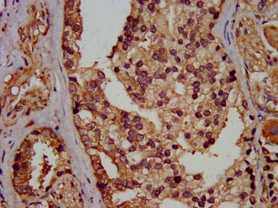
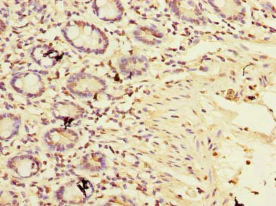
 IHC (Immunohiostchemistry)
(Immunohistochemistry of paraffin-embedded human colon cancer using AAA118464 at dilution 1:100)
IHC (Immunohiostchemistry)
(Immunohistochemistry of paraffin-embedded human colon cancer using AAA118464 at dilution 1:100)
Homeobox protein VENTX, Polyclonal Antibody (Cat# AAA118464)
 IHC (Immunohistochemisry)
(Immunohistochemistry of paraffin-embedded human kidney using AAA118466 at dilution 1:100)
IHC (Immunohistochemisry)
(Immunohistochemistry of paraffin-embedded human kidney using AAA118466 at dilution 1:100)
Dual specificity protein phosphatase 7, Polyclonal Antibody (Cat# AAA118466)
serum albumin, Polyclonal Antibody (Cat# AAA118472)
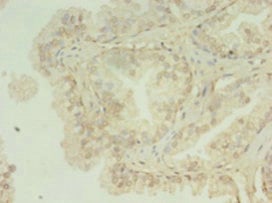
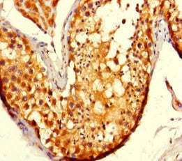
 IHC (Immunohiostchemistry)
(Immunohistochemistry of paraffin-embedded human testis tissue using AAA118476 at dilution of 1:100)
IHC (Immunohiostchemistry)
(Immunohistochemistry of paraffin-embedded human testis tissue using AAA118476 at dilution of 1:100)
MSTO1, Polyclonal Antibody (Cat# AAA118476)
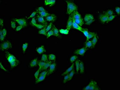
 IHC (Immunohistochemisry)
(Immunofluorescent analysis of PC3 cells using AAA118481 at a dilution of 1:100 and Alexa Fluor 488-congugated AffiniPure Goat Anti-Rabbit IgG(H+L))
IHC (Immunohistochemisry)
(Immunofluorescent analysis of PC3 cells using AAA118481 at a dilution of 1:100 and Alexa Fluor 488-congugated AffiniPure Goat Anti-Rabbit IgG(H+L))
HPCAL1, Polyclonal Antibody (Cat# AAA118481)
 IHC (Immunohiostchemistry)
(Immunohistochemistry of paraffin-embedded human breast cancer using AAA118482 at dilution 1:100)
IHC (Immunohiostchemistry)
(Immunohistochemistry of paraffin-embedded human breast cancer using AAA118482 at dilution 1:100)
Protein atonal homolog 1, Polyclonal Antibody (Cat# AAA118482)
 IHC (Immunohistochemistry)
(Immunohistochemistry of paraffin-embedded human rectal cancer using AAA118485 at dilution 1:100)
IHC (Immunohistochemistry)
(Immunohistochemistry of paraffin-embedded human rectal cancer using AAA118485 at dilution 1:100)
STRN, Polyclonal Antibody (Cat# AAA118485)
 IHC (Immunohiostchemistry)
(Immunohistochemistry of paraffin-embedded human adrenal gland tissue using AAA118486 at dilution of 1:100)
IHC (Immunohiostchemistry)
(Immunohistochemistry of paraffin-embedded human adrenal gland tissue using AAA118486 at dilution of 1:100)
TIMM9, Polyclonal Antibody (Cat# AAA118486)
 IHC (Immunohiostchemistry)
(Immunohistochemistry of paraffin-embedded human rectal cancer using AAA118487 at dilution 1:100)
IHC (Immunohiostchemistry)
(Immunohistochemistry of paraffin-embedded human rectal cancer using AAA118487 at dilution 1:100)
Cyclin-dependent kinase 9, Polyclonal Antibody (Cat# AAA118487)
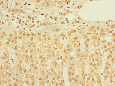
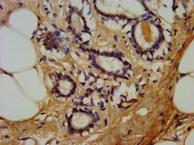
 IHC (Immunohiostchemistry)
(Immunohistochemistry of paraffin-embedded human gastric cancer using AAA118492 at dilution of 1:100)
IHC (Immunohiostchemistry)
(Immunohistochemistry of paraffin-embedded human gastric cancer using AAA118492 at dilution of 1:100)
NRIP3, Polyclonal Antibody (Cat# AAA118492)
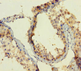
 IHC (Immunohiostchemistry)
(IHC image of AAA118493 diluted at 1:1200 and staining in paraffin-embedded human testis tissue performed on a Leica BondTM system. After dewaxing and hydration, antigen retrieval was mediated by high pressure in a citrate buffer (pH 6.0). Section was blocked with 10% normal goat serum 30min at RT. Then primary antibody (1% BSA) was incubated at 4 degree C overnight. The primary is detected by a biotinylated secondary antibody and visualized using an HRP conjugated SP system.)
IHC (Immunohiostchemistry)
(IHC image of AAA118493 diluted at 1:1200 and staining in paraffin-embedded human testis tissue performed on a Leica BondTM system. After dewaxing and hydration, antigen retrieval was mediated by high pressure in a citrate buffer (pH 6.0). Section was blocked with 10% normal goat serum 30min at RT. Then primary antibody (1% BSA) was incubated at 4 degree C overnight. The primary is detected by a biotinylated secondary antibody and visualized using an HRP conjugated SP system.)
PSMC6, Polyclonal Antibody (Cat# AAA118493)
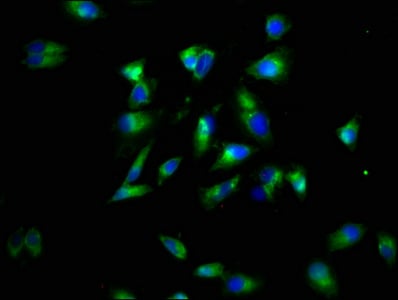
 IHC (Immunohistochemisry)
(Immunofluorescent analysis of Hela cells using AAA118494 at a dilution of 1:100 and Alexa Fluor 488-congugated AffiniPure Goat Anti-Rabbit IgG(H+L))
IHC (Immunohistochemisry)
(Immunofluorescent analysis of Hela cells using AAA118494 at a dilution of 1:100 and Alexa Fluor 488-congugated AffiniPure Goat Anti-Rabbit IgG(H+L))
HNRNPL, Polyclonal Antibody (Cat# AAA118494)
 IHC (Immunohistochemisry)
(Immunofluorescent analysis of hepg2 using AAA118496 at a dilution of 1:100 and Alexa Fluor 488-congugated AffiniPure Goat Anti-Rabbit IgG(H+L))
IHC (Immunohistochemisry)
(Immunofluorescent analysis of hepg2 using AAA118496 at a dilution of 1:100 and Alexa Fluor 488-congugated AffiniPure Goat Anti-Rabbit IgG(H+L))
C-type lectin domain family 11 member A, Polyclonal Antibody (Cat# AAA118496)
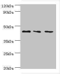
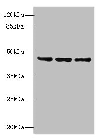
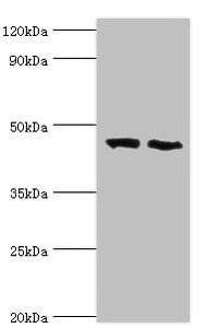
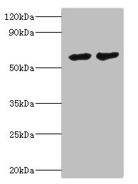
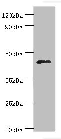
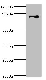
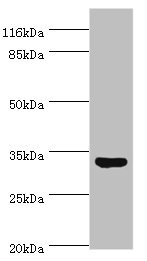
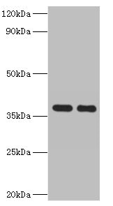
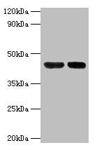
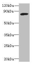 WB (Western Blot)
(Western blotAll lanes: PBXIP1 antibody at 4ug/ml+ A549 whole cell lysateSecondaryGoat polyclonal to rabbit at 1/10000 dilutionPredicted band size: 81,78,73 kDaObserved band size: 81 kDa)
WB (Western Blot)
(Western blotAll lanes: PBXIP1 antibody at 4ug/ml+ A549 whole cell lysateSecondaryGoat polyclonal to rabbit at 1/10000 dilutionPredicted band size: 81,78,73 kDaObserved band size: 81 kDa)
PBXIP1, Polyclonal Antibody (Cat# AAA118502)
 IHC (Immunohiostchemistry)
(Immunohistochemistry of paraffin-embedded human skeletal muscle tissue using AAA117918 at dilution of 1:100)
IHC (Immunohiostchemistry)
(Immunohistochemistry of paraffin-embedded human skeletal muscle tissue using AAA117918 at dilution of 1:100)
Upstream stimulatory factor 1, Polyclonal Antibody (Cat# AAA117918)
Adiponectin, Polyclonal Antibody (Cat# AAA117920)
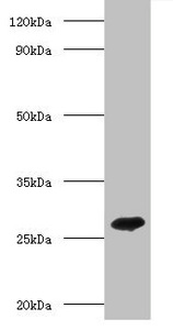 WB (Western Blot)
(Western blotAll lanes: Adiponectin antibody at 2ug/ml+recombinant Adiponectin protein 0.1ugSecondaryGoat polyclonal to Rabbit IgG at 1/10000 dilutionPredicted band size: 26kDaObserved band size: 26kDa)
WB (Western Blot)
(Western blotAll lanes: Adiponectin antibody at 2ug/ml+recombinant Adiponectin protein 0.1ugSecondaryGoat polyclonal to Rabbit IgG at 1/10000 dilutionPredicted band size: 26kDaObserved band size: 26kDa)
Adiponectin, Polyclonal Antibody (Cat# AAA117921)
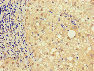
 IHC (Immunohiostchemistry)
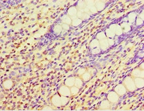
(Immunohistochemistry of paraffin-embedded human pancreatic cancer using AAA117923 at dilution of 1:100)
IHC (Immunohiostchemistry)
(Immunohistochemistry of paraffin-embedded human pancreatic cancer using AAA117923 at dilution of 1:100)
Fos-related antigen 2, Polyclonal Antibody (Cat# AAA117923)
 IHC (Immunohistochemisry)
(Immunohistochemistry of paraffin-embedded human prostate cancer using AAA117925 at dilution of 1:100)
IHC (Immunohistochemisry)
(Immunohistochemistry of paraffin-embedded human prostate cancer using AAA117925 at dilution of 1:100)
ATP-dependent RNA helicase DDX19A, Polyclonal Antibody (Cat# AAA117925)
Prostate-specific antigen, Polyclonal Antibody (Cat# AAA117927)

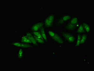
 IF (Immunofluorescence)
(Immunofluorescent analysis of HepG2 cells using AAA117928 at a dilution of 1:100 and Alexa Fluor 488-congugated AffiniPure Goat Anti-Rabbit IgG(H+L))
IF (Immunofluorescence)
(Immunofluorescent analysis of HepG2 cells using AAA117928 at a dilution of 1:100 and Alexa Fluor 488-congugated AffiniPure Goat Anti-Rabbit IgG(H+L))
Selenoprotein P, Polyclonal Antibody (Cat# AAA117928)
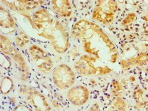
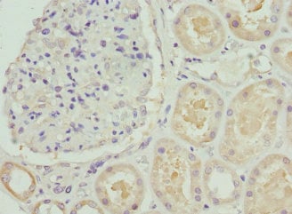
 IHC (Immunohistochemisry)
(Immunohistochemistry of paraffin-embedded human kidney tissue using AAA117947 at dilution of 1:100)
IHC (Immunohistochemisry)
(Immunohistochemistry of paraffin-embedded human kidney tissue using AAA117947 at dilution of 1:100)
Aldose 1-epimerase, Polyclonal Antibody (Cat# AAA117947)
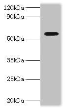
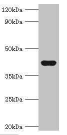
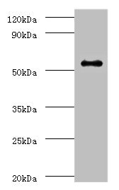
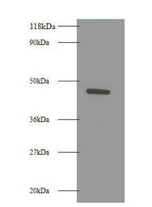
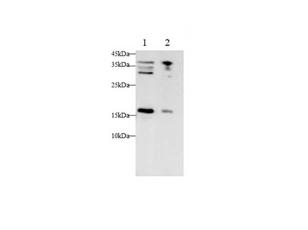
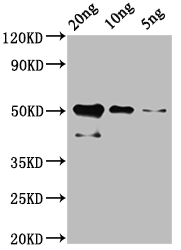 WB (Western Blot)
(Western BlotPositive WB detected in: Recombinant cry1Ac protein at 20ng, 10ng, 5ngAll lanes: cry1Ac antibody at 1:2000SecondaryGoat polyclonal to rabbit IgG at 1/50000 dilutionPredicted band size: 51 kDaObserved band size: 51 kDa)
WB (Western Blot)
(Western BlotPositive WB detected in: Recombinant cry1Ac protein at 20ng, 10ng, 5ngAll lanes: cry1Ac antibody at 1:2000SecondaryGoat polyclonal to rabbit IgG at 1/50000 dilutionPredicted band size: 51 kDaObserved band size: 51 kDa)
Bacillus thuringiensis subsp. Kurstaki Pesticidal crystal protein cry1Ac, Polyclonal Antibody (Cat# AAA117949)
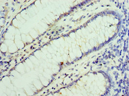
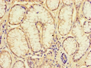
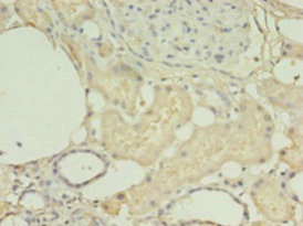
 IHC (Immunohiostchemistry)
(Immunohistochemistry of paraffin-embedded human small intestine tissue using AAA117952 at dilution 1:100)
IHC (Immunohiostchemistry)
(Immunohistochemistry of paraffin-embedded human small intestine tissue using AAA117952 at dilution 1:100)
Clathrin light chain B, Polyclonal Antibody (Cat# AAA117952)
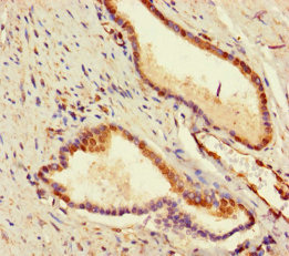 IHC (Immunohistochemisry)
(Immunohistochemistry of paraffin-embedded human prostate cancer using AAA117953 at dilution of 1:100)
IHC (Immunohistochemisry)
(Immunohistochemistry of paraffin-embedded human prostate cancer using AAA117953 at dilution of 1:100)
Uncharacterized aarF domain-containing protein kinase 2, Polyclonal Antibody (Cat# AAA117953)
RNA-splicing ligase RtcB, Polyclonal Antibody (Cat# AAA117956)
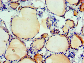
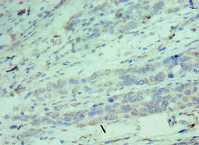
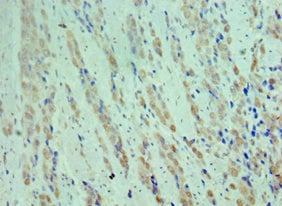
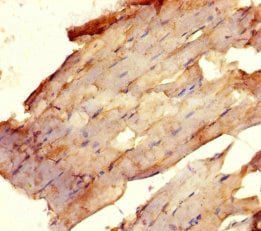
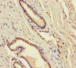
 IHC (Immunohiostchemistry)
(Immunohistochemistry of paraffin-embedded human heart tissue using AAA117958 at dilution of 1:100)
IHC (Immunohiostchemistry)
(Immunohistochemistry of paraffin-embedded human heart tissue using AAA117958 at dilution of 1:100)
Collagen alpha-1, Polyclonal Antibody (Cat# AAA117958)
 IF (Immunofluorescence)
(Immunofluorescent analysis of A549 cells using AAA117959 at a dilution of 1:100 and Alexa Fluor 488-congugated AffiniPure Goat Anti-Rabbit IgG(H+L))
IF (Immunofluorescence)
(Immunofluorescent analysis of A549 cells using AAA117959 at a dilution of 1:100 and Alexa Fluor 488-congugated AffiniPure Goat Anti-Rabbit IgG(H+L))
Dehydrogenase/reductase SDR family member 9, Polyclonal Antibody (Cat# AAA117959)
Torpedo californica Acetylcholine receptor subunit alpha, Polyclonal Antibody (Cat# AAA117960)
 IHC (Immunohiostchemistry)
(Immunohistochemistry of paraffin-embedded human spleen tissue using AAA117968 at dilution 1:100)
IHC (Immunohiostchemistry)
(Immunohistochemistry of paraffin-embedded human spleen tissue using AAA117968 at dilution 1:100)
Tumor necrosis factor receptor superfamily member 19L, Polyclonal Antibody (Cat# AAA117968)
 IHC (Immunohistochemisry)
(Immunofluorescence staining of Hela cells with AAA117970 at 1:60,counter-stained with DAPI. The cells were fixed in 4% formaldehyde, permeabilized using 0.2% Triton X-100 and blocked in 10% normal Goat Serum. The cells were then incubated with the antibody overnight at 4 degree C.The secondary antibody was Alexa Fluor 488-congugated AffiniPure Goat Anti-Rabbit IgG (H+L).)
IHC (Immunohistochemisry)
(Immunofluorescence staining of Hela cells with AAA117970 at 1:60,counter-stained with DAPI. The cells were fixed in 4% formaldehyde, permeabilized using 0.2% Triton X-100 and blocked in 10% normal Goat Serum. The cells were then incubated with the antibody overnight at 4 degree C.The secondary antibody was Alexa Fluor 488-congugated AffiniPure Goat Anti-Rabbit IgG (H+L).)
Tumor necrosis factor receptor superfamily member 3, Polyclonal Antibody (Cat# AAA117970)
Angiopoietin-1, Polyclonal Antibody (Cat# AAA117971)
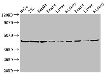
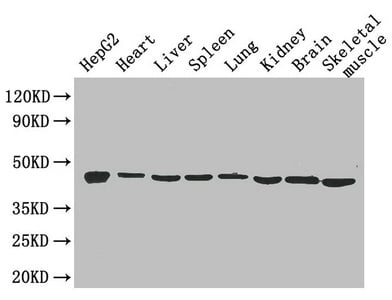 WB (Western Blot)
(Western BlotPositive WB detected in: hepG2 cell,mouse heart,mouse liver,mouse spleen,mouse lung,mouse kidney,mouse brain,mouse skeletal muscleAll lanes: Eno1 antibody at 2.6ug/mlSecondaryGoat polyclonal to rabbit IgG at 1/50000 dilutionPredicted band size: 47kDaObserved band size: 47 kDa)
WB (Western Blot)
(Western BlotPositive WB detected in: hepG2 cell,mouse heart,mouse liver,mouse spleen,mouse lung,mouse kidney,mouse brain,mouse skeletal muscleAll lanes: Eno1 antibody at 2.6ug/mlSecondaryGoat polyclonal to rabbit IgG at 1/50000 dilutionPredicted band size: 47kDaObserved band size: 47 kDa)
Alpha-enolase, Polyclonal Antibody (Cat# AAA117972)
Aerobic glycerol-3-phosphate dehydrogenase, Polyclonal Antibody (Cat# AAA117974)
Bacillus subtilis Alpha-galactosidase, Polyclonal Antibody (Cat# AAA117975)
Arabidopsis thaliana Protein ABSCISIC ACID-INSENSITIVE 5, Polyclonal Antibody (Cat# AAA117976)
Mycobacterium tuberculosis Antigen 85-A, Polyclonal Antibody (Cat# AAA117980)
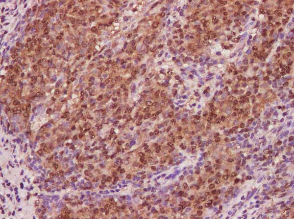
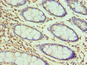
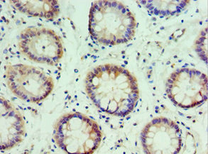
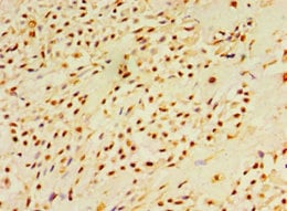
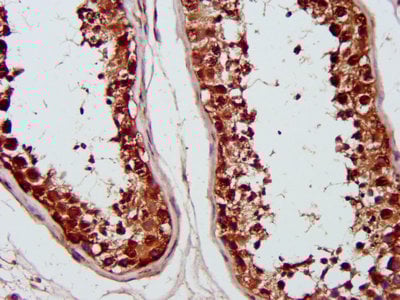
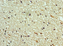
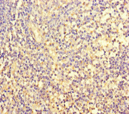
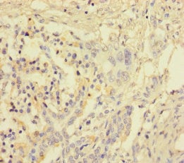
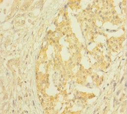
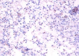
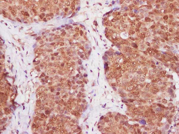 IHC (Immunohistochemisry)
(Immunohistochemistry of paraffin-embedded Human endometrium cancer using CDKN2A Polyclonal Antibody at dilution of 1:600.)
IHC (Immunohistochemisry)
(Immunohistochemistry of paraffin-embedded Human endometrium cancer using CDKN2A Polyclonal Antibody at dilution of 1:600.)
CDKN2A, Polyclonal Antibody (Cat# AAA179743)
What are Polyclonal Antibodies?
Polyclonal antibodies are antibodies that come from multiple B cell clones of a host animal. The typical hosts used for the majority of polyclonal antibody production are rabbits, goats, sheep, and donkeys. These polyclonal antibodies, once having identified their target, will bind to different epitopes located at different regions or sequences on the same protein/antigen. As a result, they are ideal at locating and binding to the target, even if the target is in very low concentrations (due to many different antibodies being able to bind to the same target molecule, which allows for significant amplification of a downstream signal).
Polyclonal antibodies are typically produced by injecting an antigen into a host animal, which causes the animal’s immune system to attack the foreign antigen by mass generating antibodies against it. After a period of time, serum is collected from the animal and purified using physicochemical fractionation, class-specific affinity purification, and/or antigen-affinity purification.
Key Uses of Polyclonal Antibodies
- Western Blotting: This method is used to find specific proteins in biological samples after separating them by size.
- Immunohistochemistry: IHC helps visualize the location of proteins in tissue sections using various staining techniques.
- ELISA: (Enzyme-Linked Immunosorbent Assay) is typically used to identify specific protein quantities in a sample. ELISAs can be either “Quantitative” or “Qualitative”.
- Flow Cytometry: technique that identifies and measures the specific protein on the surface or inside the cells in a fluid suspension.
- Immunoprecipitation: IP isolates and studies a specific protein from a complex mixture using antibodies.
Why Buy Polyclonal Antibodies from AAA Biotech?
1. Ideal for Various Applications
Our antibodies are generally going to be validated for use in multiple types of assays, including ELISA, Western Blotting, Immunohistochemistry, Immunoprecipitation, amongst others. They are ideal for a wide range of research applications.
2. Rigorous Quality Control
All of the antibodies in our catalog undergo strict quality testing to ensure specificity, sensitivity, and consistent performance. We are confident in the ability of our antibodies to provide you with accurate results.
3. Wide Assortment of Antibodies
Antibodies in are catalog can be found for both common and exotic species, and these antibodies are also available in both conjugated and recombinant forms to suit many diverse experimental needs.
4. Highly Purified
Our antibodies are available in purified forms with over 85% purity, as confirmed by SDS-PAGE. They are also available with tags such as His, Flag, GST, or MBP. We cater to customers worldwide.
FAQ
1. How are polyclonal antibodies produced?
Traditionally, polyclonal antibodies are produced by injecting an antigen into a host animal (such as a rabbit or goat), which then triggers an immune response from the host animal. The animal’s B cells produce antibodies that will recognize different parts of the injected antigen. These antibodies are then collected from the animal’s blood and purified for use.
2. How do polyclonal antibodies differ from monoclonal antibodies?
Polyclonal antibodies are a mix of antibodies that bind to different locations (epitopes) of the same antigen, while monoclonal antibodies are identical and bind to just one specific epitope. This makes polyclonal antibodies more versatile and better at detecting proteins that may be present in low quantities or in altered/modified forms.
3. How should I store polyclonal antibodies?
Polyclonal antibodies should be stored at 4°C for short-term use (up to a few weeks) and at -20°C or -80°C for long-term storage. Avoid repeated freeze-thaw cycles by dividing them into small aliquots. Always check the datasheet for specific storage instructions.