Filters
▼Clonality
▼Type
▼Reactivity
▼Gene Name
▼Isotype
▼Host
▼Application
▼Clone
▼Polyclonal Antibodies
At AAA Biotech also known as AAA Bio or AAABio, we provide a broad range of purified polyclonal antibodies (pAbs) that are able to all be browsed online through our website. Due to their high specificity and strong binding affinity, these antibodies are ideal for wide swathes of research and experimental applications.
Our polyclonal antibodies can easily support your work, whether you use them for Western Blotting, Immunocytochemistry (with or without Immunofluorescence used in conjunction), Immunohistochemistry, Immunoprecipitation, and ELISA tests. We highly encourage you to browse our range of pAbs and choose the one that best suits your experimental model.
Viewing 7550-7600 of 96805 product results
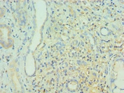
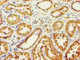
 IHC (Immunohistochemisry)
(Immunohistochemistry of paraffin-embedded human kidney tissue using AAA118783 at dilution 1:100)
IHC (Immunohistochemisry)
(Immunohistochemistry of paraffin-embedded human kidney tissue using AAA118783 at dilution 1:100)
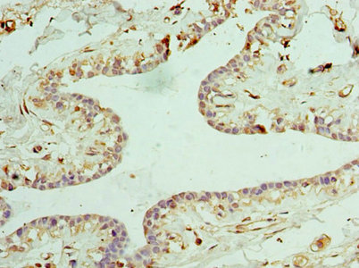
ACAT1, Polyclonal Antibody (Cat# AAA118783)
 IHC (Immunohiostchemistry)
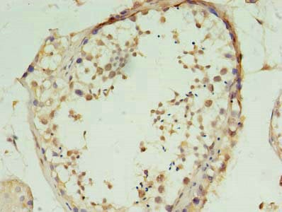
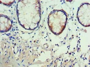
(Immunohistochemistry of paraffin-embedded human testis tissue using AAA118785 at dilution of 1:100)
IHC (Immunohiostchemistry)
(Immunohistochemistry of paraffin-embedded human testis tissue using AAA118785 at dilution of 1:100)
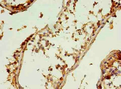
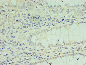
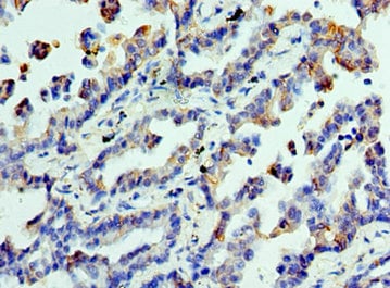
Collagen alpha-6, Polyclonal Antibody (Cat# AAA118785)
 IHC (Immunohistochemisry)
(Immunohistochemistry of paraffin-embedded human brain tissuegland using AAA118789 at dilution 1:100)
IHC (Immunohistochemisry)
(Immunohistochemistry of paraffin-embedded human brain tissuegland using AAA118789 at dilution 1:100)
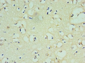
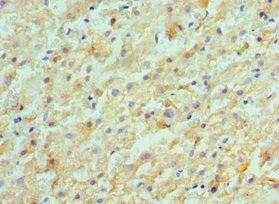
CROT, Polyclonal Antibody (Cat# AAA118789)
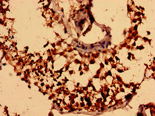
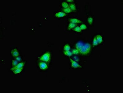
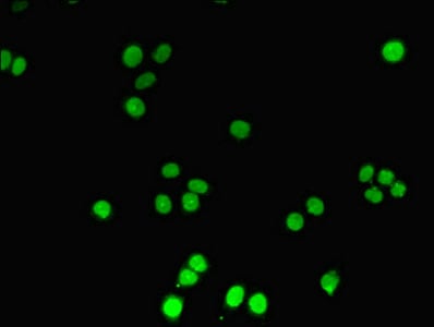 IF (Immunofluorescence)
(Immunofluorescent analysis of PC3 cells using AAA118791 at a dilution of 1:100 and Alexa Fluor 488-congugated AffiniPure Goat Anti-Rabbit IgG(H+L))
IF (Immunofluorescence)
(Immunofluorescent analysis of PC3 cells using AAA118791 at a dilution of 1:100 and Alexa Fluor 488-congugated AffiniPure Goat Anti-Rabbit IgG(H+L))
DNTT, Polyclonal Antibody (Cat# AAA118791)
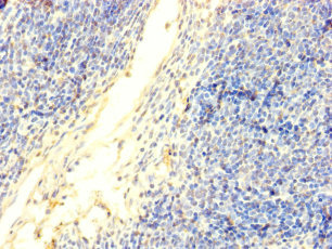
 IHC (Immunohiostchemistry)
(Immunohistochemistry of paraffin-embedded human testis using AAA118794 at dilution 1:100)
IHC (Immunohiostchemistry)
(Immunohistochemistry of paraffin-embedded human testis using AAA118794 at dilution 1:100)
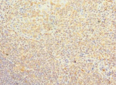
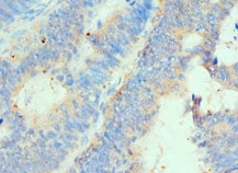
MAGEC2, Polyclonal Antibody (Cat# AAA118794)
 IHC (Immunohiostchemistry)
(Immunohistochemistry of paraffin-embedded human brain tissue at dilution 1:100)
IHC (Immunohiostchemistry)
(Immunohistochemistry of paraffin-embedded human brain tissue at dilution 1:100)
SOX6, Polyclonal Antibody (Cat# AAA118795)
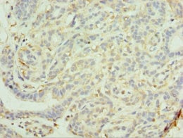
 IHC (Immunohiostchemistry)
(Immunohistochemistry of paraffin-embedded human breast cancer using AAA118796 at dilution of 1:100)
IHC (Immunohiostchemistry)
(Immunohistochemistry of paraffin-embedded human breast cancer using AAA118796 at dilution of 1:100)
PSD4, Polyclonal Antibody (Cat# AAA118796)
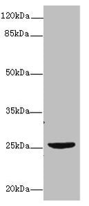
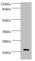
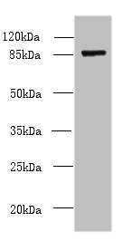
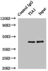 IP (Immunoprecipitation)
(Immunoprecipitating TIA1 in Hela whole cell lysateLane 1: Rabbit monoclonal IgG(1ug)instead of AAA118799 in Hela whole cell lysate. For western blotting, a HRP-conjugated anti-rabbit IgG, specific to the non-reduced form of IgG was used as the Secondary antibody (1/50000)Lane 2: AAA118799(4ug)+ Hela whole cell lysate(500ug)Lane 3: Hela whole cell lysate (20ug))
IP (Immunoprecipitation)
(Immunoprecipitating TIA1 in Hela whole cell lysateLane 1: Rabbit monoclonal IgG(1ug)instead of AAA118799 in Hela whole cell lysate. For western blotting, a HRP-conjugated anti-rabbit IgG, specific to the non-reduced form of IgG was used as the Secondary antibody (1/50000)Lane 2: AAA118799(4ug)+ Hela whole cell lysate(500ug)Lane 3: Hela whole cell lysate (20ug))
Nucleolysin TIA-1 isoform p40, Polyclonal Antibody (Cat# AAA118799)

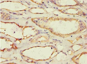
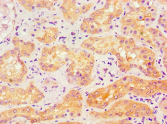
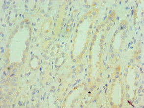
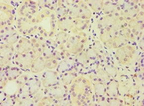
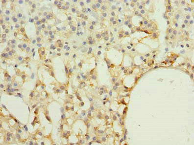
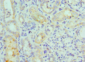
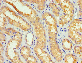
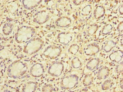
 IHC (Immunohiostchemistry)
(Immunohistochemistry of paraffin-embedded human kidney using AAA118801 at dilution 1:100)
IHC (Immunohiostchemistry)
(Immunohistochemistry of paraffin-embedded human kidney using AAA118801 at dilution 1:100)
Myc box-dependent-interacting protein 1, Polyclonal Antibody (Cat# AAA118801)
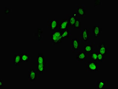
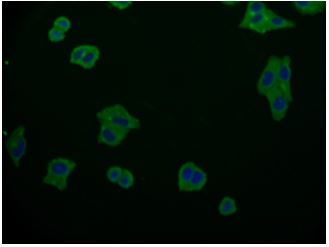
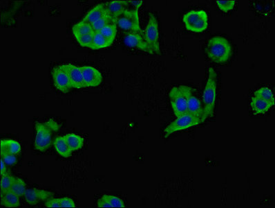 IF (Immunofluorescence)
(Immunofluorescent analysis of PC3 cells using AAA118802 at a dilution of 1:100 and Alexa Fluor 488-congugated AffiniPure Goat Anti-Rabbit IgG(H+L))
IF (Immunofluorescence)
(Immunofluorescent analysis of PC3 cells using AAA118802 at a dilution of 1:100 and Alexa Fluor 488-congugated AffiniPure Goat Anti-Rabbit IgG(H+L))
NEGR1, Polyclonal Antibody (Cat# AAA118802)

 IHC (Immunohistochemisry)
(Immunofluorescent analysis of PC3 cells using AAA118810 at a dilution of 1:100 and Alexa Fluor 488-congugated AffiniPure Goat Anti-Rabbit IgG(H+L))
IHC (Immunohistochemisry)
(Immunofluorescent analysis of PC3 cells using AAA118810 at a dilution of 1:100 and Alexa Fluor 488-congugated AffiniPure Goat Anti-Rabbit IgG(H+L))
ACVR1, Polyclonal Antibody (Cat# AAA118810)
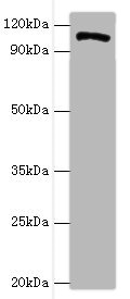
Chlamydia trachomatis Major outer membrane porin, Polyclonal Antibody (Cat# AAA118819)
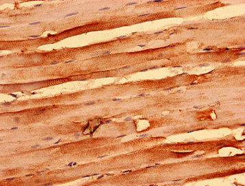
Troponin T, Polyclonal Antibody (Cat# AAA118822)
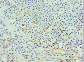
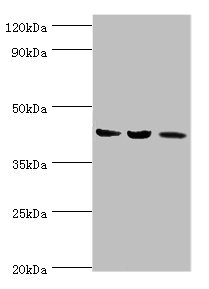
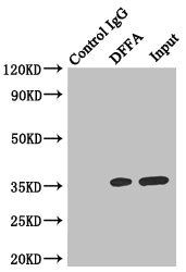 IP (Immunoprecipitation)
(Immunoprecipitating DFFA in HeLa whole cell lysateLane 1: Rabbit monoclonal IgG(1ug)instead of AAA118823 in HeLa whole cell lysate. For western blotting, a HRP-conjugated anti-rabbit IgG, specific to the non-reduced form of IgG was used as the Secondary antibody (1/50000)Lane 2: AAA118823(4ug)+ HeLa whole cell lysate(500ug)Lane 3: HeLa whole cell lysate (20ug))
IP (Immunoprecipitation)
(Immunoprecipitating DFFA in HeLa whole cell lysateLane 1: Rabbit monoclonal IgG(1ug)instead of AAA118823 in HeLa whole cell lysate. For western blotting, a HRP-conjugated anti-rabbit IgG, specific to the non-reduced form of IgG was used as the Secondary antibody (1/50000)Lane 2: AAA118823(4ug)+ HeLa whole cell lysate(500ug)Lane 3: HeLa whole cell lysate (20ug))
DFFA, Polyclonal Antibody (Cat# AAA118823)
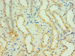
 IHC (Immunohiostchemistry)
(Immunohistochemistry of paraffin-embedded human kidney tissue using AAA118824 at dilution 1:100)
IHC (Immunohiostchemistry)
(Immunohistochemistry of paraffin-embedded human kidney tissue using AAA118824 at dilution 1:100)
TNXB, Polyclonal Antibody (Cat# AAA118824)

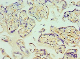
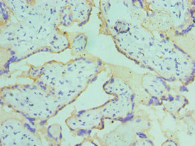
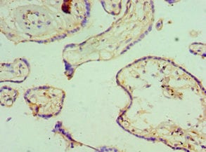 IHC (Immunohistochemisry)
(Immunohistochemistry of paraffin-embedded human placenta using AAA118669 at dilution 1:100)
IHC (Immunohistochemisry)
(Immunohistochemistry of paraffin-embedded human placenta using AAA118669 at dilution 1:100)
Intermediate conductance calcium-activated potassium channel protein 4, Polyclonal Antibody (Cat# AAA118669)
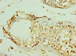
 IHC (Immunohiostchemistry)
(Immunohistochemistry of paraffin-embedded human testis tissue using AAA118670 at dilution of 1:100)
IHC (Immunohiostchemistry)
(Immunohistochemistry of paraffin-embedded human testis tissue using AAA118670 at dilution of 1:100)
TTC30B, Polyclonal Antibody (Cat# AAA118670)
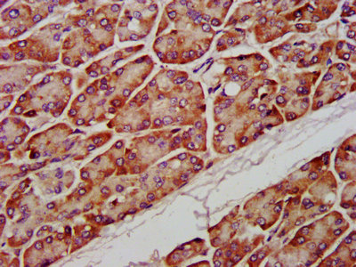
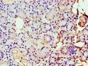
 IHC (Immunohistochemisry)
(Immunohistochemistry of paraffin-embedded human pancreas tissue using AAA118671 at dilution 1:100)
IHC (Immunohistochemisry)
(Immunohistochemistry of paraffin-embedded human pancreas tissue using AAA118671 at dilution 1:100)
ARF3, Polyclonal Antibody (Cat# AAA118671)
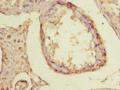
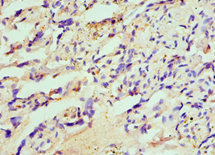
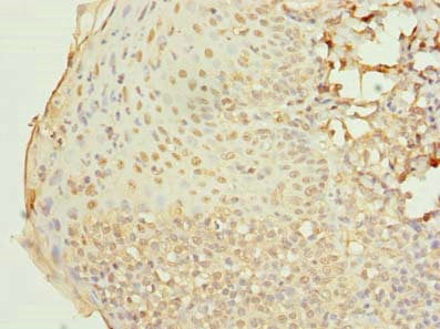
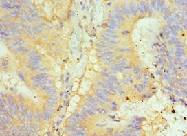
 IHC (Immunohiostchemistry)
(Immunohistochemistry of paraffin-embedded human lung cancer using AAA118676 at dilution 1:100)
IHC (Immunohiostchemistry)
(Immunohistochemistry of paraffin-embedded human lung cancer using AAA118676 at dilution 1:100)
CLTA, Polyclonal Antibody (Cat# AAA118676)

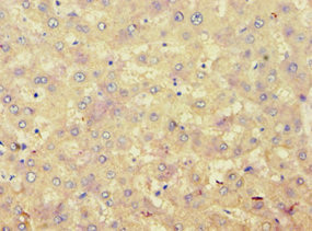
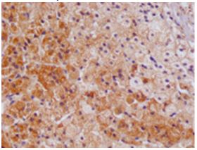
 IHC (Immunohiostchemistry)
(Immunohistochemistry of paraffin-embedded human liver cancer using AAA118685 at dilution 1:100)
IHC (Immunohiostchemistry)
(Immunohistochemistry of paraffin-embedded human liver cancer using AAA118685 at dilution 1:100)
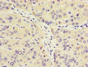
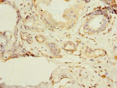
Activin receptor type-2A, Polyclonal Antibody (Cat# AAA118685)
 IHC (Immunohistochemisry)
(Immunohistochemistry of paraffin-embedded human placenta using AAA118688 at dilution 1:100)
IHC (Immunohistochemisry)
(Immunohistochemistry of paraffin-embedded human placenta using AAA118688 at dilution 1:100)
Huntingtin-interacting protein 1, Polyclonal Antibody (Cat# AAA118688)
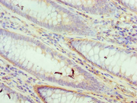
 IHC (Immunohiostchemistry)
(Immunohistochemistry of paraffin-embedded human epityphlon using AAA118689 at dilution 1:100)
IHC (Immunohiostchemistry)
(Immunohistochemistry of paraffin-embedded human epityphlon using AAA118689 at dilution 1:100)
PSEN1, Polyclonal Antibody (Cat# AAA118689)
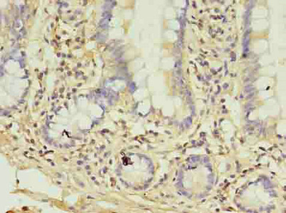
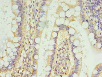
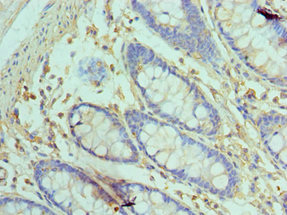
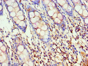
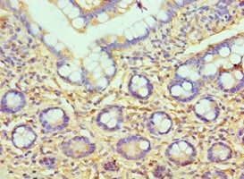 IHC (Immunohiostchemistry)
(Immunohistochemistry of paraffin-embedded human small intestine using AAA118690 at dilution 1:100)
IHC (Immunohiostchemistry)
(Immunohistochemistry of paraffin-embedded human small intestine using AAA118690 at dilution 1:100)
HYAL3, Polyclonal Antibody (Cat# AAA118690)
 IHC (Immunohiostchemistry)
(Immunohistochemistry of paraffin-embedded human adrenal gland tissue using AAA118691 at dilution of 1:100)
IHC (Immunohiostchemistry)
(Immunohistochemistry of paraffin-embedded human adrenal gland tissue using AAA118691 at dilution of 1:100)
RAB33A, Polyclonal Antibody (Cat# AAA118691)
 IHC (Immunohiostchemistry)
(IHC image of AAA118692 diluted at 1:300 and staining in paraffin-embedded human lung cancer performed on a Leica BondTM system. After dewaxing and hydration, antigen retrieval was mediated by high pressure in a citrate buffer (pH 6.0). Section was blocked with 10% normal goat serum 30min at RT. Then primary antibody (1% BSA) was incubated at 4°C overnight. The primary is detected by a biotinylated secondary antibody and visualized using an HRP conjugated SP system.)
IHC (Immunohiostchemistry)
(IHC image of AAA118692 diluted at 1:300 and staining in paraffin-embedded human lung cancer performed on a Leica BondTM system. After dewaxing and hydration, antigen retrieval was mediated by high pressure in a citrate buffer (pH 6.0). Section was blocked with 10% normal goat serum 30min at RT. Then primary antibody (1% BSA) was incubated at 4°C overnight. The primary is detected by a biotinylated secondary antibody and visualized using an HRP conjugated SP system.)
Neurotensin/neuromedin N, Polyclonal Antibody (Cat# AAA118692)
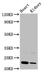
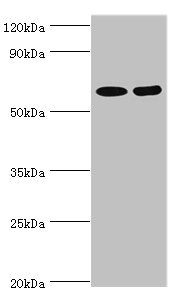
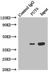 IP (Immunoprecipitation)
(Immunoprecipitating FUT6 in Hela whole cell lysateLane 1: Rabbit monoclonal IgG(1ug)instead of AAA118696 in Hela whole cell lysate. For western blotting, a HRP-conjugated anti-rabbit IgG, specific to the non-reduced form of IgG was used as the Secondary antibody (1/50000)Lane 2: AAA118696(4ug)+ Hela whole cell lysate(500ug)Lane 3: Hela whole cell lysate (20ug))
IP (Immunoprecipitation)
(Immunoprecipitating FUT6 in Hela whole cell lysateLane 1: Rabbit monoclonal IgG(1ug)instead of AAA118696 in Hela whole cell lysate. For western blotting, a HRP-conjugated anti-rabbit IgG, specific to the non-reduced form of IgG was used as the Secondary antibody (1/50000)Lane 2: AAA118696(4ug)+ Hela whole cell lysate(500ug)Lane 3: Hela whole cell lysate (20ug))
FUT6, Polyclonal Antibody (Cat# AAA118696)
Cryptomeria japonica Polygalacturonase, Polyclonal Antibody (Cat# AAA118697)
 IHC (Immunohiostchemistry)
(Immunohistochemistry of paraffin-embedded human placenta using AAA118698 at dilution 1:100)
IHC (Immunohiostchemistry)
(Immunohistochemistry of paraffin-embedded human placenta using AAA118698 at dilution 1:100)
Glutaredoxin-2, Polyclonal Antibody (Cat# AAA118698)
 IHC (Immunohistochemisry)
(Immunohistochemistry of paraffin-embedded human pancreas using AAA118699 at dilution 1:100)
IHC (Immunohistochemisry)
(Immunohistochemistry of paraffin-embedded human pancreas using AAA118699 at dilution 1:100)
Pancreatic alpha-amylase, Polyclonal Antibody (Cat# AAA118699)
Peptoclostridium difficile Toxin B, Polyclonal Antibody (Cat# AAA118703)
 IHC (Immunohiostchemistry)
(Immunohistochemistry of paraffin-embedded human liver tissue using AAA118704 at dilution 1:100)
IHC (Immunohiostchemistry)
(Immunohistochemistry of paraffin-embedded human liver tissue using AAA118704 at dilution 1:100)
Alpha/beta hydrolase domain-containing protein 14B, Polyclonal Antibody (Cat# AAA118704)
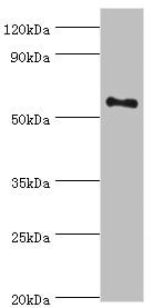
 IF (Immunofluorescence)
(Immunofluorescent analysis of Hela cells using AAA118706 at a dilution of 1:100 and Alexa Fluor 488-congugated AffiniPure Goat Anti-Rabbit IgG(H+L))
IF (Immunofluorescence)
(Immunofluorescent analysis of Hela cells using AAA118706 at a dilution of 1:100 and Alexa Fluor 488-congugated AffiniPure Goat Anti-Rabbit IgG(H+L))
Mitogen-activated protein kinase 7, Polyclonal Antibody (Cat# AAA118706)
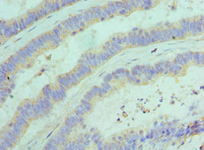
 IHC (Immunohistochemisry)
(Immunohistochemistry of paraffin-embedded human small intestine tissue using AAA118707 at dilution of 1:100)
IHC (Immunohistochemisry)
(Immunohistochemistry of paraffin-embedded human small intestine tissue using AAA118707 at dilution of 1:100)
FSCB, Polyclonal Antibody (Cat# AAA118707)
Plasmodium falciparum L-lactate dehydrogenase, Polyclonal Antibody (Cat# AAA118711)
Toxoplasma gondii Inflammatory profilin, Polyclonal Antibody (Cat# AAA118717)
Peanut Arachin Ahy-3, Polyclonal Antibody (Cat# AAA118718)
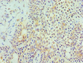
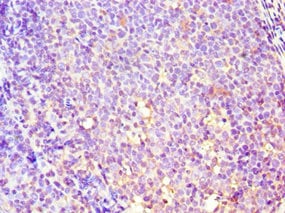
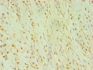 IHC (Immunohiostchemistry)
(Immunohistochemistry of paraffin-embedded human breast cancer using AAA118723 at dilution 1:100)
IHC (Immunohiostchemistry)
(Immunohistochemistry of paraffin-embedded human breast cancer using AAA118723 at dilution 1:100)
Tyrosine-protein kinase Blk, Polyclonal Antibody (Cat# AAA118723)
 IHC (Immunohiostchemistry)
(IHC image of AAA118730 diluted at 1:1200 and staining in paraffin-embedded human pancreatic tissue performed on a Leica BondTM system. After dewaxing and hydration, antigen retrieval was mediated by high pressure in a citrate buffer (pH 6.0). Section was blocked with 10% normal goat serum 30min at RT. Then primary antibody (1% BSA) was incubated at 4 degree C overnight. The primary is detected by a biotinylated secondary antibody and visualized using an HRP conjugated SP system.)
IHC (Immunohiostchemistry)
(IHC image of AAA118730 diluted at 1:1200 and staining in paraffin-embedded human pancreatic tissue performed on a Leica BondTM system. After dewaxing and hydration, antigen retrieval was mediated by high pressure in a citrate buffer (pH 6.0). Section was blocked with 10% normal goat serum 30min at RT. Then primary antibody (1% BSA) was incubated at 4 degree C overnight. The primary is detected by a biotinylated secondary antibody and visualized using an HRP conjugated SP system.)
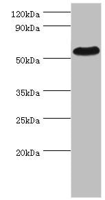
Phosphatidylglycerophosphatase and protein-tyrosine phosphatase 1, Polyclonal Antibody (Cat# AAA118730)
Epstein-Barr virus DNA polymerase catalytic subunit, Polyclonal Antibody (Cat# AAA118731)

 IHC (Immunohiostchemistry)
(Immunohistochemistry of paraffin-embedded human placenta using AAA118734 at dilution 1:100)
IHC (Immunohiostchemistry)
(Immunohistochemistry of paraffin-embedded human placenta using AAA118734 at dilution 1:100)
Cysteine/serine-rich nuclear protein 1, Polyclonal Antibody (Cat# AAA118734)

 IHC (Immunohistochemistry)
(Immunohistochemistry of paraffin-embedded human pancreas using AAA118735 at dilution 1:100)
IHC (Immunohistochemistry)
(Immunohistochemistry of paraffin-embedded human pancreas using AAA118735 at dilution 1:100)
Cytosolic beta-glucosidase, Polyclonal Antibody (Cat# AAA118735)
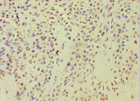
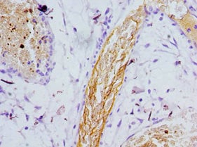
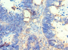
 IHC (Immunohiostchemistry)
(Immunohistochemistry of paraffin-embedded human colon cancer using AAA118736 at dilution 1:100)
IHC (Immunohiostchemistry)
(Immunohistochemistry of paraffin-embedded human colon cancer using AAA118736 at dilution 1:100)
Golgi membrane protein 1, Polyclonal Antibody (Cat# AAA118736)
 IHC (Immunohiostchemistry)
(Immunohistochemistry of paraffin-embedded human kidney using AAA118738 at dilution 1:100)
IHC (Immunohiostchemistry)
(Immunohistochemistry of paraffin-embedded human kidney using AAA118738 at dilution 1:100)
Aflatoxin B1 aldehyde reductase member 3, Polyclonal Antibody (Cat# AAA118738)
Arabidopsis thaliana FACT complex subunit SPT16, Polyclonal Antibody (Cat# AAA118739)
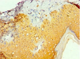
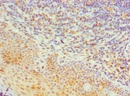
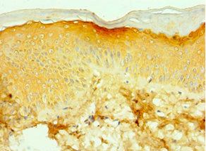
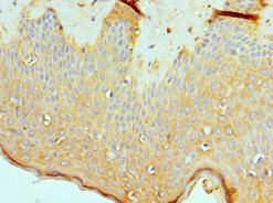
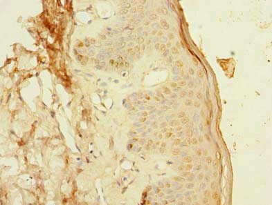 IHC (Immunohiostchemistry)
(Immunohistochemistry of paraffin-embedded human skin tissue using AAA118740 at dilution of 1:100)
IHC (Immunohiostchemistry)
(Immunohistochemistry of paraffin-embedded human skin tissue using AAA118740 at dilution of 1:100)
PCIF1, Polyclonal Antibody (Cat# AAA118740)
 IF (Immunofluorescence)
(IF analysis of HepG2 cells using AMHR2 antibody at dilution 1-100)
IF (Immunofluorescence)
(IF analysis of HepG2 cells using AMHR2 antibody at dilution 1-100)
Anti-Muellerian hormone type-2 receptor, Polyclonal Antibody (Cat# AAA119143)
Tyrosinase, Polyclonal Antibody (Cat# AAA119145)
 IHC (Immunohistochemisry)
(Immunohistochemistry of paraffin-embedded human colon cancer using AAA119146 at dilution 1:100)
IHC (Immunohistochemisry)
(Immunohistochemistry of paraffin-embedded human colon cancer using AAA119146 at dilution 1:100)
N-acetylserotonin O-methyltransferase-like protein, Polyclonal Antibody (Cat# AAA119146)
KIAA0319L, Polyclonal Antibody (Cat# AAA119151)
 IHC (Immunohiostchemistry)
(Immunohistochemistry of paraffin-embedded human colon cancer using AAA119155 at dilution 1:100)
IHC (Immunohiostchemistry)
(Immunohistochemistry of paraffin-embedded human colon cancer using AAA119155 at dilution 1:100)
A-kinase anchor protein 5, Polyclonal Antibody (Cat# AAA119155)
What are Polyclonal Antibodies?
Polyclonal antibodies are antibodies that come from multiple B cell clones of a host animal. The typical hosts used for the majority of polyclonal antibody production are rabbits, goats, sheep, and donkeys. These polyclonal antibodies, once having identified their target, will bind to different epitopes located at different regions or sequences on the same protein/antigen. As a result, they are ideal at locating and binding to the target, even if the target is in very low concentrations (due to many different antibodies being able to bind to the same target molecule, which allows for significant amplification of a downstream signal).
Polyclonal antibodies are typically produced by injecting an antigen into a host animal, which causes the animal’s immune system to attack the foreign antigen by mass generating antibodies against it. After a period of time, serum is collected from the animal and purified using physicochemical fractionation, class-specific affinity purification, and/or antigen-affinity purification.
Key Uses of Polyclonal Antibodies
- Western Blotting: This method is used to find specific proteins in biological samples after separating them by size.
- Immunohistochemistry: IHC helps visualize the location of proteins in tissue sections using various staining techniques.
- ELISA: (Enzyme-Linked Immunosorbent Assay) is typically used to identify specific protein quantities in a sample. ELISAs can be either “Quantitative” or “Qualitative”.
- Flow Cytometry: technique that identifies and measures the specific protein on the surface or inside the cells in a fluid suspension.
- Immunoprecipitation: IP isolates and studies a specific protein from a complex mixture using antibodies.
Why Buy Polyclonal Antibodies from AAA Biotech?
1. Ideal for Various Applications
Our antibodies are generally going to be validated for use in multiple types of assays, including ELISA, Western Blotting, Immunohistochemistry, Immunoprecipitation, amongst others. They are ideal for a wide range of research applications.
2. Rigorous Quality Control
All of the antibodies in our catalog undergo strict quality testing to ensure specificity, sensitivity, and consistent performance. We are confident in the ability of our antibodies to provide you with accurate results.
3. Wide Assortment of Antibodies
Antibodies in are catalog can be found for both common and exotic species, and these antibodies are also available in both conjugated and recombinant forms to suit many diverse experimental needs.
4. Highly Purified
Our antibodies are available in purified forms with over 85% purity, as confirmed by SDS-PAGE. They are also available with tags such as His, Flag, GST, or MBP. We cater to customers worldwide.
FAQ
1. How are polyclonal antibodies produced?
Traditionally, polyclonal antibodies are produced by injecting an antigen into a host animal (such as a rabbit or goat), which then triggers an immune response from the host animal. The animal’s B cells produce antibodies that will recognize different parts of the injected antigen. These antibodies are then collected from the animal’s blood and purified for use.
2. How do polyclonal antibodies differ from monoclonal antibodies?
Polyclonal antibodies are a mix of antibodies that bind to different locations (epitopes) of the same antigen, while monoclonal antibodies are identical and bind to just one specific epitope. This makes polyclonal antibodies more versatile and better at detecting proteins that may be present in low quantities or in altered/modified forms.
3. How should I store polyclonal antibodies?
Polyclonal antibodies should be stored at 4°C for short-term use (up to a few weeks) and at -20°C or -80°C for long-term storage. Avoid repeated freeze-thaw cycles by dividing them into small aliquots. Always check the datasheet for specific storage instructions.