Filters
▼Clonality
▼Type
▼Reactivity
▼Gene Name
▼Isotype
▼Host
▼Application
▼Clone
▼Polyclonal Antibodies
At AAA Biotech also known as AAA Bio or AAABio, we provide a broad range of purified polyclonal antibodies (pAbs) that are able to all be browsed online through our website. Due to their high specificity and strong binding affinity, these antibodies are ideal for wide swathes of research and experimental applications.
Our polyclonal antibodies can easily support your work, whether you use them for Western Blotting, Immunocytochemistry (with or without Immunofluorescence used in conjunction), Immunohistochemistry, Immunoprecipitation, and ELISA tests. We highly encourage you to browse our range of pAbs and choose the one that best suits your experimental model.
Viewing 7850-7900 of 96805 product results
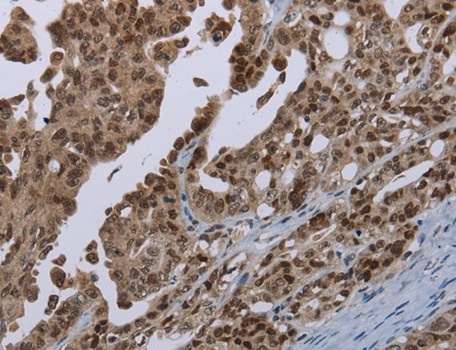
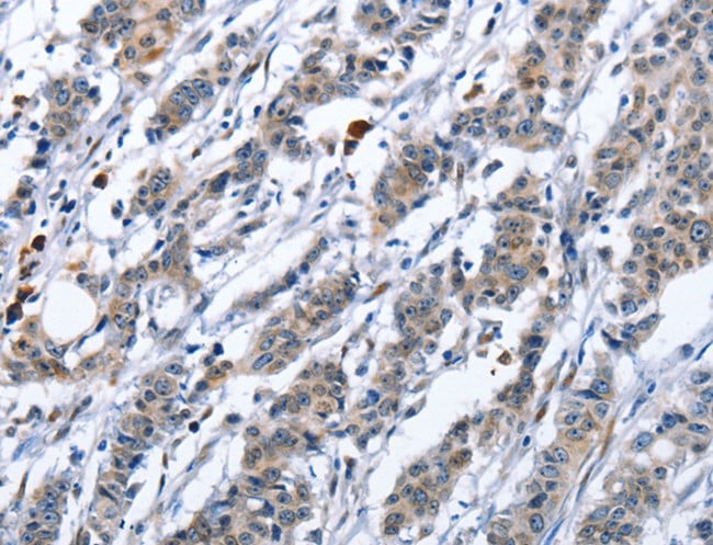
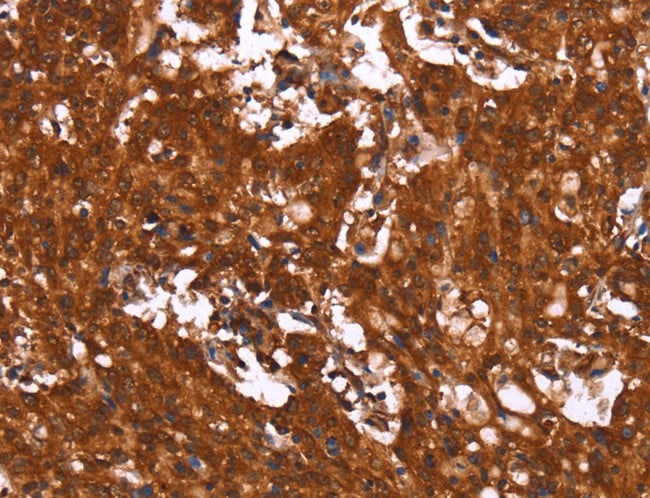
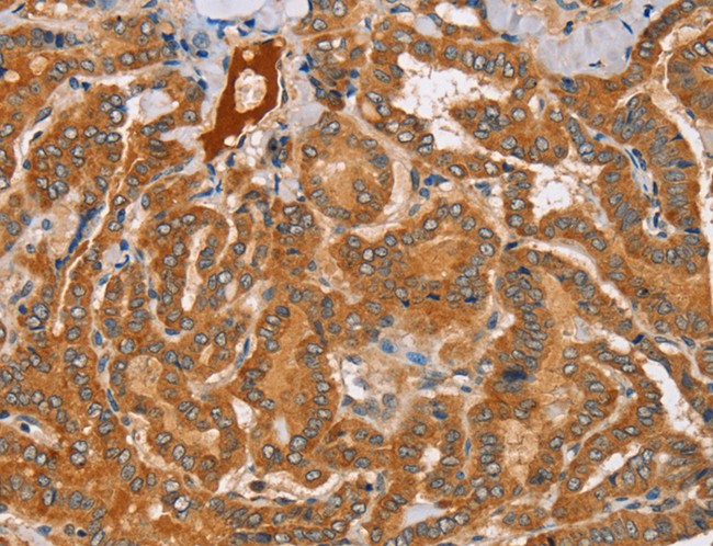
 IHC (Immunohiostchemistry)
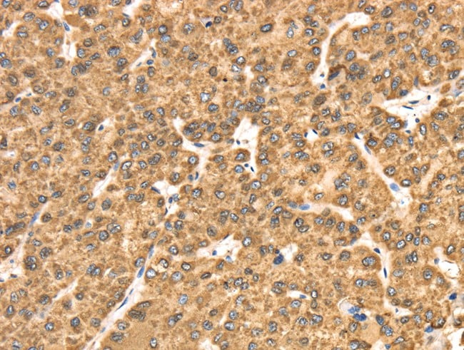
(Immunohistochemistry of paraffin-embedded Human thyroid cancer tissue using DSC1 Polyclonal Antibody at dilution 1:40)
IHC (Immunohiostchemistry)
(Immunohistochemistry of paraffin-embedded Human thyroid cancer tissue using DSC1 Polyclonal Antibody at dilution 1:40)
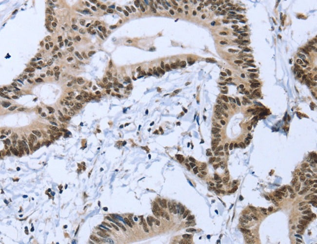
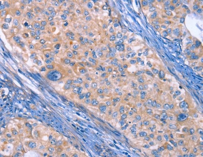
DSC1, Polyclonal Antibody (Cat# AAA166394)
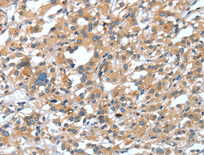
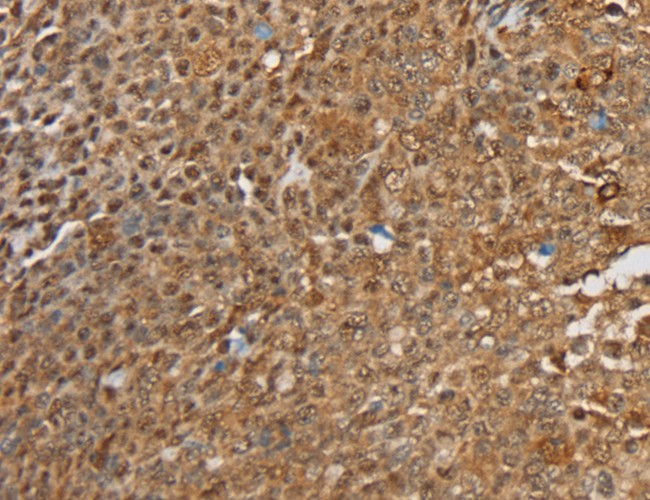
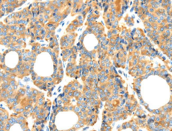
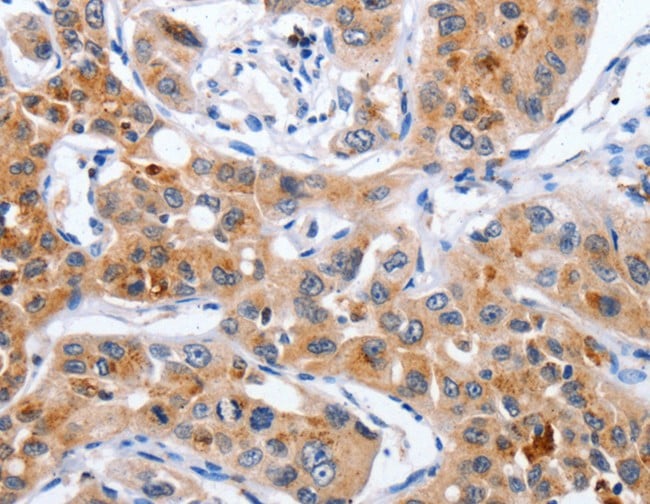
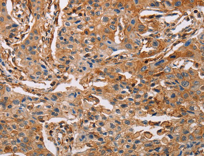
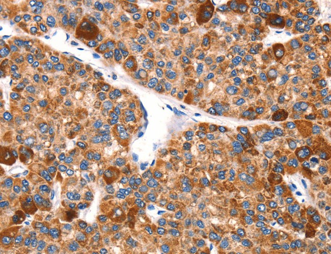
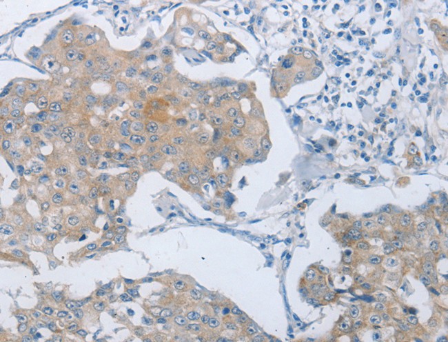
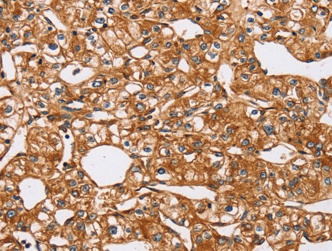
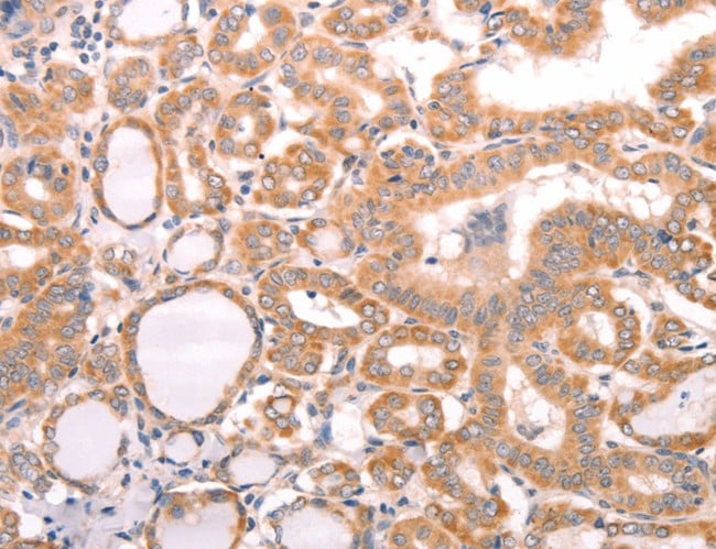
 IHC (Immunohistochemisry)
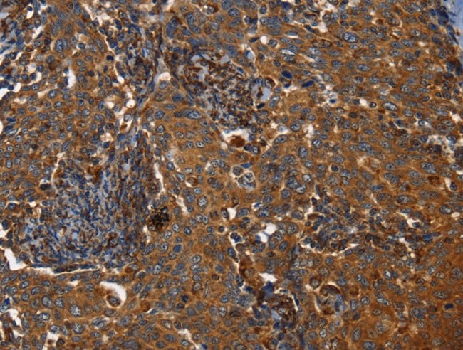
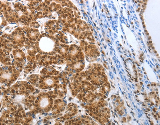
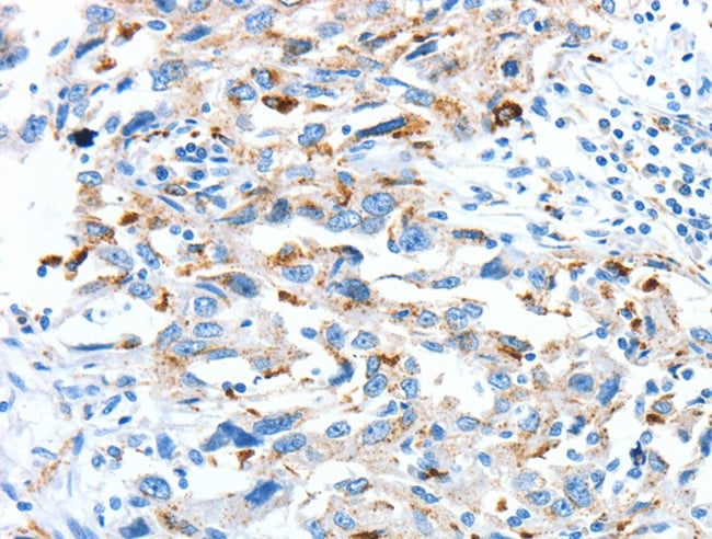
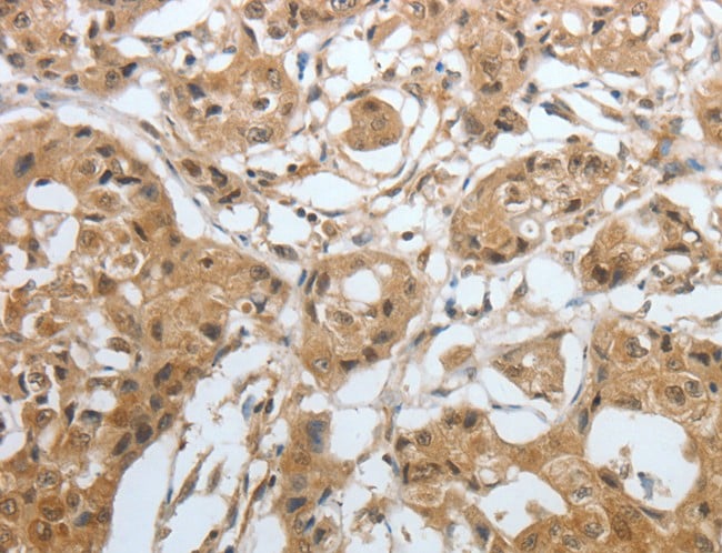
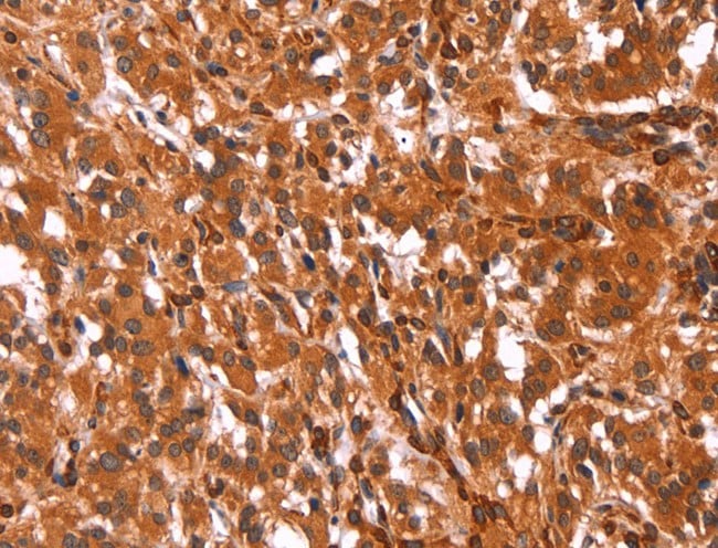
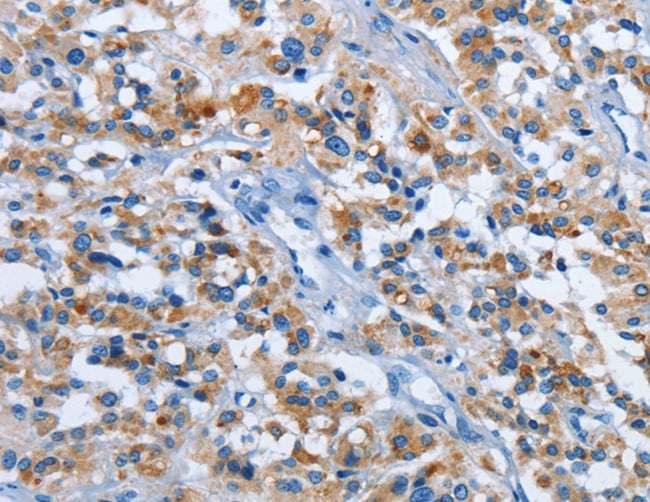
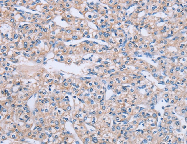
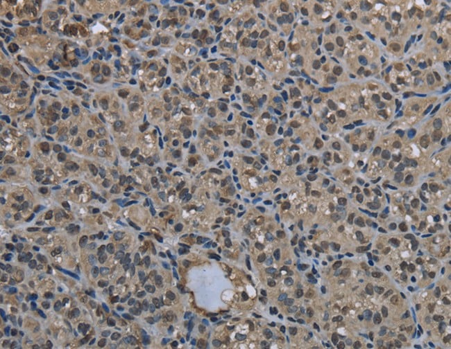
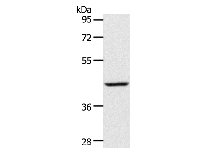
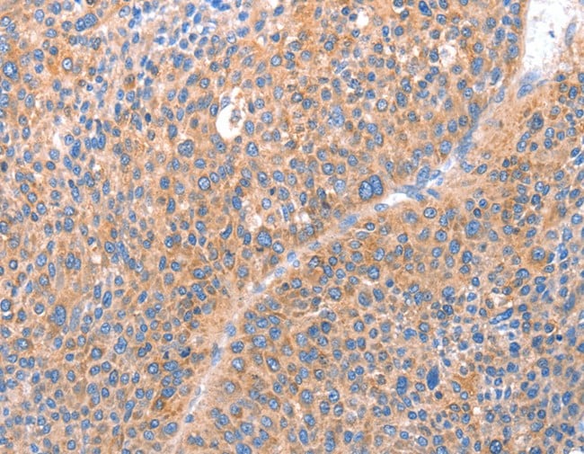
(Immunohistochemistry of paraffin-embedded Human liver cancer using RRBP1 Polyclonal Antibody at dilution of 1:25)
IHC (Immunohistochemisry)
(Immunohistochemistry of paraffin-embedded Human liver cancer using RRBP1 Polyclonal Antibody at dilution of 1:25)
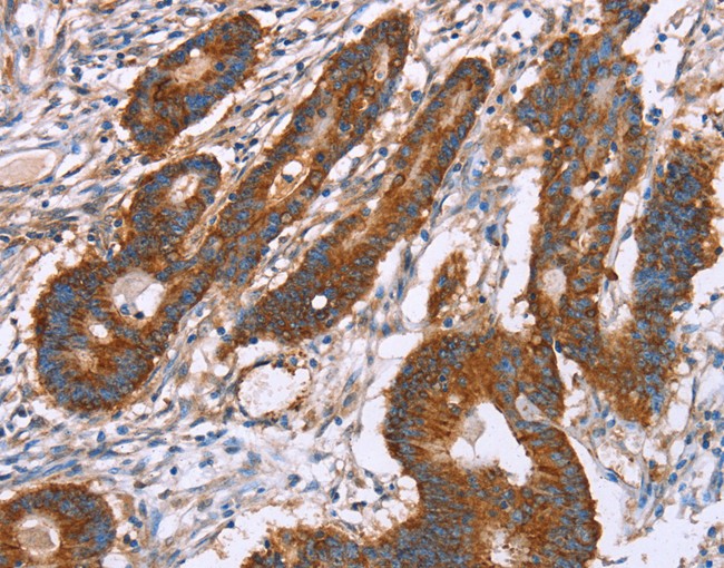
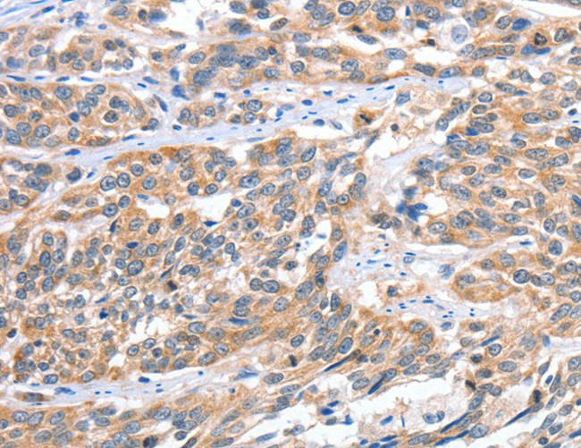
RRBP1, Polyclonal Antibody (Cat# AAA166403)
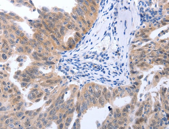
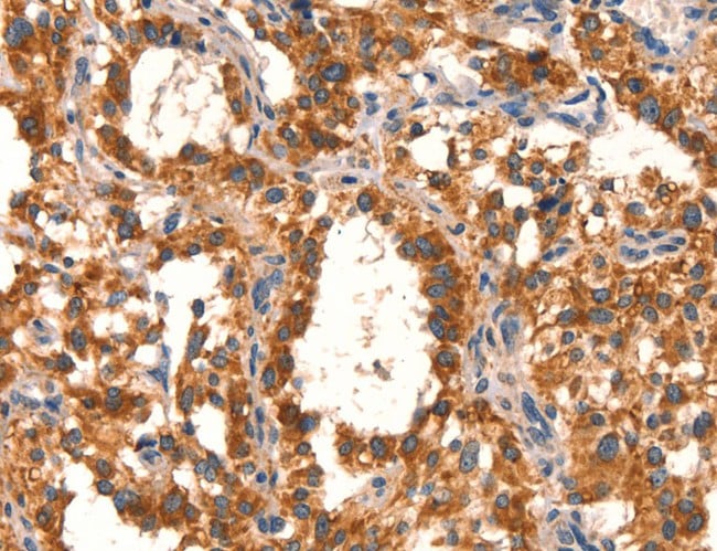
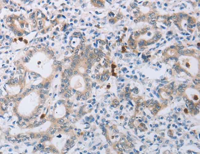
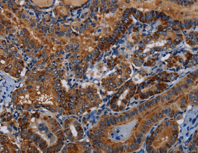
 IHC (Immunohistochemisry)
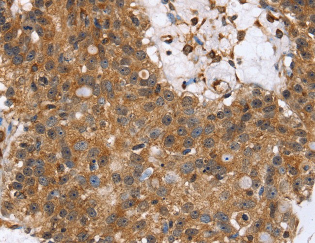
(Immunohistochemistry of paraffin-embedded Human lung cancer using TPM2 Polyclonal Antibody at dilution of 1:35)
IHC (Immunohistochemisry)
(Immunohistochemistry of paraffin-embedded Human lung cancer using TPM2 Polyclonal Antibody at dilution of 1:35)
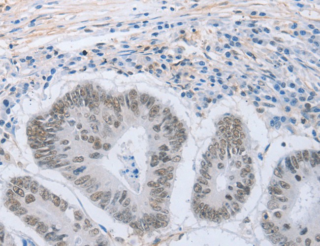
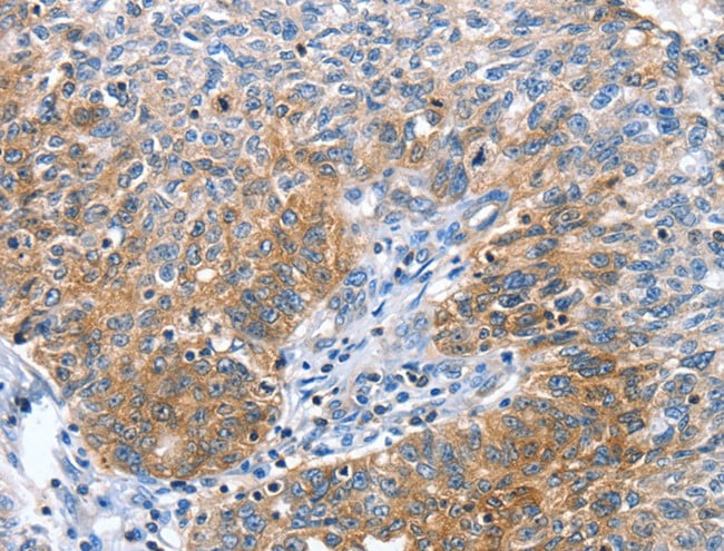
TPM2, Polyclonal Antibody (Cat# AAA166406)
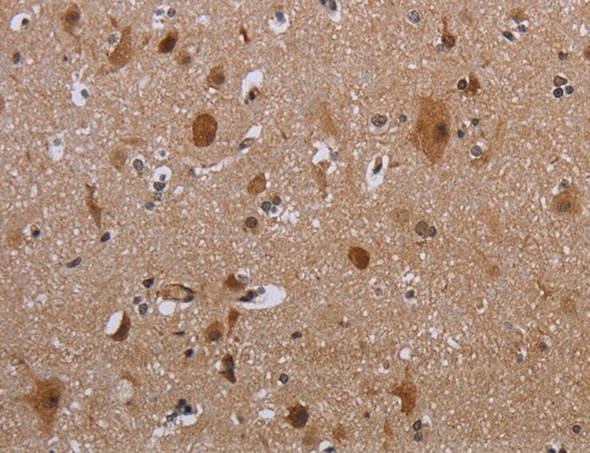
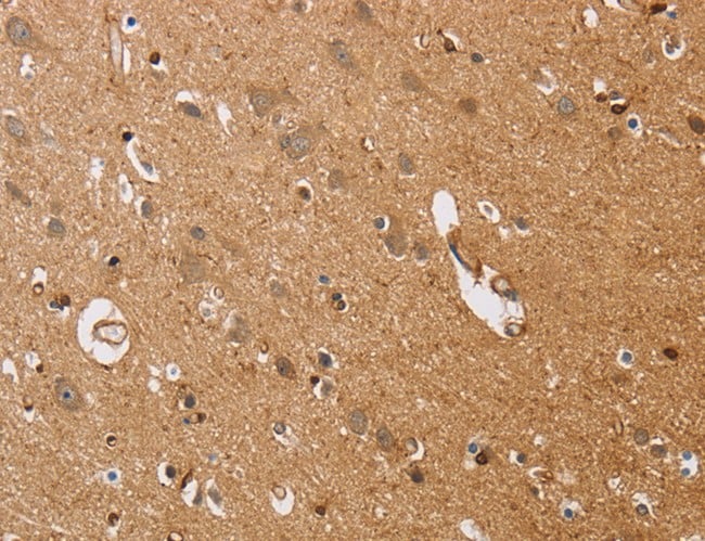
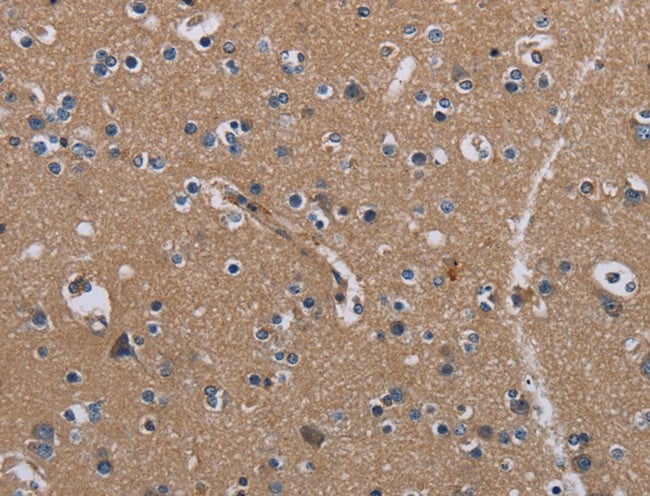
 IHC (Immunohistochemisry)
(Immunohistochemistry of paraffin-embedded Human brain using PDE5A Polyclonal Antibody at dilution of 1:50)
IHC (Immunohistochemisry)
(Immunohistochemistry of paraffin-embedded Human brain using PDE5A Polyclonal Antibody at dilution of 1:50)
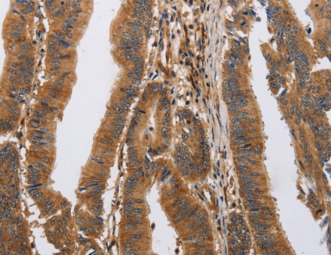
PDE5A, Polyclonal Antibody (Cat# AAA166412)
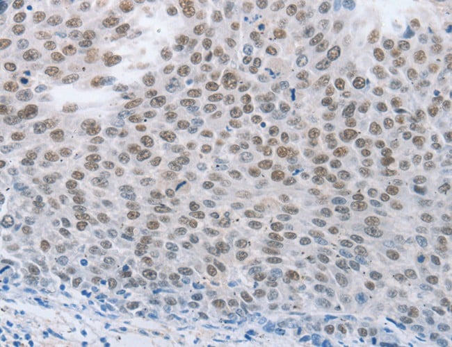
 IHC (Immunohiostchemistry)
(Immunohistochemistry of paraffin-embedded Human colon cancer tissue using ZMAT3 Polyclonal Antibody at dilution 1:30)
IHC (Immunohiostchemistry)
(Immunohistochemistry of paraffin-embedded Human colon cancer tissue using ZMAT3 Polyclonal Antibody at dilution 1:30)
ZMAT3, Polyclonal Antibody (Cat# AAA166422)
 IHC (Immunohiostchemistry)
(Immunohistochemistry of paraffin-embedded Human colon cancer using PDLIM1 Polyclonal Antibody at dilution of 1:30)
IHC (Immunohiostchemistry)
(Immunohistochemistry of paraffin-embedded Human colon cancer using PDLIM1 Polyclonal Antibody at dilution of 1:30)
PDLIM1, Polyclonal Antibody (Cat# AAA166427)
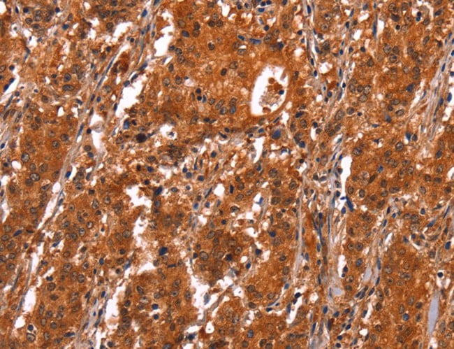
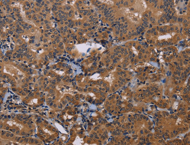
 IHC (Immunohiostchemistry)
(Immunohistochemistry of paraffin-embedded Human liver cancer tissue using NEUROG2 Polyclonal Antibody at dilution 1:15)
IHC (Immunohiostchemistry)
(Immunohistochemistry of paraffin-embedded Human liver cancer tissue using NEUROG2 Polyclonal Antibody at dilution 1:15)
NEUROG2, Polyclonal Antibody (Cat# AAA166435)
 IHC (Immunohiostchemistry)
(Immunohistochemistry of paraffin-embedded Human brain tissue using SIGLEC14 Polyclonal Antibody at dilution 1:30)
IHC (Immunohiostchemistry)
(Immunohistochemistry of paraffin-embedded Human brain tissue using SIGLEC14 Polyclonal Antibody at dilution 1:30)
SIGLEC14, Polyclonal Antibody (Cat# AAA166436)
 IHC (Immunohistochemisry)
(Immunohistochemistry of paraffin-embedded Human ovarian cancer using MAP2K1 Polyclonal Antibody at dilution of 1:25)
IHC (Immunohistochemisry)
(Immunohistochemistry of paraffin-embedded Human ovarian cancer using MAP2K1 Polyclonal Antibody at dilution of 1:25)
MAP2K1, Polyclonal Antibody (Cat# AAA166450)
 IHC (Immunohiostchemistry)
(Immunohistochemistry of paraffin-embedded Human esophagus cancer tissue using RIPK4 Polyclonal Antibody at dilution 1:40)
IHC (Immunohiostchemistry)
(Immunohistochemistry of paraffin-embedded Human esophagus cancer tissue using RIPK4 Polyclonal Antibody at dilution 1:40)
RIPK4, Polyclonal Antibody (Cat# AAA166452)
 IHC (Immunohiostchemistry)
(Immunohistochemistry of paraffin-embedded Human brain tissue using MAGEB18 Polyclonal Antibody at dilution 1:20)
IHC (Immunohiostchemistry)
(Immunohistochemistry of paraffin-embedded Human brain tissue using MAGEB18 Polyclonal Antibody at dilution 1:20)
MAGEB18, Polyclonal Antibody (Cat# AAA166453)
 IHC (Immunohistochemisry)
(Immunohistochemistry of paraffin-embedded Human colon cancer using NCK1 Polyclonal Antibody at dilution of 1:30)
IHC (Immunohistochemisry)
(Immunohistochemistry of paraffin-embedded Human colon cancer using NCK1 Polyclonal Antibody at dilution of 1:30)
NCK1, Polyclonal Antibody (Cat# AAA166289)
 IHC (Immunohistochemisry)
(Immunohistochemistry of paraffin-embedded Human lung cancer using SMARCA4 Polyclonal Antibody at dilution of 1:50)
IHC (Immunohistochemisry)
(Immunohistochemistry of paraffin-embedded Human lung cancer using SMARCA4 Polyclonal Antibody at dilution of 1:50)
SMARCA4, Polyclonal Antibody (Cat# AAA166294)
 IHC (Immunohiostchemistry)
(Immunohistochemistry of paraffin-embedded Human lung cancer using MAPK3 Polyclonal Antibody at dilution of 1:15)
IHC (Immunohiostchemistry)
(Immunohistochemistry of paraffin-embedded Human lung cancer using MAPK3 Polyclonal Antibody at dilution of 1:15)
MAPK3, Polyclonal Antibody (Cat# AAA166310)
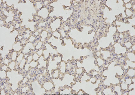
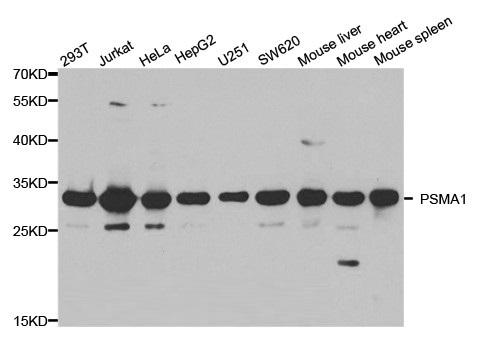
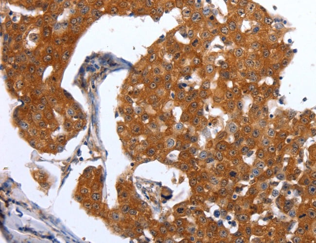
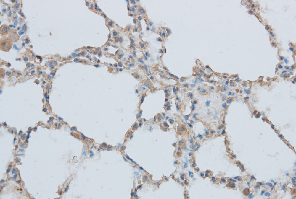
 IHC (Immunohistochemisry)
(Immunohistochemical of paraffin-embedded rat lung using PSMA1 antibody at dilution of 1:100 (200x lens).)
IHC (Immunohistochemisry)
(Immunohistochemical of paraffin-embedded rat lung using PSMA1 antibody at dilution of 1:100 (200x lens).)
PSMA1, Polyclonal Antibody (Cat# AAA166314)
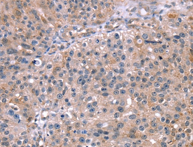
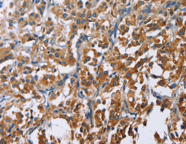
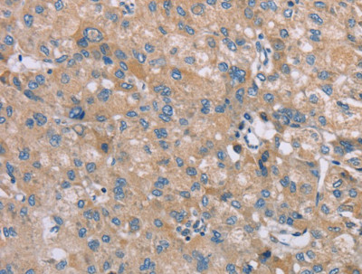 IHC (Immunohistochemistry)
(Immunohistochemistry of paraffin-embedded Human liver cancer tissue using SAMD9 Polyclonal Antibody at dilution 1:35)
IHC (Immunohistochemistry)
(Immunohistochemistry of paraffin-embedded Human liver cancer tissue using SAMD9 Polyclonal Antibody at dilution 1:35)
SAMD9, Polyclonal Antibody (Cat# AAA166318)

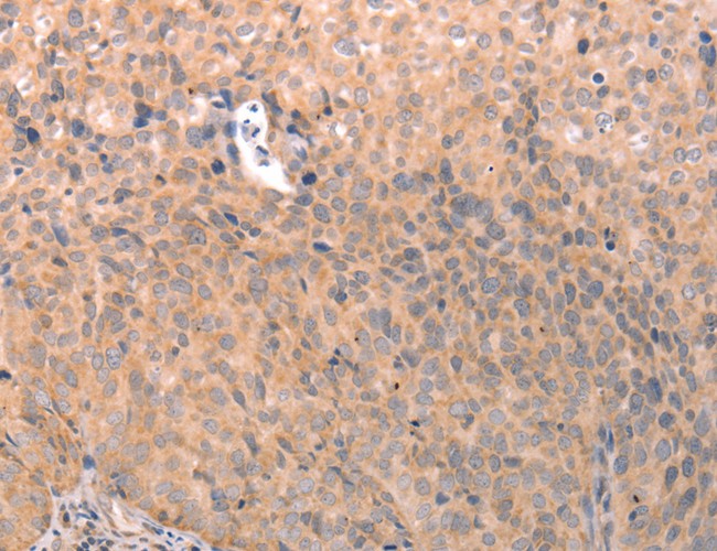
 IHC (Immunohiostchemistry)
(Immunohistochemistry of paraffin-embedded Human lung cancer tissue using ASCL1 Polyclonal Antibody at dilution 1:30)
IHC (Immunohiostchemistry)
(Immunohistochemistry of paraffin-embedded Human lung cancer tissue using ASCL1 Polyclonal Antibody at dilution 1:30)
ASCL1, Polyclonal Antibody (Cat# AAA166320)
 IHC (Immunohistochemisry)
(Immunohistochemistry of paraffin-embedded Human lung cancer using ACTA1 Polyclonal Antibody at dilution of 1:65)
IHC (Immunohistochemisry)
(Immunohistochemistry of paraffin-embedded Human lung cancer using ACTA1 Polyclonal Antibody at dilution of 1:65)
ACTA1, Polyclonal Antibody (Cat# AAA166322)
 IHC (Immunohiostchemistry)
(Immunohistochemistry of paraffin-embedded Human esophagus cancer tissue using CLPTM1L Polyclonal Antibody at dilution 1:60)
IHC (Immunohiostchemistry)
(Immunohistochemistry of paraffin-embedded Human esophagus cancer tissue using CLPTM1L Polyclonal Antibody at dilution 1:60)
CLPTM1L, Polyclonal Antibody (Cat# AAA166325)
 IHC (Immunohiostchemistry)
(Immunohistochemistry of paraffin-embedded Human cervical cancer tissue using TNFRSF9 Polyclonal Antibody at dilution 1:50)
IHC (Immunohiostchemistry)
(Immunohistochemistry of paraffin-embedded Human cervical cancer tissue using TNFRSF9 Polyclonal Antibody at dilution 1:50)
TNFRSF9, Polyclonal Antibody (Cat# AAA166329)

 IHC (Immunohistochemisry)
(Immunohistochemistry of paraffin-embedded Human lung cancer using AMPH Polyclonal Antibody at dilution of 1:30)
IHC (Immunohistochemisry)
(Immunohistochemistry of paraffin-embedded Human lung cancer using AMPH Polyclonal Antibody at dilution of 1:30)
AMPH, Polyclonal Antibody (Cat# AAA166330)
 IHC (Immunohiostchemistry)
(Immunohistochemistry of paraffin-embedded Human thyroid cancer tissue using DGAT1 Polyclonal Antibody at dilution 1:40)
IHC (Immunohiostchemistry)
(Immunohistochemistry of paraffin-embedded Human thyroid cancer tissue using DGAT1 Polyclonal Antibody at dilution 1:40)
DGAT1, Polyclonal Antibody (Cat# AAA166342)
 IHC (Immunohiostchemistry)
(Immunohistochemistry of paraffin-embedded Human thyroid cancer tissue using GCG Polyclonal Antibody at dilution 1:20)
IHC (Immunohiostchemistry)
(Immunohistochemistry of paraffin-embedded Human thyroid cancer tissue using GCG Polyclonal Antibody at dilution 1:20)
GCG, Polyclonal Antibody (Cat# AAA166347)
 IHC (Immunohiostchemistry)
(Immunohistochemistry of paraffin-embedded Human brain tissue using DCTN5 Polyclonal Antibody at dilution 1:25)
IHC (Immunohiostchemistry)
(Immunohistochemistry of paraffin-embedded Human brain tissue using DCTN5 Polyclonal Antibody at dilution 1:25)
DCTN5, Polyclonal Antibody (Cat# AAA166353)
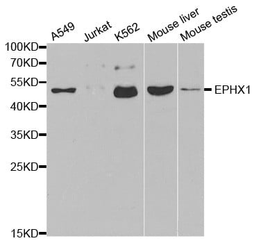
 IHC (Immunohistochemisry)
(Immunohistochemical of paraffin-embedded human esophageal cancer using EPHX1 antibody at dilution of 1:100 (200x lens).)
IHC (Immunohistochemisry)
(Immunohistochemical of paraffin-embedded human esophageal cancer using EPHX1 antibody at dilution of 1:100 (200x lens).)
EPHX1, Polyclonal Antibody (Cat# AAA166360)
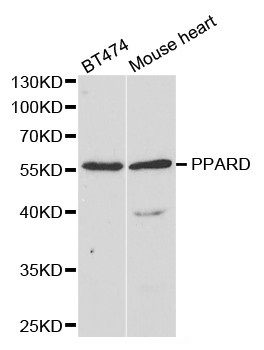
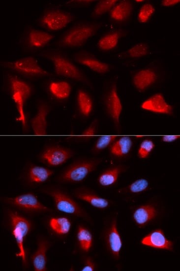 IF (Immunofluorescence)
(Immunofluorescence analysis of U20S cell using PPARD antibody. Blue: DAPI for nuclear staining.)
IF (Immunofluorescence)
(Immunofluorescence analysis of U20S cell using PPARD antibody. Blue: DAPI for nuclear staining.)
PPARD, Polyclonal Antibody (Cat# AAA166362)
 IHC (Immunohiostchemistry)
(Immunohistochemistry of paraffin-embedded Human thyroid cancer using ETHE1 Polyclonal Antibody at dilution of 1:20)
IHC (Immunohiostchemistry)
(Immunohistochemistry of paraffin-embedded Human thyroid cancer using ETHE1 Polyclonal Antibody at dilution of 1:20)
ETHE1, Polyclonal Antibody (Cat# AAA166456)
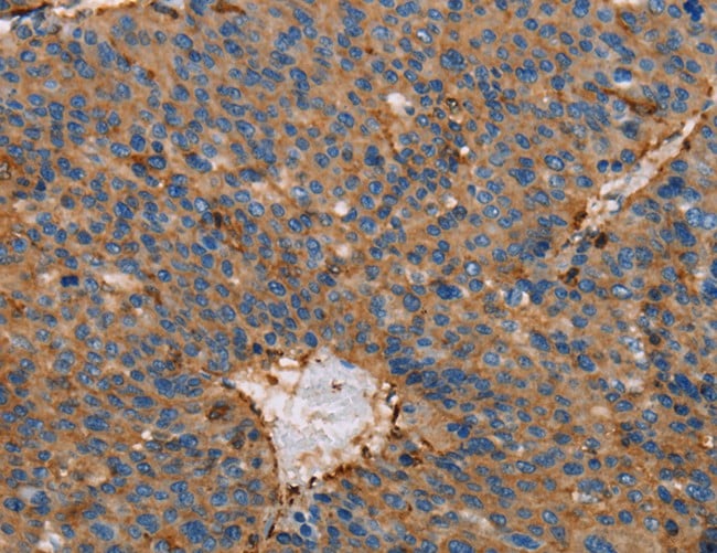
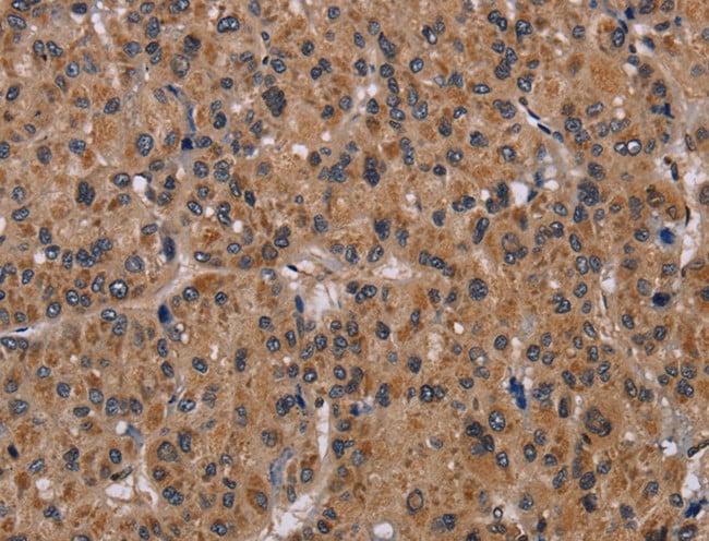
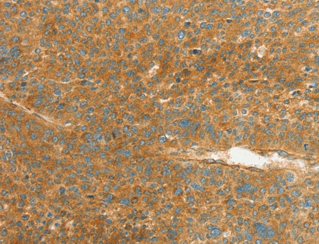
 IHC (Immunohiostchemistry)
(Immunohistochemistry of paraffin-embedded Human liver cancer tissue using NR1D1 Polyclonal Antibody at dilution 1:60)
IHC (Immunohiostchemistry)
(Immunohistochemistry of paraffin-embedded Human liver cancer tissue using NR1D1 Polyclonal Antibody at dilution 1:60)
NR1D1, Polyclonal Antibody (Cat# AAA166464)
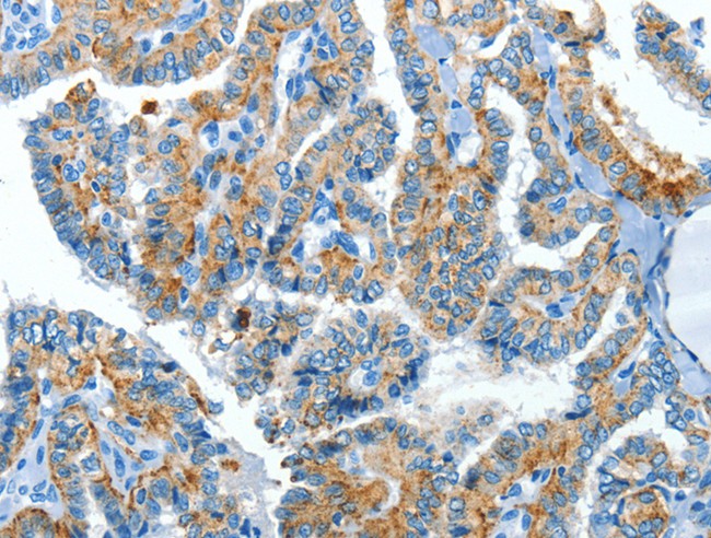
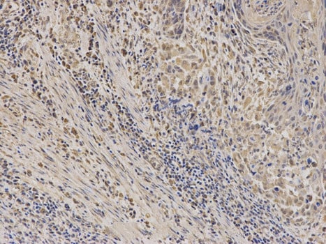
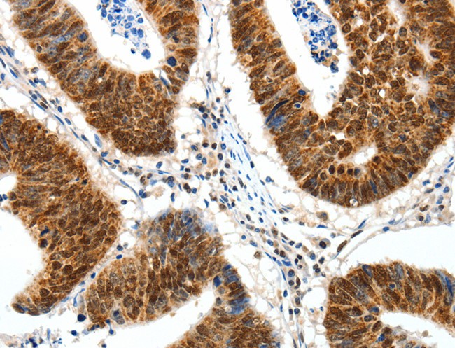
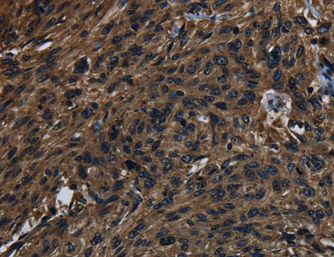
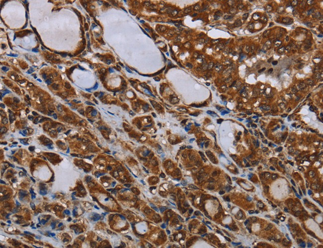
 IHC (Immunohiostchemistry)
(Immunohistochemistry of paraffin-embedded Human prostate cancer using TF Polyclonal Antibody at dilution of 1:20)
IHC (Immunohiostchemistry)
(Immunohistochemistry of paraffin-embedded Human prostate cancer using TF Polyclonal Antibody at dilution of 1:20)
TF, Polyclonal Antibody (Cat# AAA166465)
 IHC (Immunohiostchemistry)
(Immunohistochemistry of paraffin-embedded Human cervical cancer tissue using CARD9 Polyclonal Antibody at dilution 1:60)
IHC (Immunohiostchemistry)
(Immunohistochemistry of paraffin-embedded Human cervical cancer tissue using CARD9 Polyclonal Antibody at dilution 1:60)
CARD9, Polyclonal Antibody (Cat# AAA166480)
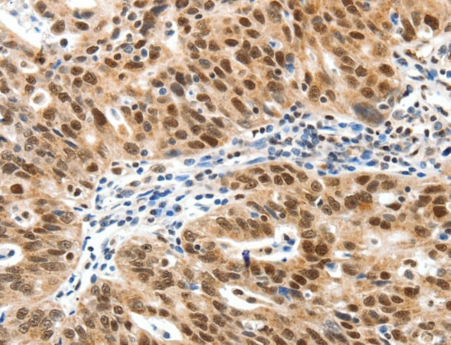
 IHC (Immunohistochemisry)
(Immunohistochemistry of paraffin-embedded Human thyroid cancer using BCAR1 Polyclonal Antibody at dilution of 1:15)
IHC (Immunohistochemisry)
(Immunohistochemistry of paraffin-embedded Human thyroid cancer using BCAR1 Polyclonal Antibody at dilution of 1:15)
BCAR1, Polyclonal Antibody (Cat# AAA166483)
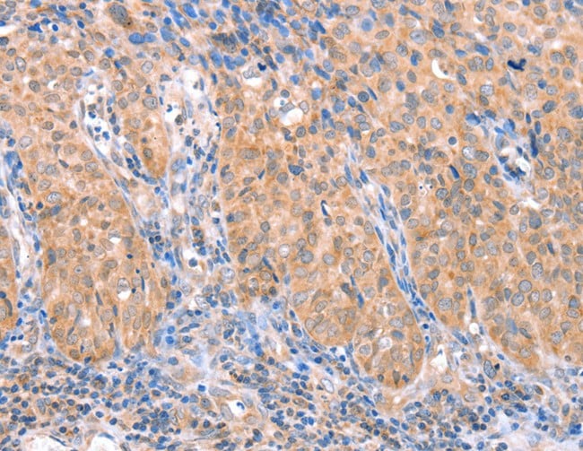
 IHC (Immunohistochemisry)
(Immunohistochemistry of paraffin-embedded Human cervical cancer using ITGB3 Polyclonal Antibody at dilution of 1:30)
IHC (Immunohistochemisry)
(Immunohistochemistry of paraffin-embedded Human cervical cancer using ITGB3 Polyclonal Antibody at dilution of 1:30)
ITGB3, Polyclonal Antibody (Cat# AAA166484)
 IHC (Immunohiostchemistry)
(Immunohistochemistry of paraffin-embedded Human ovarian cancer tissue using CGB Polyclonal Antibody at dilution 1:60)
IHC (Immunohiostchemistry)
(Immunohistochemistry of paraffin-embedded Human ovarian cancer tissue using CGB Polyclonal Antibody at dilution 1:60)
CGB, Polyclonal Antibody (Cat# AAA166487)
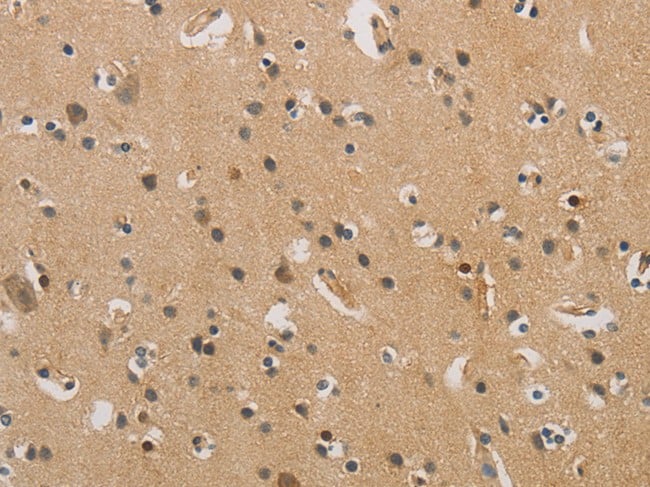
 IHC (Immunohiostchemistry)
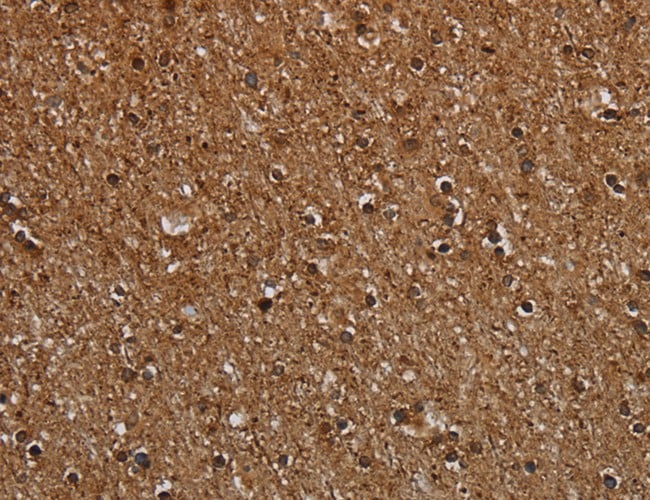
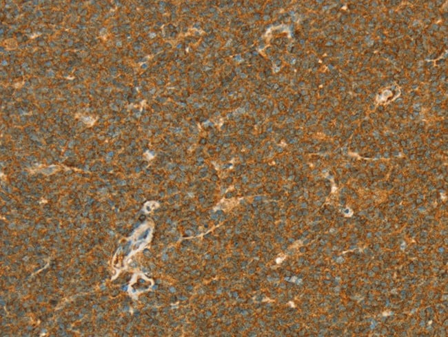
(Immunohistochemistry of paraffin-embedded Human brain tissue using ITPR1 Polyclonal Antibody at dilution 1:40)
IHC (Immunohiostchemistry)
(Immunohistochemistry of paraffin-embedded Human brain tissue using ITPR1 Polyclonal Antibody at dilution 1:40)
ITPR1, Polyclonal Antibody (Cat# AAA166496)
 IHC (Immunohiostchemistry)
(Immunohistochemistry of paraffin-embedded Human thyroid cancer tissue using EPO Polyclonal Antibody at dilution 1:25)
IHC (Immunohiostchemistry)
(Immunohistochemistry of paraffin-embedded Human thyroid cancer tissue using EPO Polyclonal Antibody at dilution 1:25)
EPO, Polyclonal Antibody (Cat# AAA166507)
 IHC (Immunohiostchemistry)
(Immunohistochemistry of paraffin-embedded Human thyroid cancer tissue using HCAR2 Polyclonal Antibody at dilution 1:40)
IHC (Immunohiostchemistry)
(Immunohistochemistry of paraffin-embedded Human thyroid cancer tissue using HCAR2 Polyclonal Antibody at dilution 1:40)
HCAR2, Polyclonal Antibody (Cat# AAA166517)
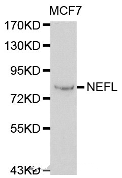
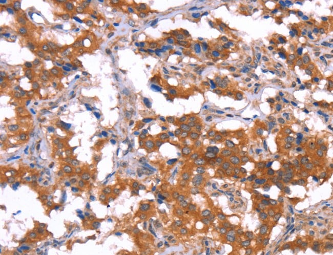
 IHC (Immunohiostchemistry)
(Immunohistochemistry of paraffin-embedded H-tonsil using NEFL antibody.)
IHC (Immunohiostchemistry)
(Immunohistochemistry of paraffin-embedded H-tonsil using NEFL antibody.)
NEFL, Polyclonal Antibody (Cat# AAA166519)
 IHC (Immunohiostchemistry)
(Immunohistochemistry of paraffin-embedded Human ovarian cancer tissue using SP1 Polyclonal Antibody at dilution 1:30)
IHC (Immunohiostchemistry)
(Immunohistochemistry of paraffin-embedded Human ovarian cancer tissue using SP1 Polyclonal Antibody at dilution 1:30)
SP1, Polyclonal Antibody (Cat# AAA166521)
 IHC (Immunohistochemisry)
(Immunohistochemistry of paraffin-embedded Human breast cancer using ABCB9 Polyclonal Antibody at dilution of 1:15)
IHC (Immunohistochemisry)
(Immunohistochemistry of paraffin-embedded Human breast cancer using ABCB9 Polyclonal Antibody at dilution of 1:15)
ABCB9, Polyclonal Antibody (Cat# AAA166523)
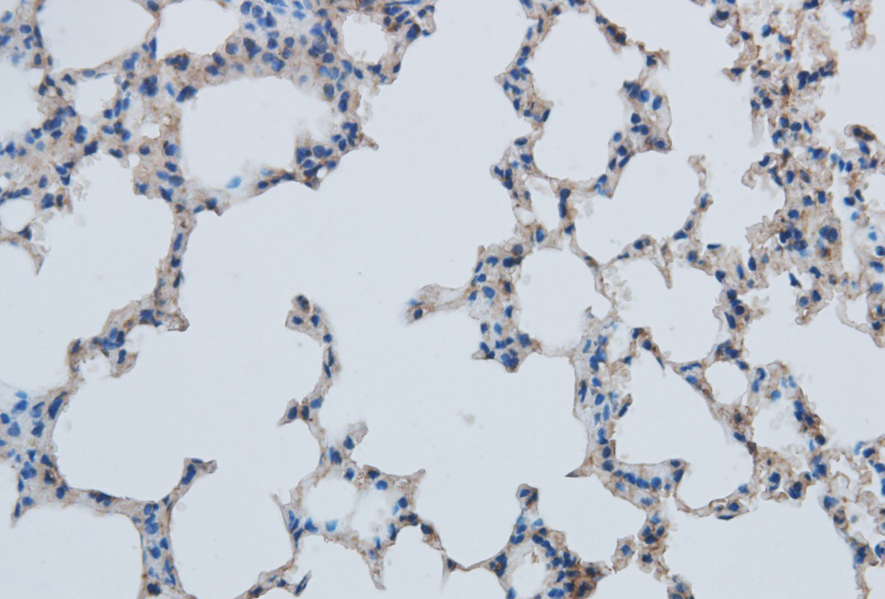
 IHC (Immunohiostchemistry)
(Immunohistochemistry of paraffin-embedded Rat lung using MMP-1 Polyclonal Antibody at dilution of 1:50)
IHC (Immunohiostchemistry)
(Immunohistochemistry of paraffin-embedded Rat lung using MMP-1 Polyclonal Antibody at dilution of 1:50)
MMP1, Polyclonal Antibody (Cat# AAA166526)
 IHC (Immunohistochemisry)
(Immunohistochemistry of paraffin-embedded Human prostate cancer using KLK14 Polyclonal Antibody at dilution of 1:40)
IHC (Immunohistochemisry)
(Immunohistochemistry of paraffin-embedded Human prostate cancer using KLK14 Polyclonal Antibody at dilution of 1:40)
KLK14, Polyclonal Antibody (Cat# AAA166528)
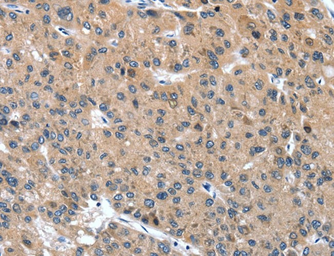
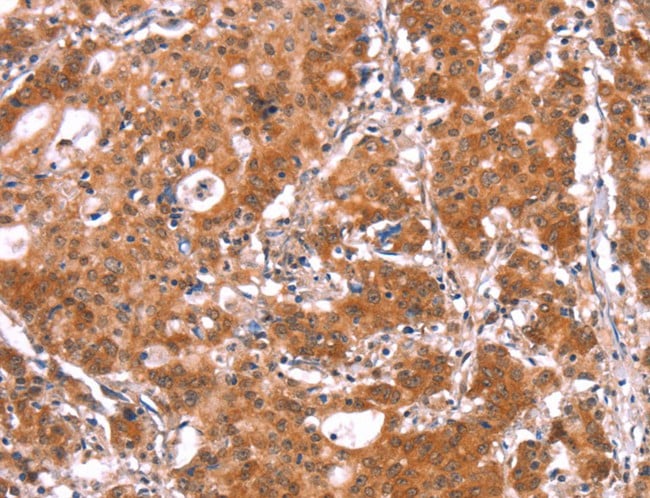
 IHC (Immunohistochemisry)
(Immunohistochemistry of paraffin-embedded Human liver cancer using UBB Polyclonal Antibody at dilution of 1:20)
IHC (Immunohistochemisry)
(Immunohistochemistry of paraffin-embedded Human liver cancer using UBB Polyclonal Antibody at dilution of 1:20)
UBB, Polyclonal Antibody (Cat# AAA166530)
 IHC (Immunohiostchemistry)
(Immunohistochemistry of paraffin-embedded Human brain tissue using MT-ND1 Polyclonal Antibody at dilution 1:30)
IHC (Immunohiostchemistry)
(Immunohistochemistry of paraffin-embedded Human brain tissue using MT-ND1 Polyclonal Antibody at dilution 1:30)
MT-ND1, Polyclonal Antibody (Cat# AAA166533)

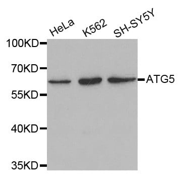
 IHC (Immunohistochemisry)
(Immunohistochemistry of paraffin-embedded human lung cancer using ATG5 antibody at dilution of 1:200 (x400 lens))
IHC (Immunohistochemisry)
(Immunohistochemistry of paraffin-embedded human lung cancer using ATG5 antibody at dilution of 1:200 (x400 lens))
ATG5, Polyclonal Antibody (Cat# AAA166538)
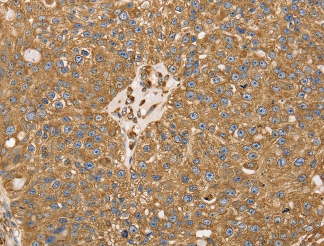
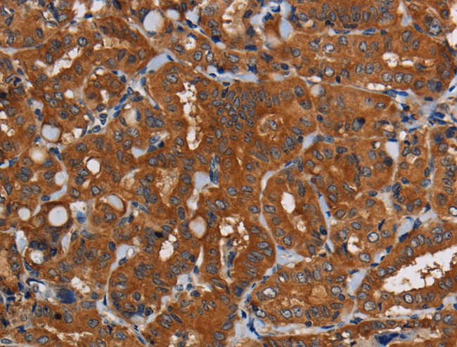
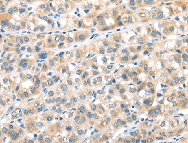
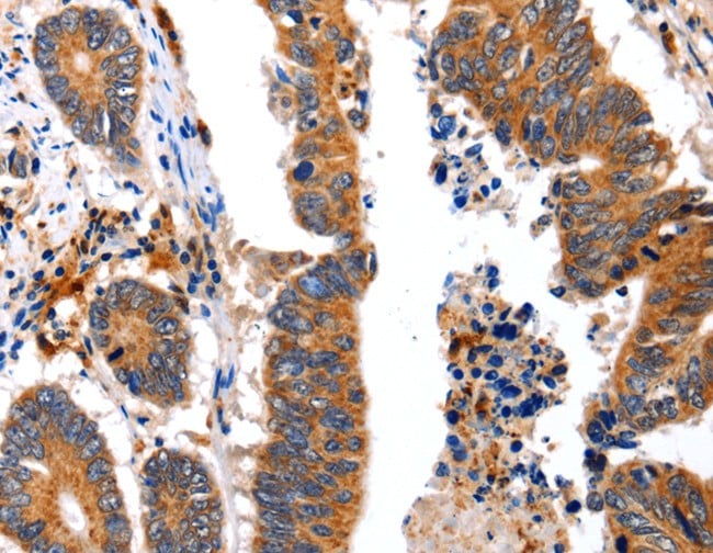
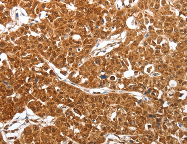
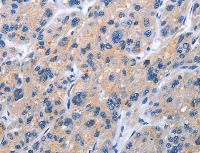
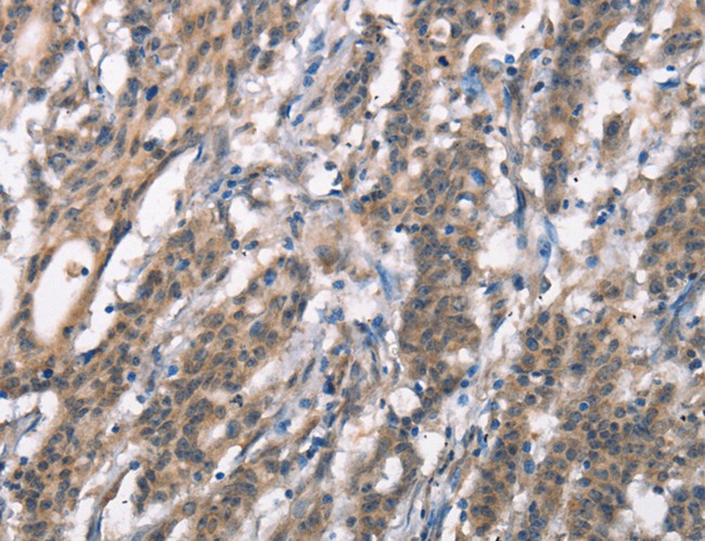
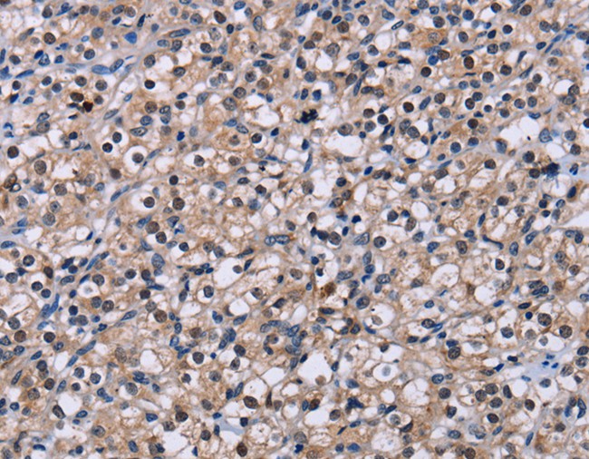
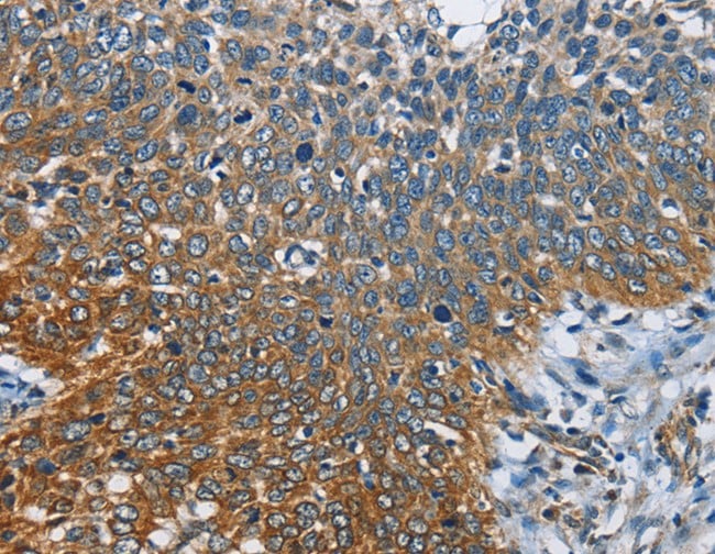
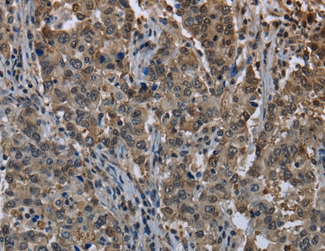
 IHC (Immunohistochemisry)
(Immunohistochemistry of paraffin-embedded Human liver cancer using ACADS Polyclonal Antibody at dilution of 1:15)
IHC (Immunohistochemisry)
(Immunohistochemistry of paraffin-embedded Human liver cancer using ACADS Polyclonal Antibody at dilution of 1:15)
ACADS, Polyclonal Antibody (Cat# AAA166539)
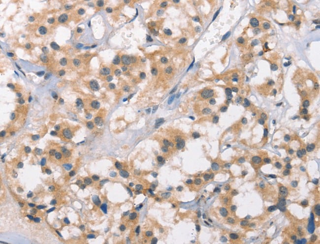
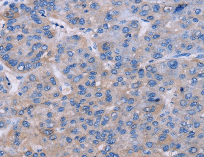
 IHC (Immunohiostchemistry)
(Immunohistochemistry of paraffin-embedded Human liver cancer tissue using GRK1 Polyclonal Antibody at dilution 1:30)
IHC (Immunohiostchemistry)
(Immunohistochemistry of paraffin-embedded Human liver cancer tissue using GRK1 Polyclonal Antibody at dilution 1:30)
GRK1, Polyclonal Antibody (Cat# AAA166831)
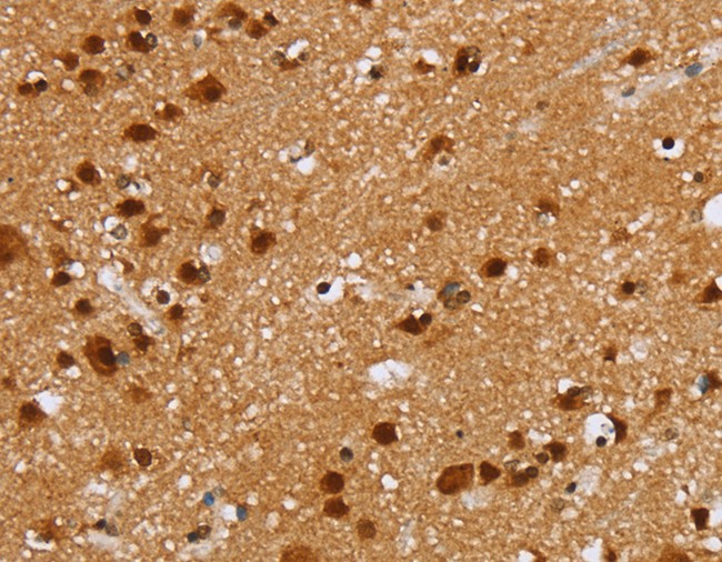
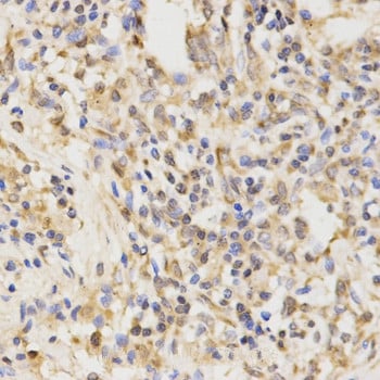
 IHC (Immunohiostchemistry)
(Immunohistochemistry of paraffin-embedded Human prostate cancer tissue using PEA15 Polyclonal Antibody at dilution 1:25)
IHC (Immunohiostchemistry)
(Immunohistochemistry of paraffin-embedded Human prostate cancer tissue using PEA15 Polyclonal Antibody at dilution 1:25)
PEA15, Polyclonal Antibody (Cat# AAA166832)
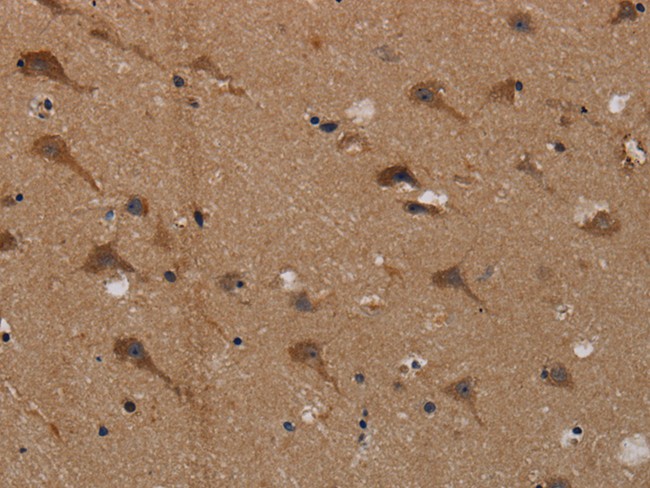
 IHC (Immunohiostchemistry)
(Immunohistochemistry of paraffin-embedded Human brain tissue using PIDD1 Polyclonal Antibody at dilution 1:30)
IHC (Immunohiostchemistry)
(Immunohistochemistry of paraffin-embedded Human brain tissue using PIDD1 Polyclonal Antibody at dilution 1:30)
PIDD1, Polyclonal Antibody (Cat# AAA166834)
 IHC (Immunohiostchemistry)
(Immunohistochemistry of paraffin-embedded Human thyroid cancer tissue using ABCF2 Polyclonal Antibody at dilution 1:15)
IHC (Immunohiostchemistry)
(Immunohistochemistry of paraffin-embedded Human thyroid cancer tissue using ABCF2 Polyclonal Antibody at dilution 1:15)
ABCF2, Polyclonal Antibody (Cat# AAA166839)
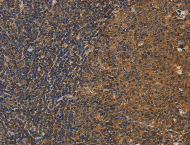
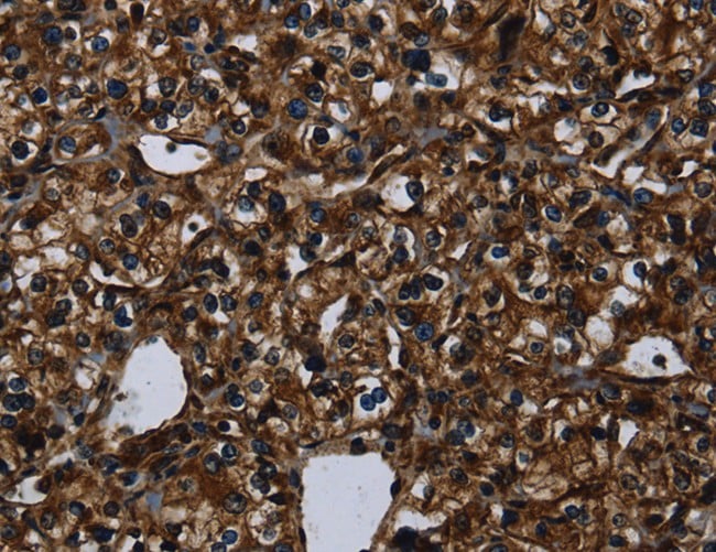
 IHC (Immunohistochemisry)
(Immunohistochemistry of paraffin-embedded Human lymphoma using APOL2 Polyclonal Antibody at dilution of 1:80)
IHC (Immunohistochemisry)
(Immunohistochemistry of paraffin-embedded Human lymphoma using APOL2 Polyclonal Antibody at dilution of 1:80)
APOL2, Polyclonal Antibody (Cat# AAA166842)
What are Polyclonal Antibodies?
Polyclonal antibodies are antibodies that come from multiple B cell clones of a host animal. The typical hosts used for the majority of polyclonal antibody production are rabbits, goats, sheep, and donkeys. These polyclonal antibodies, once having identified their target, will bind to different epitopes located at different regions or sequences on the same protein/antigen. As a result, they are ideal at locating and binding to the target, even if the target is in very low concentrations (due to many different antibodies being able to bind to the same target molecule, which allows for significant amplification of a downstream signal).
Polyclonal antibodies are typically produced by injecting an antigen into a host animal, which causes the animal’s immune system to attack the foreign antigen by mass generating antibodies against it. After a period of time, serum is collected from the animal and purified using physicochemical fractionation, class-specific affinity purification, and/or antigen-affinity purification.
Key Uses of Polyclonal Antibodies
- Western Blotting: This method is used to find specific proteins in biological samples after separating them by size.
- Immunohistochemistry: IHC helps visualize the location of proteins in tissue sections using various staining techniques.
- ELISA: (Enzyme-Linked Immunosorbent Assay) is typically used to identify specific protein quantities in a sample. ELISAs can be either “Quantitative” or “Qualitative”.
- Flow Cytometry: technique that identifies and measures the specific protein on the surface or inside the cells in a fluid suspension.
- Immunoprecipitation: IP isolates and studies a specific protein from a complex mixture using antibodies.
Why Buy Polyclonal Antibodies from AAA Biotech?
1. Ideal for Various Applications
Our antibodies are generally going to be validated for use in multiple types of assays, including ELISA, Western Blotting, Immunohistochemistry, Immunoprecipitation, amongst others. They are ideal for a wide range of research applications.
2. Rigorous Quality Control
All of the antibodies in our catalog undergo strict quality testing to ensure specificity, sensitivity, and consistent performance. We are confident in the ability of our antibodies to provide you with accurate results.
3. Wide Assortment of Antibodies
Antibodies in are catalog can be found for both common and exotic species, and these antibodies are also available in both conjugated and recombinant forms to suit many diverse experimental needs.
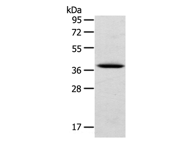
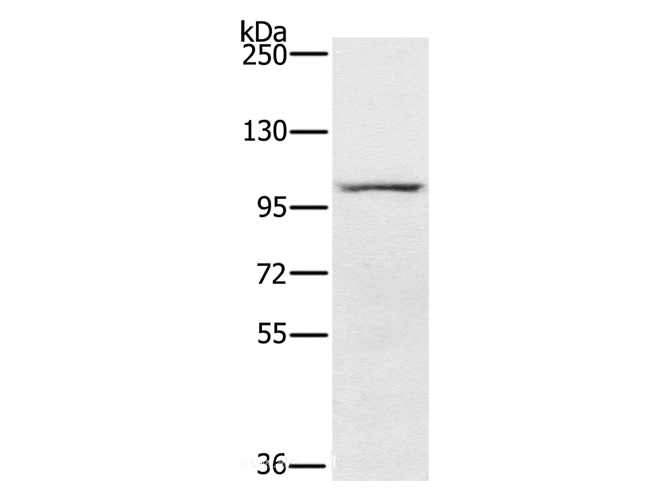
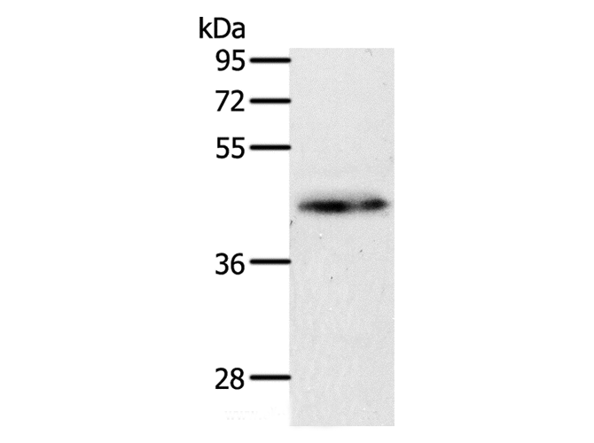
4. Highly Purified
Our antibodies are available in purified forms with over 85% purity, as confirmed by SDS-PAGE. They are also available with tags such as His, Flag, GST, or MBP. We cater to customers worldwide.
FAQ
1. How are polyclonal antibodies produced?
Traditionally, polyclonal antibodies are produced by injecting an antigen into a host animal (such as a rabbit or goat), which then triggers an immune response from the host animal. The animal’s B cells produce antibodies that will recognize different parts of the injected antigen. These antibodies are then collected from the animal’s blood and purified for use.
2. How do polyclonal antibodies differ from monoclonal antibodies?
Polyclonal antibodies are a mix of antibodies that bind to different locations (epitopes) of the same antigen, while monoclonal antibodies are identical and bind to just one specific epitope. This makes polyclonal antibodies more versatile and better at detecting proteins that may be present in low quantities or in altered/modified forms.
3. How should I store polyclonal antibodies?
Polyclonal antibodies should be stored at 4°C for short-term use (up to a few weeks) and at -20°C or -80°C for long-term storage. Avoid repeated freeze-thaw cycles by dividing them into small aliquots. Always check the datasheet for specific storage instructions.