Filters
▼Clonality
▼Type
▼Reactivity
▼Gene Name
▼Isotype
▼Host
▼Application
▼Clone
▼Polyclonal Antibodies
At AAA Biotech also known as AAA Bio or AAABio, we provide a broad range of purified polyclonal antibodies (pAbs) that are able to all be browsed online through our website. Due to their high specificity and strong binding affinity, these antibodies are ideal for wide swathes of research and experimental applications.
Our polyclonal antibodies can easily support your work, whether you use them for Western Blotting, Immunocytochemistry (with or without Immunofluorescence used in conjunction), Immunohistochemistry, Immunoprecipitation, and ELISA tests. We highly encourage you to browse our range of pAbs and choose the one that best suits your experimental model.
Viewing 8050-8100 of 96805 product results


 IHC (Immunohistochemistry)
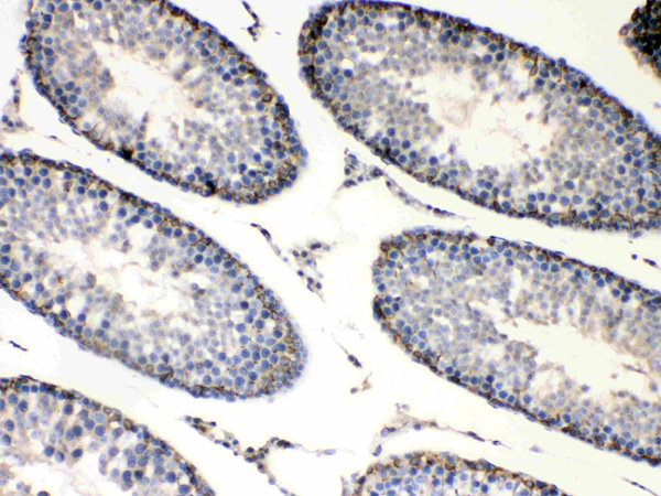
(Figure 4. IHC analysis of Bag1 using anti-Bag1 antibody (AAA124688).Bag1 was detected in paraffin-embedded section of rat testis tissues. Heat mediated antigen retrieval was performed in citrate buffer (pH6, epitope retrieval solution) for 20 mins. The tissue section was blocked with 10% goat serum. The tissue section was then incubated with 1ug/ml rabbit anti- Bag1 Antibody (AAA124688) overnight at 4 degree C. Biotinylated goat anti-rabbit IgG was used as secondary antibody and incubated for 30 minutes at 37 degree C. The tissue section was developed using Strepavidin-Biotin-Complex (SABC) with DAB as the chromogen.)
IHC (Immunohistochemistry)
(Figure 4. IHC analysis of Bag1 using anti-Bag1 antibody (AAA124688).Bag1 was detected in paraffin-embedded section of rat testis tissues. Heat mediated antigen retrieval was performed in citrate buffer (pH6, epitope retrieval solution) for 20 mins. The tissue section was blocked with 10% goat serum. The tissue section was then incubated with 1ug/ml rabbit anti- Bag1 Antibody (AAA124688) overnight at 4 degree C. Biotinylated goat anti-rabbit IgG was used as secondary antibody and incubated for 30 minutes at 37 degree C. The tissue section was developed using Strepavidin-Biotin-Complex (SABC) with DAB as the chromogen.)
Bag1, Polyclonal Antibody (Cat# AAA124688)
No cross reactivity with other proteins
 IHC (Immunohistochemistry)
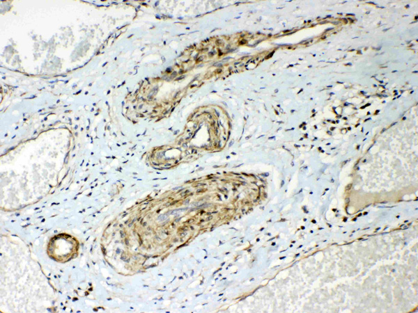
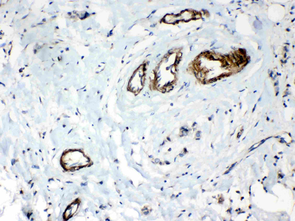
(Figure 5. IHC analysis of HSPB8/Hsp22 using anti- HSPB8/Hsp22 antibody (AAA124689).HSPB8/Hsp22 was detected in paraffin-embedded section of human mammary cancer tissues. Heat mediated antigen retrieval was performed in citrate buffer (pH6, epitope retrieval solution) for 20 mins. The tissue section was blocked with 10% goat serum. The tissue section was then incubated with 1ug/ml rabbit anti- HSPB8/Hsp22 Antibody (AAA124689) overnight at 4 degree C. Biotinylated goat anti-rabbit IgG was used as secondary antibody and incubated for 30 minutes at 37 degree C. The tissue section was developed using Strepavidin-Biotin-Complex (SABC) with DAB as the chromogen.)
IHC (Immunohistochemistry)
(Figure 5. IHC analysis of HSPB8/Hsp22 using anti- HSPB8/Hsp22 antibody (AAA124689).HSPB8/Hsp22 was detected in paraffin-embedded section of human mammary cancer tissues. Heat mediated antigen retrieval was performed in citrate buffer (pH6, epitope retrieval solution) for 20 mins. The tissue section was blocked with 10% goat serum. The tissue section was then incubated with 1ug/ml rabbit anti- HSPB8/Hsp22 Antibody (AAA124689) overnight at 4 degree C. Biotinylated goat anti-rabbit IgG was used as secondary antibody and incubated for 30 minutes at 37 degree C. The tissue section was developed using Strepavidin-Biotin-Complex (SABC) with DAB as the chromogen.)
HSPB8/Hsp22, Polyclonal Antibody (Cat# AAA124689)
No cross reactivity with other proteins
 IHC (Immunohiostchemistry)
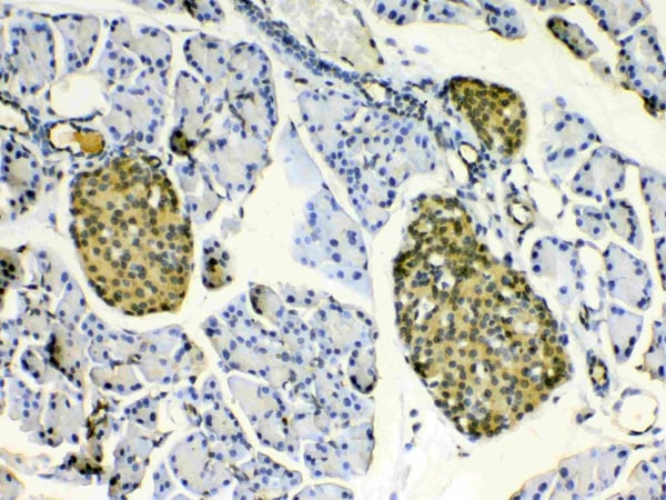
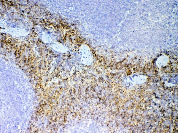
(Figure 2. IHC analysis of NAP2 using anti-NAP2 antibody (AAA124694).NAP2 was detected in paraffin-embedded section of mouse spleen tissue. Heat mediated antigen retrieval was performed in citrate buffer (pH6, epitope retrieval solution) for 20 mins. The tissue section was blocked with 10% goat serum. The tissue section was then incubated with 1ug/ml rabbit anti-NAP2 Antibody (AAA124694) overnight at 4 degree C. Biotinylated goat anti-rabbit IgG was used as secondary antibody and incubated for 30 minutes at 37 degree C. The tissue section was developed using Strepavidin-Biotin-Complex (SABC) with DAB as the chromogen.)
IHC (Immunohiostchemistry)
(Figure 2. IHC analysis of NAP2 using anti-NAP2 antibody (AAA124694).NAP2 was detected in paraffin-embedded section of mouse spleen tissue. Heat mediated antigen retrieval was performed in citrate buffer (pH6, epitope retrieval solution) for 20 mins. The tissue section was blocked with 10% goat serum. The tissue section was then incubated with 1ug/ml rabbit anti-NAP2 Antibody (AAA124694) overnight at 4 degree C. Biotinylated goat anti-rabbit IgG was used as secondary antibody and incubated for 30 minutes at 37 degree C. The tissue section was developed using Strepavidin-Biotin-Complex (SABC) with DAB as the chromogen.)
NAP2, Polyclonal Antibody (Cat# AAA124694)
No cross reactivity with other proteins.
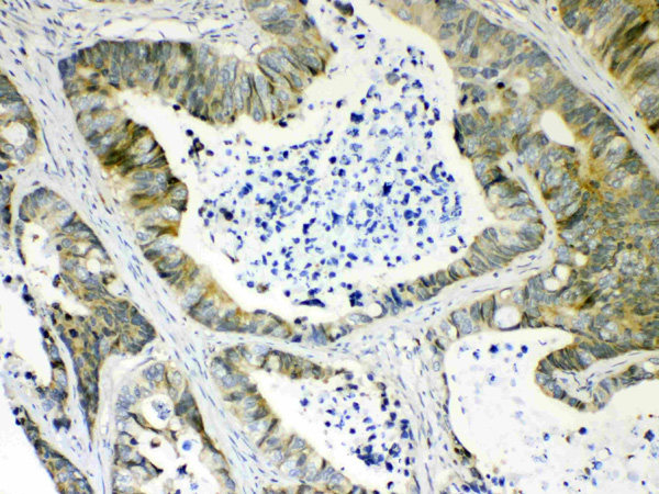
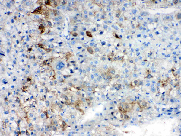
 IHC (Immunohistochemisry)
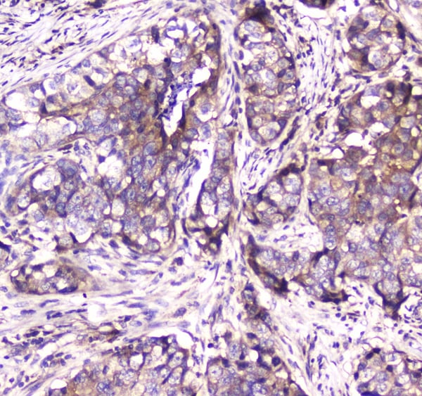
(Figure 3. IHC analysis of Caspase 4 using anti-Caspase 4 antibody (AAA124698).Caspase 4 was detected in paraffin-embedded section of human mammary cancer tissue. Heat mediated antigen retrieval was performed in citrate buffer (pH6, epitope retrieval solution) for 20 mins. The tissue section was blocked with 10% goat serum. The tissue section was then incubated with 1ug/ml rabbit anti-Caspase 4 Antibody (AAA124698) overnight at 4 degree C. Biotinylated goat anti-rabbit IgG was used as secondary antibody and incubated for 30 minutes at 37 degree C. The tissue section was developed using Strepavidin-Biotin-Complex (SABC) with DAB as the chromogen.)
IHC (Immunohistochemisry)
(Figure 3. IHC analysis of Caspase 4 using anti-Caspase 4 antibody (AAA124698).Caspase 4 was detected in paraffin-embedded section of human mammary cancer tissue. Heat mediated antigen retrieval was performed in citrate buffer (pH6, epitope retrieval solution) for 20 mins. The tissue section was blocked with 10% goat serum. The tissue section was then incubated with 1ug/ml rabbit anti-Caspase 4 Antibody (AAA124698) overnight at 4 degree C. Biotinylated goat anti-rabbit IgG was used as secondary antibody and incubated for 30 minutes at 37 degree C. The tissue section was developed using Strepavidin-Biotin-Complex (SABC) with DAB as the chromogen.)
Caspase 4, Polyclonal Antibody (Cat# AAA124698)
No cross reactivity with other proteins.

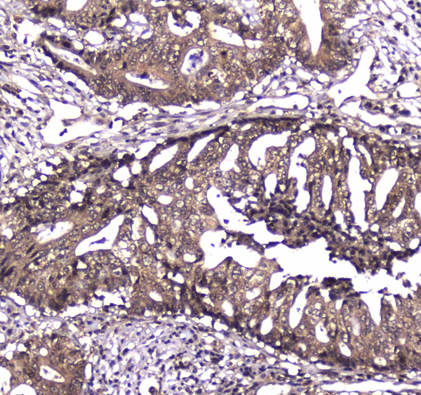
 IHC (Immunohistochemisry)
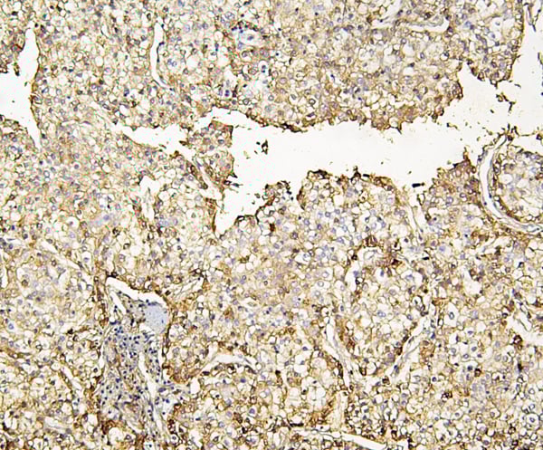
(Figure 3. IHC analysis of AKR1B10 using anti- AKR1B10 antibody (AAA124700).AKR1B10 was detected in paraffin-embedded section of human liver cancer tissues. Heat mediated antigen retrieval was performed in citrate buffer (pH6, epitope retrieval solution) for 20 mins. The tissue section was blocked with 10% goat serum. The tissue section was then incubated with 1ug/ml rabbit anti- AKR1B10 Antibody (AAA124700) overnight at 4 degree C. Biotinylated goat anti-rabbit IgG was used as secondary antibody and incubated for 30 minutes at 37 degree C. The tissue section was developed using Strepavidin-Biotin-Complex (SABC) with DAB as the chromogen.)
IHC (Immunohistochemisry)
(Figure 3. IHC analysis of AKR1B10 using anti- AKR1B10 antibody (AAA124700).AKR1B10 was detected in paraffin-embedded section of human liver cancer tissues. Heat mediated antigen retrieval was performed in citrate buffer (pH6, epitope retrieval solution) for 20 mins. The tissue section was blocked with 10% goat serum. The tissue section was then incubated with 1ug/ml rabbit anti- AKR1B10 Antibody (AAA124700) overnight at 4 degree C. Biotinylated goat anti-rabbit IgG was used as secondary antibody and incubated for 30 minutes at 37 degree C. The tissue section was developed using Strepavidin-Biotin-Complex (SABC) with DAB as the chromogen.)
AKR1B10, Polyclonal Antibody (Cat# AAA124700)
No cross reactivity with other proteins
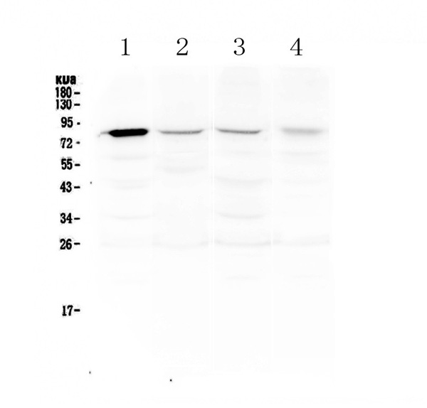
 WB (Western Blot)
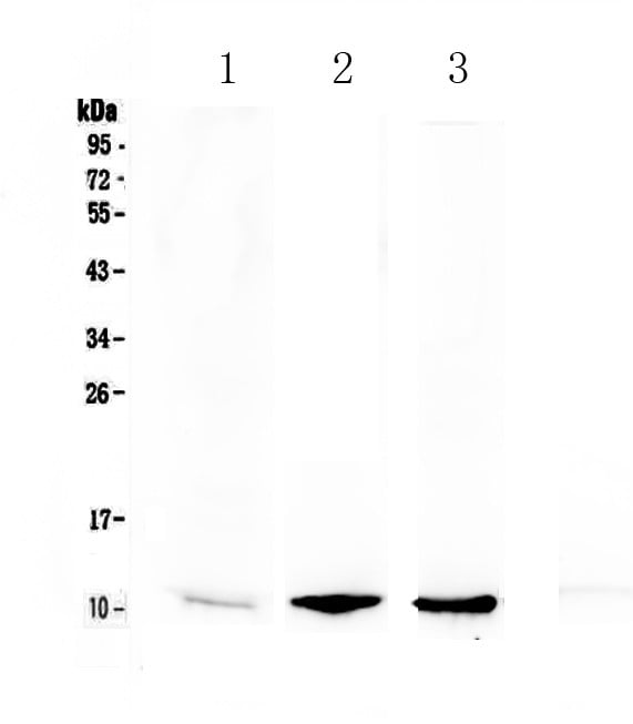
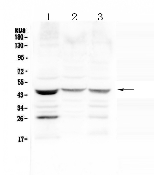
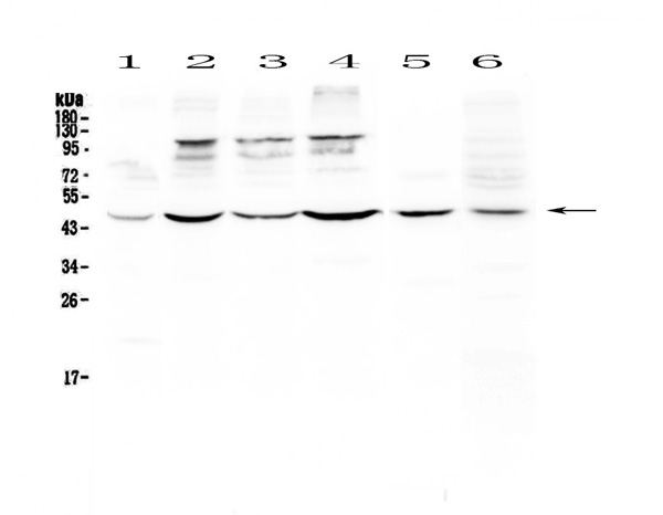
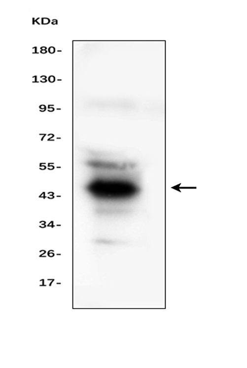
(Figure 1. Western blot analysis of SFTPB using anti-SFTPB antibody (AAA124707).Electrophoresis was performed on a 5-20% SDS-PAGE gel at 70V (Stacking gel) / 90V (Resolving gel) for 2-3 hours. The sample well of each lane was loaded with 50ug of sample under reducing conditions.Lane 1: human MCF-7 whole cell lysates,Lane 2: human COLO-320 whole cell lysates,Lane 3: human HepG2 whole cell lysates.After Electrophoresis, proteins were transferred to a Nitrocellulose membrane at 150mA for 50-90 minutes. Blocked the membrane with 5% Non-fat Milk/ TBS for 1.5 hour at RT. The membrane was incubated with rabbit anti-SFTPB antigen affinity purified polyclonal antibody at 0.5ug/mL overnight at 4 degree C, then washed with TBS-0.1%Tween 3 times with 5 minutes each and probed with a goat anti-rabbit IgG-HRP secondary antibody at a dilution of 1:10000 for 1.5 hour at RT. The signal is developed using an Enhanced Chemiluminescent detection (ECL) kit with Tanon 5200 system. A specific band was detected for SFTPB at approximately 46KD. The expected band size for SFTPB is at 42KD.)
WB (Western Blot)
(Figure 1. Western blot analysis of SFTPB using anti-SFTPB antibody (AAA124707).Electrophoresis was performed on a 5-20% SDS-PAGE gel at 70V (Stacking gel) / 90V (Resolving gel) for 2-3 hours. The sample well of each lane was loaded with 50ug of sample under reducing conditions.Lane 1: human MCF-7 whole cell lysates,Lane 2: human COLO-320 whole cell lysates,Lane 3: human HepG2 whole cell lysates.After Electrophoresis, proteins were transferred to a Nitrocellulose membrane at 150mA for 50-90 minutes. Blocked the membrane with 5% Non-fat Milk/ TBS for 1.5 hour at RT. The membrane was incubated with rabbit anti-SFTPB antigen affinity purified polyclonal antibody at 0.5ug/mL overnight at 4 degree C, then washed with TBS-0.1%Tween 3 times with 5 minutes each and probed with a goat anti-rabbit IgG-HRP secondary antibody at a dilution of 1:10000 for 1.5 hour at RT. The signal is developed using an Enhanced Chemiluminescent detection (ECL) kit with Tanon 5200 system. A specific band was detected for SFTPB at approximately 46KD. The expected band size for SFTPB is at 42KD.)
SFTPB/Surfactant Protein B, Polyclonal Antibody (Cat# AAA124707)
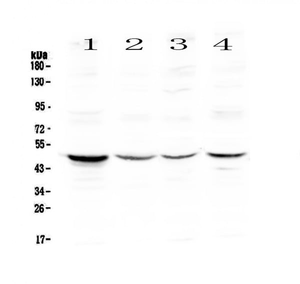
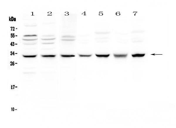
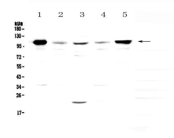
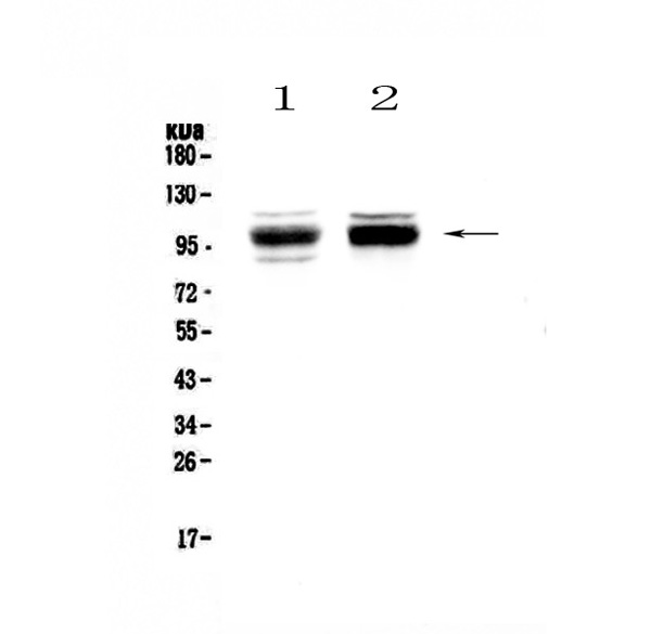
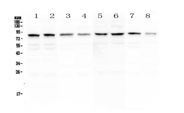
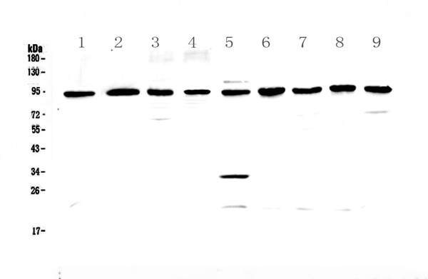 WB (Western Blot)
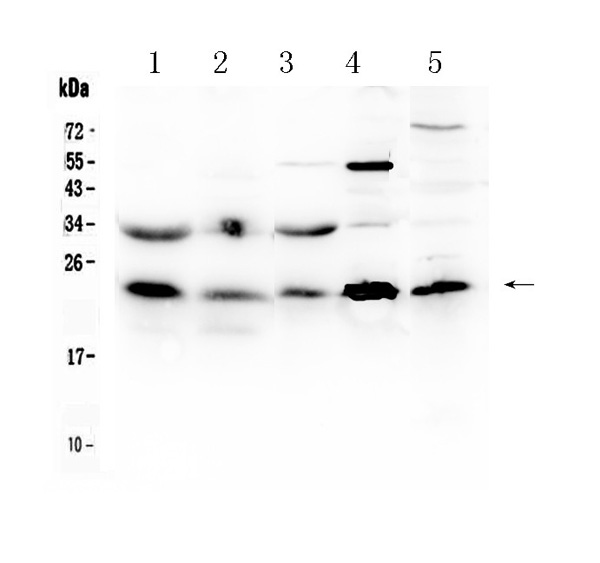
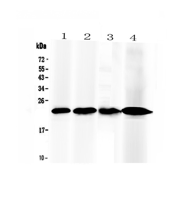
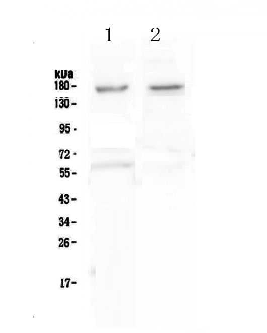
(Figure 2. Western blot analysis of GNS using anti-GNS antibody (AAA124609).Electrophoresis was performed on a 5-20% SDS-PAGE gel at 70V (Stacking gel) / 90V (Resolving gel) for 2-3 hours. The sample well of each lane was loaded with 50ug of sample under reducing conditions.Lane 1: rat lung tissue lysates,Lane 2: rat kidney tissue lysates,Lane 3: rat testis tissue lysates,Lane 4: rat PC-12 whole cell lysates,Lane 5: mouse lung tissue lysates,Lane 6: mouse kidney tissue lysates,Lane 7: mouse testis tissue lysates,Lane 8: mouse spleen tissue lysates,Lane 9: mouse thymus tissue lysates.After Electrophoresis, proteins were transferred to a Nitrocellulose membrane at 150mA for 50-90 minutes. Blocked the membrane with 5% Non-fat Milk/ TBS for 1.5 hour at RT. The membrane was incubated with rabbit anti-GNS antigen affinity purified polyclonal antibody at 0.5ug/mL overnight at 4 degree C, then washed with TBS-0.1%Tween 3 times with 5 minutes each and probed with a goat anti-rabbit IgG-HRP secondary antibody at a dilution of 1:10000 for 1.5 hour at RT. The signal is developed using an Enhanced Chemiluminescent detection (ECL) kit with Tanon 5200 system. A specific band was detected for GNS at approximately 90KD. The expected band size for GNS is at 62KD.)
WB (Western Blot)
(Figure 2. Western blot analysis of GNS using anti-GNS antibody (AAA124609).Electrophoresis was performed on a 5-20% SDS-PAGE gel at 70V (Stacking gel) / 90V (Resolving gel) for 2-3 hours. The sample well of each lane was loaded with 50ug of sample under reducing conditions.Lane 1: rat lung tissue lysates,Lane 2: rat kidney tissue lysates,Lane 3: rat testis tissue lysates,Lane 4: rat PC-12 whole cell lysates,Lane 5: mouse lung tissue lysates,Lane 6: mouse kidney tissue lysates,Lane 7: mouse testis tissue lysates,Lane 8: mouse spleen tissue lysates,Lane 9: mouse thymus tissue lysates.After Electrophoresis, proteins were transferred to a Nitrocellulose membrane at 150mA for 50-90 minutes. Blocked the membrane with 5% Non-fat Milk/ TBS for 1.5 hour at RT. The membrane was incubated with rabbit anti-GNS antigen affinity purified polyclonal antibody at 0.5ug/mL overnight at 4 degree C, then washed with TBS-0.1%Tween 3 times with 5 minutes each and probed with a goat anti-rabbit IgG-HRP secondary antibody at a dilution of 1:10000 for 1.5 hour at RT. The signal is developed using an Enhanced Chemiluminescent detection (ECL) kit with Tanon 5200 system. A specific band was detected for GNS at approximately 90KD. The expected band size for GNS is at 62KD.)
GNS, Polyclonal Antibody (Cat# AAA124609)
No cross reactivity with other proteins.

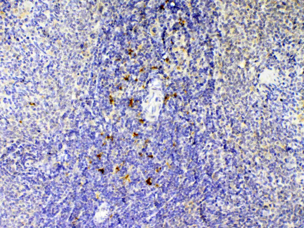
 IHC (Immunohiostchemistry)
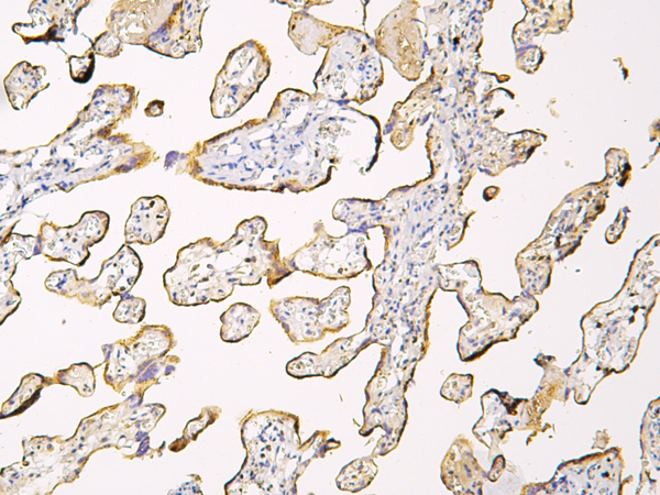
(Figure 2. IHC analysis of BCMA using anti-BCMA antibody (AAA124612).BCMA was detected in paraffin-embedded section of human tonsil tissue. Heat mediated antigen retrieval was performed in citrate buffer (pH6, epitope retrieval solution) for 20 mins. The tissue section was blocked with 10% goat serum. The tissue section was then incubated with 2ug/ml rabbit anti-BCMA Antibody (AAA124612) overnight at 4 degree C. Biotinylated goat anti-rabbit IgG was used as secondary antibody and incubated for 30 minutes at 37 degree C. The tissue section was developed using Strepavidin-Biotin-Complex (SABC) with DAB as the chromogen.)
IHC (Immunohiostchemistry)
(Figure 2. IHC analysis of BCMA using anti-BCMA antibody (AAA124612).BCMA was detected in paraffin-embedded section of human tonsil tissue. Heat mediated antigen retrieval was performed in citrate buffer (pH6, epitope retrieval solution) for 20 mins. The tissue section was blocked with 10% goat serum. The tissue section was then incubated with 2ug/ml rabbit anti-BCMA Antibody (AAA124612) overnight at 4 degree C. Biotinylated goat anti-rabbit IgG was used as secondary antibody and incubated for 30 minutes at 37 degree C. The tissue section was developed using Strepavidin-Biotin-Complex (SABC) with DAB as the chromogen.)
BCMA, Polyclonal Antibody (Cat# AAA124612)
No cross reactivity with other proteins.
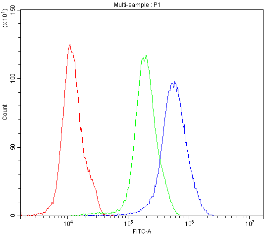
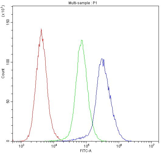
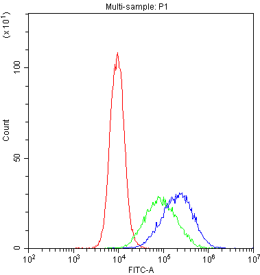 FCM/FACS (Flow Cytometry)
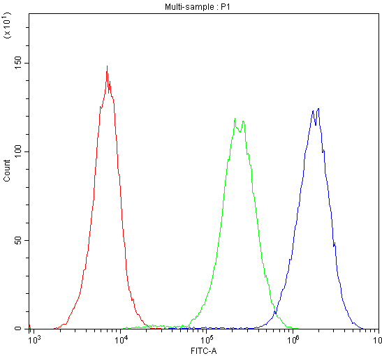
(Figure 4. Flow Cytometry analysis of THP-1 cells using anti-DC-SIGN antibody (AAA124613).Overlay histogram showing THP-1 cells stained with AAA124613 (Blue line).The cells were blocked with 10% normal goat serum. And then incubated with rabbit anti-DC-SIGN Antibody (AAA124613,1ug/1x10^6 cells) for 30 min at 20 degree C. DyLight®488 conjugated goat anti-rabbit IgG (5-10ug/1x10^6 cells) was used as secondary antibody for 30 minutes at 20 degree C. Isotype control antibody (Green line) was rabbit IgG (1ug/1x106) used under the same conditions. Unlabelled sample (Red line) was also used as a control.)
FCM/FACS (Flow Cytometry)
(Figure 4. Flow Cytometry analysis of THP-1 cells using anti-DC-SIGN antibody (AAA124613).Overlay histogram showing THP-1 cells stained with AAA124613 (Blue line).The cells were blocked with 10% normal goat serum. And then incubated with rabbit anti-DC-SIGN Antibody (AAA124613,1ug/1x10^6 cells) for 30 min at 20 degree C. DyLight®488 conjugated goat anti-rabbit IgG (5-10ug/1x10^6 cells) was used as secondary antibody for 30 minutes at 20 degree C. Isotype control antibody (Green line) was rabbit IgG (1ug/1x106) used under the same conditions. Unlabelled sample (Red line) was also used as a control.)
DC-SIGN, Polyclonal Antibody (Cat# AAA124613)
No cross reactivity with other proteins.
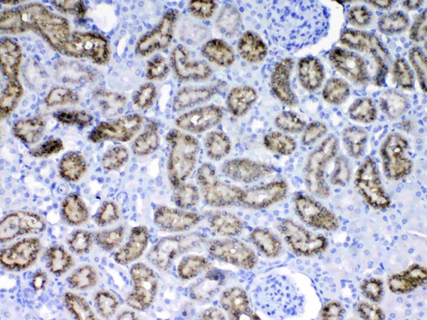
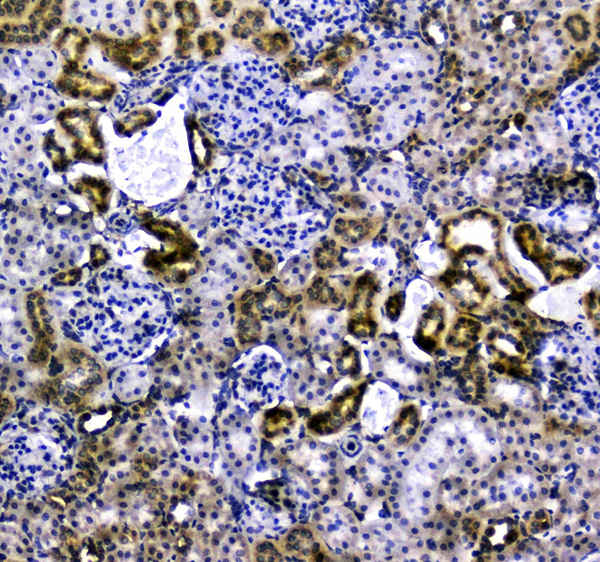
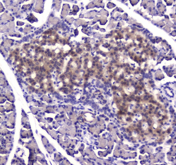
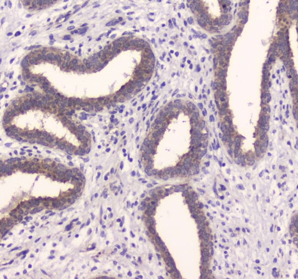
 IHC (Immunohistochemistry)
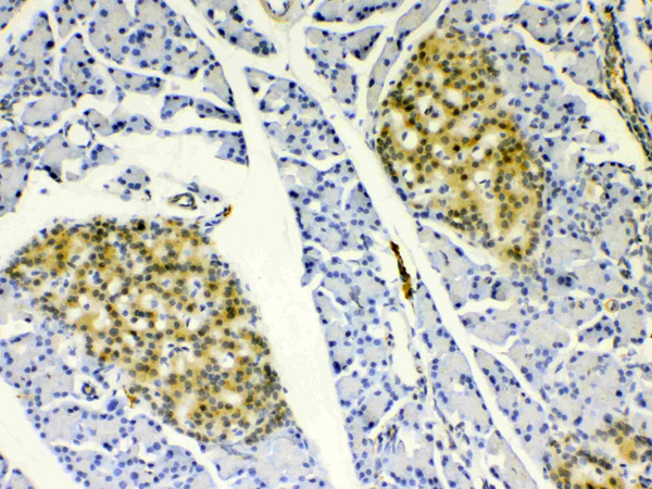
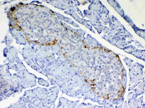
(Figure 5. IHC analysis of RBP4 using anti-RBP4 antibody (AAA124617).RBP4 was detected in paraffin-embedded section of rat pancreas tissue. Heat mediated antigen retrieval was performed in citrate buffer (pH6, epitope retrieval solution) for 20 mins. The tissue section was blocked with 10% goat serum. The tissue section was then incubated with 1ug/ml rabbit anti-RBP4 Antibody (AAA124617) overnight at 4 degree C. Biotinylated goat anti-rabbit IgG was used as secondary antibody and incubated for 30 minutes at 37 degree C. The tissue section was developed using Strepavidin-Biotin-Complex (SABC) with DAB as the chromogen.)
IHC (Immunohistochemistry)
(Figure 5. IHC analysis of RBP4 using anti-RBP4 antibody (AAA124617).RBP4 was detected in paraffin-embedded section of rat pancreas tissue. Heat mediated antigen retrieval was performed in citrate buffer (pH6, epitope retrieval solution) for 20 mins. The tissue section was blocked with 10% goat serum. The tissue section was then incubated with 1ug/ml rabbit anti-RBP4 Antibody (AAA124617) overnight at 4 degree C. Biotinylated goat anti-rabbit IgG was used as secondary antibody and incubated for 30 minutes at 37 degree C. The tissue section was developed using Strepavidin-Biotin-Complex (SABC) with DAB as the chromogen.)
RBP4/Retinol Binding Protein 4, Polyclonal Antibody (Cat# AAA124617)
No cross reactivity with other proteins.
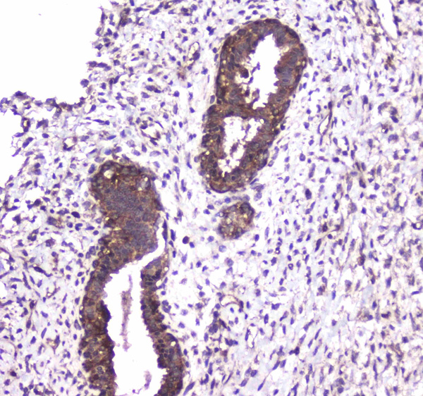
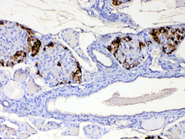
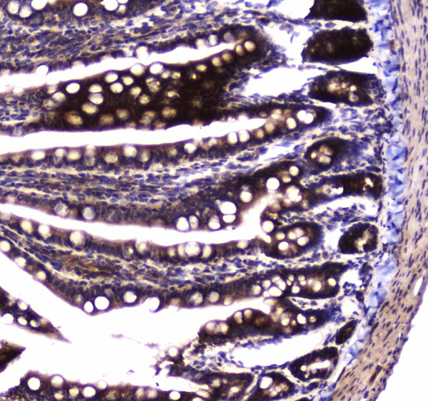 IHC (Immunohistochemistry)
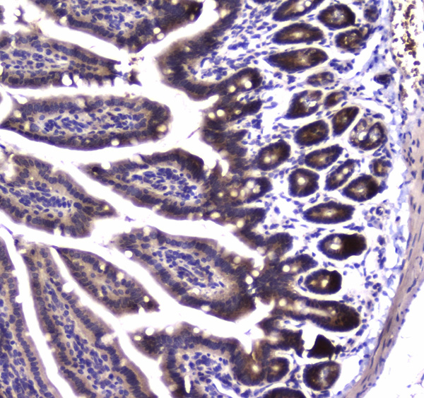
(Figure 4. IHC analysis of SDHB using anti-SDHB antibody (AAA124618).SDHB was detected in paraffin-embedded section of rat small intestine tissue. Heat mediated antigen retrieval was performed in citrate buffer (pH6, epitope retrieval solution) for 20 mins. The tissue section was blocked with 10% goat serum. The tissue section was then incubated with 1ug/ml rabbit anti-SDHB Antibody (AAA124618) overnight at 4 degree C. Biotinylated goat anti-rabbit IgG was used as secondary antibody and incubated for 30 minutes at 37 degree C. The tissue section was developed using Strepavidin-Biotin-Complex (SABC) with DAB as the chromogen.)
IHC (Immunohistochemistry)
(Figure 4. IHC analysis of SDHB using anti-SDHB antibody (AAA124618).SDHB was detected in paraffin-embedded section of rat small intestine tissue. Heat mediated antigen retrieval was performed in citrate buffer (pH6, epitope retrieval solution) for 20 mins. The tissue section was blocked with 10% goat serum. The tissue section was then incubated with 1ug/ml rabbit anti-SDHB Antibody (AAA124618) overnight at 4 degree C. Biotinylated goat anti-rabbit IgG was used as secondary antibody and incubated for 30 minutes at 37 degree C. The tissue section was developed using Strepavidin-Biotin-Complex (SABC) with DAB as the chromogen.)
SDHB, Polyclonal Antibody (Cat# AAA124618)
No cross reactivity with other proteins.
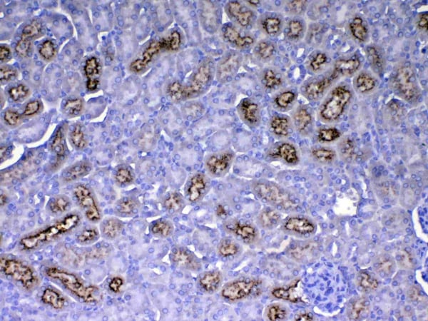
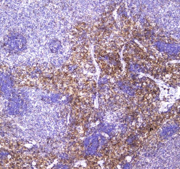
 IHC (Immunohiostchemistry)
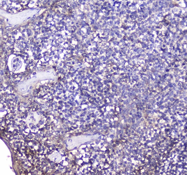
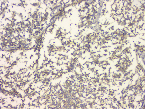
(Figure 2. IHC analysis of IL12B using anti-IL12B antibody (AAA124625).IL12B was detected in paraffin-embedded section of rat spleen tissue. Heat mediated antigen retrieval was performed in citrate buffer (pH6, epitope retrieval solution) for 20 mins. The tissue section was blocked with 10% goat serum. The tissue section was then incubated with 1ug/ml rabbit anti-IL12B Antibody (AAA124625) overnight at 4 degree C. Biotinylated goat anti-rabbit IgG was used as secondary antibody and incubated for 30 minutes at 37 degree C. The tissue section was developed using Strepavidin-Biotin-Complex (SABC) with DAB as the chromogen.)
IHC (Immunohiostchemistry)
(Figure 2. IHC analysis of IL12B using anti-IL12B antibody (AAA124625).IL12B was detected in paraffin-embedded section of rat spleen tissue. Heat mediated antigen retrieval was performed in citrate buffer (pH6, epitope retrieval solution) for 20 mins. The tissue section was blocked with 10% goat serum. The tissue section was then incubated with 1ug/ml rabbit anti-IL12B Antibody (AAA124625) overnight at 4 degree C. Biotinylated goat anti-rabbit IgG was used as secondary antibody and incubated for 30 minutes at 37 degree C. The tissue section was developed using Strepavidin-Biotin-Complex (SABC) with DAB as the chromogen.)
IL12B/Il 12, Polyclonal Antibody (Cat# AAA124625)
No cross reactivity with other proteins.
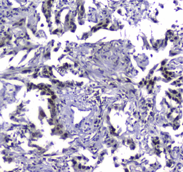
 IHC (Immunohistochemisry)
(Figure 3. IHC analysis of VCAM1 using anti-VCAM1 antibody (AAA124628).VCAM1 was detected in paraffin-embedded section of rat spleen tissue. Heat mediated antigen retrieval was performed in citrate buffer (pH6, epitope retrieval solution) for 20 mins. The tissue section was blocked with 10% goat serum. The tissue section was then incubated with 1ug/ml rabbit anti-VCAM1 Antibody (AAA124628) overnight at 4 degree C. Biotinylated goat anti-rabbit IgG was used as secondary antibody and incubated for 30 minutes at 37 degree C. The tissue section was developed using Strepavidin-Biotin-Complex (SABC) with DAB as the chromogen.)
IHC (Immunohistochemisry)
(Figure 3. IHC analysis of VCAM1 using anti-VCAM1 antibody (AAA124628).VCAM1 was detected in paraffin-embedded section of rat spleen tissue. Heat mediated antigen retrieval was performed in citrate buffer (pH6, epitope retrieval solution) for 20 mins. The tissue section was blocked with 10% goat serum. The tissue section was then incubated with 1ug/ml rabbit anti-VCAM1 Antibody (AAA124628) overnight at 4 degree C. Biotinylated goat anti-rabbit IgG was used as secondary antibody and incubated for 30 minutes at 37 degree C. The tissue section was developed using Strepavidin-Biotin-Complex (SABC) with DAB as the chromogen.)
VCAM1/Cd106, Polyclonal Antibody (Cat# AAA124628)
No cross reactivity with other proteins.
 IHC (Immunohistochemisry)
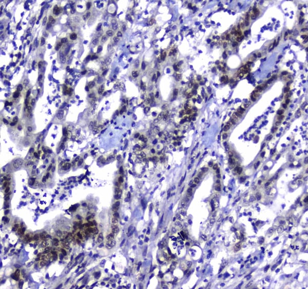
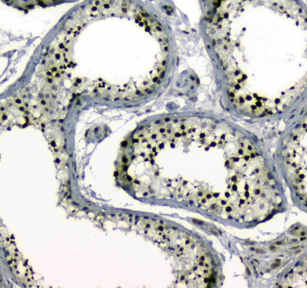
(Figure 3. IHC analysis of MDC1 using anti-MDC1 antibody (AAA124633).MDC1 was detected in paraffin-embedded section of human testis tissue. Heat mediated antigen retrieval was performed in citrate buffer (pH6, epitope retrieval solution) for 20 mins. The tissue section was blocked with 10% goat serum. The tissue section was then incubated with 1ug/ml rabbit anti-MDC1 Antibody (AAA124633) overnight at 4 degree C. Biotinylated goat anti-rabbit IgG was used as secondary antibody and incubated for 30 minutes at 37 degree C. The tissue section was developed using Strepavidin-Biotin-Complex (SABC) with DAB as the chromogen.)
IHC (Immunohistochemisry)
(Figure 3. IHC analysis of MDC1 using anti-MDC1 antibody (AAA124633).MDC1 was detected in paraffin-embedded section of human testis tissue. Heat mediated antigen retrieval was performed in citrate buffer (pH6, epitope retrieval solution) for 20 mins. The tissue section was blocked with 10% goat serum. The tissue section was then incubated with 1ug/ml rabbit anti-MDC1 Antibody (AAA124633) overnight at 4 degree C. Biotinylated goat anti-rabbit IgG was used as secondary antibody and incubated for 30 minutes at 37 degree C. The tissue section was developed using Strepavidin-Biotin-Complex (SABC) with DAB as the chromogen.)
MDC1, Polyclonal Antibody (Cat# AAA124633)
No cross reactivity with other proteins.

 IHC (Immunohistochemisry)
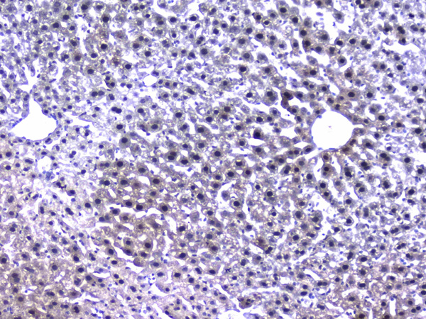
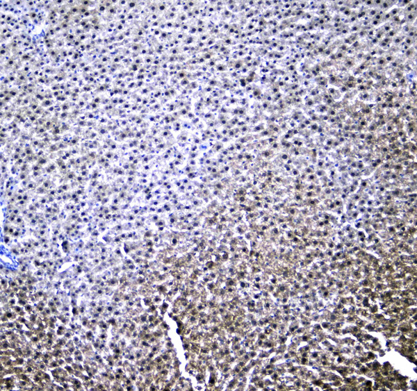
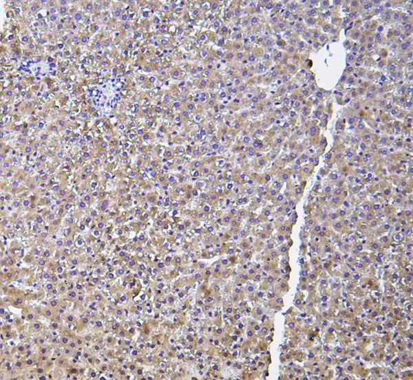
(Figure 3. IHC analysis of RXRA using anti-RXRA antibody (AAA124639).RXRA was detected in paraffin-embedded section of rat liver tissue. Heat mediated antigen retrieval was performed in citrate buffer (pH6, epitope retrieval solution) for 20 mins. The tissue section was blocked with 10% goat serum. The tissue section was then incubated with 1ug/ml rabbit anti-RXRA Antibody (AAA124639) overnight at 4 degree C. Biotinylated goat anti-rabbit IgG was used as secondary antibody and incubated for 30 minutes at 37 degree C. The tissue section was developed using Strepavidin-Biotin-Complex (SABC) with DAB as the chromogen.)
IHC (Immunohistochemisry)
(Figure 3. IHC analysis of RXRA using anti-RXRA antibody (AAA124639).RXRA was detected in paraffin-embedded section of rat liver tissue. Heat mediated antigen retrieval was performed in citrate buffer (pH6, epitope retrieval solution) for 20 mins. The tissue section was blocked with 10% goat serum. The tissue section was then incubated with 1ug/ml rabbit anti-RXRA Antibody (AAA124639) overnight at 4 degree C. Biotinylated goat anti-rabbit IgG was used as secondary antibody and incubated for 30 minutes at 37 degree C. The tissue section was developed using Strepavidin-Biotin-Complex (SABC) with DAB as the chromogen.)
RXRA/Rxr Alpha, Polyclonal Antibody (Cat# AAA124639)
No cross reactivity with other proteins.
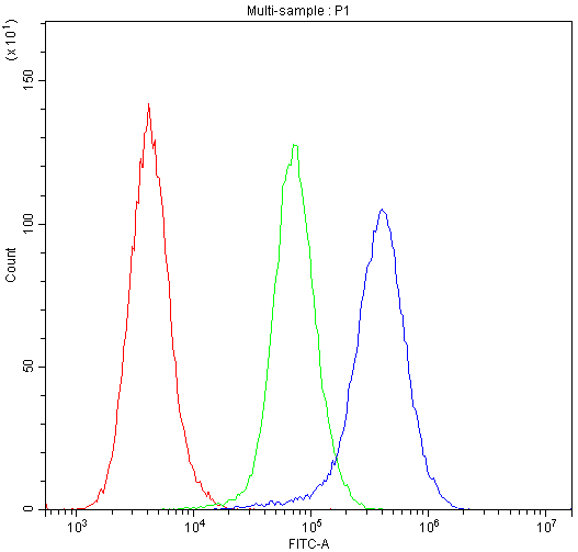
 FCM/FACS (Flow Cytometry)
(Figure 2. Flow Cytometry analysis of PC-3 cells using anti-SATB1 antibody (AAA124640).Overlay histogram showing PC-3 cells stained with AAA124640 (Blue line).The cells were blocked with 10% normal goat serum. And then incubated with rabbit anti-SATB1 Antibody (AAA124640,1ug/1x10^6 cells) for 30 min at 20 degree C. DyLight®488 conjugated goat anti-rabbit IgG (5-10ug/1x10^6 cells) was used as secondary antibody for 30 minutes at 20 degree C. Isotype control antibody (Green line) was rabbit IgG (1ug/1x106) used under the same conditions. Unlabelled sample (Red line) was also used as a control.)
FCM/FACS (Flow Cytometry)
(Figure 2. Flow Cytometry analysis of PC-3 cells using anti-SATB1 antibody (AAA124640).Overlay histogram showing PC-3 cells stained with AAA124640 (Blue line).The cells were blocked with 10% normal goat serum. And then incubated with rabbit anti-SATB1 Antibody (AAA124640,1ug/1x10^6 cells) for 30 min at 20 degree C. DyLight®488 conjugated goat anti-rabbit IgG (5-10ug/1x10^6 cells) was used as secondary antibody for 30 minutes at 20 degree C. Isotype control antibody (Green line) was rabbit IgG (1ug/1x106) used under the same conditions. Unlabelled sample (Red line) was also used as a control.)
SATB1, Polyclonal Antibody (Cat# AAA124640)
No cross reactivity with other proteins.

 FCM/FACS (Flow Cytometry)
(Figure 5. Flow Cytometry analysis of CACO-2 cells using anti-IDE antibody (AAA124641).Overlay histogram showing CACO-2 cells stained with AAA124641 (Blue line).The cells were blocked with 10% normal goat serum. And then incubated with rabbit anti-IDE Antibody (AAA124641,1ug/1x10^6 cells) for 30 min at 20 degree C. DyLight®488 conjugated goat anti-rabbit IgG (5-10ug/1x10^6 cells) was used as secondary antibody for 30 minutes at 20 degree C. Isotype control antibody (Green line) was rabbit IgG (1ug/1x106) used under the same conditions. Unlabelled sample (Red line) was also used as a control.)
FCM/FACS (Flow Cytometry)
(Figure 5. Flow Cytometry analysis of CACO-2 cells using anti-IDE antibody (AAA124641).Overlay histogram showing CACO-2 cells stained with AAA124641 (Blue line).The cells were blocked with 10% normal goat serum. And then incubated with rabbit anti-IDE Antibody (AAA124641,1ug/1x10^6 cells) for 30 min at 20 degree C. DyLight®488 conjugated goat anti-rabbit IgG (5-10ug/1x10^6 cells) was used as secondary antibody for 30 minutes at 20 degree C. Isotype control antibody (Green line) was rabbit IgG (1ug/1x106) used under the same conditions. Unlabelled sample (Red line) was also used as a control.)
IDE, Polyclonal Antibody (Cat# AAA124641)
No cross reactivity with other proteins.
 WB (Western Blot)
(Figure 2. Western blot analysis of DNA Polymerase iota using anti-DNA Polymerase iota antibody (AAA124644).Electrophoresis was performed on a 5-20% SDS-PAGE gel at 70V (Stacking gel) / 90V (Resolving gel) for 2-3 hours. The sample well of each lane was loaded with 50ug of sample under reducing conditions.Lane 1: rat testis tissue lysates,Lane 2: rat testis tissue lysates,Lane 3: rat kidney tissue lysates,Lane 4: rat stomach tissue lysates,Lane 5: mouse testis tissue lysates,Lane 6: mouse testis tissue lysates,Lane 7: mouse kidney tissue lysates,Lane 8: mouse stomach tissue lysates.After Electrophoresis, proteins were transferred to a Nitrocellulose membrane at 150mA for 50-90 minutes. Blocked the membrane with 5% Non-fat Milk/ TBS for 1.5 hour at RT. The membrane was incubated with rabbit anti-DNA Polymerase iota antigen affinity purified polyclonal antibody at 0.5ug/mL overnight at 4 degree C, then washed with TBS-0.1%Tween 3 times with 5 minutes each and probed with a goat anti-rabbit IgG-HRP secondary antibody at a dilution of 1:10000 for 1.5 hour at RT. The signal is developed using an Enhanced Chemiluminescent detection (ECL) kit with Tanon 5200 system. A specific band was detected for DNA Polymerase iota at approximately 83KD. The expected band size for DNA Polymerase iota is at 83KD.)
WB (Western Blot)
(Figure 2. Western blot analysis of DNA Polymerase iota using anti-DNA Polymerase iota antibody (AAA124644).Electrophoresis was performed on a 5-20% SDS-PAGE gel at 70V (Stacking gel) / 90V (Resolving gel) for 2-3 hours. The sample well of each lane was loaded with 50ug of sample under reducing conditions.Lane 1: rat testis tissue lysates,Lane 2: rat testis tissue lysates,Lane 3: rat kidney tissue lysates,Lane 4: rat stomach tissue lysates,Lane 5: mouse testis tissue lysates,Lane 6: mouse testis tissue lysates,Lane 7: mouse kidney tissue lysates,Lane 8: mouse stomach tissue lysates.After Electrophoresis, proteins were transferred to a Nitrocellulose membrane at 150mA for 50-90 minutes. Blocked the membrane with 5% Non-fat Milk/ TBS for 1.5 hour at RT. The membrane was incubated with rabbit anti-DNA Polymerase iota antigen affinity purified polyclonal antibody at 0.5ug/mL overnight at 4 degree C, then washed with TBS-0.1%Tween 3 times with 5 minutes each and probed with a goat anti-rabbit IgG-HRP secondary antibody at a dilution of 1:10000 for 1.5 hour at RT. The signal is developed using an Enhanced Chemiluminescent detection (ECL) kit with Tanon 5200 system. A specific band was detected for DNA Polymerase iota at approximately 83KD. The expected band size for DNA Polymerase iota is at 83KD.)
DNA Polymerase iota, Polyclonal Antibody (Cat# AAA124644)
No cross reactivity with other proteins.
 FCM/FACS (Flow Cytometry)
(Figure 2. Flow Cytometry analysis of U251 cells using anti-NRXN1 antibody (AAA124650).Overlay histogram showing U251 cells stained with AAA124650 (Blue line).The cells were blocked with 10% normal goat serum. And then incubated with rabbit anti-NRXN1 Antibody (AAA124650,1ug/1x10^6 cells) for 30 min at 20 degree C. DyLight®488 conjugated goat anti-rabbit IgG (5-10ug/1x10^6 cells) was used as secondary antibody for 30 minutes at 20 degree C. Isotype control antibody (Green line) was rabbit IgG (1ug/1x106) used under the same conditions. Unlabelled sample (Red line) was also used as a control.)
FCM/FACS (Flow Cytometry)
(Figure 2. Flow Cytometry analysis of U251 cells using anti-NRXN1 antibody (AAA124650).Overlay histogram showing U251 cells stained with AAA124650 (Blue line).The cells were blocked with 10% normal goat serum. And then incubated with rabbit anti-NRXN1 Antibody (AAA124650,1ug/1x10^6 cells) for 30 min at 20 degree C. DyLight®488 conjugated goat anti-rabbit IgG (5-10ug/1x10^6 cells) was used as secondary antibody for 30 minutes at 20 degree C. Isotype control antibody (Green line) was rabbit IgG (1ug/1x106) used under the same conditions. Unlabelled sample (Red line) was also used as a control.)
Neurexin 1, Polyclonal Antibody (Cat# AAA124650)
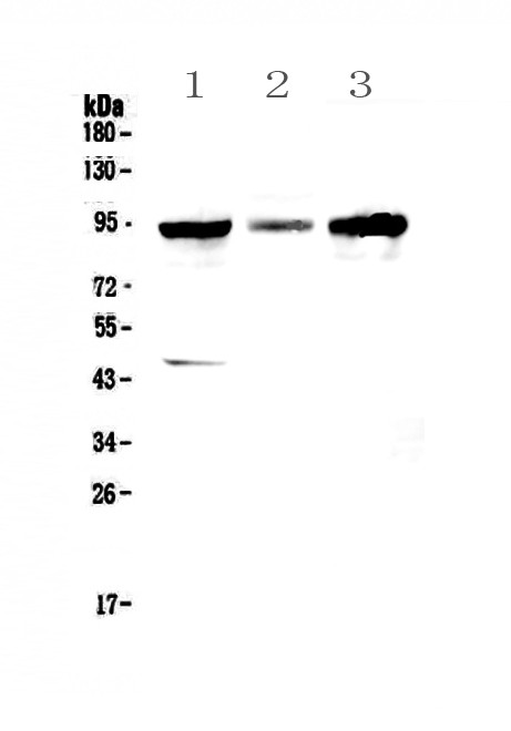
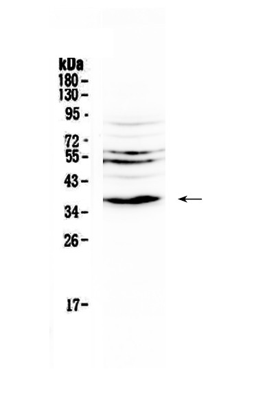
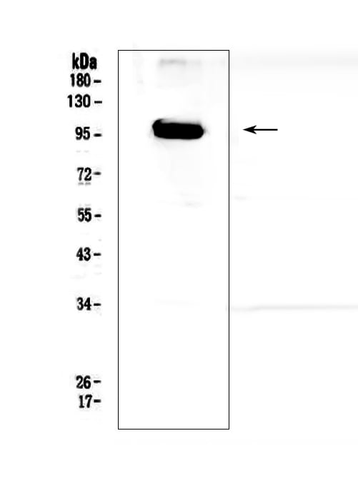
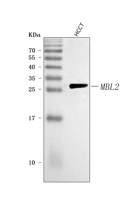
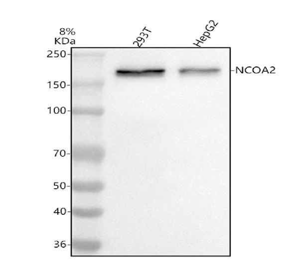
 WB (Western Blot)
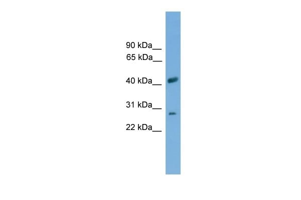
(Western blot analysis of SLC25A37 expression in Human THP-1. SLC25A37 at 37KD was detected using rabbit anti-SLC25A37 Antigen Affinity purified polyclonal antibody at Dilution: 0.2-1ug/ml. The blot was developed using chemiluminescence (ECL) method)
WB (Western Blot)
(Western blot analysis of SLC25A37 expression in Human THP-1. SLC25A37 at 37KD was detected using rabbit anti-SLC25A37 Antigen Affinity purified polyclonal antibody at Dilution: 0.2-1ug/ml. The blot was developed using chemiluminescence (ECL) method)
SLC25A37/Mitoferrin, Polyclonal Antibody (Cat# AAA124885)
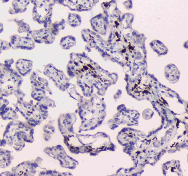
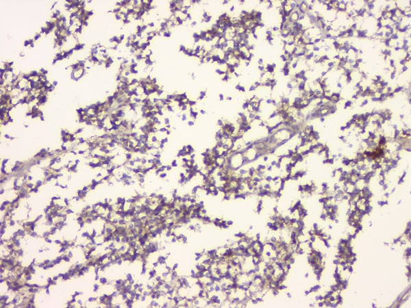
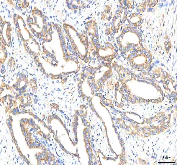
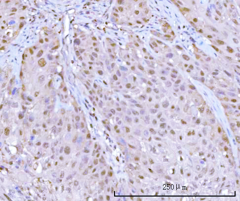
 IHC (Immunohistochemistry)
(Figure 4. IHC analysis of HP using anti-HP antibod.)
IHC (Immunohistochemistry)
(Figure 4. IHC analysis of HP using anti-HP antibod.)
Haptoglobin/HP, Polyclonal Antibody (Cat# AAA124887)
No cross reactivity with other proteins.
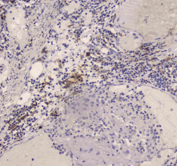
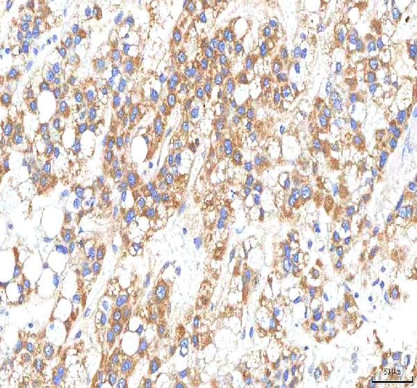
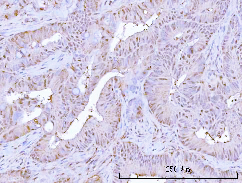
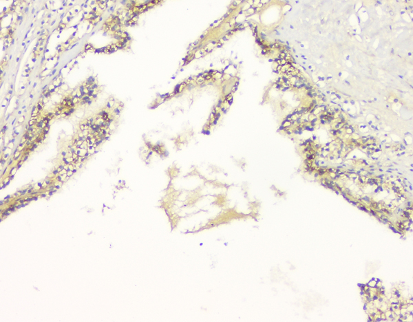 IHC (Immunohistochemistry)
(Figure 4. IHC analysis of C99 using anti-C99 antibody.)
IHC (Immunohistochemistry)
(Figure 4. IHC analysis of C99 using anti-C99 antibody.)
APP/C99, Polyclonal Antibody (Cat# AAA124888)
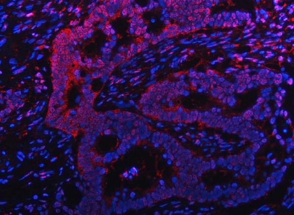
 IF (Immunofluorescence)
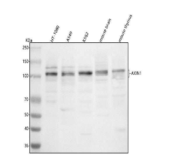
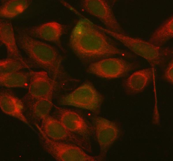
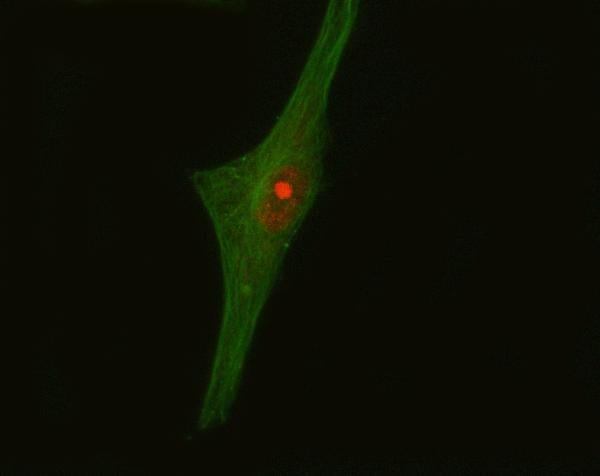
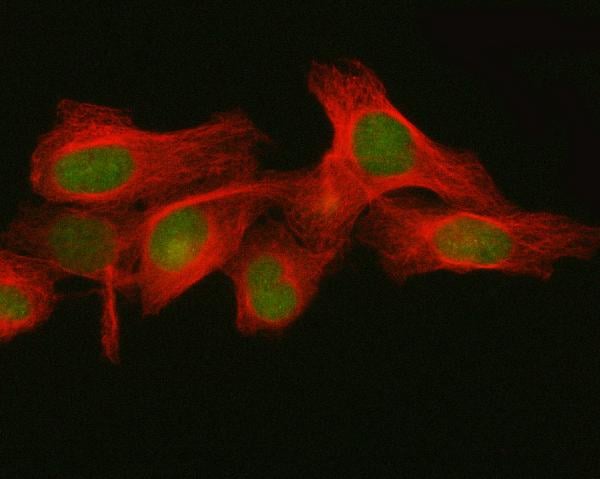
(Figure 2. IF analysis of AXIN1 using anti-AXIN1 antibody (AAA127024) and anti-Beta Tubulin antibody (M01857-3).AXIN1 was detected in immunocytochemical section of HELA cell. Enzyme antigen retrieval was performed using IHC enzyme antigen retrieval reagent for 15 mins. The cells were blocked with 10% goat serum. And then incubated with 5ug/mL rabbit anti-AXIN1 Antibody (AAA127024) and mouse anti-Beta Tubulin antibody (M01857-3) overnight at 4 degree C. Cy3 Conjugated Goat Anti-Rabbit IgG (BA1032) and DyLight488 Conjugated Goat Anti-Mouse IgG (BA1126) were used as secondary antibody at 1:500 dilution and incubated for 30 minutes at 37 degree C. Visualize using a fluorescence microscope and filter sets appropriate for the label used.)
IF (Immunofluorescence)
(Figure 2. IF analysis of AXIN1 using anti-AXIN1 antibody (AAA127024) and anti-Beta Tubulin antibody (M01857-3).AXIN1 was detected in immunocytochemical section of HELA cell. Enzyme antigen retrieval was performed using IHC enzyme antigen retrieval reagent for 15 mins. The cells were blocked with 10% goat serum. And then incubated with 5ug/mL rabbit anti-AXIN1 Antibody (AAA127024) and mouse anti-Beta Tubulin antibody (M01857-3) overnight at 4 degree C. Cy3 Conjugated Goat Anti-Rabbit IgG (BA1032) and DyLight488 Conjugated Goat Anti-Mouse IgG (BA1126) were used as secondary antibody at 1:500 dilution and incubated for 30 minutes at 37 degree C. Visualize using a fluorescence microscope and filter sets appropriate for the label used.)
AXIN1, Polyclonal Antibody (Cat# AAA127024)
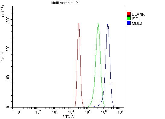 FCM/FACS (Flow Cytometry)
(Figure 2. Flow Cytometry analysis of HepG2 cells using anti-MBL2 antibody (AAA127027).Overlay histogram showing HepG2 cells stained with AAA127027 (Blue line). The cells were fixed with 4% paraformaldehyde and blocked with 10% normal goat serum. And then incubated with rabbit anti-MBL2 Antibody (AAA127027, 1ug/1x106 cells) for 30 min at 20 degree C. DyLight488 conjugated goat anti-rabbit IgG was used as secondary antibody for 30 minutes at 20 degree C. Isotype control antibody (Green line) was rabbit IgG (1ug/1x106) used under the same conditions. Unlabelled sample (Red line) was also used as a control.)
FCM/FACS (Flow Cytometry)
(Figure 2. Flow Cytometry analysis of HepG2 cells using anti-MBL2 antibody (AAA127027).Overlay histogram showing HepG2 cells stained with AAA127027 (Blue line). The cells were fixed with 4% paraformaldehyde and blocked with 10% normal goat serum. And then incubated with rabbit anti-MBL2 Antibody (AAA127027, 1ug/1x106 cells) for 30 min at 20 degree C. DyLight488 conjugated goat anti-rabbit IgG was used as secondary antibody for 30 minutes at 20 degree C. Isotype control antibody (Green line) was rabbit IgG (1ug/1x106) used under the same conditions. Unlabelled sample (Red line) was also used as a control.)
MBL2, Polyclonal Antibody (Cat# AAA127027)
 FCM/FACS (Flow Cytometry)
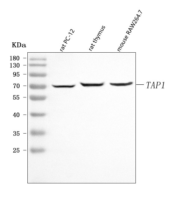
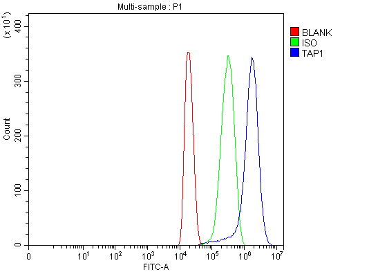
(Figure 3. Flow Cytometry analysis of HEPA1-6 cells using anti-Tap1 antibody (AAA127036).Overlay histogram showing HEPA1-6 cells stained with AAA127036 (Blue line). To facilitate intracellular staining, cells were fixed with 4% paraformaldehyde and permeabilized with permeabilization buffer. The cells were blocked with 10% normal goat serum. And then incubated with rabbit anti-Tap1 Antibody (AAA127036, 1ug/1x106 cells) for 30 min at 20 degree C. DyLight488 conjugated goat anti-rabbit IgG was used as secondary antibody for 30 minutes at 20 degree C. Isotype control antibody (Green line) was rabbit IgG (1ug/1x106) used under the same conditions. Unlabelled sample without incubation with primary antibody and secondary antibody (Red line) was used as a blank control.)
FCM/FACS (Flow Cytometry)
(Figure 3. Flow Cytometry analysis of HEPA1-6 cells using anti-Tap1 antibody (AAA127036).Overlay histogram showing HEPA1-6 cells stained with AAA127036 (Blue line). To facilitate intracellular staining, cells were fixed with 4% paraformaldehyde and permeabilized with permeabilization buffer. The cells were blocked with 10% normal goat serum. And then incubated with rabbit anti-Tap1 Antibody (AAA127036, 1ug/1x106 cells) for 30 min at 20 degree C. DyLight488 conjugated goat anti-rabbit IgG was used as secondary antibody for 30 minutes at 20 degree C. Isotype control antibody (Green line) was rabbit IgG (1ug/1x106) used under the same conditions. Unlabelled sample without incubation with primary antibody and secondary antibody (Red line) was used as a blank control.)
Tap1, Polyclonal Antibody (Cat# AAA127036)
 FCM/FACS (Flow Cytometry)
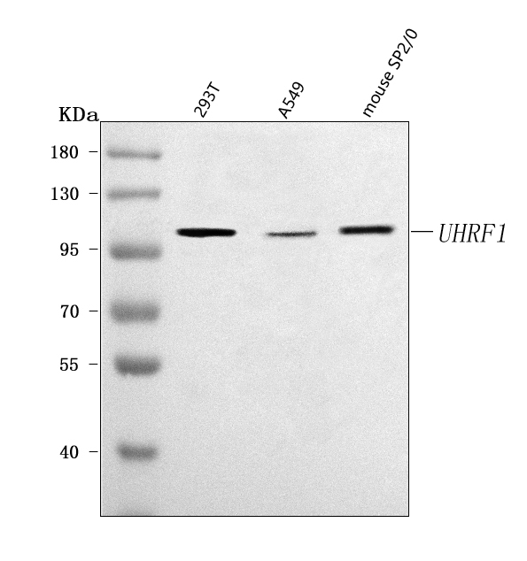
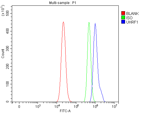
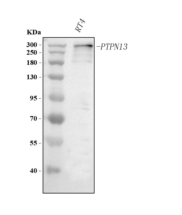
(Figure 3. Flow Cytometry analysis of RT4 cells using anti-UHRF1 antibody (AAA127040).Overlay histogram showing RT4 cells stained with AAA127040 (Blue line). To facilitate intracellular staining, cells were fixed with 4% paraformaldehyde and permeabilized with permeabilization buffer. The cells were blocked with 10% normal goat serum. And then incubated with rabbit anti-UHRF1 Antibody (AAA127040, 1ug/1x106 cells) for 30 min at 20 degree C. DyLight488 conjugated goat anti-rabbit IgG was used as secondary antibody for 30 minutes at 20 degree C. Isotype control antibody (Green line) was rabbit IgG (1ug/1x106) used under the same conditions. Unlabelled sample without incubation with primary antibody and secondary antibody (Red line) was used as a blank control.)
FCM/FACS (Flow Cytometry)
(Figure 3. Flow Cytometry analysis of RT4 cells using anti-UHRF1 antibody (AAA127040).Overlay histogram showing RT4 cells stained with AAA127040 (Blue line). To facilitate intracellular staining, cells were fixed with 4% paraformaldehyde and permeabilized with permeabilization buffer. The cells were blocked with 10% normal goat serum. And then incubated with rabbit anti-UHRF1 Antibody (AAA127040, 1ug/1x106 cells) for 30 min at 20 degree C. DyLight488 conjugated goat anti-rabbit IgG was used as secondary antibody for 30 minutes at 20 degree C. Isotype control antibody (Green line) was rabbit IgG (1ug/1x106) used under the same conditions. Unlabelled sample without incubation with primary antibody and secondary antibody (Red line) was used as a blank control.)
UHRF1, Polyclonal Antibody (Cat# AAA127040)
 FCM/FACS (Flow Cytometry)
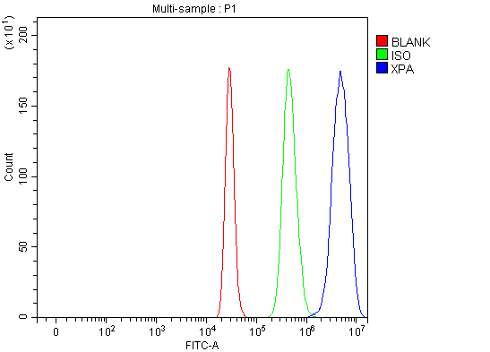
(Figure 3. Flow Cytometry analysis of 293T cells using anti-XPA antibody (AAA127042).Overlay histogram showing 293T cells stained with AAA127042 (Blue line). To facilitate intracellular staining, cells were fixed with 4% paraformaldehyde and permeabilized with permeabilization buffer. The cells were blocked with 10% normal goat serum. And then incubated with rabbit anti-XPA Antibody (AAA127042, 1ug/1x106 cells) for 30 min at 20 degree C. DyLight488 conjugated goat anti-rabbit IgG was used as secondary antibody for 30 minutes at 20 degree C. Isotype control antibody (Green line) was rabbit IgG (1ug/1x106) used under the same conditions. Unlabelled sample (Red line) was also used as a control.)
FCM/FACS (Flow Cytometry)
(Figure 3. Flow Cytometry analysis of 293T cells using anti-XPA antibody (AAA127042).Overlay histogram showing 293T cells stained with AAA127042 (Blue line). To facilitate intracellular staining, cells were fixed with 4% paraformaldehyde and permeabilized with permeabilization buffer. The cells were blocked with 10% normal goat serum. And then incubated with rabbit anti-XPA Antibody (AAA127042, 1ug/1x106 cells) for 30 min at 20 degree C. DyLight488 conjugated goat anti-rabbit IgG was used as secondary antibody for 30 minutes at 20 degree C. Isotype control antibody (Green line) was rabbit IgG (1ug/1x106) used under the same conditions. Unlabelled sample (Red line) was also used as a control.)
XPA, Polyclonal Antibody (Cat# AAA127042)
 FCM/FACS (Flow Cytometry)
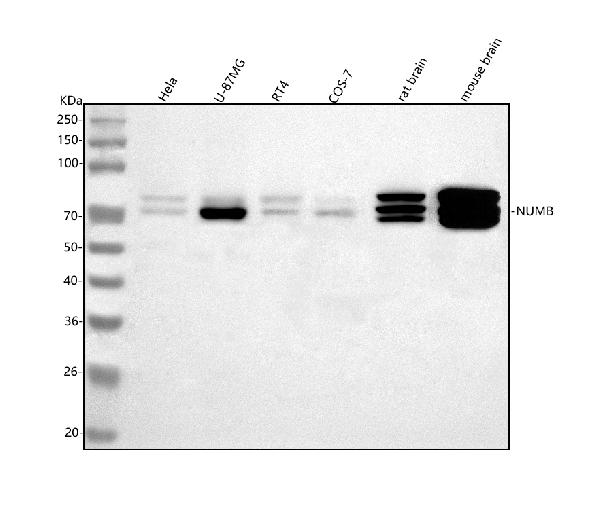
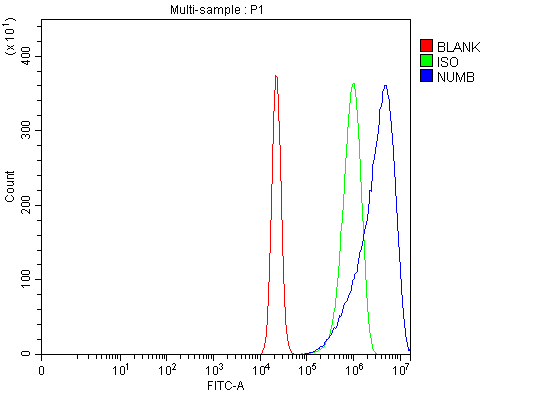
(Figure 2. Flow Cytometry analysis of RT4 cells using anti-NUMB antibody (AAA127043).Overlay histogram showing RT4 cells stained with AAA127043 (Blue line). To facilitate intracellular staining, cells were fixed with 4% paraformaldehyde and permeabilized with permeabilization buffer. The cells were blocked with 10% normal goat serum. And then incubated with rabbit anti-NUMB Antibody (AAA127043, 1ug/1x106 cells) for 30 min at 20 degree C. DyLight488 conjugated goat anti-rabbit IgG was used as secondary antibody for 30 minutes at 20 degree C. Isotype control antibody (Green line) was rabbit IgG (1ug/1x106) used under the same conditions. Unlabelled sample (Red line) was also used as a control.)
FCM/FACS (Flow Cytometry)
(Figure 2. Flow Cytometry analysis of RT4 cells using anti-NUMB antibody (AAA127043).Overlay histogram showing RT4 cells stained with AAA127043 (Blue line). To facilitate intracellular staining, cells were fixed with 4% paraformaldehyde and permeabilized with permeabilization buffer. The cells were blocked with 10% normal goat serum. And then incubated with rabbit anti-NUMB Antibody (AAA127043, 1ug/1x106 cells) for 30 min at 20 degree C. DyLight488 conjugated goat anti-rabbit IgG was used as secondary antibody for 30 minutes at 20 degree C. Isotype control antibody (Green line) was rabbit IgG (1ug/1x106) used under the same conditions. Unlabelled sample (Red line) was also used as a control.)
NUMB, Polyclonal Antibody (Cat# AAA127043)
 FCM/FACS (Flow Cytometry)
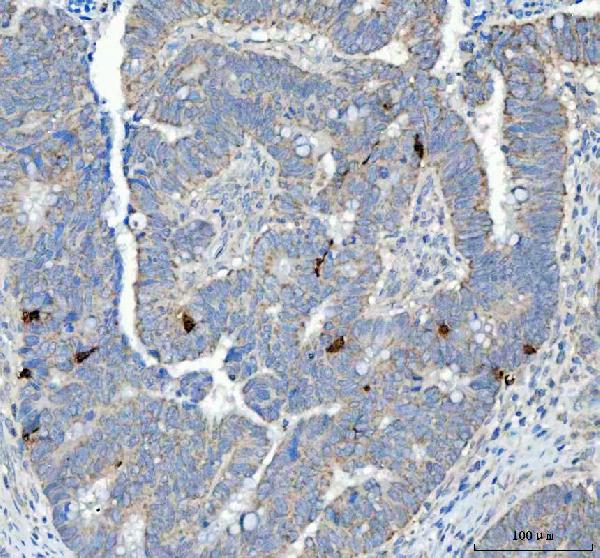
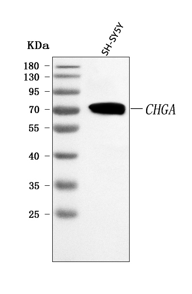
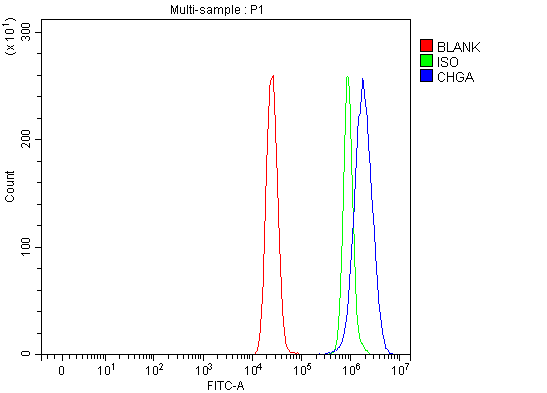
(Figure 5. Flow Cytometry analysis of MCF-7 cells using anti-Chromogranin A/CHGA antibody (AAA127045).Overlay histogram showing MCF-7 cells stained with AAA127045 (Blue line). To facilitate intracellular staining, cells were fixed with 4% paraformaldehyde and permeabilized with permeabilization buffer. The cells were blocked with 10% normal goat serum. And then incubated with rabbit anti-Chromogranin A/CHGA Antibody (AAA127045, 1ug/1x106 cells) for 30 min at 20 degree C. DyLight488 conjugated goat anti-rabbit IgG was used as secondary antibody for 30 minutes at 20 degree C. Isotype control antibody (Green line) was rabbit IgG (1ug/1x106) used under the same conditions. Unlabelled sample without incubation with primary antibody and secondary antibody (Red line) was used as a blank control.)
FCM/FACS (Flow Cytometry)
(Figure 5. Flow Cytometry analysis of MCF-7 cells using anti-Chromogranin A/CHGA antibody (AAA127045).Overlay histogram showing MCF-7 cells stained with AAA127045 (Blue line). To facilitate intracellular staining, cells were fixed with 4% paraformaldehyde and permeabilized with permeabilization buffer. The cells were blocked with 10% normal goat serum. And then incubated with rabbit anti-Chromogranin A/CHGA Antibody (AAA127045, 1ug/1x106 cells) for 30 min at 20 degree C. DyLight488 conjugated goat anti-rabbit IgG was used as secondary antibody for 30 minutes at 20 degree C. Isotype control antibody (Green line) was rabbit IgG (1ug/1x106) used under the same conditions. Unlabelled sample without incubation with primary antibody and secondary antibody (Red line) was used as a blank control.)
Chromogranin A/CHGA, Polyclonal Antibody (Cat# AAA127045)
 FCM/FACS (Flow Cytometry)
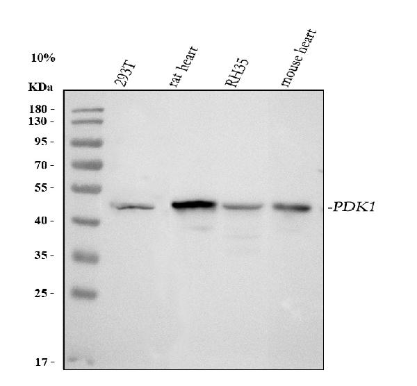
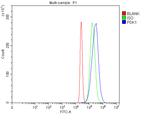
(Figure 2. Flow Cytometry analysis of 293T cells using anti-PDK1 antibody (AAA127048).Overlay histogram showing 293T cells stained with AAA127048 (Blue line). To facilitate intracellular staining, cells were fixed with 4% paraformaldehyde and permeabilized with permeabilization buffer. The cells were blocked with 10% normal goat serum. And then incubated with rabbit anti-PDK1 Antibody (AAA127048, 1ug/1x106 cells) for 30 min at 20 degree C. DyLight488 conjugated goat anti-rabbit IgG was used as secondary antibody for 30 minutes at 20 degree C. Isotype control antibody (Green line) was rabbit IgG (1ug/1x106) used under the same conditions. Unlabelled sample (Red line) was also used as a control.)
FCM/FACS (Flow Cytometry)
(Figure 2. Flow Cytometry analysis of 293T cells using anti-PDK1 antibody (AAA127048).Overlay histogram showing 293T cells stained with AAA127048 (Blue line). To facilitate intracellular staining, cells were fixed with 4% paraformaldehyde and permeabilized with permeabilization buffer. The cells were blocked with 10% normal goat serum. And then incubated with rabbit anti-PDK1 Antibody (AAA127048, 1ug/1x106 cells) for 30 min at 20 degree C. DyLight488 conjugated goat anti-rabbit IgG was used as secondary antibody for 30 minutes at 20 degree C. Isotype control antibody (Green line) was rabbit IgG (1ug/1x106) used under the same conditions. Unlabelled sample (Red line) was also used as a control.)
PDK1, Polyclonal Antibody (Cat# AAA127048)
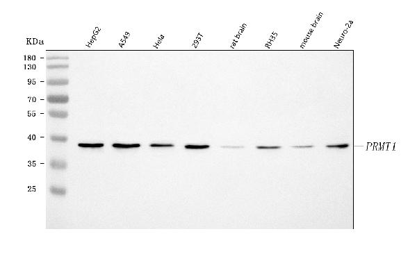
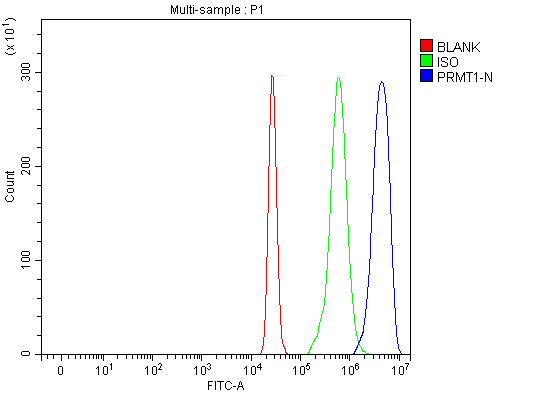 FCM/FACS (Flow Cytometry)
(Figure 3. Flow Cytometry analysis of U937 cells using anti-PRMT1 antibody (AAA127059).Overlay histogram showing U937 cells stained with AAA127059 (Blue line). To facilitate intracellular staining, cells were fixed with 4% paraformaldehyde and permeabilized with permeabilization buffer. The cells were blocked with 10% normal goat serum. And then incubated with rabbit anti-PRMT1 Antibody (AAA127059, 1ug/1x106 cells) for 30 min at 20 degree C. DyLight488 conjugated goat anti-rabbit IgG was used as secondary antibody for 30 minutes at 20 degree C. Isotype control antibody (Green line) was rabbit IgG (1ug/1x106) used under the same conditions. Unlabelled sample (Red line) was also used as a control.)
FCM/FACS (Flow Cytometry)
(Figure 3. Flow Cytometry analysis of U937 cells using anti-PRMT1 antibody (AAA127059).Overlay histogram showing U937 cells stained with AAA127059 (Blue line). To facilitate intracellular staining, cells were fixed with 4% paraformaldehyde and permeabilized with permeabilization buffer. The cells were blocked with 10% normal goat serum. And then incubated with rabbit anti-PRMT1 Antibody (AAA127059, 1ug/1x106 cells) for 30 min at 20 degree C. DyLight488 conjugated goat anti-rabbit IgG was used as secondary antibody for 30 minutes at 20 degree C. Isotype control antibody (Green line) was rabbit IgG (1ug/1x106) used under the same conditions. Unlabelled sample (Red line) was also used as a control.)
PRMT1, Polyclonal Antibody (Cat# AAA127059)
 FCM/FACS (Flow Cytometry)
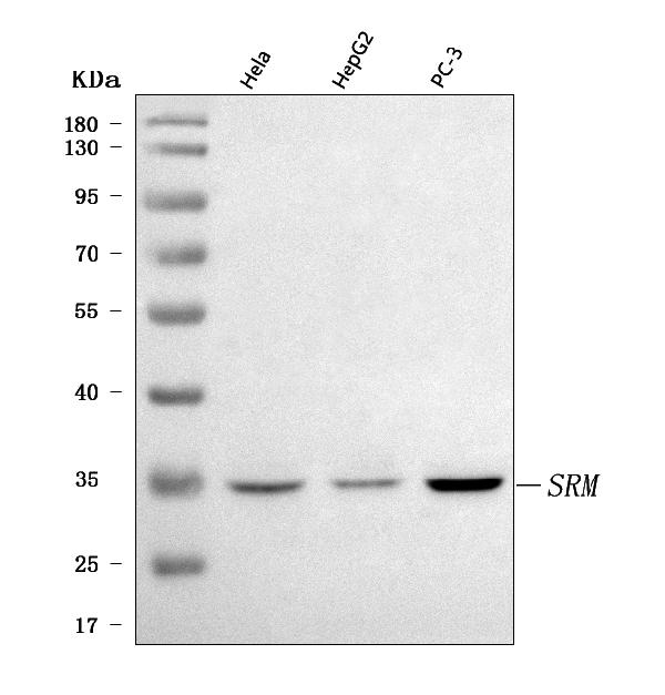
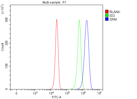
(Figure 2. Flow Cytometry analysis of HepG2 cells using anti-Spermidine synthase/SRM antibody (AAA127068).Overlay histogram showing HepG2 cells stained with AAA127068 (Blue line). To facilitate intracellular staining, cells were fixed with 4% paraformaldehyde and permeabilized with permeabilization buffer. The cells were blocked with 10% normal goat serum. And then incubated with rabbit anti-Spermidine synthase/SRM Antibody (AAA127068, 1ug/1x106 cells) for 30 min at 20 degree C. DyLight488 conjugated goat anti-rabbit IgG was used as secondary antibody for 30 minutes at 20 degree C. Isotype control antibody (Green line) was rabbit IgG (1ug/1x106) used under the same conditions. Unlabelled sample without incubation with primary antibody and secondary antibody (Red line) was used as a blank control.)
FCM/FACS (Flow Cytometry)
(Figure 2. Flow Cytometry analysis of HepG2 cells using anti-Spermidine synthase/SRM antibody (AAA127068).Overlay histogram showing HepG2 cells stained with AAA127068 (Blue line). To facilitate intracellular staining, cells were fixed with 4% paraformaldehyde and permeabilized with permeabilization buffer. The cells were blocked with 10% normal goat serum. And then incubated with rabbit anti-Spermidine synthase/SRM Antibody (AAA127068, 1ug/1x106 cells) for 30 min at 20 degree C. DyLight488 conjugated goat anti-rabbit IgG was used as secondary antibody for 30 minutes at 20 degree C. Isotype control antibody (Green line) was rabbit IgG (1ug/1x106) used under the same conditions. Unlabelled sample without incubation with primary antibody and secondary antibody (Red line) was used as a blank control.)
Spermidine synthase/SRM, Polyclonal Antibody (Cat# AAA127068)
 FCM/FACS (Flow Cytometry)
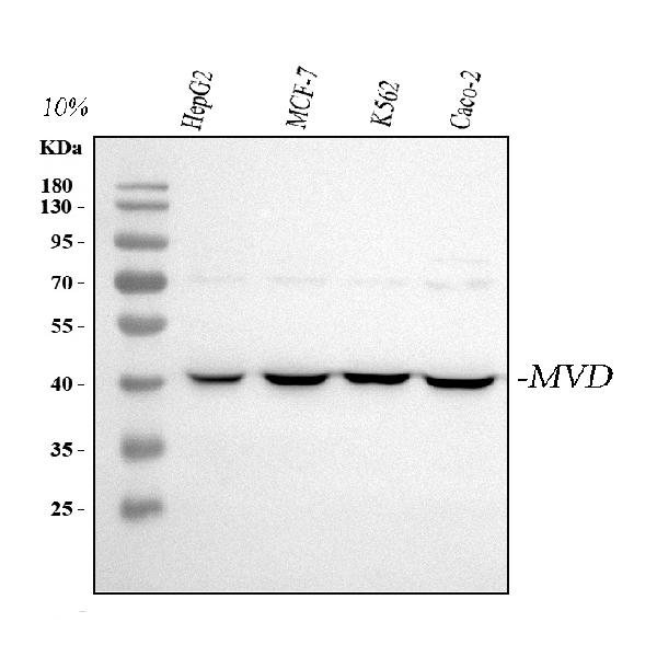
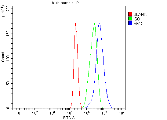
(Figure 3. Flow Cytometry analysis of MCF-7 cells using anti-MVD antibody (AAA127073).Overlay histogram showing MCF-7 cells stained with AAA127073 (Blue line). To facilitate intracellular staining, cells were fixed with 4% paraformaldehyde and permeabilized with permeabilization buffer. The cells were blocked with 10% normal goat serum. And then incubated with rabbit anti-MVD Antibody (AAA127073, 1ug/1x106 cells) for 30 min at 20 degree C. DyLight488 conjugated goat anti-rabbit IgG was used as secondary antibody for 30 minutes at 20 degree C. Isotype control antibody (Green line) was rabbit IgG (1ug/1x106) used under the same conditions. Unlabelled sample (Red line) was also used as a control.)
FCM/FACS (Flow Cytometry)
(Figure 3. Flow Cytometry analysis of MCF-7 cells using anti-MVD antibody (AAA127073).Overlay histogram showing MCF-7 cells stained with AAA127073 (Blue line). To facilitate intracellular staining, cells were fixed with 4% paraformaldehyde and permeabilized with permeabilization buffer. The cells were blocked with 10% normal goat serum. And then incubated with rabbit anti-MVD Antibody (AAA127073, 1ug/1x106 cells) for 30 min at 20 degree C. DyLight488 conjugated goat anti-rabbit IgG was used as secondary antibody for 30 minutes at 20 degree C. Isotype control antibody (Green line) was rabbit IgG (1ug/1x106) used under the same conditions. Unlabelled sample (Red line) was also used as a control.)
MVD, Polyclonal Antibody (Cat# AAA127073)
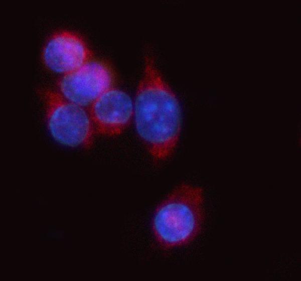
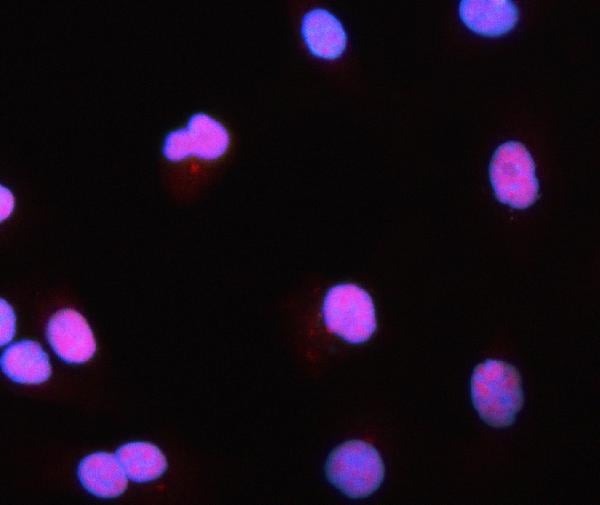
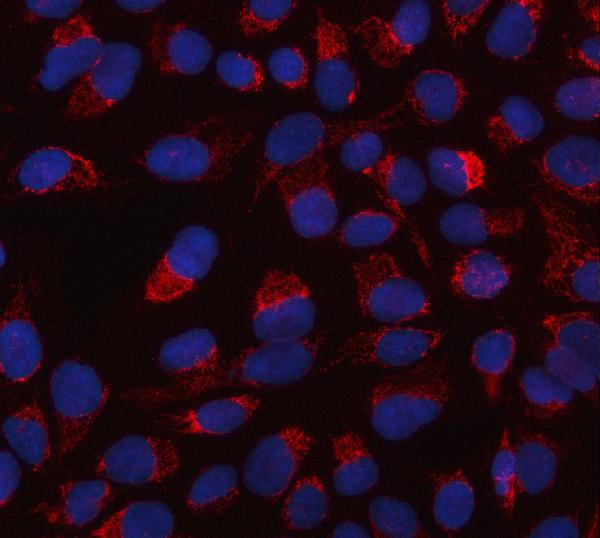
 IF (Immunofluorescence)
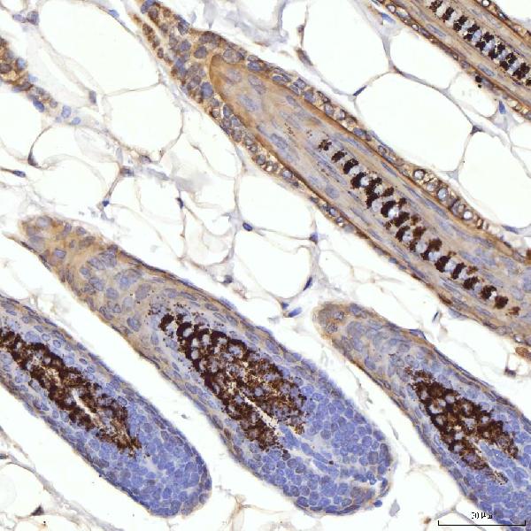
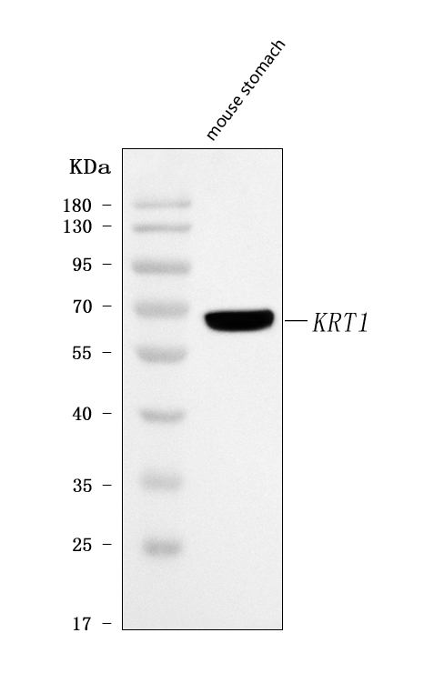
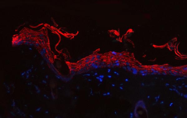
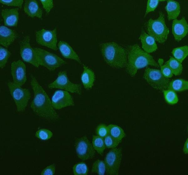
(Figure 3. IF analysis of Cytokeratin 1/Krt1 using anti-Cytokeratin 1/Krt1 antibody (AAA127075).Cytokeratin 1/Krt1 was detected in a paraffin-embedded section of mouse skin tissue. Heat mediated antigen retrieval was performed in EDTA buffer (pH 8.0, epitope retrieval solution). The tissue section was blocked with 10% goat serum. The tissue section was then incubated with 5ug/mL rabbit anti-Cytokeratin 1/Krt1 Antibody (AAA127075) overnight at 4 degree C. Cy3 Conjugated Goat Anti-Rabbit IgG (BA1032) was used as secondary antibody at 1:500 dilution and incubated for 30 minutes at 37 degree C. The section was counterstained with DAPI. Visualize using a fluorescence microscope and filter sets appropriate for the label used.)
IF (Immunofluorescence)
(Figure 3. IF analysis of Cytokeratin 1/Krt1 using anti-Cytokeratin 1/Krt1 antibody (AAA127075).Cytokeratin 1/Krt1 was detected in a paraffin-embedded section of mouse skin tissue. Heat mediated antigen retrieval was performed in EDTA buffer (pH 8.0, epitope retrieval solution). The tissue section was blocked with 10% goat serum. The tissue section was then incubated with 5ug/mL rabbit anti-Cytokeratin 1/Krt1 Antibody (AAA127075) overnight at 4 degree C. Cy3 Conjugated Goat Anti-Rabbit IgG (BA1032) was used as secondary antibody at 1:500 dilution and incubated for 30 minutes at 37 degree C. The section was counterstained with DAPI. Visualize using a fluorescence microscope and filter sets appropriate for the label used.)
Cytokeratin 1/Krt1, Polyclonal Antibody (Cat# AAA127075)
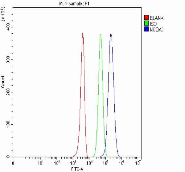 FCM/FACS (Flow Cytometry)
(Figure 3. Flow Cytometry analysis of 293T cells using anti-NCOA2 antibody (AAA127079).Overlay histogram showing 293T cells stained with AAA127079 (Blue line). To facilitate intracellular staining, cells were fixed with 4% paraformaldehyde and permeabilized with permeabilization buffer. The cells were blocked with 10% normal goat serum. And then incubated with rabbit anti-NCOA2 Antibody (AAA127079, 1ug/1x106 cells) for 30 min at 20 degree C. DyLight488 conjugated goat anti-rabbit IgG was used as secondary antibody for 30 minutes at 20 degree C. Isotype control antibody (Green line) was rabbit IgG (1ug/1x106) used under the same conditions. Unlabelled sample without incubation with primary antibody and secondary antibody (Red line) was used as a blank control.)
FCM/FACS (Flow Cytometry)
(Figure 3. Flow Cytometry analysis of 293T cells using anti-NCOA2 antibody (AAA127079).Overlay histogram showing 293T cells stained with AAA127079 (Blue line). To facilitate intracellular staining, cells were fixed with 4% paraformaldehyde and permeabilized with permeabilization buffer. The cells were blocked with 10% normal goat serum. And then incubated with rabbit anti-NCOA2 Antibody (AAA127079, 1ug/1x106 cells) for 30 min at 20 degree C. DyLight488 conjugated goat anti-rabbit IgG was used as secondary antibody for 30 minutes at 20 degree C. Isotype control antibody (Green line) was rabbit IgG (1ug/1x106) used under the same conditions. Unlabelled sample without incubation with primary antibody and secondary antibody (Red line) was used as a blank control.)
NCOA2, Polyclonal Antibody (Cat# AAA127079)
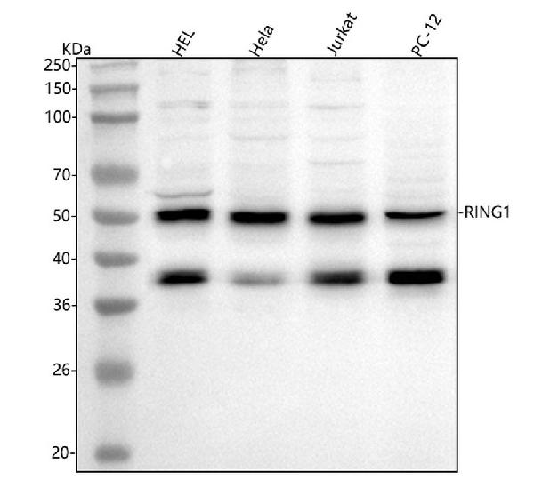
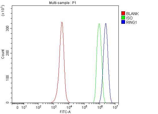 FCM/FACS (Flow Cytometry)
(Figure 3. Flow Cytometry analysis of JK cells using anti-RING1 antibody (AAA127083).Overlay histogram showing JK cells stained with AAA127083 (Blue line). To facilitate intracellular staining, cells were fixed with 4% paraformaldehyde and permeabilized with permeabilization buffer. The cells were blocked with 10% normal goat serum. And then incubated with rabbit anti-RING1 Antibody (AAA127083, 1ug/1x106 cells) for 30 min at 20 degree C. DyLight488 conjugated goat anti-rabbit IgG was used as secondary antibody for 30 minutes at 20 degree C. Isotype control antibody (Green line) was rabbit IgG (1ug/1x106) used under the same conditions. Unlabelled sample (Red line) was also used as a control.)
FCM/FACS (Flow Cytometry)
(Figure 3. Flow Cytometry analysis of JK cells using anti-RING1 antibody (AAA127083).Overlay histogram showing JK cells stained with AAA127083 (Blue line). To facilitate intracellular staining, cells were fixed with 4% paraformaldehyde and permeabilized with permeabilization buffer. The cells were blocked with 10% normal goat serum. And then incubated with rabbit anti-RING1 Antibody (AAA127083, 1ug/1x106 cells) for 30 min at 20 degree C. DyLight488 conjugated goat anti-rabbit IgG was used as secondary antibody for 30 minutes at 20 degree C. Isotype control antibody (Green line) was rabbit IgG (1ug/1x106) used under the same conditions. Unlabelled sample (Red line) was also used as a control.)
RING1, Polyclonal Antibody (Cat# AAA127083)
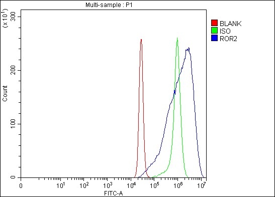
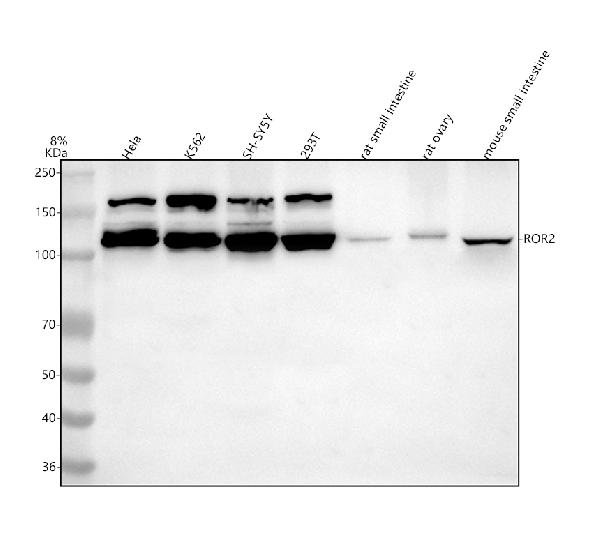
 FCM/FACS (Flow Cytometry)
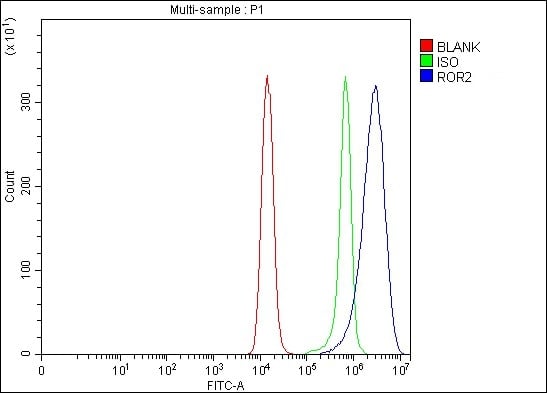
(Figure 3. Flow Cytometry analysis of Raji cells using anti-ROR2 antibody (AAA127084).Overlay histogram showing Raji cells stained with AAA127084 (Blue line). The cells were fixed with 4% paraformaldehyde and blocked with 10% normal goat serum. And then incubated with rabbit anti-ROR2 Antibody (AAA127084, 1ug/1x106 cells) for 30 min at 20 degree C. DyLight488 conjugated goat anti-rabbit IgG was used as secondary antibody for 30 minutes at 20 degree C. Isotype control antibody (Green line) was rabbit IgG (1ug/1x106) used under the same conditions. Unlabelled sample without incubation with primary antibody and secondary antibody (Red line) was used as a blank control.)
FCM/FACS (Flow Cytometry)
(Figure 3. Flow Cytometry analysis of Raji cells using anti-ROR2 antibody (AAA127084).Overlay histogram showing Raji cells stained with AAA127084 (Blue line). The cells were fixed with 4% paraformaldehyde and blocked with 10% normal goat serum. And then incubated with rabbit anti-ROR2 Antibody (AAA127084, 1ug/1x106 cells) for 30 min at 20 degree C. DyLight488 conjugated goat anti-rabbit IgG was used as secondary antibody for 30 minutes at 20 degree C. Isotype control antibody (Green line) was rabbit IgG (1ug/1x106) used under the same conditions. Unlabelled sample without incubation with primary antibody and secondary antibody (Red line) was used as a blank control.)
ROR2, Polyclonal Antibody (Cat# AAA127084)
 FCM/FACS (Flow Cytometry)
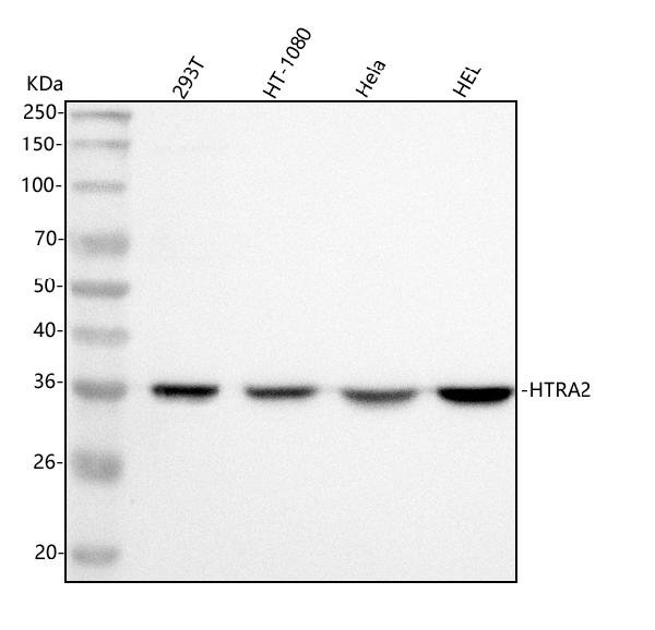
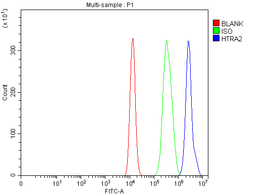
(Figure 5. Flow Cytometry analysis of HEL cells using anti-HTRA2 antibody (AAA127088).Overlay histogram showing HEL cells stained with AAA127088 (Blue line). To facilitate intracellular staining, cells were fixed with 4% paraformaldehyde and permeabilized with permeabilization buffer. The cells were blocked with 10% normal goat serum. And then incubated with rabbit anti-HTRA2 Antibody (AAA127088, 1ug/1x106 cells) for 30 min at 20 degree C. DyLight488 conjugated goat anti-rabbit IgG was used as secondary antibody for 30 minutes at 20 degree C. Isotype control antibody (Green line) was rabbit IgG (1ug/1x106) used under the same conditions. Unlabelled sample (Red line) was also used as a control.)
FCM/FACS (Flow Cytometry)
(Figure 5. Flow Cytometry analysis of HEL cells using anti-HTRA2 antibody (AAA127088).Overlay histogram showing HEL cells stained with AAA127088 (Blue line). To facilitate intracellular staining, cells were fixed with 4% paraformaldehyde and permeabilized with permeabilization buffer. The cells were blocked with 10% normal goat serum. And then incubated with rabbit anti-HTRA2 Antibody (AAA127088, 1ug/1x106 cells) for 30 min at 20 degree C. DyLight488 conjugated goat anti-rabbit IgG was used as secondary antibody for 30 minutes at 20 degree C. Isotype control antibody (Green line) was rabbit IgG (1ug/1x106) used under the same conditions. Unlabelled sample (Red line) was also used as a control.)
HTRA2, Polyclonal Antibody (Cat# AAA127088)
 FCM/FACS (Flow Cytometry)
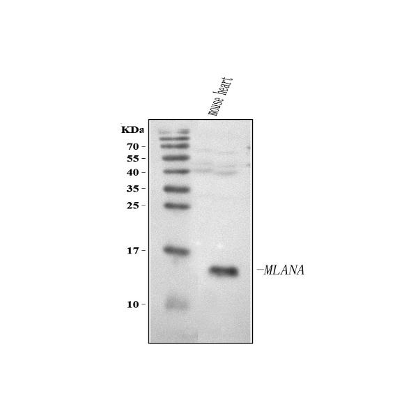
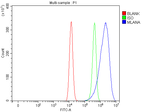
(Figure 2. Flow Cytometry analysis of HEL cells using anti-Melan-A/MLANA antibody (AAA127095).Overlay histogram showing HEL cells stained with AAA127095 (Blue line). To facilitate intracellular staining, cells were fixed with 4% paraformaldehyde and permeabilized with permeabilization buffer. The cells were blocked with 10% normal goat serum. And then incubated with rabbit anti-Melan-A/MLANA Antibody (AAA127095, 1ug/1x106 cells) for 30 min at 20 degree C. DyLight488 conjugated goat anti-rabbit IgG was used as secondary antibody for 30 minutes at 20 degree C. Isotype control antibody (Green line) was rabbit IgG (1ug/1x106) used under the same conditions. Unlabelled sample (Red line) was also used as a control.)
FCM/FACS (Flow Cytometry)
(Figure 2. Flow Cytometry analysis of HEL cells using anti-Melan-A/MLANA antibody (AAA127095).Overlay histogram showing HEL cells stained with AAA127095 (Blue line). To facilitate intracellular staining, cells were fixed with 4% paraformaldehyde and permeabilized with permeabilization buffer. The cells were blocked with 10% normal goat serum. And then incubated with rabbit anti-Melan-A/MLANA Antibody (AAA127095, 1ug/1x106 cells) for 30 min at 20 degree C. DyLight488 conjugated goat anti-rabbit IgG was used as secondary antibody for 30 minutes at 20 degree C. Isotype control antibody (Green line) was rabbit IgG (1ug/1x106) used under the same conditions. Unlabelled sample (Red line) was also used as a control.)
Melan-A/MLANA, Polyclonal Antibody (Cat# AAA127095)
 FCM/FACS (Flow Cytometry)
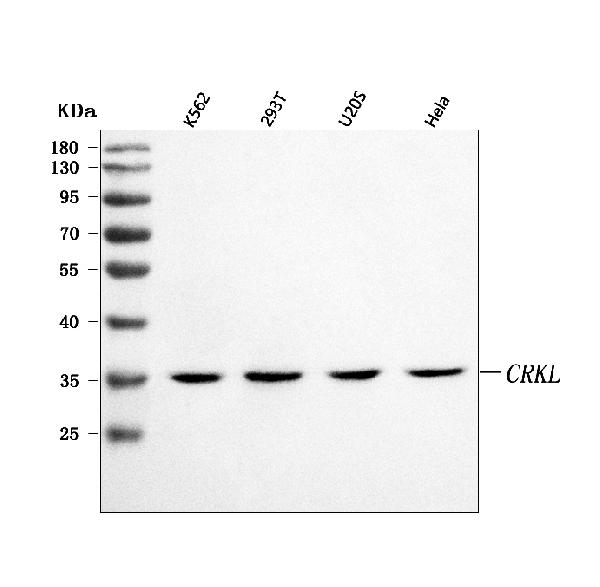
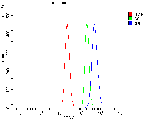
(Figure 3. Flow Cytometry analysis of RT4 cells using anti-CRKL antibody (AAA127100).Overlay histogram showing RT4 cells stained with AAA127100 (Blue line). To facilitate intracellular staining, cells were fixed with 4% paraformaldehyde and permeabilized with permeabilization buffer. The cells were blocked with 10% normal goat serum. And then incubated with rabbit anti-CRKL Antibody (AAA127100, 1ug/1x106 cells) for 30 min at 20 degree C. DyLight488 conjugated goat anti-rabbit IgG was used as secondary antibody for 30 minutes at 20 degree C. Isotype control antibody (Green line) was rabbit IgG (1ug/1x106) used under the same conditions. Unlabelled sample without incubation with primary antibody and secondary antibody (Red line) was used as a blank control.)
FCM/FACS (Flow Cytometry)
(Figure 3. Flow Cytometry analysis of RT4 cells using anti-CRKL antibody (AAA127100).Overlay histogram showing RT4 cells stained with AAA127100 (Blue line). To facilitate intracellular staining, cells were fixed with 4% paraformaldehyde and permeabilized with permeabilization buffer. The cells were blocked with 10% normal goat serum. And then incubated with rabbit anti-CRKL Antibody (AAA127100, 1ug/1x106 cells) for 30 min at 20 degree C. DyLight488 conjugated goat anti-rabbit IgG was used as secondary antibody for 30 minutes at 20 degree C. Isotype control antibody (Green line) was rabbit IgG (1ug/1x106) used under the same conditions. Unlabelled sample without incubation with primary antibody and secondary antibody (Red line) was used as a blank control.)
CRKL, Polyclonal Antibody (Cat# AAA127100)
 FCM/FACS (Flow Cytometry)
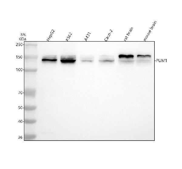
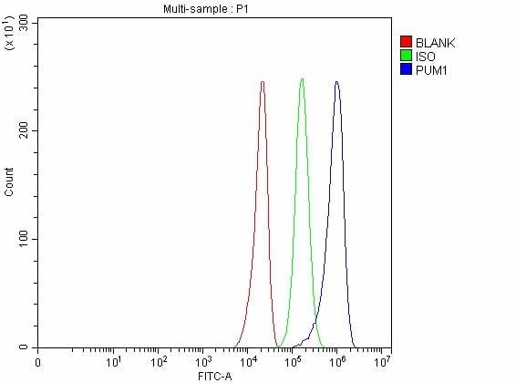
(Figure 3. Flow Cytometry analysis of U251 cells using anti-PUM1 antibody (AAA127102).Overlay histogram showing U251 cells stained with AAA127102 (Blue line). To facilitate intracellular staining, cells were fixed with 4% paraformaldehyde and permeabilized with permeabilization buffer. The cells were blocked with 10% normal goat serum. And then incubated with rabbit anti-PUM1 Antibody (AAA127102, 1ug/1x106 cells) for 30 min at 20 degree C. DyLight488 conjugated goat anti-rabbit IgG was used as secondary antibody for 30 minutes at 20 degree C. Isotype control antibody (Green line) was rabbit IgG (1ug/1x106) used under the same conditions. Unlabelled sample without incubation with primary antibody and secondary antibody (Red line) was used as a blank control.)
FCM/FACS (Flow Cytometry)
(Figure 3. Flow Cytometry analysis of U251 cells using anti-PUM1 antibody (AAA127102).Overlay histogram showing U251 cells stained with AAA127102 (Blue line). To facilitate intracellular staining, cells were fixed with 4% paraformaldehyde and permeabilized with permeabilization buffer. The cells were blocked with 10% normal goat serum. And then incubated with rabbit anti-PUM1 Antibody (AAA127102, 1ug/1x106 cells) for 30 min at 20 degree C. DyLight488 conjugated goat anti-rabbit IgG was used as secondary antibody for 30 minutes at 20 degree C. Isotype control antibody (Green line) was rabbit IgG (1ug/1x106) used under the same conditions. Unlabelled sample without incubation with primary antibody and secondary antibody (Red line) was used as a blank control.)
PUM1, Polyclonal Antibody (Cat# AAA127102)
 FCM/FACS (Flow Cytometry)
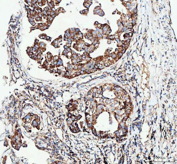
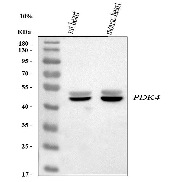
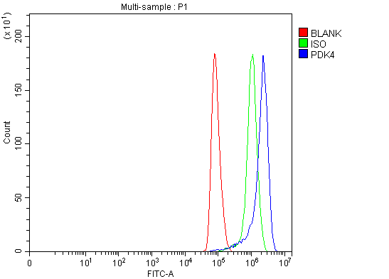
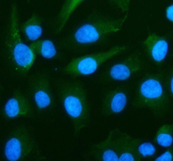
(Figure 5. Flow Cytometry analysis of Hela cells using anti-PDK4 antibody (AAA127103).Overlay histogram showing Hela cells stained with AAA127103 (Blue line). To facilitate intracellular staining, cells were fixed with 4% paraformaldehyde and permeabilized with permeabilization buffer. The cells were blocked with 10% normal goat serum. And then incubated with rabbit anti-PDK4 Antibody (AAA127103, 1ug/1x106 cells) for 30 min at 20 degree C. DyLight488 conjugated goat anti-rabbit IgG was used as secondary antibody for 30 minutes at 20 degree C. Isotype control antibody (Green line) was rabbit IgG (1ug/1x106) used under the same conditions. Unlabelled sample (Red line) was also used as a control.)
FCM/FACS (Flow Cytometry)
(Figure 5. Flow Cytometry analysis of Hela cells using anti-PDK4 antibody (AAA127103).Overlay histogram showing Hela cells stained with AAA127103 (Blue line). To facilitate intracellular staining, cells were fixed with 4% paraformaldehyde and permeabilized with permeabilization buffer. The cells were blocked with 10% normal goat serum. And then incubated with rabbit anti-PDK4 Antibody (AAA127103, 1ug/1x106 cells) for 30 min at 20 degree C. DyLight488 conjugated goat anti-rabbit IgG was used as secondary antibody for 30 minutes at 20 degree C. Isotype control antibody (Green line) was rabbit IgG (1ug/1x106) used under the same conditions. Unlabelled sample (Red line) was also used as a control.)
PDK4, Polyclonal Antibody (Cat# AAA127103)
 FCM/FACS (Flow Cytometry)
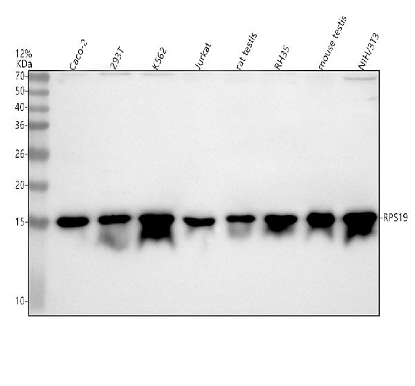
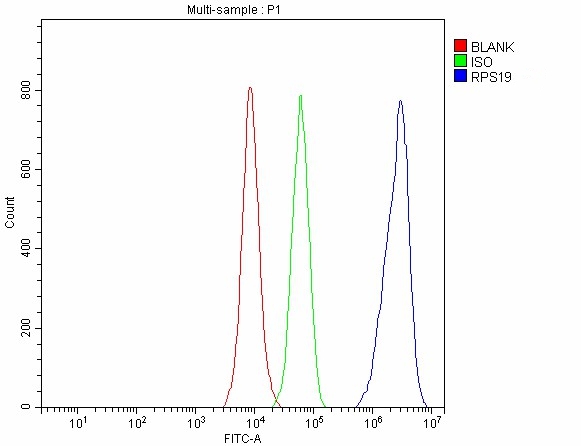
(Figure 3. Flow Cytometry analysis of 293T cells using anti-RPS19 antibody (AAA127107).Overlay histogram showing 293T cells stained with AAA127107 (Blue line). To facilitate intracellular staining, cells were fixed with 4% paraformaldehyde and permeabilized with permeabilization buffer. The cells were blocked with 10% normal goat serum. And then incubated with rabbit anti-RPS19 Antibody (AAA127107, 1ug/1x106 cells) for 30 min at 20 degree C. DyLight488 conjugated goat anti-rabbit IgG was used as secondary antibody for 30 minutes at 20 degree C. Isotype control antibody (Green line) was rabbit IgG (1ug/1x106) used under the same conditions. Unlabelled sample without incubation with primary antibody and secondary antibody (Red line) was used as a blank control.)
FCM/FACS (Flow Cytometry)
(Figure 3. Flow Cytometry analysis of 293T cells using anti-RPS19 antibody (AAA127107).Overlay histogram showing 293T cells stained with AAA127107 (Blue line). To facilitate intracellular staining, cells were fixed with 4% paraformaldehyde and permeabilized with permeabilization buffer. The cells were blocked with 10% normal goat serum. And then incubated with rabbit anti-RPS19 Antibody (AAA127107, 1ug/1x106 cells) for 30 min at 20 degree C. DyLight488 conjugated goat anti-rabbit IgG was used as secondary antibody for 30 minutes at 20 degree C. Isotype control antibody (Green line) was rabbit IgG (1ug/1x106) used under the same conditions. Unlabelled sample without incubation with primary antibody and secondary antibody (Red line) was used as a blank control.)
RPS19, Polyclonal Antibody (Cat# AAA127107)
 FCM/FACS (Flow Cytometry)
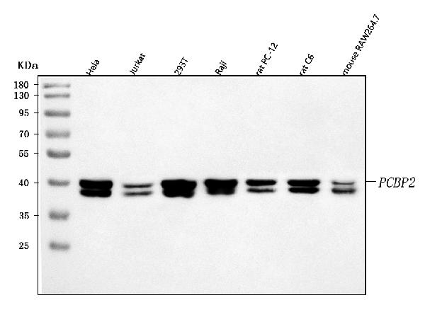
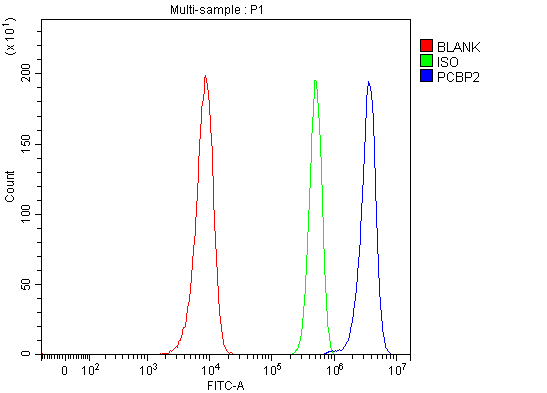
(Figure 3. Flow Cytometry analysis of Hela cells using anti-hnRNP E2/PCBP2 antibody (AAA127115).Overlay histogram showing Hela cells stained with AAA127115 (Blue line). To facilitate intracellular staining, cells were fixed with 4% paraformaldehyde and permeabilized with permeabilization buffer. The cells were blocked with 10% normal goat serum. And then incubated with rabbit anti-hnRNP E2/PCBP2 Antibody (AAA127115, 1ug/1x106 cells) for 30 min at 20 degree C. DyLight488 conjugated goat anti-rabbit IgG was used as secondary antibody for 30 minutes at 20 degree C. Isotype control antibody (Green line) was rabbit IgG (1ug/1x106) used under the same conditions. Unlabelled sample without incubation with primary antibody and secondary antibody (Red line) was used as a blank control.)
FCM/FACS (Flow Cytometry)
(Figure 3. Flow Cytometry analysis of Hela cells using anti-hnRNP E2/PCBP2 antibody (AAA127115).Overlay histogram showing Hela cells stained with AAA127115 (Blue line). To facilitate intracellular staining, cells were fixed with 4% paraformaldehyde and permeabilized with permeabilization buffer. The cells were blocked with 10% normal goat serum. And then incubated with rabbit anti-hnRNP E2/PCBP2 Antibody (AAA127115, 1ug/1x106 cells) for 30 min at 20 degree C. DyLight488 conjugated goat anti-rabbit IgG was used as secondary antibody for 30 minutes at 20 degree C. Isotype control antibody (Green line) was rabbit IgG (1ug/1x106) used under the same conditions. Unlabelled sample without incubation with primary antibody and secondary antibody (Red line) was used as a blank control.)
hnRNP E2/PCBP2, Polyclonal Antibody (Cat# AAA127115)
 FCM/FACS (Flow Cytometry)
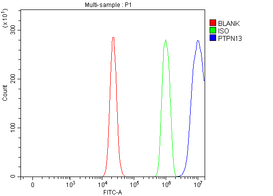
(Figure 2. Flow Cytometry analysis of RT4 cells using anti-PTPN13 antibody (AAA127121).Overlay histogram showing RT4 cells stained with AAA127121 (Blue line). To facilitate intracellular staining, cells were fixed with 4% paraformaldehyde and permeabilized with permeabilization buffer. The cells were blocked with 10% normal goat serum. And then incubated with rabbit anti-PTPN13 Antibody (AAA127121, 1ug/1x106 cells) for 30 min at 20 degree C. DyLight488 conjugated goat anti-rabbit IgG was used as secondary antibody for 30 minutes at 20 degree C. Isotype control antibody (Green line) was rabbit IgG (1ug/1x106) used under the same conditions. Unlabelled sample (Red line) was also used as a control.)
FCM/FACS (Flow Cytometry)
(Figure 2. Flow Cytometry analysis of RT4 cells using anti-PTPN13 antibody (AAA127121).Overlay histogram showing RT4 cells stained with AAA127121 (Blue line). To facilitate intracellular staining, cells were fixed with 4% paraformaldehyde and permeabilized with permeabilization buffer. The cells were blocked with 10% normal goat serum. And then incubated with rabbit anti-PTPN13 Antibody (AAA127121, 1ug/1x106 cells) for 30 min at 20 degree C. DyLight488 conjugated goat anti-rabbit IgG was used as secondary antibody for 30 minutes at 20 degree C. Isotype control antibody (Green line) was rabbit IgG (1ug/1x106) used under the same conditions. Unlabelled sample (Red line) was also used as a control.)
PTPN13, Polyclonal Antibody (Cat# AAA127121)
 FCM/FACS (Flow Cytometry)
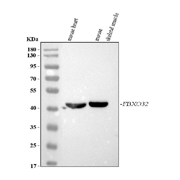
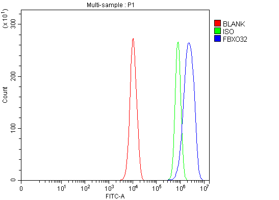
(Figure 3. Flow Cytometry analysis of PC-3 cells using anti-Fbx32/FBXO32 antibody (AAA127124).Overlay histogram showing PC-3 cells stained with AAA127124 (Blue line). To facilitate intracellular staining, cells were fixed with 4% paraformaldehyde and permeabilized with permeabilization buffer. The cells were blocked with 10% normal goat serum. And then incubated with rabbit anti-Fbx32/FBXO32 Antibody (AAA127124, 1ug/1x106 cells) for 30 min at 20 degree C. DyLight488 conjugated goat anti-rabbit IgG was used as secondary antibody for 30 minutes at 20 degree C. Isotype control antibody (Green line) was rabbit IgG (1ug/1x106) used under the same conditions. Unlabelled sample (Red line) was also used as a control.)
FCM/FACS (Flow Cytometry)
(Figure 3. Flow Cytometry analysis of PC-3 cells using anti-Fbx32/FBXO32 antibody (AAA127124).Overlay histogram showing PC-3 cells stained with AAA127124 (Blue line). To facilitate intracellular staining, cells were fixed with 4% paraformaldehyde and permeabilized with permeabilization buffer. The cells were blocked with 10% normal goat serum. And then incubated with rabbit anti-Fbx32/FBXO32 Antibody (AAA127124, 1ug/1x106 cells) for 30 min at 20 degree C. DyLight488 conjugated goat anti-rabbit IgG was used as secondary antibody for 30 minutes at 20 degree C. Isotype control antibody (Green line) was rabbit IgG (1ug/1x106) used under the same conditions. Unlabelled sample (Red line) was also used as a control.)
FBXO32, Polyclonal Antibody (Cat# AAA127124)
 FCM/FACS (Flow Cytometry)
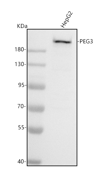
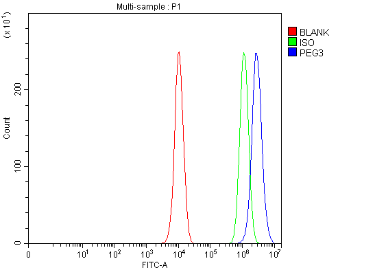
(Figure 3. Flow Cytometry analysis of PC-3 cells using anti-PEG3 antibody (AAA127125).Overlay histogram showing PC-3 cells stained with AAA127125 (Blue line). To facilitate intracellular staining, cells were fixed with 4% paraformaldehyde and permeabilized with permeabilization buffer. The cells were blocked with 10% normal goat serum. And then incubated with rabbit anti-PEG3 Antibody (AAA127125, 1ug/1x106 cells) for 30 min at 20 degree C. DyLight488 conjugated goat anti-rabbit IgG was used as secondary antibody for 30 minutes at 20 degree C. Isotype control antibody (Green line) was rabbit IgG (1ug/1x106) used under the same conditions. Unlabelled sample (Red line) was also used as a control.)
FCM/FACS (Flow Cytometry)
(Figure 3. Flow Cytometry analysis of PC-3 cells using anti-PEG3 antibody (AAA127125).Overlay histogram showing PC-3 cells stained with AAA127125 (Blue line). To facilitate intracellular staining, cells were fixed with 4% paraformaldehyde and permeabilized with permeabilization buffer. The cells were blocked with 10% normal goat serum. And then incubated with rabbit anti-PEG3 Antibody (AAA127125, 1ug/1x106 cells) for 30 min at 20 degree C. DyLight488 conjugated goat anti-rabbit IgG was used as secondary antibody for 30 minutes at 20 degree C. Isotype control antibody (Green line) was rabbit IgG (1ug/1x106) used under the same conditions. Unlabelled sample (Red line) was also used as a control.)
PEG3, Polyclonal Antibody (Cat# AAA127125)
 FCM/FACS (Flow Cytometry)
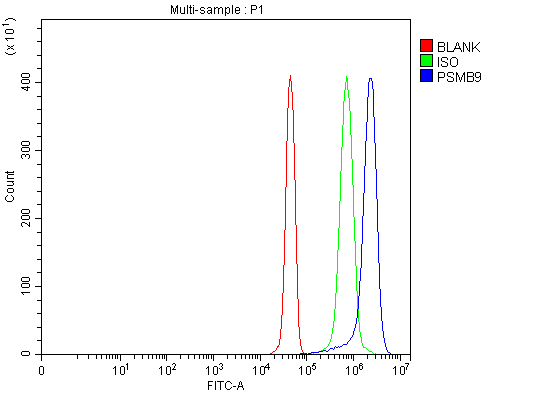
(Figure 5. Flow Cytometry analysis of JK cells using anti-PSMB9 antibody (AAA127146).Overlay histogram showing JK cells stained with AAA127146 (Blue line). To facilitate intracellular staining, cells were fixed with 4% paraformaldehyde and permeabilized with permeabilization buffer. The cells were blocked with 10% normal goat serum. And then incubated with rabbit anti-PSMB9 Antibody (AAA127146, 1ug/1x106 cells) for 30 min at 20 degree C. DyLight488 conjugated goat anti-rabbit IgG was used as secondary antibody for 30 minutes at 20 degree C. Isotype control antibody (Green line) was rabbit IgG (1ug/1x106) used under the same conditions. Unlabelled sample (Red line) was also used as a control.)
FCM/FACS (Flow Cytometry)
(Figure 5. Flow Cytometry analysis of JK cells using anti-PSMB9 antibody (AAA127146).Overlay histogram showing JK cells stained with AAA127146 (Blue line). To facilitate intracellular staining, cells were fixed with 4% paraformaldehyde and permeabilized with permeabilization buffer. The cells were blocked with 10% normal goat serum. And then incubated with rabbit anti-PSMB9 Antibody (AAA127146, 1ug/1x106 cells) for 30 min at 20 degree C. DyLight488 conjugated goat anti-rabbit IgG was used as secondary antibody for 30 minutes at 20 degree C. Isotype control antibody (Green line) was rabbit IgG (1ug/1x106) used under the same conditions. Unlabelled sample (Red line) was also used as a control.)
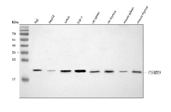
PSMB9, Polyclonal Antibody (Cat# AAA127146)
 FCM/FACS (Flow Cytometry)
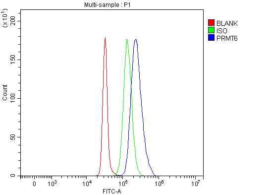
(Figure 2. Flow Cytometry analysis of SH-SY5Y cells using anti-PRMT6 antibody (AAA127150).Overlay histogram showing SH-SY5Y cells stained with AAA127150 (Blue line). To facilitate intracellular staining, cells were fixed with 4% paraformaldehyde and permeabilized with permeabilization buffer. The cells were blocked with 10% normal goat serum. And then incubated with rabbit anti-PRMT6 Antibody (AAA127150, 1ug/1x106 cells) for 30 min at 20 degree C. DyLight488 conjugated goat anti-rabbit IgG was used as secondary antibody for 30 minutes at 20 degree C. Isotype control antibody (Green line) was rabbit IgG (1ug/1x106) used under the same conditions. Unlabelled sample (Red line) was also used as a control.)
FCM/FACS (Flow Cytometry)
(Figure 2. Flow Cytometry analysis of SH-SY5Y cells using anti-PRMT6 antibody (AAA127150).Overlay histogram showing SH-SY5Y cells stained with AAA127150 (Blue line). To facilitate intracellular staining, cells were fixed with 4% paraformaldehyde and permeabilized with permeabilization buffer. The cells were blocked with 10% normal goat serum. And then incubated with rabbit anti-PRMT6 Antibody (AAA127150, 1ug/1x106 cells) for 30 min at 20 degree C. DyLight488 conjugated goat anti-rabbit IgG was used as secondary antibody for 30 minutes at 20 degree C. Isotype control antibody (Green line) was rabbit IgG (1ug/1x106) used under the same conditions. Unlabelled sample (Red line) was also used as a control.)
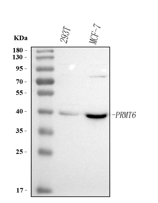
PRMT6, Polyclonal Antibody (Cat# AAA127150)
 FCM/FACS (Flow Cytometry)
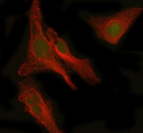
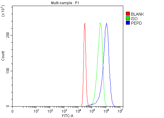
(Figure 3. Flow Cytometry analysis of 293T cells using anti-PEPD antibody (AAA127175).Overlay histogram showing 293T cells stained with AAA127175 (Blue line). The cells were fixed with 4% paraformaldehyde and blocked with 10% normal goat serum. And then incubated with rabbit anti-PEPD Antibody (AAA127175, 1ug/1x106 cells) for 30 min at 20 degree C. DyLight488 conjugated goat anti-rabbit IgG was used as secondary antibody for 30 minutes at 20 degree C. Isotype control antibody (Green line) was rabbit IgG (1ug/1x106) used under the same conditions. Unlabelled sample (Red line) was also used as a control.)
FCM/FACS (Flow Cytometry)
(Figure 3. Flow Cytometry analysis of 293T cells using anti-PEPD antibody (AAA127175).Overlay histogram showing 293T cells stained with AAA127175 (Blue line). The cells were fixed with 4% paraformaldehyde and blocked with 10% normal goat serum. And then incubated with rabbit anti-PEPD Antibody (AAA127175, 1ug/1x106 cells) for 30 min at 20 degree C. DyLight488 conjugated goat anti-rabbit IgG was used as secondary antibody for 30 minutes at 20 degree C. Isotype control antibody (Green line) was rabbit IgG (1ug/1x106) used under the same conditions. Unlabelled sample (Red line) was also used as a control.)
PEPD, Polyclonal Antibody (Cat# AAA127175)
What are Polyclonal Antibodies?
Polyclonal antibodies are antibodies that come from multiple B cell clones of a host animal. The typical hosts used for the majority of polyclonal antibody production are rabbits, goats, sheep, and donkeys. These polyclonal antibodies, once having identified their target, will bind to different epitopes located at different regions or sequences on the same protein/antigen. As a result, they are ideal at locating and binding to the target, even if the target is in very low concentrations (due to many different antibodies being able to bind to the same target molecule, which allows for significant amplification of a downstream signal).
Polyclonal antibodies are typically produced by injecting an antigen into a host animal, which causes the animal’s immune system to attack the foreign antigen by mass generating antibodies against it. After a period of time, serum is collected from the animal and purified using physicochemical fractionation, class-specific affinity purification, and/or antigen-affinity purification.
Key Uses of Polyclonal Antibodies
- Western Blotting: This method is used to find specific proteins in biological samples after separating them by size.
- Immunohistochemistry: IHC helps visualize the location of proteins in tissue sections using various staining techniques.
- ELISA: (Enzyme-Linked Immunosorbent Assay) is typically used to identify specific protein quantities in a sample. ELISAs can be either “Quantitative” or “Qualitative”.
- Flow Cytometry: technique that identifies and measures the specific protein on the surface or inside the cells in a fluid suspension.
- Immunoprecipitation: IP isolates and studies a specific protein from a complex mixture using antibodies.
Why Buy Polyclonal Antibodies from AAA Biotech?
1. Ideal for Various Applications
Our antibodies are generally going to be validated for use in multiple types of assays, including ELISA, Western Blotting, Immunohistochemistry, Immunoprecipitation, amongst others. They are ideal for a wide range of research applications.
2. Rigorous Quality Control
All of the antibodies in our catalog undergo strict quality testing to ensure specificity, sensitivity, and consistent performance. We are confident in the ability of our antibodies to provide you with accurate results.
3. Wide Assortment of Antibodies
Antibodies in are catalog can be found for both common and exotic species, and these antibodies are also available in both conjugated and recombinant forms to suit many diverse experimental needs.
4. Highly Purified
Our antibodies are available in purified forms with over 85% purity, as confirmed by SDS-PAGE. They are also available with tags such as His, Flag, GST, or MBP. We cater to customers worldwide.
FAQ
1. How are polyclonal antibodies produced?
Traditionally, polyclonal antibodies are produced by injecting an antigen into a host animal (such as a rabbit or goat), which then triggers an immune response from the host animal. The animal’s B cells produce antibodies that will recognize different parts of the injected antigen. These antibodies are then collected from the animal’s blood and purified for use.
2. How do polyclonal antibodies differ from monoclonal antibodies?
Polyclonal antibodies are a mix of antibodies that bind to different locations (epitopes) of the same antigen, while monoclonal antibodies are identical and bind to just one specific epitope. This makes polyclonal antibodies more versatile and better at detecting proteins that may be present in low quantities or in altered/modified forms.
3. How should I store polyclonal antibodies?
Polyclonal antibodies should be stored at 4°C for short-term use (up to a few weeks) and at -20°C or -80°C for long-term storage. Avoid repeated freeze-thaw cycles by dividing them into small aliquots. Always check the datasheet for specific storage instructions.