Filters
▼Clonality
▼Type
▼Reactivity
▼Gene Name
▼Isotype
▼Host
▼Application
▼Clone
▼Polyclonal Antibodies
At AAA Biotech also known as AAA Bio or AAABio, we provide a broad range of purified polyclonal antibodies (pAbs) that are able to all be browsed online through our website. Due to their high specificity and strong binding affinity, these antibodies are ideal for wide swathes of research and experimental applications.
Our polyclonal antibodies can easily support your work, whether you use them for Western Blotting, Immunocytochemistry (with or without Immunofluorescence used in conjunction), Immunohistochemistry, Immunoprecipitation, and ELISA tests. We highly encourage you to browse our range of pAbs and choose the one that best suits your experimental model.
Viewing 6300-6350 of 96805 product results
 SDS-PAGE
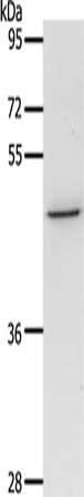
(Gel: 8%SDS-PAGE,Lysate: 40 ug,,Primary antibody: AAA239717(AQP3 Antibody) at dilution 1/200 dilution,Secondary antibody: Goat anti rabbit IgG at 1/8000 dilution,Exposure time: 1 minute)
SDS-PAGE
(Gel: 8%SDS-PAGE,Lysate: 40 ug,,Primary antibody: AAA239717(AQP3 Antibody) at dilution 1/200 dilution,Secondary antibody: Goat anti rabbit IgG at 1/8000 dilution,Exposure time: 1 minute)
AQP3, Polyclonal Antibody (Cat# AAA239717)
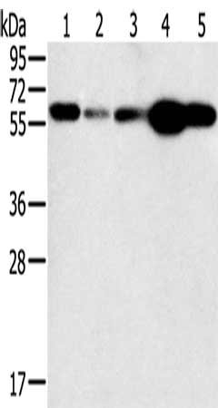
 SDS-PAGE
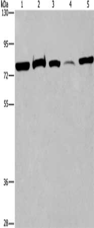
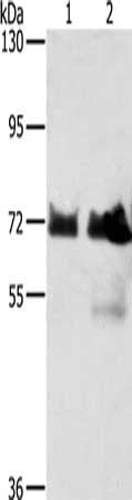
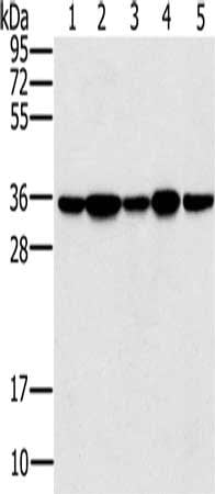
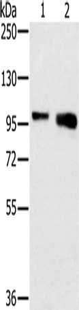
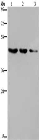
(Gel: 6%SDS-PAGE, Lysate: 40 ug, Lane 1-5: 293T cells, human fetal brain tissue, K562 cells, human fetal liver tissue, Hela cells, Primary antibody: AAA239721(ACVR2A Antibody) at dilution 1/400, Secondary antibody: Goat anti rabbit IgG at 1/8000 dilution, Exposure time: 40 seconds)
SDS-PAGE
(Gel: 6%SDS-PAGE, Lysate: 40 ug, Lane 1-5: 293T cells, human fetal brain tissue, K562 cells, human fetal liver tissue, Hela cells, Primary antibody: AAA239721(ACVR2A Antibody) at dilution 1/400, Secondary antibody: Goat anti rabbit IgG at 1/8000 dilution, Exposure time: 40 seconds)
ACVR2A, Polyclonal Antibody (Cat# AAA239721)
 SDS-PAGE
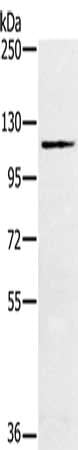
(Gel: 6%SDS-PAGE,Lysate: 40 ug,,Primary antibody: AAA239722(ACVR2A Antibody) at dilution 1/400 dilution,Secondary antibody: Goat anti rabbit IgG at 1/8000 dilution,Exposure time: 2 minutes)
SDS-PAGE
(Gel: 6%SDS-PAGE,Lysate: 40 ug,,Primary antibody: AAA239722(ACVR2A Antibody) at dilution 1/400 dilution,Secondary antibody: Goat anti rabbit IgG at 1/8000 dilution,Exposure time: 2 minutes)
ACVR2A, Polyclonal Antibody (Cat# AAA239722)
 SDS-PAGE
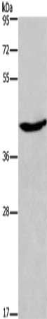
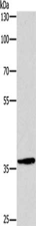
(Gel: 10%SDS-PAGE, Lysate: 40 ug, Lane: Human lung cancer tissue, Primary antibody: AAA239723(ADRA1B Antibody) at dilution 1/550, Secondary antibody: Goat anti rabbit IgG at 1/8000 dilution, Exposure time: 1 minute)
SDS-PAGE
(Gel: 10%SDS-PAGE, Lysate: 40 ug, Lane: Human lung cancer tissue, Primary antibody: AAA239723(ADRA1B Antibody) at dilution 1/550, Secondary antibody: Goat anti rabbit IgG at 1/8000 dilution, Exposure time: 1 minute)
ADRA1B, Polyclonal Antibody (Cat# AAA239723)
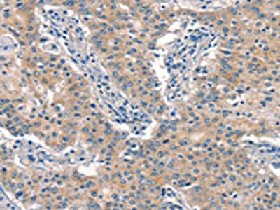
 IHC (Immunohiostchemistry)
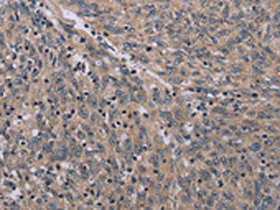
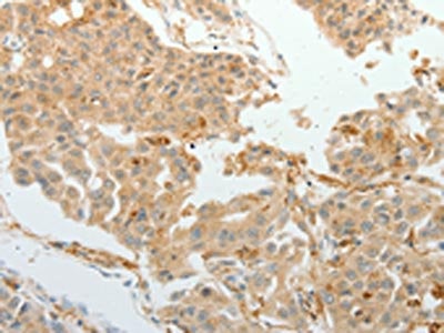
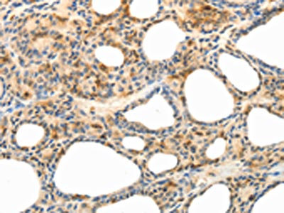
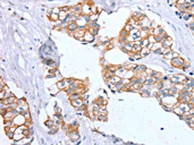
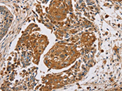
(The image on the left is immunohistochemistry of paraffin-embedded Human lung cancer tissue using AAA239724(ASCL1 Antibody) at dilution 1/30, on the right is treated with synthetic peptide. (Original magnification: ×200))
IHC (Immunohiostchemistry)
(The image on the left is immunohistochemistry of paraffin-embedded Human lung cancer tissue using AAA239724(ASCL1 Antibody) at dilution 1/30, on the right is treated with synthetic peptide. (Original magnification: ×200))
ASCL1, Polyclonal Antibody (Cat# AAA239724)
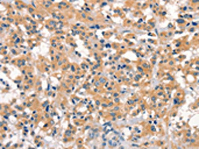
 IHC (Immunohiostchemistry)
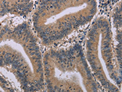
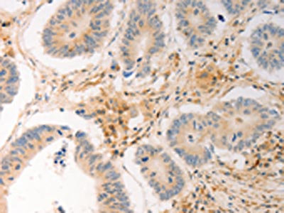
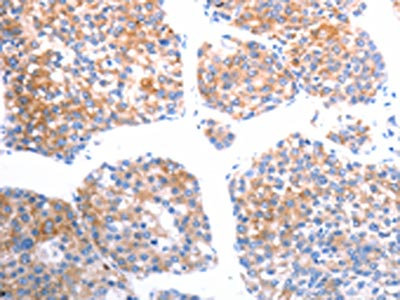
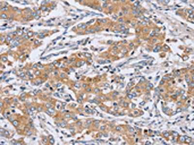
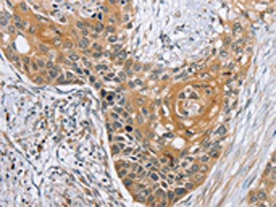
(The image on the left is immunohistochemistry of paraffin-embedded Human gastric cancer tissue using AAA239730(AVPR2 Antibody) at dilution 1/30, on the right is treated with synthetic peptide. (Original magnification: ×200))
IHC (Immunohiostchemistry)
(The image on the left is immunohistochemistry of paraffin-embedded Human gastric cancer tissue using AAA239730(AVPR2 Antibody) at dilution 1/30, on the right is treated with synthetic peptide. (Original magnification: ×200))
AVPR2, Polyclonal Antibody (Cat# AAA239730)
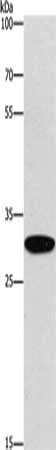
 SDS-PAGE
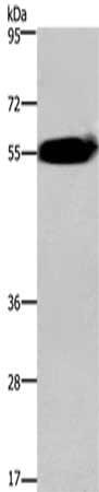
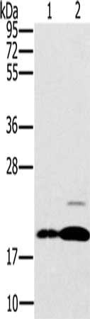
(Gel: 10%SDS-PAGE, Lysate: 50 ug, Lane: Jurkat cells, Primary antibody: AAA239732(BDKRB2 Antibody) at dilution 1/800, Secondary antibody: Goat anti rabbit IgG at 1/8000 dilution, Exposure time: 1 minute)
SDS-PAGE
(Gel: 10%SDS-PAGE, Lysate: 50 ug, Lane: Jurkat cells, Primary antibody: AAA239732(BDKRB2 Antibody) at dilution 1/800, Secondary antibody: Goat anti rabbit IgG at 1/8000 dilution, Exposure time: 1 minute)
BDKRB2, Polyclonal Antibody (Cat# AAA239732)
 IHC (Immunohiostchemistry)
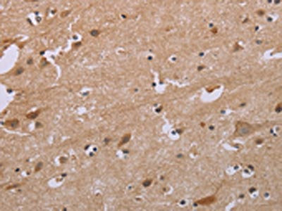
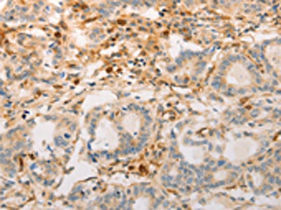
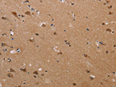
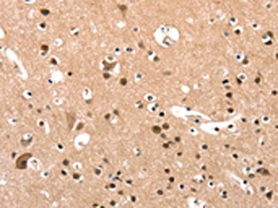
(The image on the left is immunohistochemistry of paraffin-embedded Human brain tissue using AAA239739(FGF1 Antibody) at dilution 1/50, on the right is treated with synthetic peptide. (Original magnification: ×200))
IHC (Immunohiostchemistry)
(The image on the left is immunohistochemistry of paraffin-embedded Human brain tissue using AAA239739(FGF1 Antibody) at dilution 1/50, on the right is treated with synthetic peptide. (Original magnification: ×200))
FGF1, Polyclonal Antibody (Cat# AAA239739)
 IHC (Immunohiostchemistry)
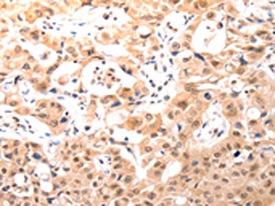
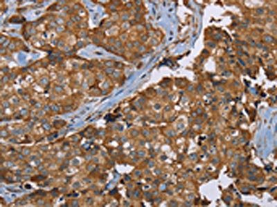
(The image on the left is immunohistochemistry of paraffin-embedded Human gastric cancer tissue using AAA239740(FGF1 Antibody) at dilution 1/25, on the right is treated with synthetic peptide. (Original magnification: ×200))
IHC (Immunohiostchemistry)
(The image on the left is immunohistochemistry of paraffin-embedded Human gastric cancer tissue using AAA239740(FGF1 Antibody) at dilution 1/25, on the right is treated with synthetic peptide. (Original magnification: ×200))
FGF1, Polyclonal Antibody (Cat# AAA239740)
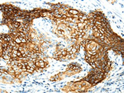
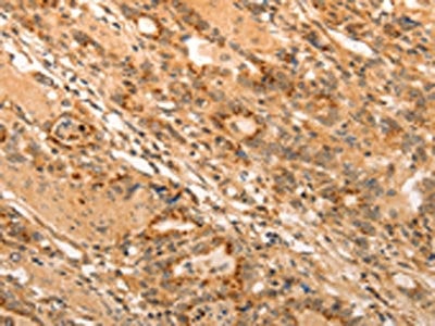
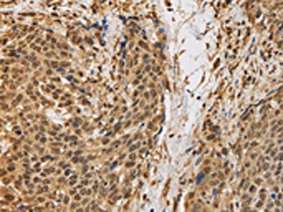
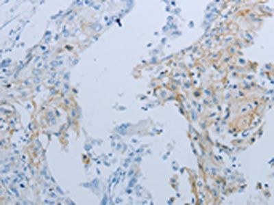
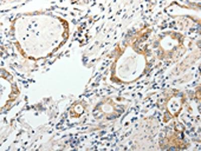
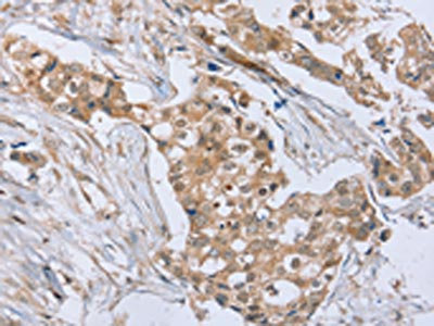
 IHC (Immunohistochemistry)
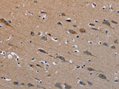
(The image on the left is immunohistochemistry of paraffin-embedded Human esophagus cancer tissue using AAA239742(FGF7 Antibody) at dilution 1/15, on the right is treated with synthetic peptide. (Original magnification: ×200))
IHC (Immunohistochemistry)
(The image on the left is immunohistochemistry of paraffin-embedded Human esophagus cancer tissue using AAA239742(FGF7 Antibody) at dilution 1/15, on the right is treated with synthetic peptide. (Original magnification: ×200))
FGF7, Polyclonal Antibody (Cat# AAA239742)
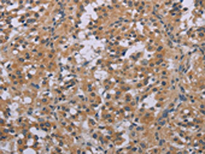
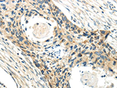
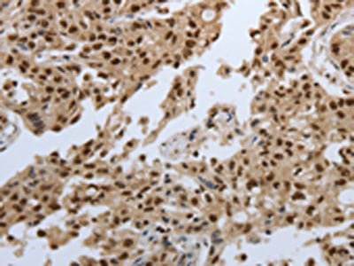
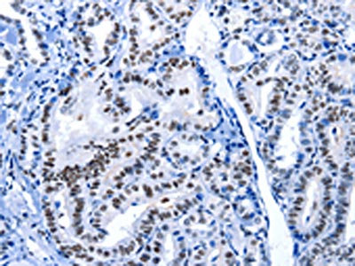 IHC (Immunohiostchemistry)
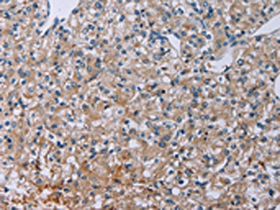
(The image on the left is immunohistochemistry of paraffin-embedded Human thyroid cancer tissue using AAA239745(POU2AF1 Antibody) at dilution 1/15, on the right is treated with synthetic peptide. (Original magnification: ×200))
IHC (Immunohiostchemistry)
(The image on the left is immunohistochemistry of paraffin-embedded Human thyroid cancer tissue using AAA239745(POU2AF1 Antibody) at dilution 1/15, on the right is treated with synthetic peptide. (Original magnification: ×200))
POU2AF1, Polyclonal Antibody (Cat# AAA239745)
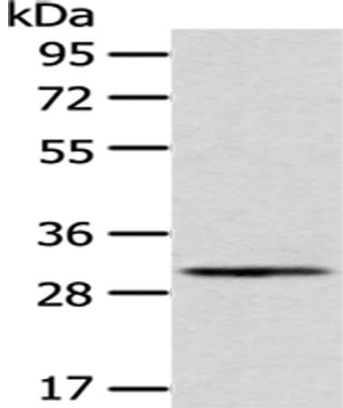
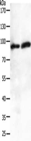
 SDS-PAGE
(Gel: 6%SDS-PAGE, Lysate: 40 ug, Lane 1-2: Hela cells, Raji cells, Primary antibody: AAA239748(BRAF Antibody) at dilution 1/400, Secondary antibody: Goat anti rabbit IgG at 1/8000 dilution, Exposure time: 1 minute)
SDS-PAGE
(Gel: 6%SDS-PAGE, Lysate: 40 ug, Lane 1-2: Hela cells, Raji cells, Primary antibody: AAA239748(BRAF Antibody) at dilution 1/400, Secondary antibody: Goat anti rabbit IgG at 1/8000 dilution, Exposure time: 1 minute)
BRAF, Polyclonal Antibody (Cat# AAA239748)
 SDS-PAGE
(Gel: 10%SDS-PAGE, Lysate: 50 ug, Lane 1-2: A549 cells, human fetal brain tissue, Primary antibody: AAA239752(JUP Antibody) at dilution 1/200, Secondary antibody: Goat anti rabbit IgG at 1/8000 dilution, Exposure time: 2 minutes)
SDS-PAGE
(Gel: 10%SDS-PAGE, Lysate: 50 ug, Lane 1-2: A549 cells, human fetal brain tissue, Primary antibody: AAA239752(JUP Antibody) at dilution 1/200, Secondary antibody: Goat anti rabbit IgG at 1/8000 dilution, Exposure time: 2 minutes)
JUP, Polyclonal Antibody (Cat# AAA239752)

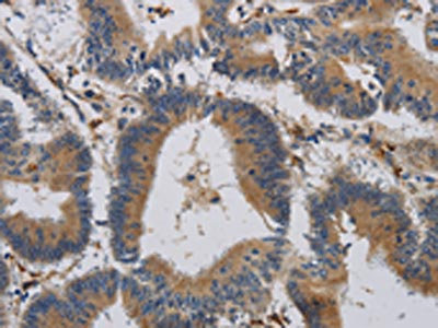
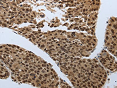
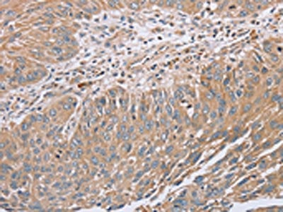
 IHC (Immunohiostchemistry)
(The image on the left is immunohistochemistry of paraffin-embedded Human esophagus cancer tissue using AAA239755(CCL13 Antibody) at dilution 1/40, on the right is treated with synthetic peptide. (Original magnification: ×200))
IHC (Immunohiostchemistry)
(The image on the left is immunohistochemistry of paraffin-embedded Human esophagus cancer tissue using AAA239755(CCL13 Antibody) at dilution 1/40, on the right is treated with synthetic peptide. (Original magnification: ×200))
CCL13, Polyclonal Antibody (Cat# AAA239755)
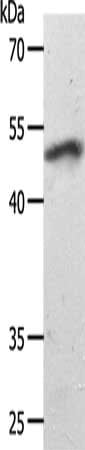
 SDS-PAGE
(Gel: 8%SDS-PAGE, Lysate: 40 ug, Lane: Mouse skin tissue, Primary antibody: AAA239759(CCR2 Antibody) at dilution 1/450, Secondary antibody: Goat anti rabbit IgG at 1/8000 dilution, Exposure time: 5 minutes)
SDS-PAGE
(Gel: 8%SDS-PAGE, Lysate: 40 ug, Lane: Mouse skin tissue, Primary antibody: AAA239759(CCR2 Antibody) at dilution 1/450, Secondary antibody: Goat anti rabbit IgG at 1/8000 dilution, Exposure time: 5 minutes)
CCR2, Polyclonal Antibody (Cat# AAA239759)
 SDS-PAGE
(Gel: 10%SDS-PAGE, Lysate: 40 ug, Lane: SKOV3 cells, Primary antibody: AAA239761(CCR3 Antibody) at dilution 1/300, Secondary antibody: Goat anti rabbit IgG at 1/8000 dilution, Exposure time: 1 minute)
SDS-PAGE
(Gel: 10%SDS-PAGE, Lysate: 40 ug, Lane: SKOV3 cells, Primary antibody: AAA239761(CCR3 Antibody) at dilution 1/300, Secondary antibody: Goat anti rabbit IgG at 1/8000 dilution, Exposure time: 1 minute)
CCR3, Polyclonal Antibody (Cat# AAA239761)
 SDS-PAGE
(Gel: 10%SDS-PAGE, Lysate: 80 ug, Lane 1-2: Hela cells, K562 cells, Primary antibody: AAA239763(CCR8 Antibody) at dilution 1/200, Secondary antibody: Goat anti rabbit IgG at 1/8000 dilution, Exposure time: 30 seconds)
SDS-PAGE
(Gel: 10%SDS-PAGE, Lysate: 80 ug, Lane 1-2: Hela cells, K562 cells, Primary antibody: AAA239763(CCR8 Antibody) at dilution 1/200, Secondary antibody: Goat anti rabbit IgG at 1/8000 dilution, Exposure time: 30 seconds)
CCR8, Polyclonal Antibody (Cat# AAA239763)
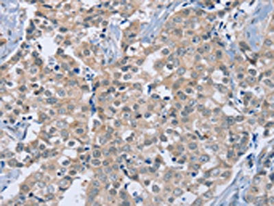
 IHC (Immunohiostchemistry)
(The image on the left is immunohistochemistry of paraffin-embedded Human ovarian cancer tissue using AAA240197(SLC44A1 Antibody) at dilution 1/25, on the right is treated with synthetic peptide. (Original magnification: ×200))
IHC (Immunohiostchemistry)
(The image on the left is immunohistochemistry of paraffin-embedded Human ovarian cancer tissue using AAA240197(SLC44A1 Antibody) at dilution 1/25, on the right is treated with synthetic peptide. (Original magnification: ×200))
SLC44A1, Polyclonal Antibody (Cat# AAA240197)
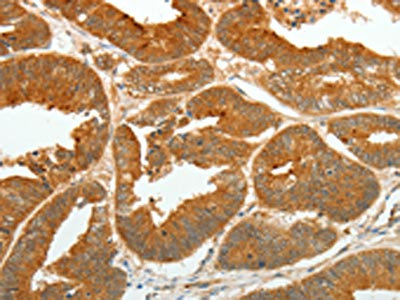
 IHC (Immunohiostchemistry)
(The image on the left is immunohistochemistry of paraffin-embedded Human ovarian cancer tissue using AAA240199(SLC34A2 Antibody) at dilution 1/50, on the right is treated with synthetic peptide. (Original magnification: ×200))
IHC (Immunohiostchemistry)
(The image on the left is immunohistochemistry of paraffin-embedded Human ovarian cancer tissue using AAA240199(SLC34A2 Antibody) at dilution 1/50, on the right is treated with synthetic peptide. (Original magnification: ×200))
SLC34A2, Polyclonal Antibody (Cat# AAA240199)
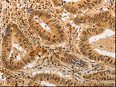
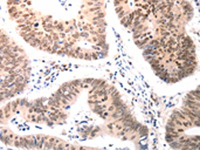
 IHC (Immunohiostchemistry)
(The image on the left is immunohistochemistry of paraffin-embedded Human gastric cancer tissue using AAA240200(GEMIN2 Antibody) at dilution 1/40, on the right is treated with synthetic peptide. (Original magnification: ×200))
IHC (Immunohiostchemistry)
(The image on the left is immunohistochemistry of paraffin-embedded Human gastric cancer tissue using AAA240200(GEMIN2 Antibody) at dilution 1/40, on the right is treated with synthetic peptide. (Original magnification: ×200))
GEMIN2, Polyclonal Antibody (Cat# AAA240200)
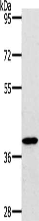
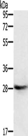
 SDS-PAGE
(Gel: 10%SDS-PAGE, Lysate: 40 ug, Lane: Mouse heart tissue, Primary antibody: AAA240202(SMAD7 Antibody) at dilution 1/1000, Secondary antibody: Goat anti rabbit IgG at 1/8000 dilution, Exposure time: 40 seconds)
SDS-PAGE
(Gel: 10%SDS-PAGE, Lysate: 40 ug, Lane: Mouse heart tissue, Primary antibody: AAA240202(SMAD7 Antibody) at dilution 1/1000, Secondary antibody: Goat anti rabbit IgG at 1/8000 dilution, Exposure time: 40 seconds)
SMAD7, Polyclonal Antibody (Cat# AAA240202)
 SDS-PAGE
(Gel: 10%SDS-PAGE, Lysate: 40 ug, Lane: 231 cells, Primary antibody: AAA240206(SOCS1 Antibody) at dilution 1/1300, Secondary antibody: Goat anti rabbit IgG at 1/8000 dilution, Exposure time: 1 minute)
SDS-PAGE
(Gel: 10%SDS-PAGE, Lysate: 40 ug, Lane: 231 cells, Primary antibody: AAA240206(SOCS1 Antibody) at dilution 1/1300, Secondary antibody: Goat anti rabbit IgG at 1/8000 dilution, Exposure time: 1 minute)
SOCS1, Polyclonal Antibody (Cat# AAA240206)
 IHC (Immunohiostchemistry)
(The image on the left is immunohistochemistry of paraffin-embedded Human lung cancer tissue using AAA240208(SOCS7 Antibody) at dilution 1/30, on the right is treated with synthetic peptide. (Original magnification: ×200))
IHC (Immunohiostchemistry)
(The image on the left is immunohistochemistry of paraffin-embedded Human lung cancer tissue using AAA240208(SOCS7 Antibody) at dilution 1/30, on the right is treated with synthetic peptide. (Original magnification: ×200))
SOCS7, Polyclonal Antibody (Cat# AAA240208)
 IHC (Immunohiostchemistry)
(The image on the left is immunohistochemistry of paraffin-embedded Human ovarian cancer tissue using AAA240209(SOCS7 Antibody) at dilution 1/30, on the right is treated with synthetic peptide. (Original magnification: ×200))
IHC (Immunohiostchemistry)
(The image on the left is immunohistochemistry of paraffin-embedded Human ovarian cancer tissue using AAA240209(SOCS7 Antibody) at dilution 1/30, on the right is treated with synthetic peptide. (Original magnification: ×200))
SOCS7, Polyclonal Antibody (Cat# AAA240209)
 IHC (Immunohiostchemistry)
(The image on the left is immunohistochemistry of paraffin-embedded Human ovarian cancer tissue using AAA240210(SPAG1 Antibody) at dilution 1/25, on the right is treated with synthetic peptide. (Original magnification: ×200))
IHC (Immunohiostchemistry)
(The image on the left is immunohistochemistry of paraffin-embedded Human ovarian cancer tissue using AAA240210(SPAG1 Antibody) at dilution 1/25, on the right is treated with synthetic peptide. (Original magnification: ×200))
SPAG1, Polyclonal Antibody (Cat# AAA240210)
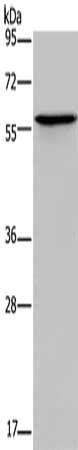
 SDS-PAGE
(Gel: 8%SDS-PAGE, Lysate: 40 ug, Lane: Human colon tissue, Primary antibody: AAA240211(SPAG1 Antibody) at dilution 1/600, Secondary antibody: Goat anti rabbit IgG at 1/8000 dilution, Exposure time: 2 minutes)
SDS-PAGE
(Gel: 8%SDS-PAGE, Lysate: 40 ug, Lane: Human colon tissue, Primary antibody: AAA240211(SPAG1 Antibody) at dilution 1/600, Secondary antibody: Goat anti rabbit IgG at 1/8000 dilution, Exposure time: 2 minutes)
SPAG1, Polyclonal Antibody (Cat# AAA240211)
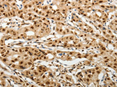
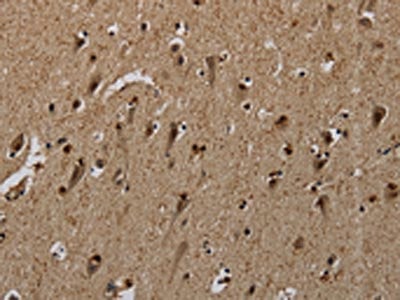 IHC (Immunohiostchemistry)
(The image on the left is immunohistochemistry of paraffin-embedded Human brain tissue using AAA241575(PNN Antibody) at dilution 1/50, on the right is treated with synthetic peptide. (Original magnification: ×200))
IHC (Immunohiostchemistry)
(The image on the left is immunohistochemistry of paraffin-embedded Human brain tissue using AAA241575(PNN Antibody) at dilution 1/50, on the right is treated with synthetic peptide. (Original magnification: ×200))
PNN, Polyclonal Antibody (Cat# AAA241575)
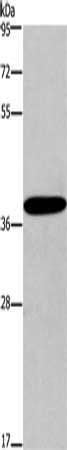
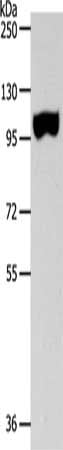
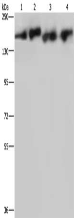 SDS-PAGE
(Gel: 6%SDS-PAGE, Lysate: 40 ug, Lane 1-4: PC3 cells, A431 cells, mouse lung tissue, Hela cells, Primary antibody: AAA241579(PLCB3 Antibody) at dilution 1/200, Secondary antibody: Goat anti rabbit IgG at 1/8000 dilution, Exposure time: 3 minutes)
SDS-PAGE
(Gel: 6%SDS-PAGE, Lysate: 40 ug, Lane 1-4: PC3 cells, A431 cells, mouse lung tissue, Hela cells, Primary antibody: AAA241579(PLCB3 Antibody) at dilution 1/200, Secondary antibody: Goat anti rabbit IgG at 1/8000 dilution, Exposure time: 3 minutes)
PLCB3, Polyclonal Antibody (Cat# AAA241579)
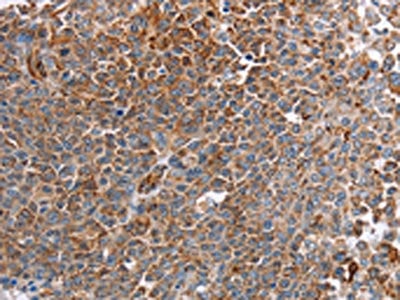
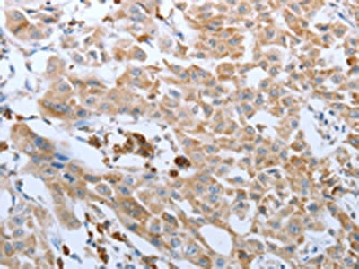
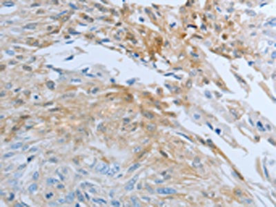
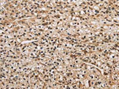
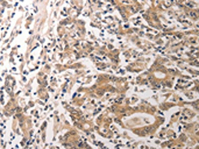
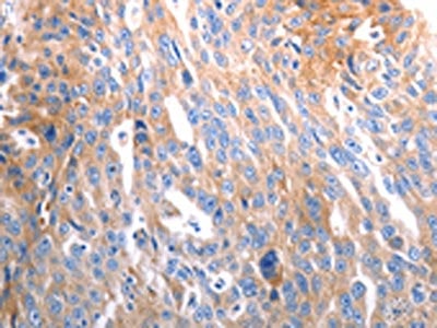
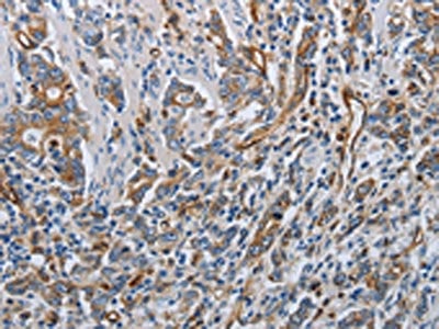
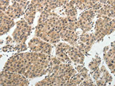
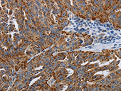
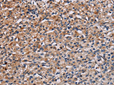
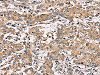
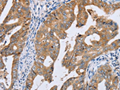
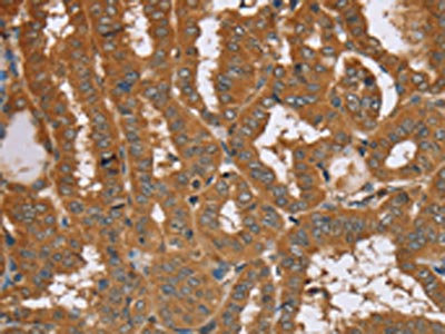
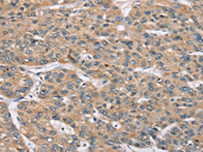
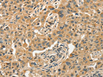
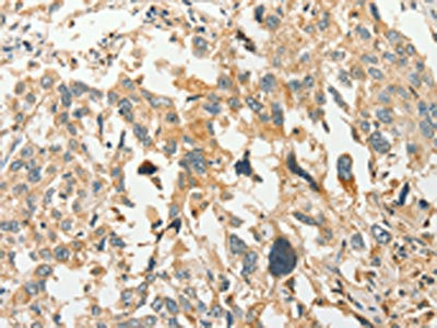
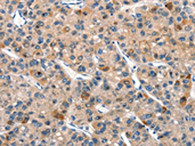
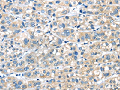 IHC (Immunohiostchemistry)
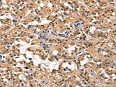
(The image on the left is immunohistochemistry of paraffin-embedded Human liver cancer tissue using AAA241581(PLXNA2 Antibody) at dilution 1/40, on the right is treated with synthetic peptide. (Original magnification: ×200))
IHC (Immunohiostchemistry)
(The image on the left is immunohistochemistry of paraffin-embedded Human liver cancer tissue using AAA241581(PLXNA2 Antibody) at dilution 1/40, on the right is treated with synthetic peptide. (Original magnification: ×200))
PLXNA2, Polyclonal Antibody (Cat# AAA241581)
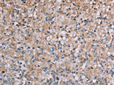
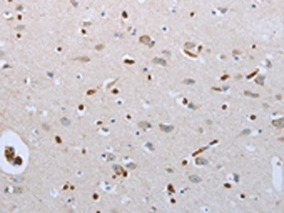
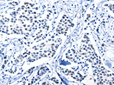
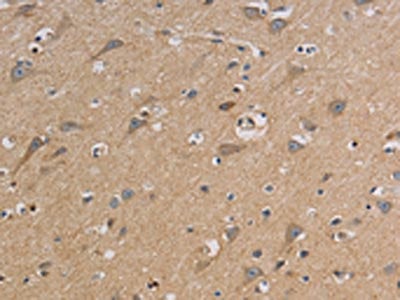
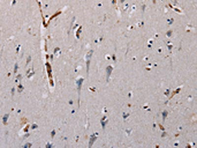
 IHC (Immunohiostchemistry)
(The image on the left is immunohistochemistry of paraffin-embedded Human brain tissue using AAA241583(PLXNA4 Antibody) at dilution 1/50, on the right is treated with synthetic peptide. (Original magnification: ×200))
IHC (Immunohiostchemistry)
(The image on the left is immunohistochemistry of paraffin-embedded Human brain tissue using AAA241583(PLXNA4 Antibody) at dilution 1/50, on the right is treated with synthetic peptide. (Original magnification: ×200))
PLXNA4, Polyclonal Antibody (Cat# AAA241583)

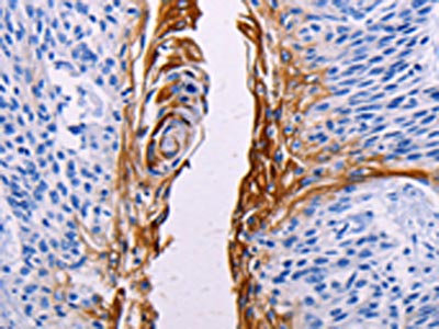
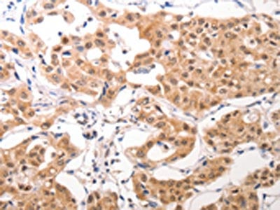
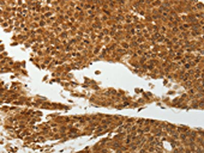
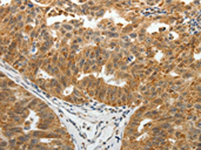
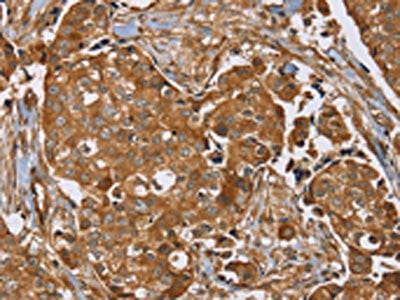
 IHC (Immunohiostchemistry)
(The image on the left is immunohistochemistry of paraffin-embedded Human breast cancer tissue using AAA241585(PLXNB1 Antibody) at dilution 1/50, on the right is treated with synthetic peptide. (Original magnification: ×200))
IHC (Immunohiostchemistry)
(The image on the left is immunohistochemistry of paraffin-embedded Human breast cancer tissue using AAA241585(PLXNB1 Antibody) at dilution 1/50, on the right is treated with synthetic peptide. (Original magnification: ×200))
PLXNB1, Polyclonal Antibody (Cat# AAA241585)
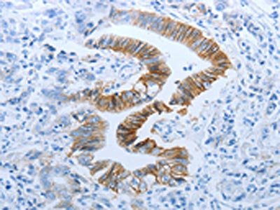
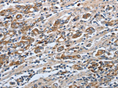
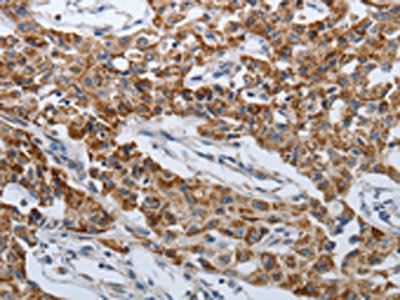
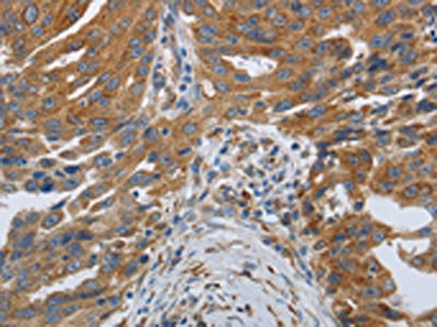
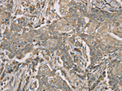
 IHC (Immunohiostchemistry)
(The image on the left is immunohistochemistry of paraffin-embedded Human prostate cancer tissue using AAA241587(PLXND1 Antibody) at dilution 1/40, on the right is treated with synthetic peptide. (Original magnification: ×200))
IHC (Immunohiostchemistry)
(The image on the left is immunohistochemistry of paraffin-embedded Human prostate cancer tissue using AAA241587(PLXND1 Antibody) at dilution 1/40, on the right is treated with synthetic peptide. (Original magnification: ×200))
PLXND1, Polyclonal Antibody (Cat# AAA241587)

 SDS-PAGE
(Gel: 10%SDS-PAGE, Lysate: 40 ug, Lane: Mouse brain tissue, Primary antibody: AAA241597(PPP1CC Antibody) at dilution 1/200, Secondary antibody: Goat anti rabbit IgG at 1/8000 dilution, Exposure time: 10 seconds)
SDS-PAGE
(Gel: 10%SDS-PAGE, Lysate: 40 ug, Lane: Mouse brain tissue, Primary antibody: AAA241597(PPP1CC Antibody) at dilution 1/200, Secondary antibody: Goat anti rabbit IgG at 1/8000 dilution, Exposure time: 10 seconds)
PPP1CC, Polyclonal Antibody (Cat# AAA241597)
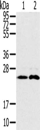
 SDS-PAGE
(Gel: 10%SDS-PAGE, Lysate: 40 ug, Lane 1-5: Jurkat cells, 293T cells, hela cells, mouse brain tissue, K562 cells, Primary antibody: AAA241598(PPP1CC Antibody) at dilution 1/200, Secondary antibody: Goat anti rabbit IgG at 1/8000 dilution, Exposure time: 1 second)
SDS-PAGE
(Gel: 10%SDS-PAGE, Lysate: 40 ug, Lane 1-5: Jurkat cells, 293T cells, hela cells, mouse brain tissue, K562 cells, Primary antibody: AAA241598(PPP1CC Antibody) at dilution 1/200, Secondary antibody: Goat anti rabbit IgG at 1/8000 dilution, Exposure time: 1 second)
PPP1CC, Polyclonal Antibody (Cat# AAA241598)
 SDS-PAGE
(Gel: 10%SDS-PAGE, Lysate: 40 ug, Lane 1-2: Mouse brain tissue, Mouse heart tissue, Primary antibody: AAA241603(PTP4A2 Antibody) at dilution 1/200, Secondary antibody: Goat anti rabbit IgG at 1/8000 dilution, Exposure time: 30 seconds)
SDS-PAGE
(Gel: 10%SDS-PAGE, Lysate: 40 ug, Lane 1-2: Mouse brain tissue, Mouse heart tissue, Primary antibody: AAA241603(PTP4A2 Antibody) at dilution 1/200, Secondary antibody: Goat anti rabbit IgG at 1/8000 dilution, Exposure time: 30 seconds)
PTP4A2, Polyclonal Antibody (Cat# AAA241603)
 SDS-PAGE
(Gel: 10%SDS-PAGE, Lysate: 40 ug, Lane 1-5: Hela cells, Jurkat cells, mouse lung tissue, PC3 cells, mouse liver tissue, Primary antibody: AAA241604(PTGER2 Antibody) at dilution 1/200, Secondary antibody: Goat anti rabbit IgG at 1/8000 dilution, Exposure time: 30 seconds)
SDS-PAGE
(Gel: 10%SDS-PAGE, Lysate: 40 ug, Lane 1-5: Hela cells, Jurkat cells, mouse lung tissue, PC3 cells, mouse liver tissue, Primary antibody: AAA241604(PTGER2 Antibody) at dilution 1/200, Secondary antibody: Goat anti rabbit IgG at 1/8000 dilution, Exposure time: 30 seconds)
PTGER2, Polyclonal Antibody (Cat# AAA241604)
 SDS-PAGE
(Gel: 6%SDS-PAGE, Lysate: 40 ug, Lane 1-2: 231 cells, K562 cells, Primary antibody: AAA241611(PTPN12 Antibody) at dilution 1/200, Secondary antibody: Goat anti rabbit IgG at 1/8000 dilution, Exposure time: 10 minutes)
SDS-PAGE
(Gel: 6%SDS-PAGE, Lysate: 40 ug, Lane 1-2: 231 cells, K562 cells, Primary antibody: AAA241611(PTPN12 Antibody) at dilution 1/200, Secondary antibody: Goat anti rabbit IgG at 1/8000 dilution, Exposure time: 10 minutes)
PTPN12, Polyclonal Antibody (Cat# AAA241611)
 IHC (Immunohiostchemistry)
(The image on the left is immunohistochemistry of paraffin-embedded Human lung cancer tissue using AAA241620(RAB8B Antibody) at dilution 1/45, on the right is treated with synthetic peptide. (Original magnification: ×200))
IHC (Immunohiostchemistry)
(The image on the left is immunohistochemistry of paraffin-embedded Human lung cancer tissue using AAA241620(RAB8B Antibody) at dilution 1/45, on the right is treated with synthetic peptide. (Original magnification: ×200))
RAB8B, Polyclonal Antibody (Cat# AAA241620)
 SDS-PAGE
(Gel: 8%SDS-PAGE, Lysate: 80 ug, Lane: Human normal colon tissue, Primary antibody: AAA241621(RAB8B Antibody) at dilution 1/400, Secondary antibody: Goat anti rabbit IgG at 1/8000 dilution, Exposure time: 4 minutes)
SDS-PAGE
(Gel: 8%SDS-PAGE, Lysate: 80 ug, Lane: Human normal colon tissue, Primary antibody: AAA241621(RAB8B Antibody) at dilution 1/400, Secondary antibody: Goat anti rabbit IgG at 1/8000 dilution, Exposure time: 4 minutes)
RAB8B, Polyclonal Antibody (Cat# AAA241621)
 SDS-PAGE
(Gel: 10%SDS-PAGE, Lysate: 40 ug, Lane 1-2: Mouse brain tissue, Raji cells, Primary antibody: AAA241622(RAB14 Antibody) at dilution 1/200, Secondary antibody: Goat anti rabbit IgG at 1/8000 dilution, Exposure time: 30 seconds)
SDS-PAGE
(Gel: 10%SDS-PAGE, Lysate: 40 ug, Lane 1-2: Mouse brain tissue, Raji cells, Primary antibody: AAA241622(RAB14 Antibody) at dilution 1/200, Secondary antibody: Goat anti rabbit IgG at 1/8000 dilution, Exposure time: 30 seconds)
RAB14, Polyclonal Antibody (Cat# AAA241622)
 SDS-PAGE
(Gel: 6%SDS-PAGE, Lysate: 40 ug, Lane 1-2: K562 cells, Jurkat cells, Primary antibody: AAA241625(TFRC Antibody) at dilution 1/200, Secondary antibody: Goat anti rabbit IgG at 1/8000 dilution, Exposure time: 20 seconds)
SDS-PAGE
(Gel: 6%SDS-PAGE, Lysate: 40 ug, Lane 1-2: K562 cells, Jurkat cells, Primary antibody: AAA241625(TFRC Antibody) at dilution 1/200, Secondary antibody: Goat anti rabbit IgG at 1/8000 dilution, Exposure time: 20 seconds)
TFRC, Polyclonal Antibody (Cat# AAA241625)
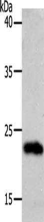
 SDS-PAGE
(Gel: 6%SDS-PAGE, Lysate: 40 ug, Lane: HT29 cells, Primary antibody: AAA241626(MUC1 Antibody) at dilution 1/200, Secondary antibody: Goat anti rabbit IgG at 1/8000 dilution, Exposure time: 30 seconds)
SDS-PAGE
(Gel: 6%SDS-PAGE, Lysate: 40 ug, Lane: HT29 cells, Primary antibody: AAA241626(MUC1 Antibody) at dilution 1/200, Secondary antibody: Goat anti rabbit IgG at 1/8000 dilution, Exposure time: 30 seconds)
MUC1, Polyclonal Antibody (Cat# AAA241626)
 SDS-PAGE
(Gel: 10%SDS-PAGE, Lysate: 40 ug, Lane: Human heart tissue, Primary antibody: AAA241636(RBM38 Antibody) at dilution 1/200, Secondary antibody: Goat anti rabbit IgG at 1/8000 dilution, Exposure time: 1 minute)
SDS-PAGE
(Gel: 10%SDS-PAGE, Lysate: 40 ug, Lane: Human heart tissue, Primary antibody: AAA241636(RBM38 Antibody) at dilution 1/200, Secondary antibody: Goat anti rabbit IgG at 1/8000 dilution, Exposure time: 1 minute)
RBM38, Polyclonal Antibody (Cat# AAA241636)
 IHC (Immunohiostchemistry)
(The image on the left is immunohistochemistry of paraffin-embedded Human thyroid cancer tissue using AAA241659(TRIM40 Antibody) at dilution 1/40, on the right is treated with synthetic peptide. (Original magnification: ×200))
IHC (Immunohiostchemistry)
(The image on the left is immunohistochemistry of paraffin-embedded Human thyroid cancer tissue using AAA241659(TRIM40 Antibody) at dilution 1/40, on the right is treated with synthetic peptide. (Original magnification: ×200))
TRIM40, Polyclonal Antibody (Cat# AAA241659)
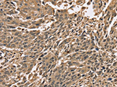
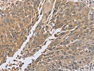
 IHC (Immunohiostchemistry)
(The image on the left is immunohistochemistry of paraffin-embedded Human esophagus cancer tissue using AAA241054(SLC11A2 Antibody) at dilution 1/50, on the right is treated with synthetic peptide. (Original magnification: ×200))
IHC (Immunohiostchemistry)
(The image on the left is immunohistochemistry of paraffin-embedded Human esophagus cancer tissue using AAA241054(SLC11A2 Antibody) at dilution 1/50, on the right is treated with synthetic peptide. (Original magnification: ×200))
SLC11A2, Polyclonal Antibody (Cat# AAA241054)

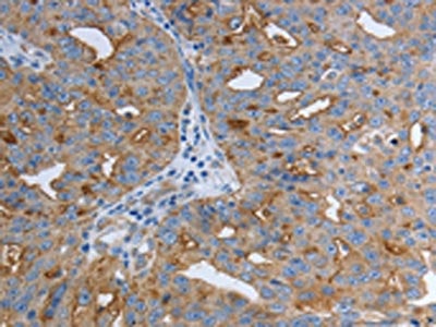
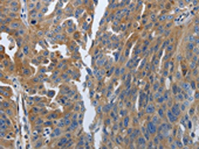
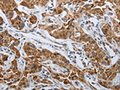
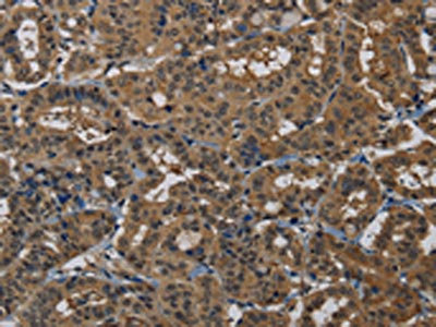
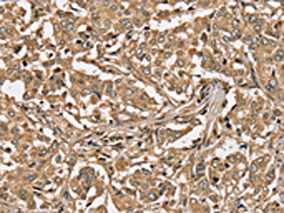
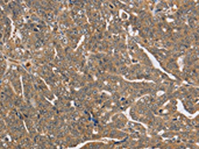
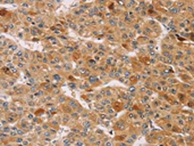
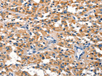
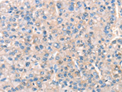 IHC (Immunohiostchemistry)
(The image on the left is immunohistochemistry of paraffin-embedded Human liver cancer tissue using AAA241061(KCNB1 Antibody) at dilution 1/30, on the right is treated with synthetic peptide. (Original magnification: ×200))
IHC (Immunohiostchemistry)
(The image on the left is immunohistochemistry of paraffin-embedded Human liver cancer tissue using AAA241061(KCNB1 Antibody) at dilution 1/30, on the right is treated with synthetic peptide. (Original magnification: ×200))
KCNB1, Polyclonal Antibody (Cat# AAA241061)
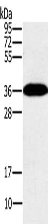
 SDS-PAGE
(Gel: 8%SDS-PAGE, Lysate: 40 ug, Lane: Hela cells, Primary antibody: AAA241062(DUSP2 Antibody) at dilution 1/300, Secondary antibody: Goat anti rabbit IgG at 1/8000 dilution, Exposure time: 1 minute)
SDS-PAGE
(Gel: 8%SDS-PAGE, Lysate: 40 ug, Lane: Hela cells, Primary antibody: AAA241062(DUSP2 Antibody) at dilution 1/300, Secondary antibody: Goat anti rabbit IgG at 1/8000 dilution, Exposure time: 1 minute)
DUSP2, Polyclonal Antibody (Cat# AAA241062)
 SDS-PAGE
(Gel: 8%SDS-PAGE, Lysate: 40 ug, Lane: Mouse kidney tissue, Primary antibody: AAA241065(EID3 Antibody) at dilution 1/200, Secondary antibody: Goat anti rabbit IgG at 1/8000 dilution, Exposure time: 1 minute)
SDS-PAGE
(Gel: 8%SDS-PAGE, Lysate: 40 ug, Lane: Mouse kidney tissue, Primary antibody: AAA241065(EID3 Antibody) at dilution 1/200, Secondary antibody: Goat anti rabbit IgG at 1/8000 dilution, Exposure time: 1 minute)
EID3, Polyclonal Antibody (Cat# AAA241065)

 SDS-PAGE
(Gel: 8%SDS-PAGE, Lysate: 40 ug, Lane 1-3: Raji cells, human fetal brain tissue, mouse brain tissue, Primary antibody: AAA241070(ELOVL6 Antibody) at dilution 1/400, Secondary antibody: Goat anti rabbit IgG at 1/8000 dilution, Exposure time: 30 seconds)
SDS-PAGE
(Gel: 8%SDS-PAGE, Lysate: 40 ug, Lane 1-3: Raji cells, human fetal brain tissue, mouse brain tissue, Primary antibody: AAA241070(ELOVL6 Antibody) at dilution 1/400, Secondary antibody: Goat anti rabbit IgG at 1/8000 dilution, Exposure time: 30 seconds)
ELOVL6, Polyclonal Antibody (Cat# AAA241070)
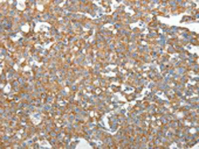
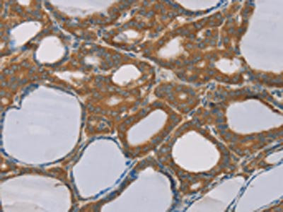
 IHC (Immunohiostchemistry)
(The image on the left is immunohistochemistry of paraffin-embedded Human thyroid cancer tissue using AAA241071(ELOVL6 Antibody) at dilution 1/25, on the right is treated with synthetic peptide. (Original magnification: ×200))
IHC (Immunohiostchemistry)
(The image on the left is immunohistochemistry of paraffin-embedded Human thyroid cancer tissue using AAA241071(ELOVL6 Antibody) at dilution 1/25, on the right is treated with synthetic peptide. (Original magnification: ×200))
ELOVL6, Polyclonal Antibody (Cat# AAA241071)
What are Polyclonal Antibodies?
Polyclonal antibodies are antibodies that come from multiple B cell clones of a host animal. The typical hosts used for the majority of polyclonal antibody production are rabbits, goats, sheep, and donkeys. These polyclonal antibodies, once having identified their target, will bind to different epitopes located at different regions or sequences on the same protein/antigen. As a result, they are ideal at locating and binding to the target, even if the target is in very low concentrations (due to many different antibodies being able to bind to the same target molecule, which allows for significant amplification of a downstream signal).
Polyclonal antibodies are typically produced by injecting an antigen into a host animal, which causes the animal’s immune system to attack the foreign antigen by mass generating antibodies against it. After a period of time, serum is collected from the animal and purified using physicochemical fractionation, class-specific affinity purification, and/or antigen-affinity purification.
Key Uses of Polyclonal Antibodies
- Western Blotting: This method is used to find specific proteins in biological samples after separating them by size.
- Immunohistochemistry: IHC helps visualize the location of proteins in tissue sections using various staining techniques.
- ELISA: (Enzyme-Linked Immunosorbent Assay) is typically used to identify specific protein quantities in a sample. ELISAs can be either “Quantitative” or “Qualitative”.
- Flow Cytometry: technique that identifies and measures the specific protein on the surface or inside the cells in a fluid suspension.
- Immunoprecipitation: IP isolates and studies a specific protein from a complex mixture using antibodies.
Why Buy Polyclonal Antibodies from AAA Biotech?
1. Ideal for Various Applications
Our antibodies are generally going to be validated for use in multiple types of assays, including ELISA, Western Blotting, Immunohistochemistry, Immunoprecipitation, amongst others. They are ideal for a wide range of research applications.
2. Rigorous Quality Control
All of the antibodies in our catalog undergo strict quality testing to ensure specificity, sensitivity, and consistent performance. We are confident in the ability of our antibodies to provide you with accurate results.
3. Wide Assortment of Antibodies
Antibodies in are catalog can be found for both common and exotic species, and these antibodies are also available in both conjugated and recombinant forms to suit many diverse experimental needs.
4. Highly Purified
Our antibodies are available in purified forms with over 85% purity, as confirmed by SDS-PAGE. They are also available with tags such as His, Flag, GST, or MBP. We cater to customers worldwide.
FAQ
1. How are polyclonal antibodies produced?
Traditionally, polyclonal antibodies are produced by injecting an antigen into a host animal (such as a rabbit or goat), which then triggers an immune response from the host animal. The animal’s B cells produce antibodies that will recognize different parts of the injected antigen. These antibodies are then collected from the animal’s blood and purified for use.
2. How do polyclonal antibodies differ from monoclonal antibodies?
Polyclonal antibodies are a mix of antibodies that bind to different locations (epitopes) of the same antigen, while monoclonal antibodies are identical and bind to just one specific epitope. This makes polyclonal antibodies more versatile and better at detecting proteins that may be present in low quantities or in altered/modified forms.
3. How should I store polyclonal antibodies?
Polyclonal antibodies should be stored at 4°C for short-term use (up to a few weeks) and at -20°C or -80°C for long-term storage. Avoid repeated freeze-thaw cycles by dividing them into small aliquots. Always check the datasheet for specific storage instructions.