Filters
▼Clonality
▼Type
▼Reactivity
▼Gene Name
▼Isotype
▼Host
▼Application
▼Clone
▼Polyclonal Antibodies
At AAA Biotech also known as AAA Bio or AAABio, we provide a broad range of purified polyclonal antibodies (pAbs) that are able to all be browsed online through our website. Due to their high specificity and strong binding affinity, these antibodies are ideal for wide swathes of research and experimental applications.
Our polyclonal antibodies can easily support your work, whether you use them for Western Blotting, Immunocytochemistry (with or without Immunofluorescence used in conjunction), Immunohistochemistry, Immunoprecipitation, and ELISA tests. We highly encourage you to browse our range of pAbs and choose the one that best suits your experimental model.
Viewing 6100-6150 of 96812 product results
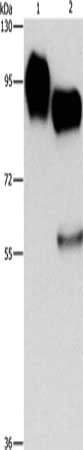
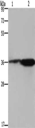
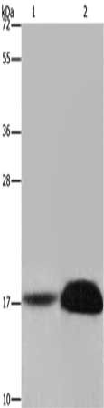
 SDS-PAGE
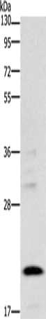
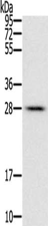
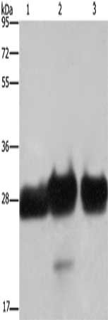
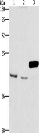
(Gel: 6%SDS-PAGE, Lysate: 40 ug, Lane 1-2: PC3 cells, Hela cells, Primary antibody: AAA238646(COMP Antibody) at dilution 1/600, Secondary antibody: Goat anti rabbit IgG at 1/8000 dilution, Exposure time: 10 seconds)
SDS-PAGE
(Gel: 6%SDS-PAGE, Lysate: 40 ug, Lane 1-2: PC3 cells, Hela cells, Primary antibody: AAA238646(COMP Antibody) at dilution 1/600, Secondary antibody: Goat anti rabbit IgG at 1/8000 dilution, Exposure time: 10 seconds)
COMP, Polyclonal Antibody (Cat# AAA238646)
 SDS-PAGE
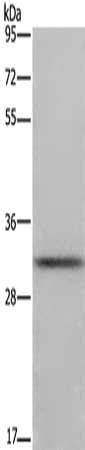
(Gel: 8%SDS-PAGE, Lysate: 40 ug, Lane: Lovo cells, Primary antibody: AAA238648(CORO1C Antibody) at dilution 1/670, Secondary antibody: Goat anti rabbit IgG at 1/8000 dilution, Exposure time: 40 seconds)
SDS-PAGE
(Gel: 8%SDS-PAGE, Lysate: 40 ug, Lane: Lovo cells, Primary antibody: AAA238648(CORO1C Antibody) at dilution 1/670, Secondary antibody: Goat anti rabbit IgG at 1/8000 dilution, Exposure time: 40 seconds)
CORO1C, Polyclonal Antibody (Cat# AAA238648)
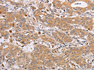
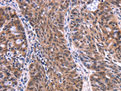
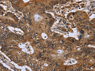
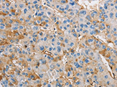
 IHC (Immunohiostchemistry)
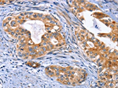
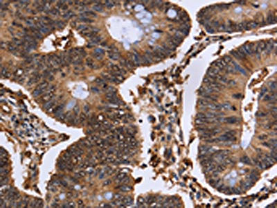
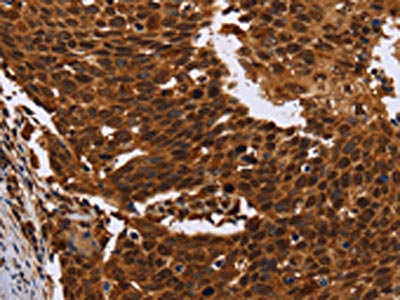
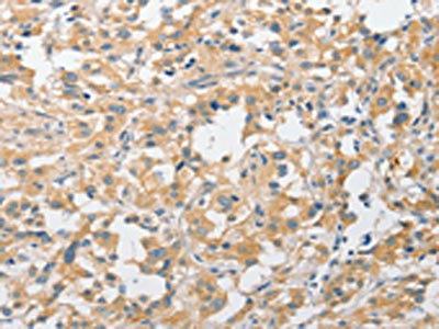
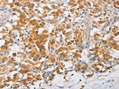
(The image on the left is immunohistochemistry of paraffin-embedded Human colon cancer tissue using AAA238330(EPHB6 Antibody) at dilution 1/40, on the right is treated with fusion protein. (Original magnification: ×200))
IHC (Immunohiostchemistry)
(The image on the left is immunohistochemistry of paraffin-embedded Human colon cancer tissue using AAA238330(EPHB6 Antibody) at dilution 1/40, on the right is treated with fusion protein. (Original magnification: ×200))
EPHB6, Polyclonal Antibody (Cat# AAA238330)
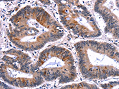
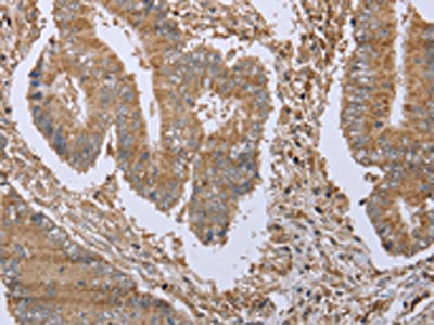
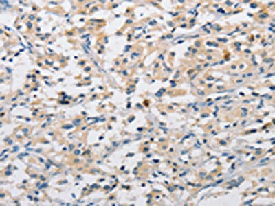
 IHC (Immunohiostchemistry)
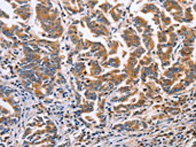
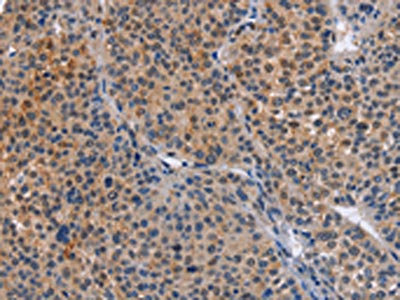
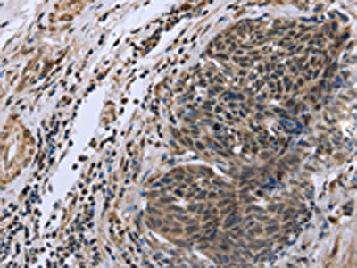
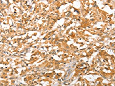
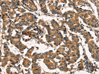
(The image on the left is immunohistochemistry of paraffin-embedded Human cervical cancer tissue using AAA238333(MUSK Antibody) at dilution 1/30, on the right is treated with fusion protein. (Original magnification: ×200))
IHC (Immunohiostchemistry)
(The image on the left is immunohistochemistry of paraffin-embedded Human cervical cancer tissue using AAA238333(MUSK Antibody) at dilution 1/30, on the right is treated with fusion protein. (Original magnification: ×200))
MUSK, Polyclonal Antibody (Cat# AAA238333)
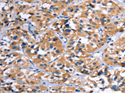
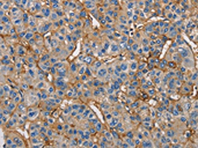
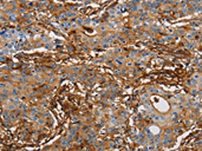
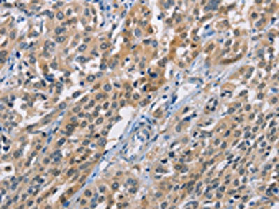
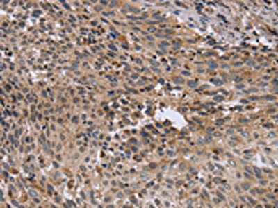
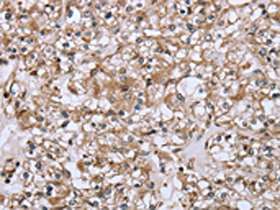
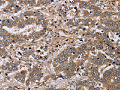
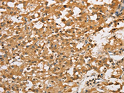
 IHC (Immunohiostchemistry)
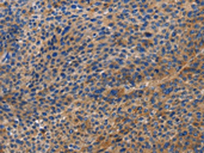
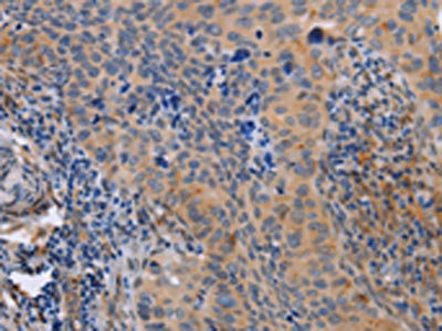
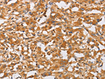
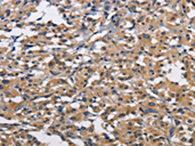
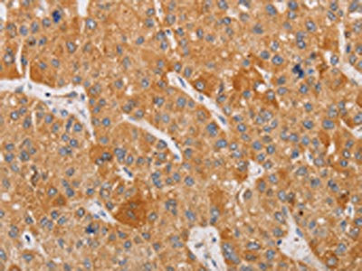
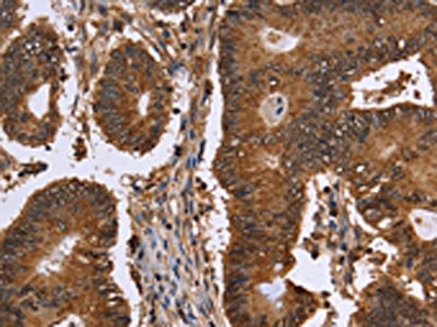
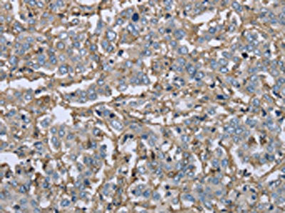
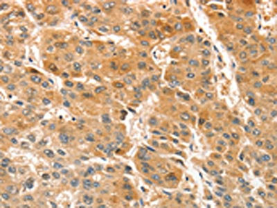
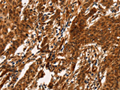
(The image on the left is immunohistochemistry of paraffin-embedded Human liver cancer tissue using AAA238336(DOK4 Antibody) at dilution 1/60, on the right is treated with fusion protein. (Original magnification: ×200))
IHC (Immunohiostchemistry)
(The image on the left is immunohistochemistry of paraffin-embedded Human liver cancer tissue using AAA238336(DOK4 Antibody) at dilution 1/60, on the right is treated with fusion protein. (Original magnification: ×200))
DOK4, Polyclonal Antibody (Cat# AAA238336)
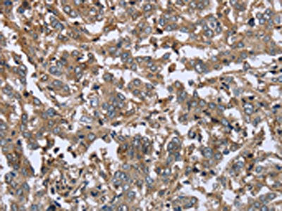
 IHC (Immunohiostchemistry)
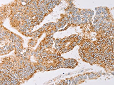
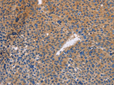
(The image on the left is immunohistochemistry of paraffin-embedded Human lung cancer tissue using AAA238339(YARS2 Antibody) at dilution 1/30, on the right is treated with fusion protein. (Original magnification: ×200))
IHC (Immunohiostchemistry)
(The image on the left is immunohistochemistry of paraffin-embedded Human lung cancer tissue using AAA238339(YARS2 Antibody) at dilution 1/30, on the right is treated with fusion protein. (Original magnification: ×200))
YARS2, Polyclonal Antibody (Cat# AAA238339)
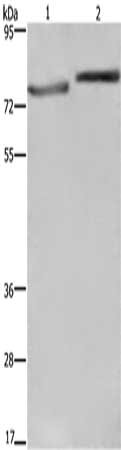
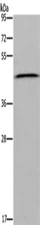
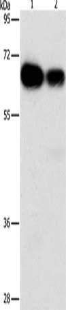
 SDS-PAGE
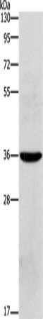
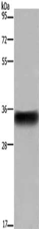
(Gel: 6%SDS-PAGE, Lysate: 40 ug, Lane: Human placenta tissue, Primary antibody: AAA238342(STK40 Antibody) at dilution 1/400, Secondary antibody: Goat anti rabbit IgG at 1/8000 dilution, Exposure time: 2 minutes)
SDS-PAGE
(Gel: 6%SDS-PAGE, Lysate: 40 ug, Lane: Human placenta tissue, Primary antibody: AAA238342(STK40 Antibody) at dilution 1/400, Secondary antibody: Goat anti rabbit IgG at 1/8000 dilution, Exposure time: 2 minutes)
STK40, Polyclonal Antibody (Cat# AAA238342)
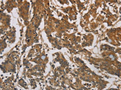
 IHC (Immunohiostchemistry)
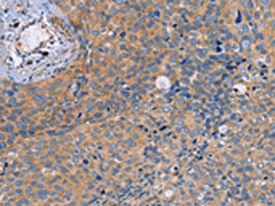
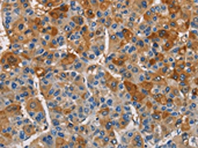
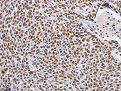
(The image on the left is immunohistochemistry of paraffin-embedded Human cervical cancer tissue using AAA238346(MAP3K13 Antibody) at dilution 1/40, on the right is treated with fusion protein. (Original magnification: ×200))
IHC (Immunohiostchemistry)
(The image on the left is immunohistochemistry of paraffin-embedded Human cervical cancer tissue using AAA238346(MAP3K13 Antibody) at dilution 1/40, on the right is treated with fusion protein. (Original magnification: ×200))
MAP3K13, Polyclonal Antibody (Cat# AAA238346)
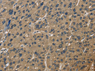
 IHC (Immunohiostchemistry)
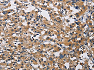
(The image on the left is immunohistochemistry of paraffin-embedded Human cervical cancer tissue using AAA238351(NEK8 Antibody) at dilution 1/30, on the right is treated with fusion protein. (Original magnification: ×200))
IHC (Immunohiostchemistry)
(The image on the left is immunohistochemistry of paraffin-embedded Human cervical cancer tissue using AAA238351(NEK8 Antibody) at dilution 1/30, on the right is treated with fusion protein. (Original magnification: ×200))
NEK8, Polyclonal Antibody (Cat# AAA238351)
 IHC (Immunohiostchemistry)
(The image on the left is immunohistochemistry of paraffin-embedded Human colon cancer tissue using AAA238352(PACSIN2 Antibody) at dilution 1/20, on the right is treated with fusion protein. (Original magnification: ×200))
IHC (Immunohiostchemistry)
(The image on the left is immunohistochemistry of paraffin-embedded Human colon cancer tissue using AAA238352(PACSIN2 Antibody) at dilution 1/20, on the right is treated with fusion protein. (Original magnification: ×200))
PACSIN2, Polyclonal Antibody (Cat# AAA238352)
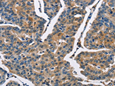
 IHC (Immunohiostchemistry)
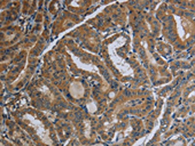
(The image on the left is immunohistochemistry of paraffin-embedded Human thyroid cancer tissue using AAA238354(UCK1 Antibody) at dilution 1/50, on the right is treated with fusion protein. (Original magnification: ×200))
IHC (Immunohiostchemistry)
(The image on the left is immunohistochemistry of paraffin-embedded Human thyroid cancer tissue using AAA238354(UCK1 Antibody) at dilution 1/50, on the right is treated with fusion protein. (Original magnification: ×200))
UCK1, Polyclonal Antibody (Cat# AAA238354)
 SDS-PAGE
(Gel: 10%SDS-PAGE, Lysate: 40 ug, Lane 1-2: Raji cells, huvec cells, Primary antibody: AAA238367(ICAM1 Antibody) at dilution 1/1150, Secondary antibody: Goat anti rabbit IgG at 1/8000 dilution, Exposure time: 1 second)
SDS-PAGE
(Gel: 10%SDS-PAGE, Lysate: 40 ug, Lane 1-2: Raji cells, huvec cells, Primary antibody: AAA238367(ICAM1 Antibody) at dilution 1/1150, Secondary antibody: Goat anti rabbit IgG at 1/8000 dilution, Exposure time: 1 second)
ICAM1, Polyclonal Antibody (Cat# AAA238367)
 SDS-PAGE
(Gel: 10%SDS-PAGE, Lysate: 40 ug, Lane: Human placenta tissue, Primary antibody: AAA238370(OLR1 Antibody) at dilution 1/450, Secondary antibody: Goat anti rabbit IgG at 1/8000 dilution, Exposure time: 20 seconds)
SDS-PAGE
(Gel: 10%SDS-PAGE, Lysate: 40 ug, Lane: Human placenta tissue, Primary antibody: AAA238370(OLR1 Antibody) at dilution 1/450, Secondary antibody: Goat anti rabbit IgG at 1/8000 dilution, Exposure time: 20 seconds)
OLR1, Polyclonal Antibody (Cat# AAA238370)
 IHC (Immunohiostchemistry)
(The image on the left is immunohistochemistry of paraffin-embedded Human thyroid cancer tissue using AAA238373(CST4 Antibody) at dilution 1/60, on the right is treated with fusion protein. (Original magnification: ×200))
IHC (Immunohiostchemistry)
(The image on the left is immunohistochemistry of paraffin-embedded Human thyroid cancer tissue using AAA238373(CST4 Antibody) at dilution 1/60, on the right is treated with fusion protein. (Original magnification: ×200))
CST4, Polyclonal Antibody (Cat# AAA238373)
 IHC (Immunohiostchemistry)
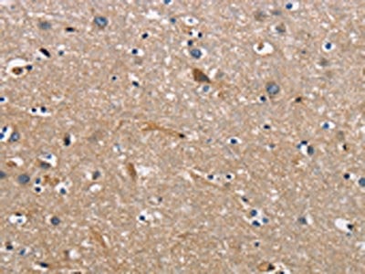
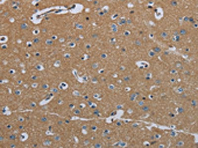
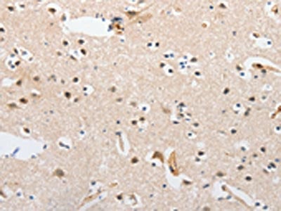
(The image on the left is immunohistochemistry of paraffin-embedded Human brain tissue using AAA238374(CBY1 Antibody) at dilution 1/60, on the right is treated with fusion protein. (Original magnification: ×200))
IHC (Immunohiostchemistry)
(The image on the left is immunohistochemistry of paraffin-embedded Human brain tissue using AAA238374(CBY1 Antibody) at dilution 1/60, on the right is treated with fusion protein. (Original magnification: ×200))
CBY1, Polyclonal Antibody (Cat# AAA238374)
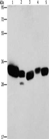
 SDS-PAGE
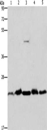
(Gel: 10%SDS-PAGE, Lysate: 40 ug, Lane 1-5: Mouse spleen tissue, human fetal liver tissue, hela cells, mouse testis tissue, A431 cells, Primary antibody: AAA238379(THOC7 Antibody) at dilution 1/500, Secondary antibody: Goat anti rabbit IgG at 1/8000 dilution, Exposure time: 15 seconds)
SDS-PAGE
(Gel: 10%SDS-PAGE, Lysate: 40 ug, Lane 1-5: Mouse spleen tissue, human fetal liver tissue, hela cells, mouse testis tissue, A431 cells, Primary antibody: AAA238379(THOC7 Antibody) at dilution 1/500, Secondary antibody: Goat anti rabbit IgG at 1/8000 dilution, Exposure time: 15 seconds)
THOC7, Polyclonal Antibody (Cat# AAA238379)
 IHC (Immunohiostchemistry)
(The image on the left is immunohistochemistry of paraffin-embedded Human thyroid cancer tissue using AAA238381(ELP2 Antibody) at dilution 1/40, on the right is treated with fusion protein. (Original magnification: ×200))
IHC (Immunohiostchemistry)
(The image on the left is immunohistochemistry of paraffin-embedded Human thyroid cancer tissue using AAA238381(ELP2 Antibody) at dilution 1/40, on the right is treated with fusion protein. (Original magnification: ×200))
ELP2, Polyclonal Antibody (Cat# AAA238381)
 SDS-PAGE
(Gel: 10%SDS-PAGE, Lysate: 40 ug, Lane: A172 cells, Primary antibody: AAA238384(BNIP3 Antibody) at dilution 1/300, Secondary antibody: Goat anti rabbit IgG at 1/8000 dilution, Exposure time: 2 minutes)
SDS-PAGE
(Gel: 10%SDS-PAGE, Lysate: 40 ug, Lane: A172 cells, Primary antibody: AAA238384(BNIP3 Antibody) at dilution 1/300, Secondary antibody: Goat anti rabbit IgG at 1/8000 dilution, Exposure time: 2 minutes)
BNIP3, Polyclonal Antibody (Cat# AAA238384)
 IHC (Immunohiostchemistry)
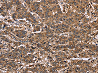
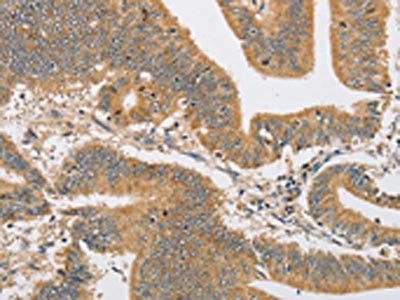
(The image on the left is immunohistochemistry of paraffin-embedded Human colon cancer tissue using AAA238395(EIF2AK4 Antibody) at dilution 1/50, on the right is treated with fusion protein. (Original magnification: ×200))
IHC (Immunohiostchemistry)
(The image on the left is immunohistochemistry of paraffin-embedded Human colon cancer tissue using AAA238395(EIF2AK4 Antibody) at dilution 1/50, on the right is treated with fusion protein. (Original magnification: ×200))
EIF2AK4, Polyclonal Antibody (Cat# AAA238395)
 SDS-PAGE
(Gel: 15%SDS-PAGE, Lysate: 40 ug, Lane 1-5: Mouse liver tissue, human fetal lung tissue, hela cells, mouse kidney tissue, human brain malignant glioma tissue, Primary antibody: AAA238399(CBR1 Antibody) at dilution 1/900, Secondary antibody: Goat anti rabbit IgG at 1/8000 dilution, Exposure time: 10 seconds)
SDS-PAGE
(Gel: 15%SDS-PAGE, Lysate: 40 ug, Lane 1-5: Mouse liver tissue, human fetal lung tissue, hela cells, mouse kidney tissue, human brain malignant glioma tissue, Primary antibody: AAA238399(CBR1 Antibody) at dilution 1/900, Secondary antibody: Goat anti rabbit IgG at 1/8000 dilution, Exposure time: 10 seconds)
CBR1, Polyclonal Antibody (Cat# AAA238399)
 IHC (Immunohiostchemistry)
(The image on the left is immunohistochemistry of paraffin-embedded Human brain tissue using AAA238402(PIP5K1B Antibody) at dilution 1/30, on the right is treated with fusion protein. (Original magnification: ×200))
IHC (Immunohiostchemistry)
(The image on the left is immunohistochemistry of paraffin-embedded Human brain tissue using AAA238402(PIP5K1B Antibody) at dilution 1/30, on the right is treated with fusion protein. (Original magnification: ×200))
PIP5K1B, Polyclonal Antibody (Cat# AAA238402)
 SDS-PAGE
(Gel: 8%SDS-PAGE, Lysate: 40 ug, Lane: Human placenta tissue, Primary antibody: AAA238825(EPSTI1 Antibody) at dilution 1/1900, Secondary antibody: Goat anti rabbit IgG at 1/8000 dilution, Exposure time: 30 seconds)
SDS-PAGE
(Gel: 8%SDS-PAGE, Lysate: 40 ug, Lane: Human placenta tissue, Primary antibody: AAA238825(EPSTI1 Antibody) at dilution 1/1900, Secondary antibody: Goat anti rabbit IgG at 1/8000 dilution, Exposure time: 30 seconds)
EPSTI1, Polyclonal Antibody (Cat# AAA238825)


 SDS-PAGE
(Gel: 8%SDS-PAGE, Lysate: 40 ug, Lane: Mouse brain tissue, Primary antibody: AAA238827(ESR2 Antibody) at dilution 1/250, Secondary antibody: Goat anti rabbit IgG at 1/8000 dilution, Exposure time: 10 seconds)
SDS-PAGE
(Gel: 8%SDS-PAGE, Lysate: 40 ug, Lane: Mouse brain tissue, Primary antibody: AAA238827(ESR2 Antibody) at dilution 1/250, Secondary antibody: Goat anti rabbit IgG at 1/8000 dilution, Exposure time: 10 seconds)
ESR2, Polyclonal Antibody (Cat# AAA238827)
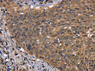
 IHC (Immunohiostchemistry)
(The image on the left is immunohistochemistry of paraffin-embedded Human esophagus cancer tissue using AAA238832(EDN1 Antibody) at dilution 1/30, on the right is treated with fusion protein. (Original magnification: ×200))
IHC (Immunohiostchemistry)
(The image on the left is immunohistochemistry of paraffin-embedded Human esophagus cancer tissue using AAA238832(EDN1 Antibody) at dilution 1/30, on the right is treated with fusion protein. (Original magnification: ×200))
EDN1, Polyclonal Antibody (Cat# AAA238832)
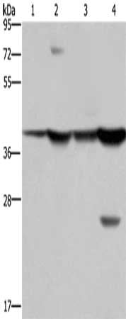
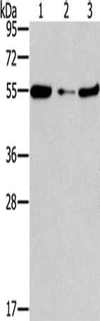
 SDS-PAGE
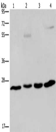
(Gel: 10%SDS-PAGE, Lysate: 40 ug, Lane 1-4: A549 cells, human hepatocellular carcinoma tissue, mouse liver tissue, HT29 cells, Primary antibody: AAA238836(ETHE1 Antibody) at dilution 1/400, Secondary antibody: Goat anti rabbit IgG at 1/8000 dilution, Exposure time: 1 minute)
SDS-PAGE
(Gel: 10%SDS-PAGE, Lysate: 40 ug, Lane 1-4: A549 cells, human hepatocellular carcinoma tissue, mouse liver tissue, HT29 cells, Primary antibody: AAA238836(ETHE1 Antibody) at dilution 1/400, Secondary antibody: Goat anti rabbit IgG at 1/8000 dilution, Exposure time: 1 minute)
ETHE1, Polyclonal Antibody (Cat# AAA238836)
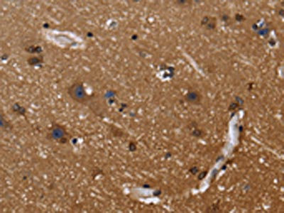
 IHC (Immunohiostchemistry)
(The image on the left is immunohistochemistry of paraffin-embedded Human brain tissue using AAA238842(EXTL3 Antibody) at dilution 1/30, on the right is treated with fusion protein. (Original magnification: ×200))
IHC (Immunohiostchemistry)
(The image on the left is immunohistochemistry of paraffin-embedded Human brain tissue using AAA238842(EXTL3 Antibody) at dilution 1/30, on the right is treated with fusion protein. (Original magnification: ×200))
EXTL3, Polyclonal Antibody (Cat# AAA238842)
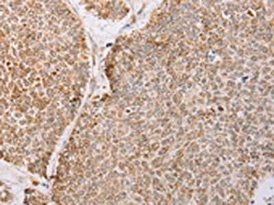
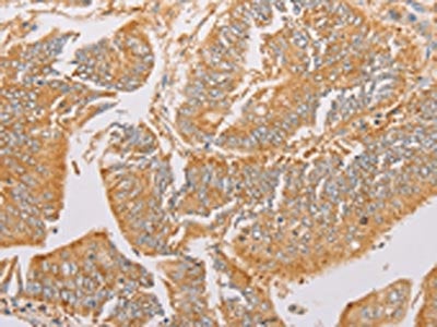
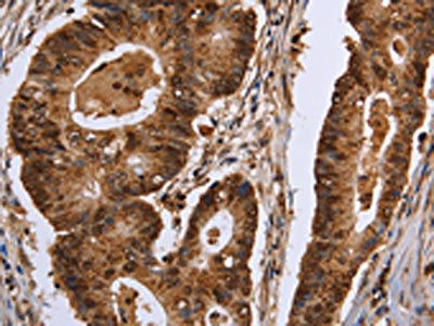
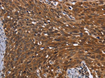
 IHC (Immunohiostchemistry)
(The image on the left is immunohistochemistry of paraffin-embedded Human colon cancer tissue using AAA238843(EXTL3 Antibody) at dilution 1/50, on the right is treated with fusion protein. (Original magnification: ×200))
IHC (Immunohiostchemistry)
(The image on the left is immunohistochemistry of paraffin-embedded Human colon cancer tissue using AAA238843(EXTL3 Antibody) at dilution 1/50, on the right is treated with fusion protein. (Original magnification: ×200))
EXTL3, Polyclonal Antibody (Cat# AAA238843)
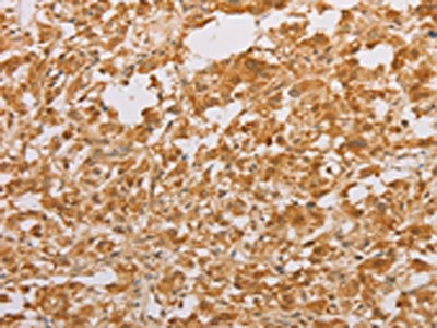
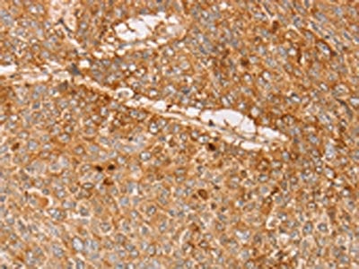
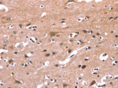
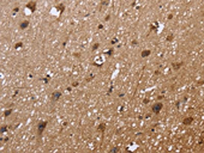
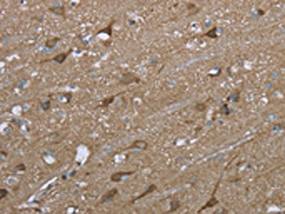
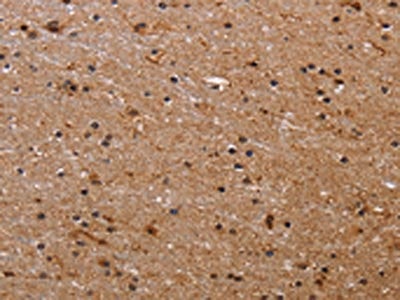
 IHC (Immunohiostchemistry)
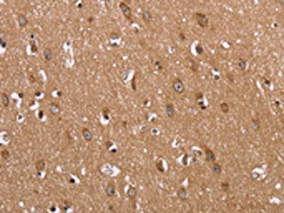
(The image on the left is immunohistochemistry of paraffin-embedded Human brain tissue using AAA238848(FAAH Antibody) at dilution 1/40, on the right is treated with fusion protein. (Original magnification: ×200))
IHC (Immunohiostchemistry)
(The image on the left is immunohistochemistry of paraffin-embedded Human brain tissue using AAA238848(FAAH Antibody) at dilution 1/40, on the right is treated with fusion protein. (Original magnification: ×200))
FAAH, Polyclonal Antibody (Cat# AAA238848)
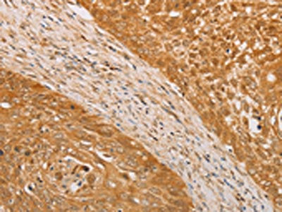
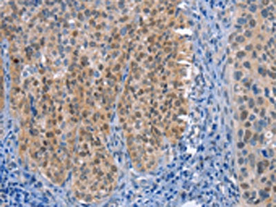
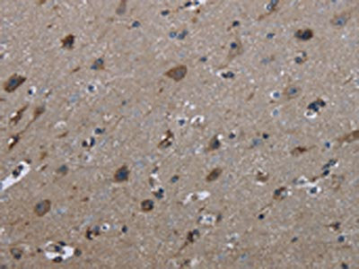
 IHC (Immunohiostchemistry)
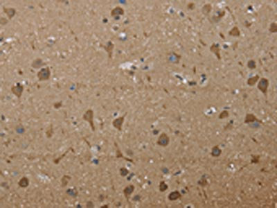
(The image on the left is immunohistochemistry of paraffin-embedded Human brain tissue using AAA238851(FABP6 Antibody) at dilution 1/30, on the right is treated with fusion protein. (Original magnification: ×200))
IHC (Immunohiostchemistry)
(The image on the left is immunohistochemistry of paraffin-embedded Human brain tissue using AAA238851(FABP6 Antibody) at dilution 1/30, on the right is treated with fusion protein. (Original magnification: ×200))
FABP6, Polyclonal Antibody (Cat# AAA238851)
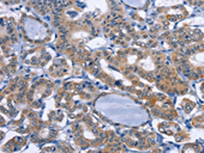
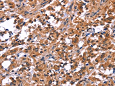
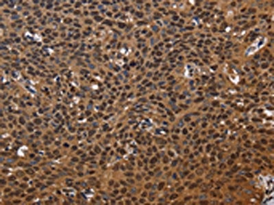
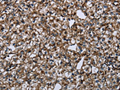
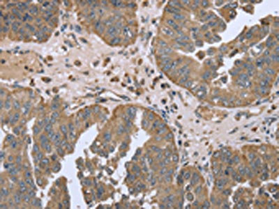
 IHC (Immunohiostchemistry)
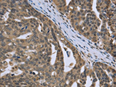
(The image on the left is immunohistochemistry of paraffin-embedded Human ovarian cancer tissue using AAA238857(PTK2B Antibody) at dilution 1/30, on the right is treated with fusion protein. (Original magnification: ×200))
IHC (Immunohiostchemistry)
(The image on the left is immunohistochemistry of paraffin-embedded Human ovarian cancer tissue using AAA238857(PTK2B Antibody) at dilution 1/30, on the right is treated with fusion protein. (Original magnification: ×200))
PTK2B, Polyclonal Antibody (Cat# AAA238857)
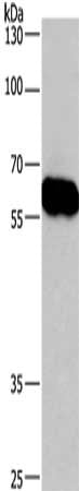
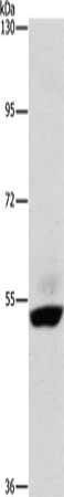
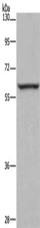
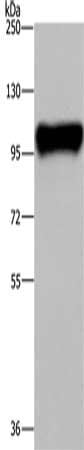 SDS-PAGE
(Gel: 6%SDS-PAGE, Lysate: 40 ug, Lane: Raji cells, Primary antibody: AAA238858(PTK2B Antibody) at dilution 1/300, Secondary antibody: Goat anti rabbit IgG at 1/8000 dilution, Exposure time: 1 minute)
SDS-PAGE
(Gel: 6%SDS-PAGE, Lysate: 40 ug, Lane: Raji cells, Primary antibody: AAA238858(PTK2B Antibody) at dilution 1/300, Secondary antibody: Goat anti rabbit IgG at 1/8000 dilution, Exposure time: 1 minute)
PTK2B, Polyclonal Antibody (Cat# AAA238858)
 SDS-PAGE
(Gel: 12%SDS-PAGE, Lysate: 40 ug, Lane: Mouse liver tissue, Primary antibody: AAA238862(FAM89B Antibody) at dilution 1/400, Secondary antibody: Goat anti rabbit IgG at 1/8000 dilution, Exposure time: 10 seconds)
SDS-PAGE
(Gel: 12%SDS-PAGE, Lysate: 40 ug, Lane: Mouse liver tissue, Primary antibody: AAA238862(FAM89B Antibody) at dilution 1/400, Secondary antibody: Goat anti rabbit IgG at 1/8000 dilution, Exposure time: 10 seconds)
FAM89B, Polyclonal Antibody (Cat# AAA238862)
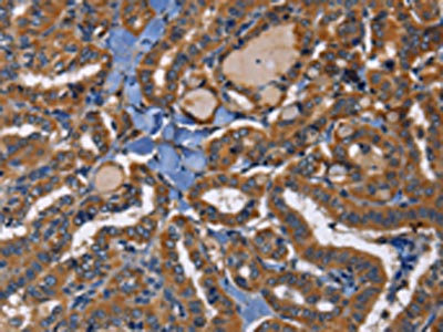
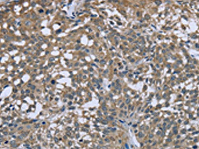
 IHC (Immunohiostchemistry)
(The image on the left is immunohistochemistry of paraffin-embedded Human colon cancer tissue using AAA238863(FANCF Antibody) at dilution 1/60, on the right is treated with fusion protein. (Original magnification: ×200))
IHC (Immunohiostchemistry)
(The image on the left is immunohistochemistry of paraffin-embedded Human colon cancer tissue using AAA238863(FANCF Antibody) at dilution 1/60, on the right is treated with fusion protein. (Original magnification: ×200))
FANCF, Polyclonal Antibody (Cat# AAA238863)
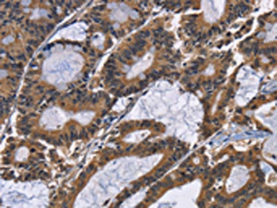
 IHC (Immunohiostchemistry)
(The image on the left is immunohistochemistry of paraffin-embedded Human ovarian cancer tissue using AAA238865(FANCG Antibody) at dilution 1/50, on the right is treated with fusion protein. (Original magnification: ×200))
IHC (Immunohiostchemistry)
(The image on the left is immunohistochemistry of paraffin-embedded Human ovarian cancer tissue using AAA238865(FANCG Antibody) at dilution 1/50, on the right is treated with fusion protein. (Original magnification: ×200))
FANCG, Polyclonal Antibody (Cat# AAA238865)

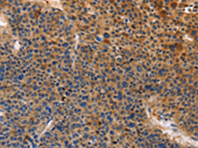
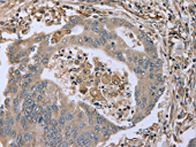 IHC (Immunohiostchemistry)
(The image on the left is immunohistochemistry of paraffin-embedded Human colon cancer tissue using AAA238867(FAP Antibody) at dilution 1/30, on the right is treated with fusion protein. (Original magnification: ×200))
IHC (Immunohiostchemistry)
(The image on the left is immunohistochemistry of paraffin-embedded Human colon cancer tissue using AAA238867(FAP Antibody) at dilution 1/30, on the right is treated with fusion protein. (Original magnification: ×200))
FAP, Polyclonal Antibody (Cat# AAA238867)
 SDS-PAGE
(Gel: 8%SDS-PAGE, Lysate: 40 ug, Lane 1-4: Mouse stomach tissue, human fetal liver tissue, MCF7 cells, mouse liver tissue tissue, Primary antibody: AAA238877(FBP1 Antibody) at dilution 1/850, Secondary antibody: Goat anti rabbit IgG at 1/8000 dilution, Exposure time: 10 seconds)
SDS-PAGE
(Gel: 8%SDS-PAGE, Lysate: 40 ug, Lane 1-4: Mouse stomach tissue, human fetal liver tissue, MCF7 cells, mouse liver tissue tissue, Primary antibody: AAA238877(FBP1 Antibody) at dilution 1/850, Secondary antibody: Goat anti rabbit IgG at 1/8000 dilution, Exposure time: 10 seconds)
FBP1, Polyclonal Antibody (Cat# AAA238877)
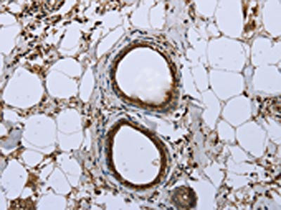
 IHC (Immunohiostchemistry)
(The image on the left is immunohistochemistry of paraffin-embedded Human brain tissue using AAA238880(FBP2 Antibody) at dilution 1/40, on the right is treated with fusion protein. (Original magnification: ×200))
IHC (Immunohiostchemistry)
(The image on the left is immunohistochemistry of paraffin-embedded Human brain tissue using AAA238880(FBP2 Antibody) at dilution 1/40, on the right is treated with fusion protein. (Original magnification: ×200))
FBP2, Polyclonal Antibody (Cat# AAA238880)
 SDS-PAGE
(Gel: 8%SDS-PAGE, Lysate: 40 ug, Lane: Human placenta tissue, Primary antibody: AAA238881(FCGR2B Antibody) at dilution 1/850, Secondary antibody: Goat anti rabbit IgG at 1/8000 dilution, Exposure time: 5 seconds)
SDS-PAGE
(Gel: 8%SDS-PAGE, Lysate: 40 ug, Lane: Human placenta tissue, Primary antibody: AAA238881(FCGR2B Antibody) at dilution 1/850, Secondary antibody: Goat anti rabbit IgG at 1/8000 dilution, Exposure time: 5 seconds)
FCGR2B, Polyclonal Antibody (Cat# AAA238881)
 SDS-PAGE
(Gel: 8%SDS-PAGE, Lysate: 40 ug, Lane: Mouse brain tissue, Primary antibody: AAA238882(FCAR Antibody) at dilution 1/700, Secondary antibody: Goat anti rabbit IgG at 1/8000 dilution, Exposure time: 10 seconds)
SDS-PAGE
(Gel: 8%SDS-PAGE, Lysate: 40 ug, Lane: Mouse brain tissue, Primary antibody: AAA238882(FCAR Antibody) at dilution 1/700, Secondary antibody: Goat anti rabbit IgG at 1/8000 dilution, Exposure time: 10 seconds)
FCAR, Polyclonal Antibody (Cat# AAA238882)
 SDS-PAGE
(Gel: 8%SDS-PAGE, Lysate: 40 ug, Lane 1-2: 293T cells, mouse brain tissue, Primary antibody: AAA238883(FCAR Antibody) at dilution 1/550, Secondary antibody: Goat anti rabbit IgG at 1/8000 dilution, Exposure time: 10 seconds)
SDS-PAGE
(Gel: 8%SDS-PAGE, Lysate: 40 ug, Lane 1-2: 293T cells, mouse brain tissue, Primary antibody: AAA238883(FCAR Antibody) at dilution 1/550, Secondary antibody: Goat anti rabbit IgG at 1/8000 dilution, Exposure time: 10 seconds)
FCAR, Polyclonal Antibody (Cat# AAA238883)
 SDS-PAGE
(Gel: 8%SDS-PAGE, Lysate: 40 ug, Lane 1-3: Mouse heart tissue, NIH/3T3 cells, mouse liver tissue, Primary antibody: AAA238890(FGFR1OP Antibody) at dilution 1/850, Secondary antibody: Goat anti rabbit IgG at 1/8000 dilution, Exposure time: 2 minutes)
SDS-PAGE
(Gel: 8%SDS-PAGE, Lysate: 40 ug, Lane 1-3: Mouse heart tissue, NIH/3T3 cells, mouse liver tissue, Primary antibody: AAA238890(FGFR1OP Antibody) at dilution 1/850, Secondary antibody: Goat anti rabbit IgG at 1/8000 dilution, Exposure time: 2 minutes)
FGFR1OP, Polyclonal Antibody (Cat# AAA238890)
 SDS-PAGE
(Gel: 8%SDS-PAGE, Lysate: 40 ug, Lane 1-2: Mouse kidney tissue, human kidney tissue, Primary antibody: AAA238896(FHIT Antibody) at dilution 1/600, Secondary antibody: Goat anti rabbit IgG at 1/8000 dilution, Exposure time: 20 seconds)
SDS-PAGE
(Gel: 8%SDS-PAGE, Lysate: 40 ug, Lane 1-2: Mouse kidney tissue, human kidney tissue, Primary antibody: AAA238896(FHIT Antibody) at dilution 1/600, Secondary antibody: Goat anti rabbit IgG at 1/8000 dilution, Exposure time: 20 seconds)
FHIT, Polyclonal Antibody (Cat# AAA238896)
 SDS-PAGE
(Gel: 8%SDS-PAGE, Lysate: 40 ug, Lane 1-3: Human adrenal pheochromocytoma tissue, Human fetal muscle tissue, Human skeletal muscle tissue, Primary antibody: AAA238898(FHL1 Antibody) at dilution 1/500, Secondary antibody: Goat anti rabbit IgG at 1/8000 dilution, Exposure time: 5 seconds)
SDS-PAGE
(Gel: 8%SDS-PAGE, Lysate: 40 ug, Lane 1-3: Human adrenal pheochromocytoma tissue, Human fetal muscle tissue, Human skeletal muscle tissue, Primary antibody: AAA238898(FHL1 Antibody) at dilution 1/500, Secondary antibody: Goat anti rabbit IgG at 1/8000 dilution, Exposure time: 5 seconds)
FHL1, Polyclonal Antibody (Cat# AAA238898)
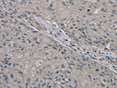
 IHC (Immunohiostchemistry)
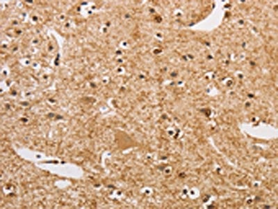
(The image on the left is immunohistochemistry of paraffin-embedded Human brain tissue using AAA238173(APPBP2 Antibody) at dilution 1/30, on the right is treated with fusion protein. (Original magnification: ×200))
IHC (Immunohiostchemistry)
(The image on the left is immunohistochemistry of paraffin-embedded Human brain tissue using AAA238173(APPBP2 Antibody) at dilution 1/30, on the right is treated with fusion protein. (Original magnification: ×200))
APPBP2, Polyclonal Antibody (Cat# AAA238173)
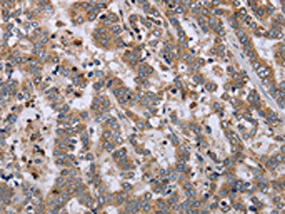
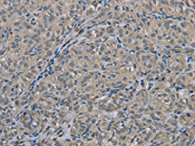
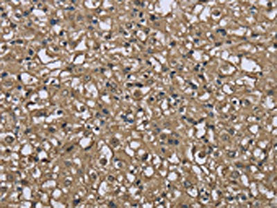
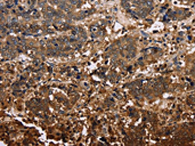
 IHC (Immunohiostchemistry)
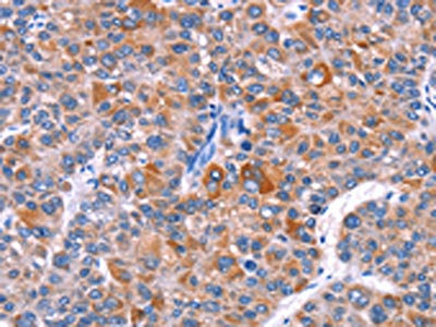
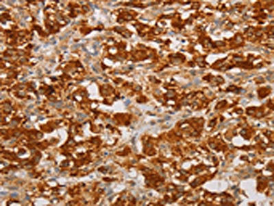
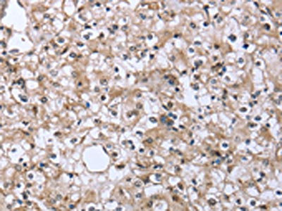
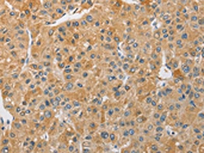
(The image on the left is immunohistochemistry of paraffin-embedded Human liver cancer tissue using AAA238176(APTX Antibody) at dilution 1/50, on the right is treated with fusion protein. (Original magnification: ×200))
IHC (Immunohiostchemistry)
(The image on the left is immunohistochemistry of paraffin-embedded Human liver cancer tissue using AAA238176(APTX Antibody) at dilution 1/50, on the right is treated with fusion protein. (Original magnification: ×200))
APTX, Polyclonal Antibody (Cat# AAA238176)
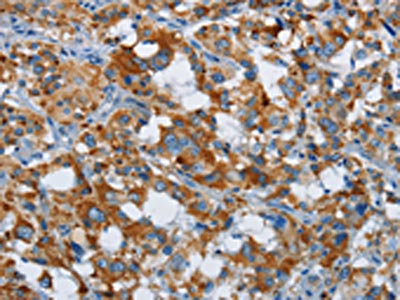
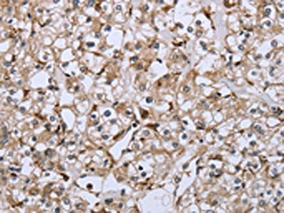
 IHC (Immunohiostchemistry)
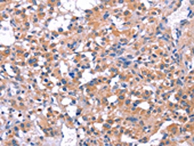
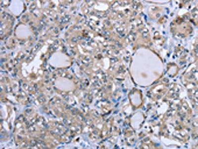
(The image on the left is immunohistochemistry of paraffin-embedded Human liver cancer tissue using AAA238192(ARHGAP15 Antibody) at dilution 1/30, on the right is treated with fusion protein. (Original magnification: ×200))
IHC (Immunohiostchemistry)
(The image on the left is immunohistochemistry of paraffin-embedded Human liver cancer tissue using AAA238192(ARHGAP15 Antibody) at dilution 1/30, on the right is treated with fusion protein. (Original magnification: ×200))
ARHGAP15, Polyclonal Antibody (Cat# AAA238192)
 SDS-PAGE
(Gel: 8%SDS-PAGE, Lysate: 40 ug, Lane 1-2: Hela cells, K562 cells, Primary antibody: AAA238196(ARHGEF5 Antibody) at dilution 1/1150, Secondary antibody: Goat anti rabbit IgG at 1/8000 dilution, Exposure time: 1 minute)
SDS-PAGE
(Gel: 8%SDS-PAGE, Lysate: 40 ug, Lane 1-2: Hela cells, K562 cells, Primary antibody: AAA238196(ARHGEF5 Antibody) at dilution 1/1150, Secondary antibody: Goat anti rabbit IgG at 1/8000 dilution, Exposure time: 1 minute)
ARHGEF5, Polyclonal Antibody (Cat# AAA238196)
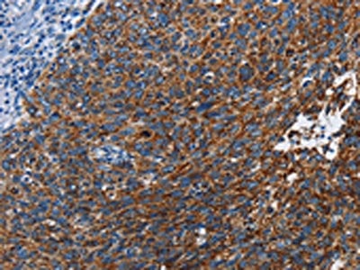
 IHC (Immunohiostchemistry)
(The image on the left is immunohistochemistry of paraffin-embedded Human thyroid cancer tissue using AAA238206(DIRAS3 Antibody) at dilution 1/25, on the right is treated with fusion protein. (Original magnification: ×200))
IHC (Immunohiostchemistry)
(The image on the left is immunohistochemistry of paraffin-embedded Human thyroid cancer tissue using AAA238206(DIRAS3 Antibody) at dilution 1/25, on the right is treated with fusion protein. (Original magnification: ×200))
DIRAS3, Polyclonal Antibody (Cat# AAA238206)
 SDS-PAGE
(Gel: 8%SDS-PAGE, Lysate: 40 ug, Lane 1-3: A172 cells, K562 cells, mouse pancreas tissue, Primary antibody: AAA238216(ARMCX3 Antibody) at dilution 1/900, Secondary antibody: Goat anti rabbit IgG at 1/8000 dilution, Exposure time: 30 seconds)
SDS-PAGE
(Gel: 8%SDS-PAGE, Lysate: 40 ug, Lane 1-3: A172 cells, K562 cells, mouse pancreas tissue, Primary antibody: AAA238216(ARMCX3 Antibody) at dilution 1/900, Secondary antibody: Goat anti rabbit IgG at 1/8000 dilution, Exposure time: 30 seconds)
ARMCX3, Polyclonal Antibody (Cat# AAA238216)
 IHC (Immunohiostchemistry)
(The image on the left is immunohistochemistry of paraffin-embedded Human gastric cancer tissue using AAA238217(ARL6IP1 Antibody) at dilution 1/20, on the right is treated with fusion protein. (Original magnification: ×200))
IHC (Immunohiostchemistry)
(The image on the left is immunohistochemistry of paraffin-embedded Human gastric cancer tissue using AAA238217(ARL6IP1 Antibody) at dilution 1/20, on the right is treated with fusion protein. (Original magnification: ×200))
ARL6IP1, Polyclonal Antibody (Cat# AAA238217)
What are Polyclonal Antibodies?
Polyclonal antibodies are antibodies that come from multiple B cell clones of a host animal. The typical hosts used for the majority of polyclonal antibody production are rabbits, goats, sheep, and donkeys. These polyclonal antibodies, once having identified their target, will bind to different epitopes located at different regions or sequences on the same protein/antigen. As a result, they are ideal at locating and binding to the target, even if the target is in very low concentrations (due to many different antibodies being able to bind to the same target molecule, which allows for significant amplification of a downstream signal).
Polyclonal antibodies are typically produced by injecting an antigen into a host animal, which causes the animal’s immune system to attack the foreign antigen by mass generating antibodies against it. After a period of time, serum is collected from the animal and purified using physicochemical fractionation, class-specific affinity purification, and/or antigen-affinity purification.
Key Uses of Polyclonal Antibodies
- Western Blotting: This method is used to find specific proteins in biological samples after separating them by size.
- Immunohistochemistry: IHC helps visualize the location of proteins in tissue sections using various staining techniques.
- ELISA: (Enzyme-Linked Immunosorbent Assay) is typically used to identify specific protein quantities in a sample. ELISAs can be either “Quantitative” or “Qualitative”.
- Flow Cytometry: technique that identifies and measures the specific protein on the surface or inside the cells in a fluid suspension.
- Immunoprecipitation: IP isolates and studies a specific protein from a complex mixture using antibodies.
Why Buy Polyclonal Antibodies from AAA Biotech?
1. Ideal for Various Applications
Our antibodies are generally going to be validated for use in multiple types of assays, including ELISA, Western Blotting, Immunohistochemistry, Immunoprecipitation, amongst others. They are ideal for a wide range of research applications.
2. Rigorous Quality Control
All of the antibodies in our catalog undergo strict quality testing to ensure specificity, sensitivity, and consistent performance. We are confident in the ability of our antibodies to provide you with accurate results.
3. Wide Assortment of Antibodies
Antibodies in are catalog can be found for both common and exotic species, and these antibodies are also available in both conjugated and recombinant forms to suit many diverse experimental needs.
4. Highly Purified
Our antibodies are available in purified forms with over 85% purity, as confirmed by SDS-PAGE. They are also available with tags such as His, Flag, GST, or MBP. We cater to customers worldwide.
FAQ
1. How are polyclonal antibodies produced?
Traditionally, polyclonal antibodies are produced by injecting an antigen into a host animal (such as a rabbit or goat), which then triggers an immune response from the host animal. The animal’s B cells produce antibodies that will recognize different parts of the injected antigen. These antibodies are then collected from the animal’s blood and purified for use.
2. How do polyclonal antibodies differ from monoclonal antibodies?
Polyclonal antibodies are a mix of antibodies that bind to different locations (epitopes) of the same antigen, while monoclonal antibodies are identical and bind to just one specific epitope. This makes polyclonal antibodies more versatile and better at detecting proteins that may be present in low quantities or in altered/modified forms.
3. How should I store polyclonal antibodies?
Polyclonal antibodies should be stored at 4°C for short-term use (up to a few weeks) and at -20°C or -80°C for long-term storage. Avoid repeated freeze-thaw cycles by dividing them into small aliquots. Always check the datasheet for specific storage instructions.