Filters
▼Clonality
▼Type
▼Reactivity
▼Gene Name
▼Isotype
▼Host
▼Application
▼Clone
▼Polyclonal Antibodies
At AAA Biotech also known as AAA Bio or AAABio, we provide a broad range of purified polyclonal antibodies (pAbs) that are able to all be browsed online through our website. Due to their high specificity and strong binding affinity, these antibodies are ideal for wide swathes of research and experimental applications.
Our polyclonal antibodies can easily support your work, whether you use them for Western Blotting, Immunocytochemistry (with or without Immunofluorescence used in conjunction), Immunohistochemistry, Immunoprecipitation, and ELISA tests. We highly encourage you to browse our range of pAbs and choose the one that best suits your experimental model.
Viewing 6150-6200 of 96812 product results
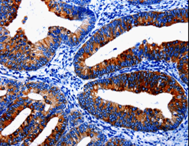
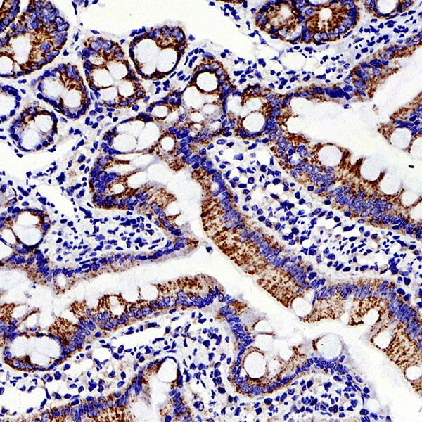
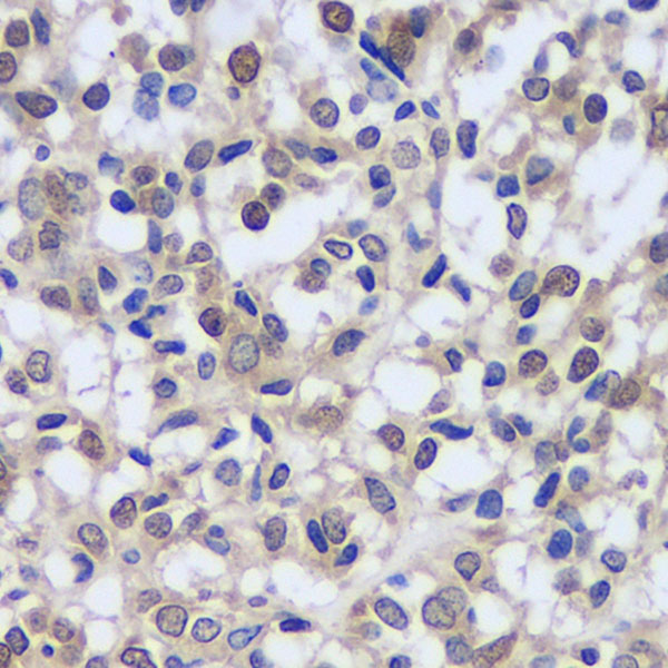
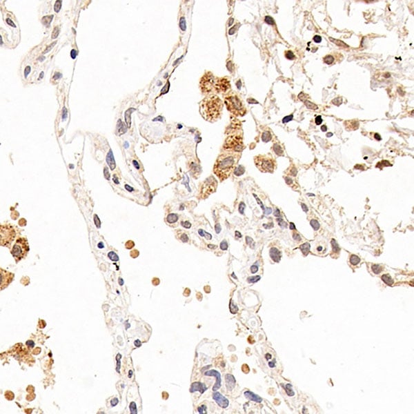
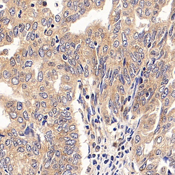
 IHC (Immunohiostchemistry)
(Immunohistochemistry of paraffin-embedded Human gastric cancer tissue using CLDN2 Polyclonal Antibody at dilution 1:50)
IHC (Immunohiostchemistry)
(Immunohistochemistry of paraffin-embedded Human gastric cancer tissue using CLDN2 Polyclonal Antibody at dilution 1:50)
CLDN2, Polyclonal Antibody (Cat# AAA170662)
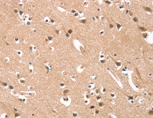 IHC (Immunohiostchemistry)
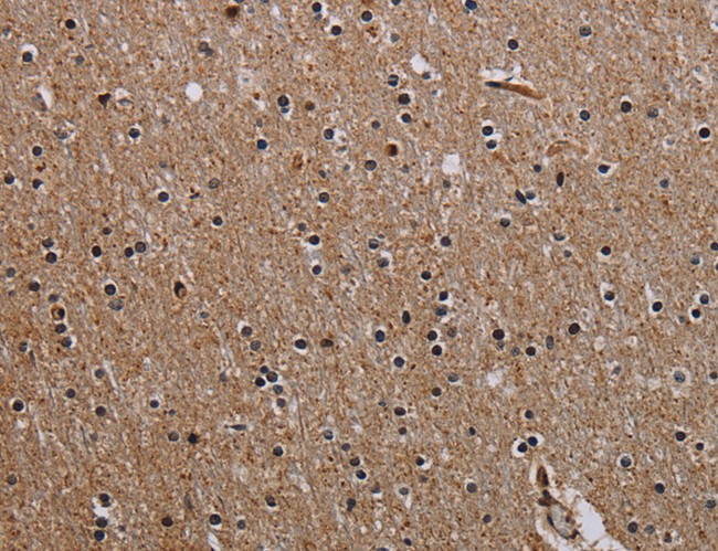
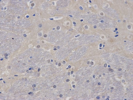
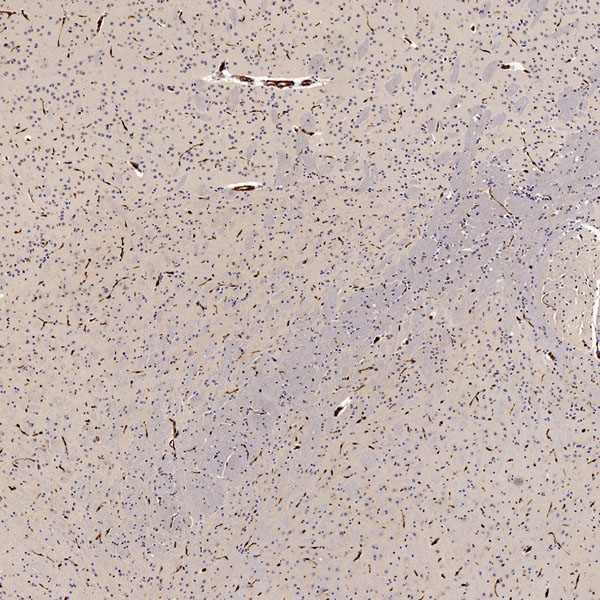
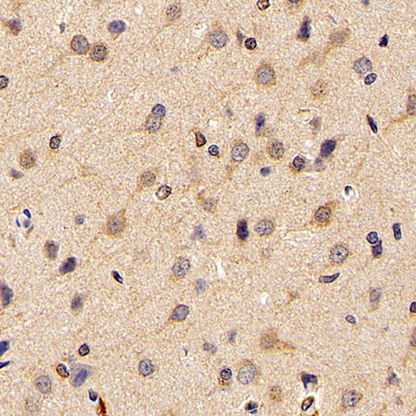
(Immunohistochemistry of paraffin-embedded Human brain tissue using CYP46A1 Polyclonal Antibody at dilution 1:15)
IHC (Immunohiostchemistry)
(Immunohistochemistry of paraffin-embedded Human brain tissue using CYP46A1 Polyclonal Antibody at dilution 1:15)
CYP46A1, Polyclonal Antibody (Cat# AAA170664)
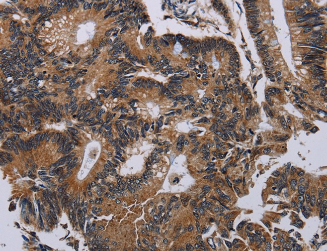
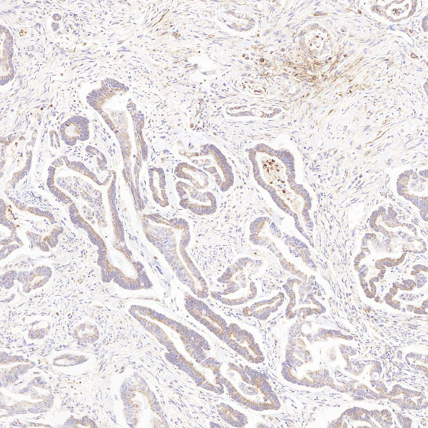
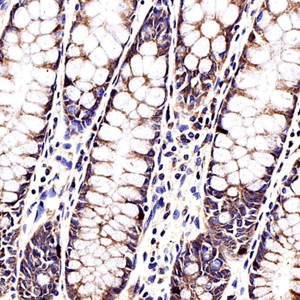
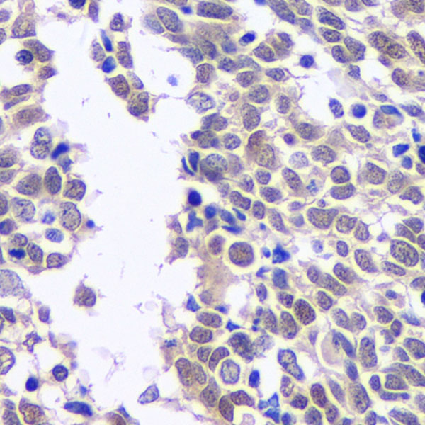
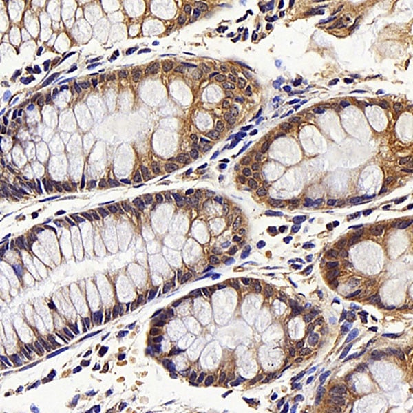
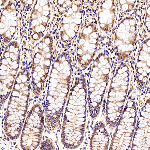
 IHC (Immunohiostchemistry)
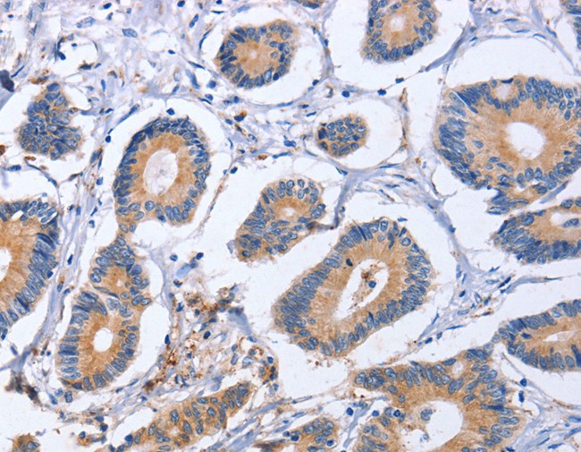
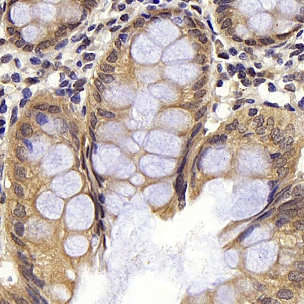
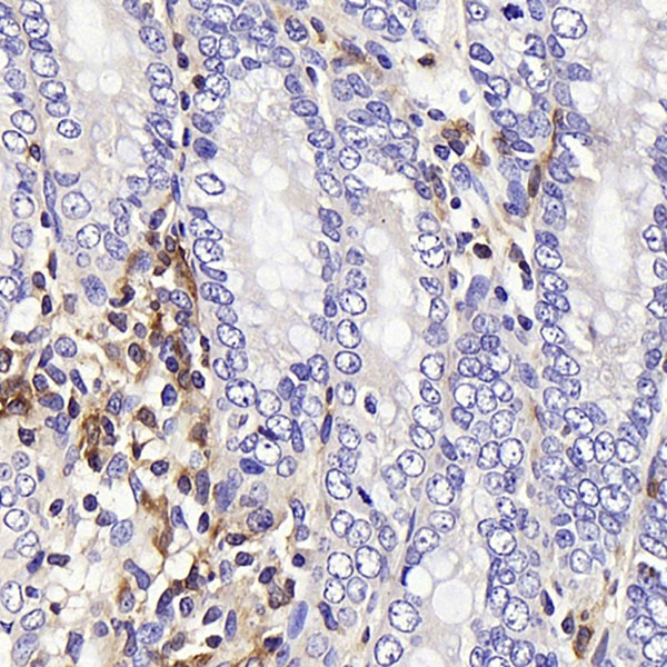
(Immunohistochemistry of paraffin-embedded Human colon cancer tissue using ITGB5 Polyclonal Antibody at dilution 1:40)
IHC (Immunohiostchemistry)
(Immunohistochemistry of paraffin-embedded Human colon cancer tissue using ITGB5 Polyclonal Antibody at dilution 1:40)
ITGB5, Polyclonal Antibody (Cat# AAA170669)
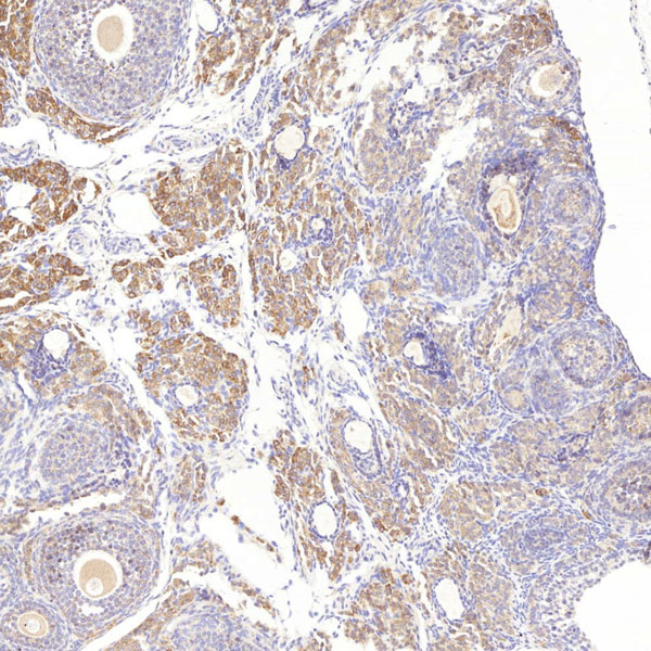
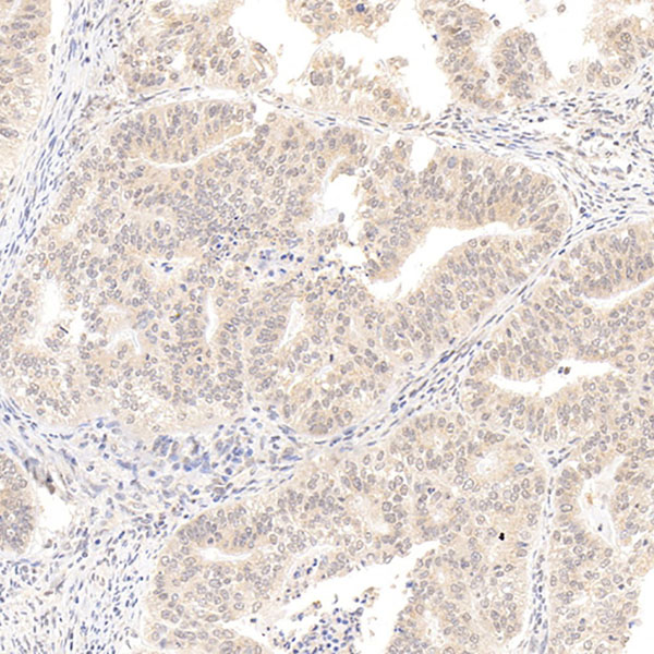
 IHC (Immunohiostchemistry)
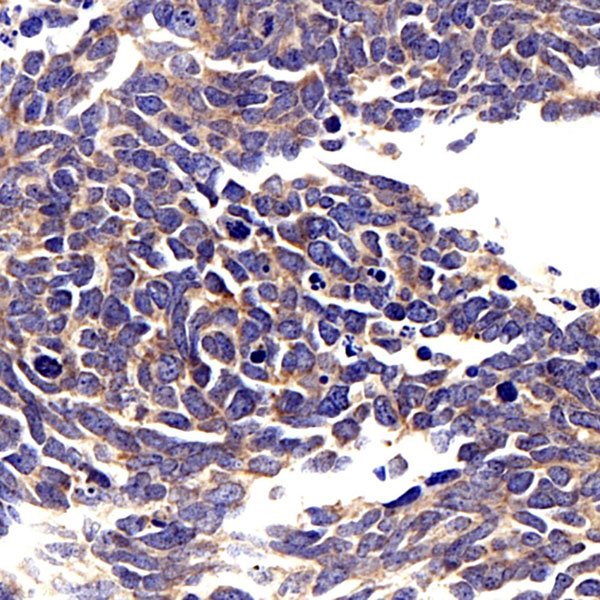
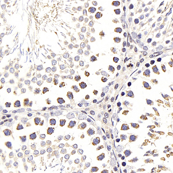
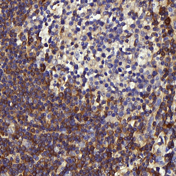
(Immunohistochemistry of paraffin-embedded Human thyroid cancer tissue using GFRA4 Polyclonal Antibody at dilution 1:40)
IHC (Immunohiostchemistry)
(Immunohistochemistry of paraffin-embedded Human thyroid cancer tissue using GFRA4 Polyclonal Antibody at dilution 1:40)
GFRA4, Polyclonal Antibody (Cat# AAA170670)
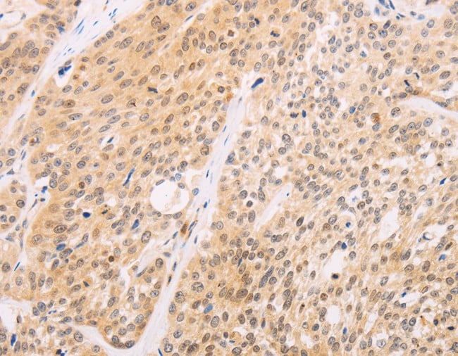
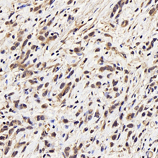
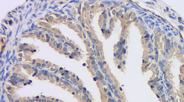
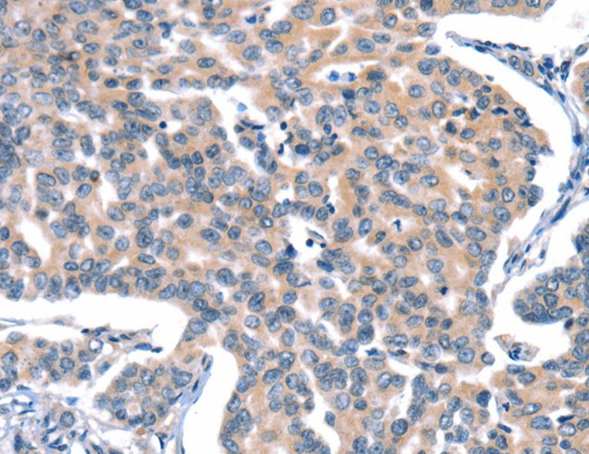 IHC (Immunohiostchemistry)
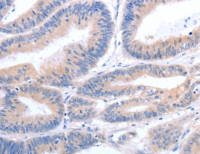
(Immunohistochemistry of paraffin-embedded Human gastric cancer tissue using CACNA1A Polyclonal Antibody at dilution 1:15)
IHC (Immunohiostchemistry)
(Immunohistochemistry of paraffin-embedded Human gastric cancer tissue using CACNA1A Polyclonal Antibody at dilution 1:15)
CACNA1A, Polyclonal Antibody (Cat# AAA170673)
 IHC (Immunohistochemisry)
(Immunohistochemistry of paraffin-embedded Human brain using PCDHAC1 Polyclonal Antibody at dilution of 1:30)
IHC (Immunohistochemisry)
(Immunohistochemistry of paraffin-embedded Human brain using PCDHAC1 Polyclonal Antibody at dilution of 1:30)
PCDHAC1, Polyclonal Antibody (Cat# AAA170674)
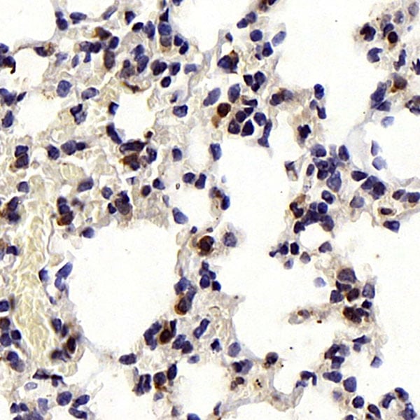
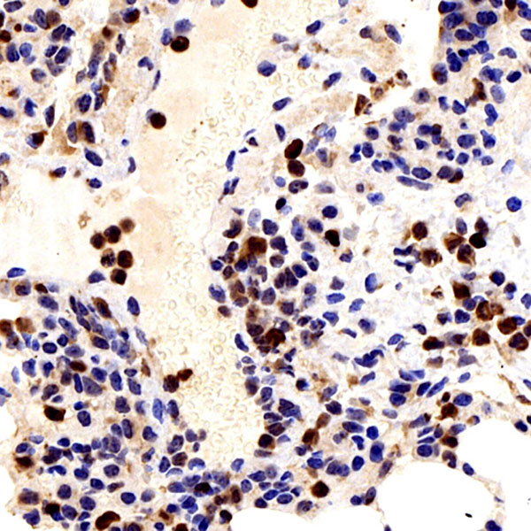
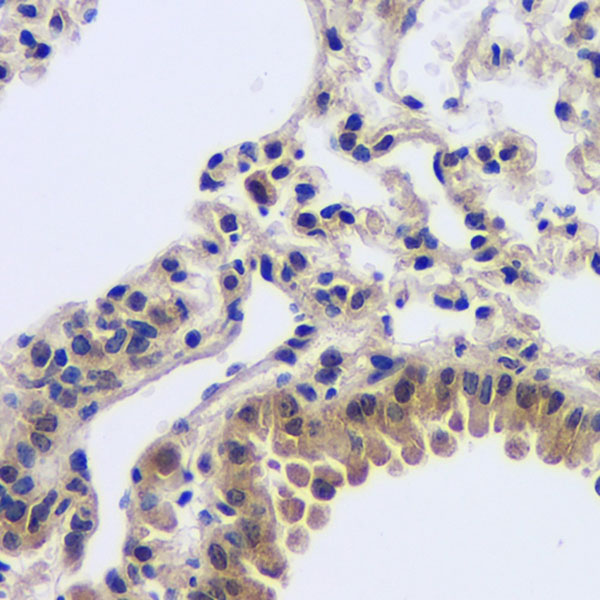
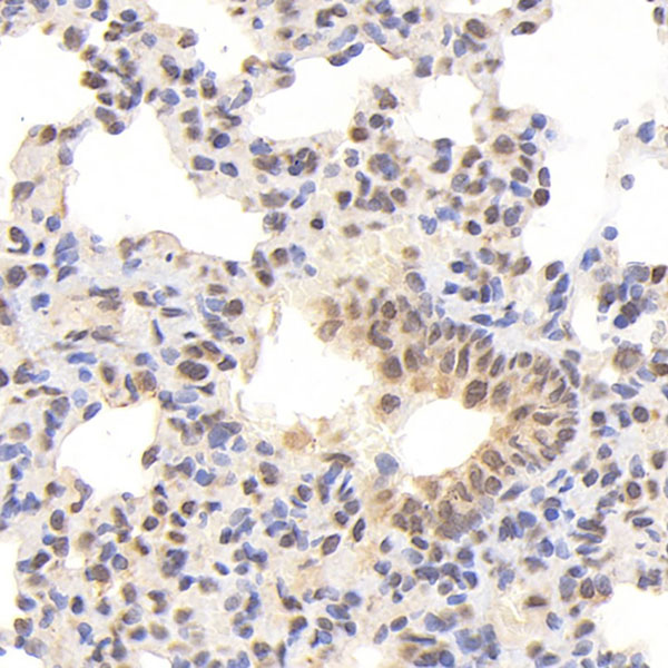
 IHC (Immunohiostchemistry)
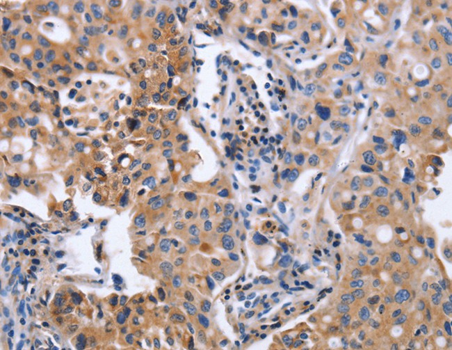
(Immunohistochemistry of paraffin-embedded human lung cancer using ANGPT2 antibody at dilution of 1:200 (x400 lens))
IHC (Immunohiostchemistry)
(Immunohistochemistry of paraffin-embedded human lung cancer using ANGPT2 antibody at dilution of 1:200 (x400 lens))
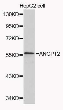
ANGPT2, Polyclonal Antibody (Cat# AAA170677)

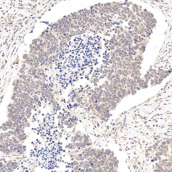
 IHC (Immunohiostchemistry)
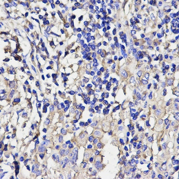
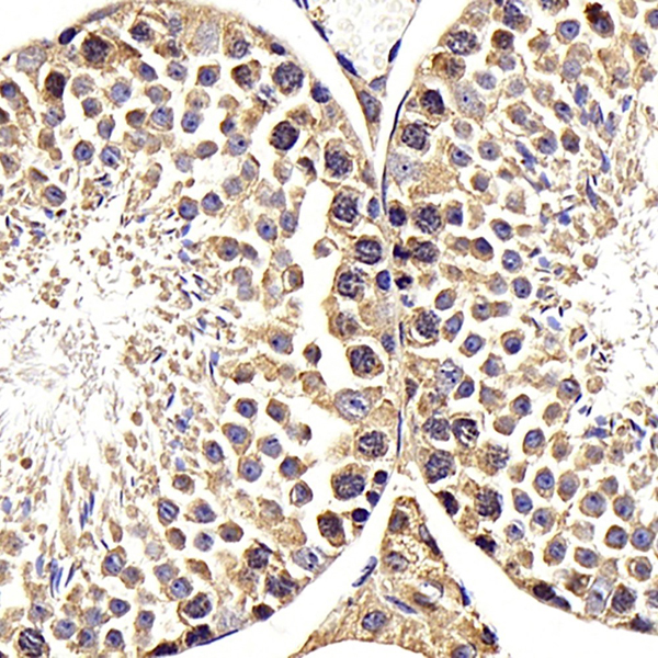
(Immunohistochemistry of paraffin-embedded Human thyroid cancer tissue using STRA8 Polyclonal Antibody at dilution 1:25)
IHC (Immunohiostchemistry)
(Immunohistochemistry of paraffin-embedded Human thyroid cancer tissue using STRA8 Polyclonal Antibody at dilution 1:25)
STRA8, Polyclonal Antibody (Cat# AAA170681)
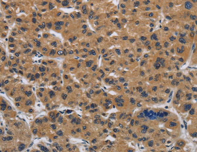
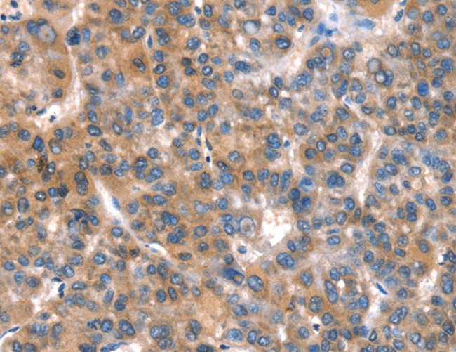
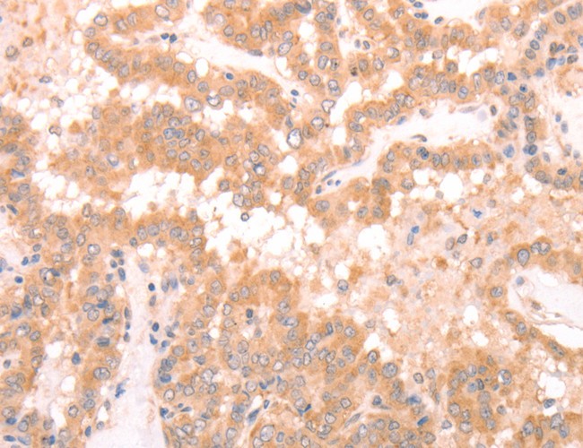
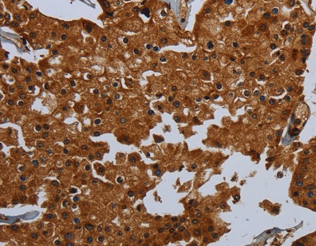
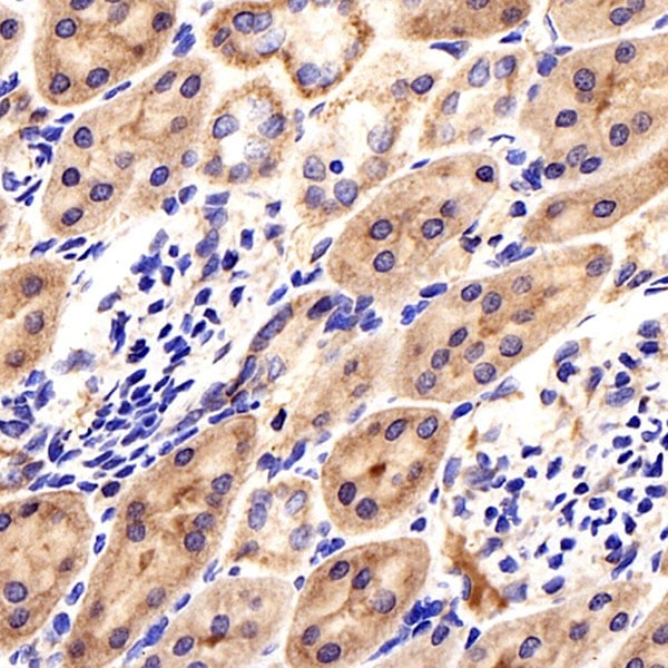
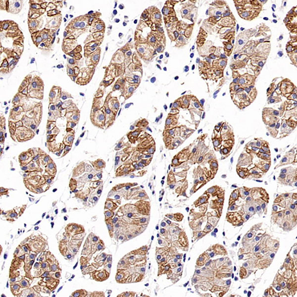
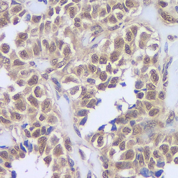
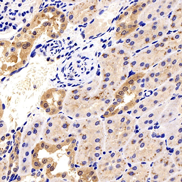
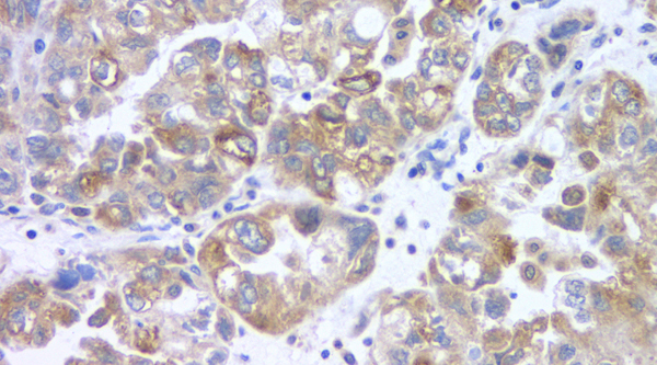
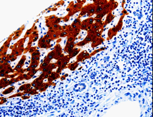 IHC (Immunohiostchemistry)
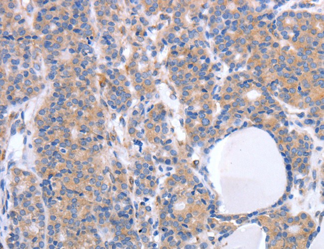
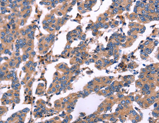
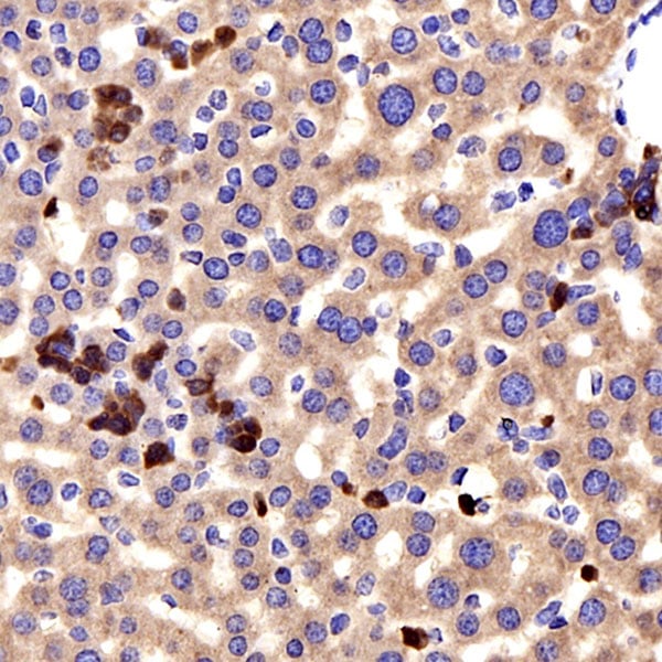
(Immunohistochemistry of paraffin-embedded Human liver cancer tissue using EPHX1 Polyclonal Antibody at dilution 1:20)
IHC (Immunohiostchemistry)
(Immunohistochemistry of paraffin-embedded Human liver cancer tissue using EPHX1 Polyclonal Antibody at dilution 1:20)
EPHX1, Polyclonal Antibody (Cat# AAA170714)
 IHC (Immunohiostchemistry)
(Immunohistochemistry of paraffin-embedded Human thyroid cancer tissue using DLL4 Polyclonal Antibody at dilution 1:50)
IHC (Immunohiostchemistry)
(Immunohistochemistry of paraffin-embedded Human thyroid cancer tissue using DLL4 Polyclonal Antibody at dilution 1:50)
DLL4, Polyclonal Antibody (Cat# AAA170717)
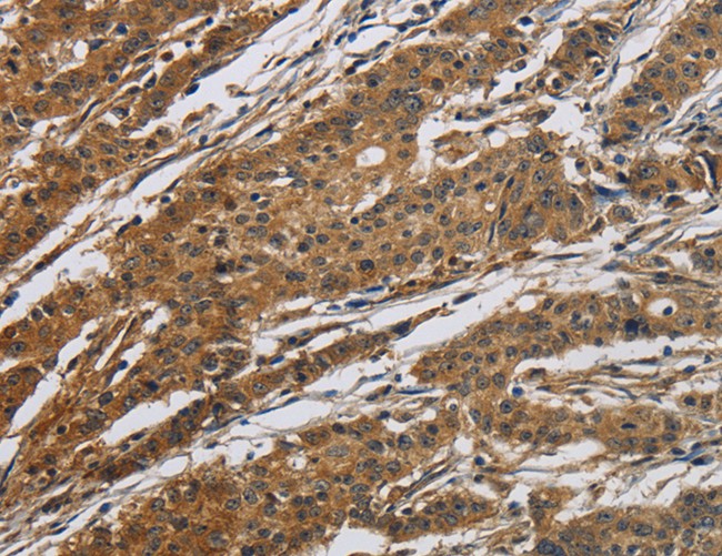
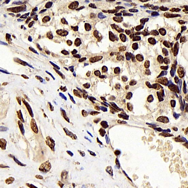
 IHC (Immunohistochemisry)
(Immunohistochemistry of paraffin-embedded Human esophagus cancer using SRC Polyclonal Antibody at dilution of 1:10)
IHC (Immunohistochemisry)
(Immunohistochemistry of paraffin-embedded Human esophagus cancer using SRC Polyclonal Antibody at dilution of 1:10)
SRC, Polyclonal Antibody (Cat# AAA170719)
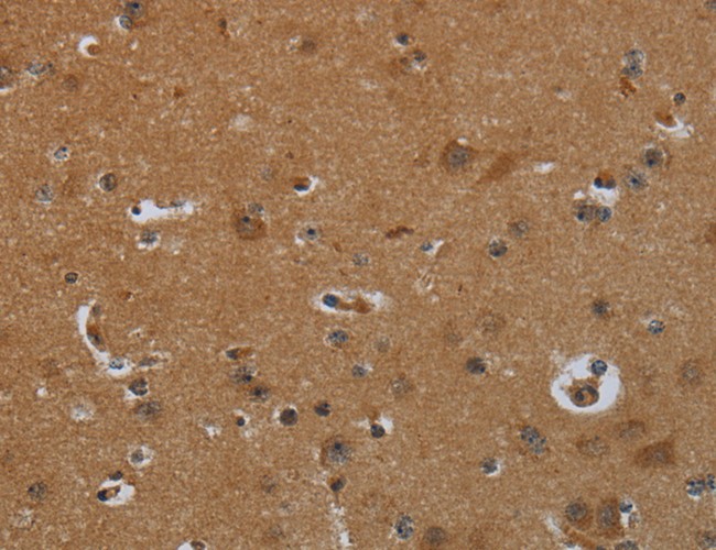 IHC (Immunohiostchemistry)
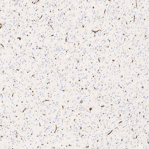
(Immunohistochemistry of paraffin-embedded Human brain tissue using ENTPD7 Polyclonal Antibody at dilution 1:60)
IHC (Immunohiostchemistry)
(Immunohistochemistry of paraffin-embedded Human brain tissue using ENTPD7 Polyclonal Antibody at dilution 1:60)
ENTPD7, Polyclonal Antibody (Cat# AAA170727)
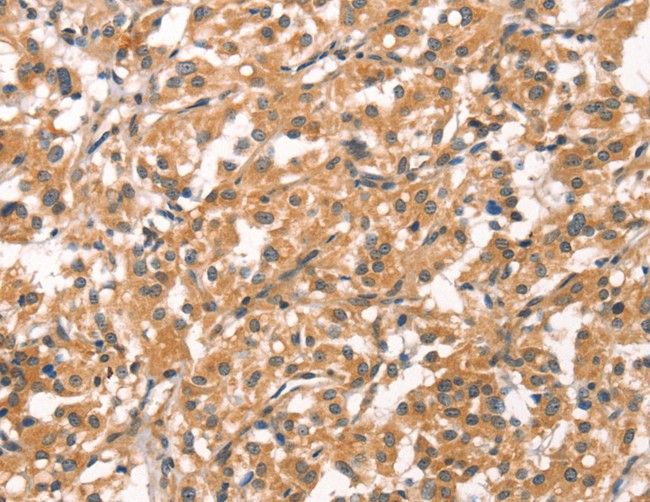
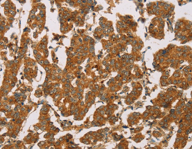
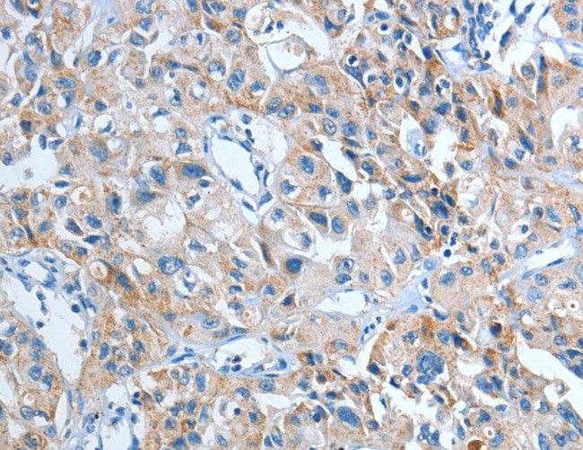
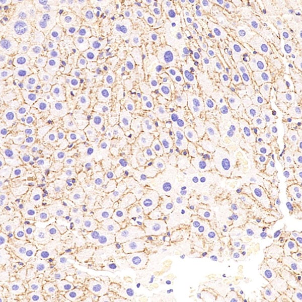
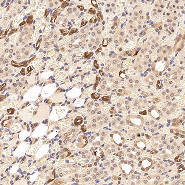
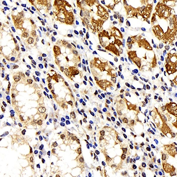
 IHC (Immunohiostchemistry)
(Immunohistochemistry of paraffin-embedded Human liver cancer using CYP1A1 Polyclonal Antibody at dilution of 1:25)
IHC (Immunohiostchemistry)
(Immunohistochemistry of paraffin-embedded Human liver cancer using CYP1A1 Polyclonal Antibody at dilution of 1:25)
CYP1A1, Polyclonal Antibody (Cat# AAA170729)
 IHC (Immunohistochemisry)
(Immunohistochemistry of paraffin-embedded Human breast cancer using RDX Polyclonal Antibody at dilution of 1:60)
IHC (Immunohistochemisry)
(Immunohistochemistry of paraffin-embedded Human breast cancer using RDX Polyclonal Antibody at dilution of 1:60)
RDX, Polyclonal Antibody (Cat# AAA170732)

 IHC (Immunohiostchemistry)
(Immunohistochemistry of paraffin-embedded Human lung cancer tissue using IL21R Polyclonal Antibody at dilution 1:40)
IHC (Immunohiostchemistry)
(Immunohistochemistry of paraffin-embedded Human lung cancer tissue using IL21R Polyclonal Antibody at dilution 1:40)
IL21R, Polyclonal Antibody (Cat# AAA170738)

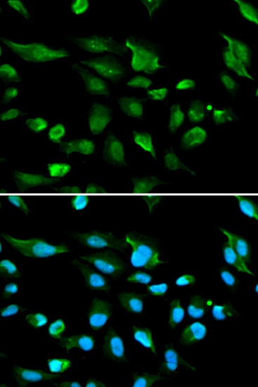 IF (Immunofluorescence)
(Immunofluorescence analysis of A549 cell using AIRE antibody. Blue: DAPI for nuclear staining.)
IF (Immunofluorescence)
(Immunofluorescence analysis of A549 cell using AIRE antibody. Blue: DAPI for nuclear staining.)
AIRE, Polyclonal Antibody (Cat# AAA170743)
 IHC (Immunohistochemisry)
(Immunohistochemistry analysis of paraffin-embedded mouse uterus using MMP2 Polyclonal Antibody at dilution of 1:500.)
IHC (Immunohistochemisry)
(Immunohistochemistry analysis of paraffin-embedded mouse uterus using MMP2 Polyclonal Antibody at dilution of 1:500.)
MMP2, Polyclonal Antibody (Cat# AAA174383)
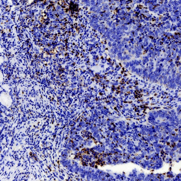
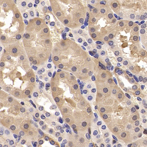
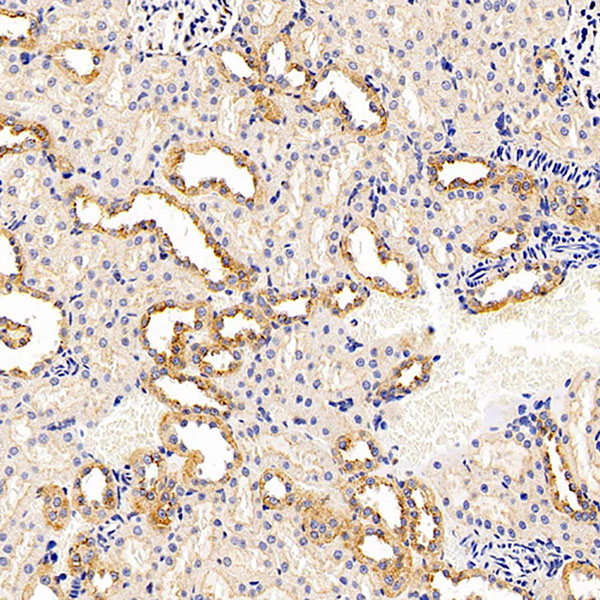
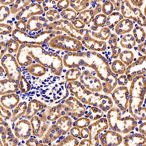
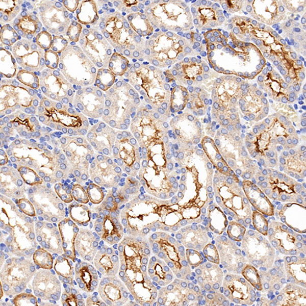
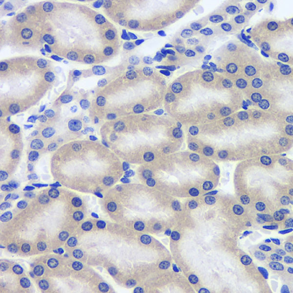
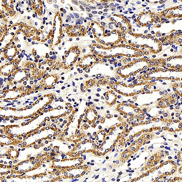
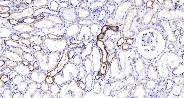 IHC (Immunohiostchemistry)
(Immunohistochemistry analysis of paraffin-embedded human kidney using MMP9 Polyclonal Antibody at dilution of 1:200.)
IHC (Immunohiostchemistry)
(Immunohistochemistry analysis of paraffin-embedded human kidney using MMP9 Polyclonal Antibody at dilution of 1:200.)
MMP9, Polyclonal Antibody (Cat# AAA174385)
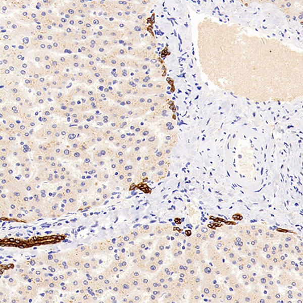
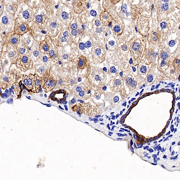
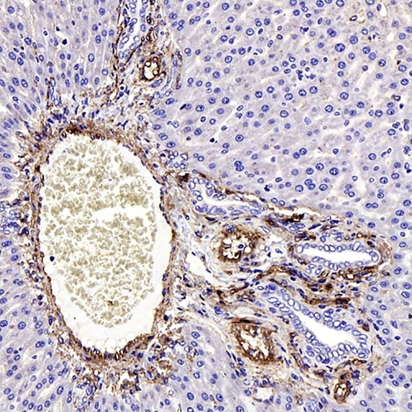
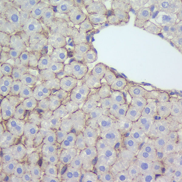 IHC (Immunohistochemistry)
(Immunohistochemistry analysis of paraffin-embedded rat liver using N-cadherin Polyclonal Antibody at dilution of 1:500.)
IHC (Immunohistochemistry)
(Immunohistochemistry analysis of paraffin-embedded rat liver using N-cadherin Polyclonal Antibody at dilution of 1:500.)
N-cadherin, Polyclonal Antibody (Cat# AAA174386)
 IHC (Immunohiostchemistry)
(Immunohistochemistry analysis of paraffin-embedded Rat tooth using TNFRSF11B Polyclonal Antibody at dilution of 1:200.)
IHC (Immunohiostchemistry)
(Immunohistochemistry analysis of paraffin-embedded Rat tooth using TNFRSF11B Polyclonal Antibody at dilution of 1:200.)
TNFRSF11B, Polyclonal Antibody (Cat# AAA174388)
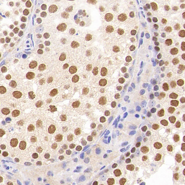
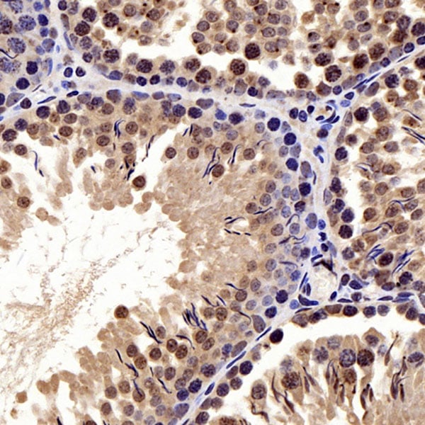
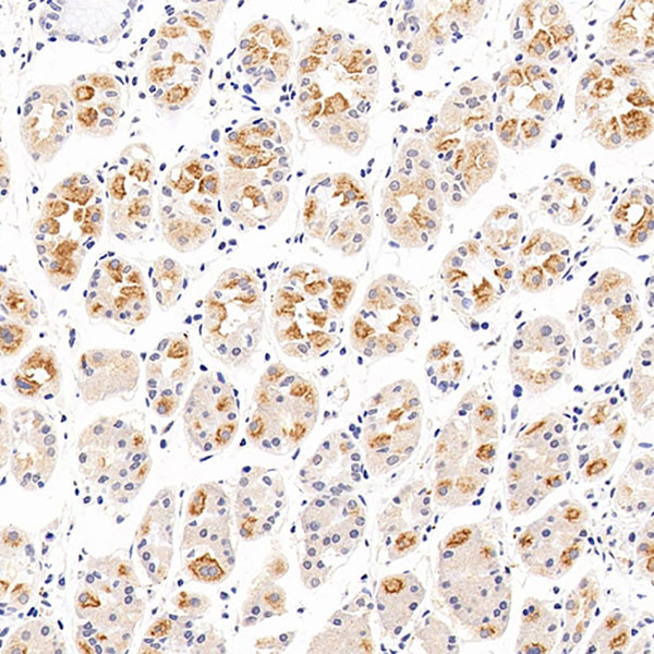
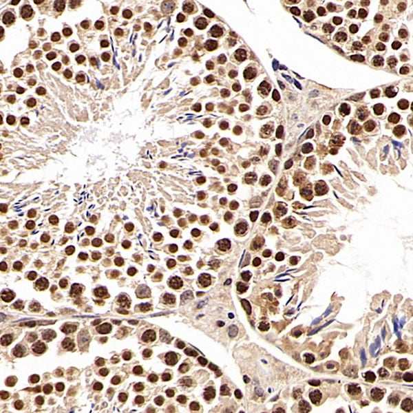
 IHC (Immunohistochemisry)
(Immunohistochemistry analysis of paraffin-embedded Rat testis using SIRT1 Polyclonal Antibody at dilution of 1:300.)
IHC (Immunohistochemisry)
(Immunohistochemistry analysis of paraffin-embedded Rat testis using SIRT1 Polyclonal Antibody at dilution of 1:300.)
SIRT1, Polyclonal Antibody (Cat# AAA174392)
 IHC (Immunohistochemistry)
(Immunohistochemistry analysis of paraffin-embedded rat kidney using CK-19 Polyclonal Antibody at dilution of 1:300.)
IHC (Immunohistochemistry)
(Immunohistochemistry analysis of paraffin-embedded rat kidney using CK-19 Polyclonal Antibody at dilution of 1:300.)
CK-19, Polyclonal Antibody (Cat# AAA174400)
 IHC (Immunohistochemistry)
(Immunohistochemistry analysis of paraffin-embedded rat spleen using BAD Polyclonal Antibody at dilution of 1:300.)
IHC (Immunohistochemistry)
(Immunohistochemistry analysis of paraffin-embedded rat spleen using BAD Polyclonal Antibody at dilution of 1:300.)
BAD, Polyclonal Antibody (Cat# AAA174401)
 IHC (Immunohistochemistry)
(Immunohistochemistry analysis of paraffin-embedded mouse kidney using CCL2 Polyclonal Antibody at dilution of 1:300.)
IHC (Immunohistochemistry)
(Immunohistochemistry analysis of paraffin-embedded mouse kidney using CCL2 Polyclonal Antibody at dilution of 1:300.)
CCL2, Polyclonal Antibody (Cat# AAA174402)

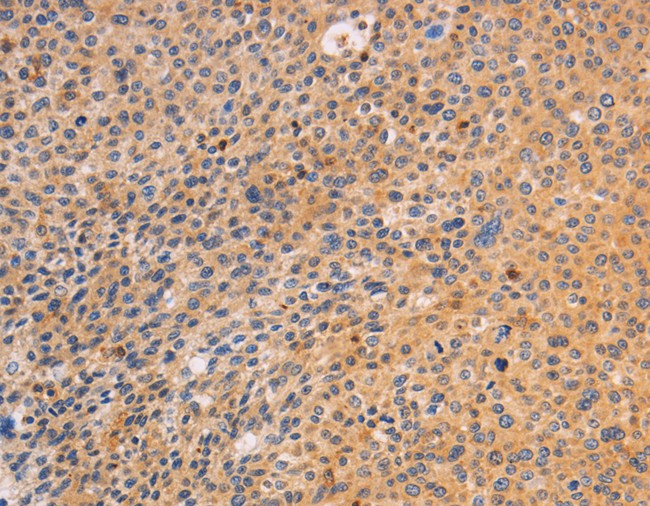
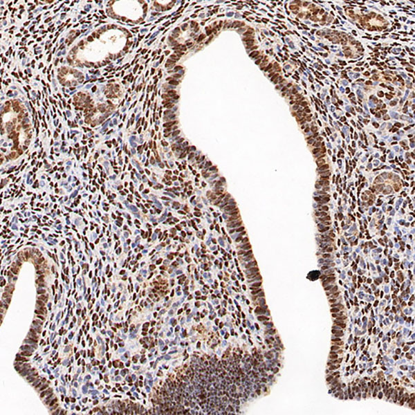
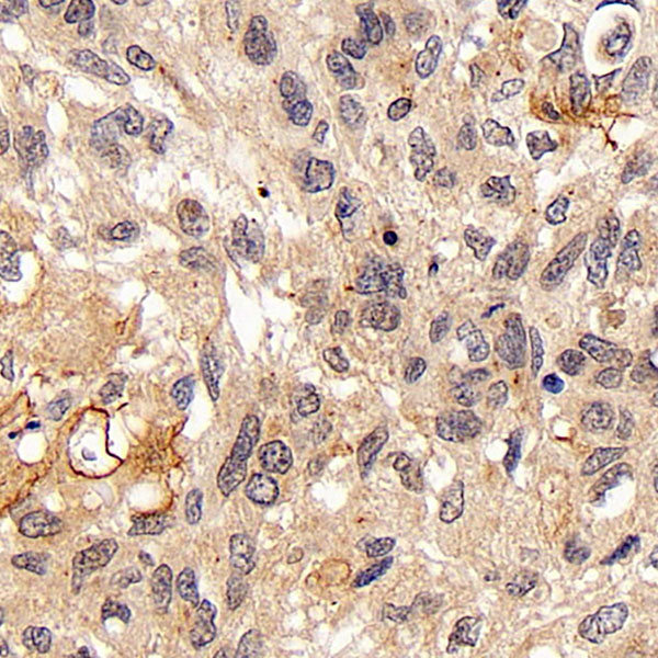
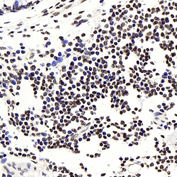
 IHC (Immunohistochemisry)
(Immunohistochemistry analysis of paraffin-embedded human colon cancer using IkB alpha Polyclonal Antibody at dilution of 1:500.)
IHC (Immunohistochemisry)
(Immunohistochemistry analysis of paraffin-embedded human colon cancer using IkB alpha Polyclonal Antibody at dilution of 1:500.)
IkB alpha, Polyclonal Antibody (Cat# AAA174404)
 IHC (Immunohistochemisry)
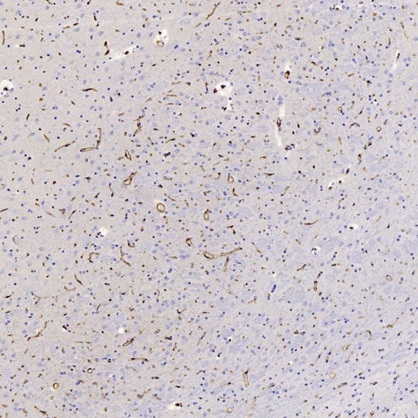
(Immunohistochemistry analysis of paraffin-embedded Rat brain using GLUT-1 Polyclonal Antibody at dilution of 1:300.)
IHC (Immunohistochemisry)
(Immunohistochemistry analysis of paraffin-embedded Rat brain using GLUT-1 Polyclonal Antibody at dilution of 1:300.)
GLUT-1, Polyclonal Antibody (Cat# AAA174405)
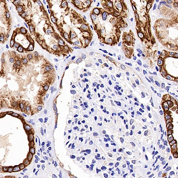
 IHC (Immunohiostchemistry)
(Immunohistochemistry analysis of paraffin-embedded rat kidney using CK-7 Polyclonal Antibody at dilution of 1:200.)
IHC (Immunohiostchemistry)
(Immunohistochemistry analysis of paraffin-embedded rat kidney using CK-7 Polyclonal Antibody at dilution of 1:200.)
CK-7, Polyclonal Antibody (Cat# AAA174407)
 IHC (Immunohistochemistry)
(Immunohistochemistry analysis of paraffin-embedded Rat lung using CK-18 Polyclonal Antibody at dilution of 1:500.)
IHC (Immunohistochemistry)
(Immunohistochemistry analysis of paraffin-embedded Rat lung using CK-18 Polyclonal Antibody at dilution of 1:500.)
CK-18, Polyclonal Antibody (Cat# AAA174409)


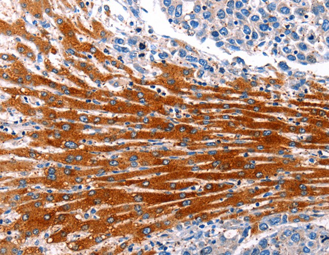
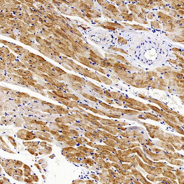
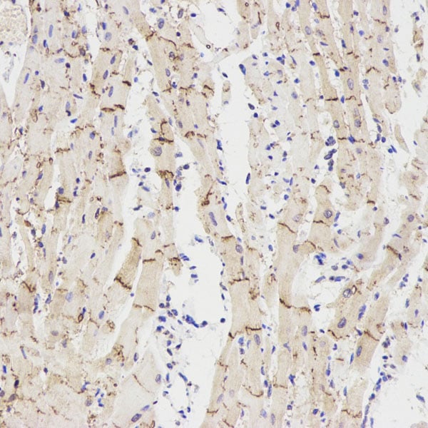 IHC (Immunohistochemisry)
(Immunohistochemistry analysis of paraffin-embedded rat heart using Connexin 43 Polyclonal Antibody at dilution of 1:300.)
IHC (Immunohistochemisry)
(Immunohistochemistry analysis of paraffin-embedded rat heart using Connexin 43 Polyclonal Antibody at dilution of 1:300.)
Connexin 43, Polyclonal Antibody (Cat# AAA174411)
 IHC (Immunohistochemistry)
(Immunohistochemistry analysis of paraffin-embedded human stomach cancer using CREB1 Polyclonal Antibody at dilution of 1:200.)
IHC (Immunohistochemistry)
(Immunohistochemistry analysis of paraffin-embedded human stomach cancer using CREB1 Polyclonal Antibody at dilution of 1:200.)
CREB1, Polyclonal Antibody (Cat# AAA174412)
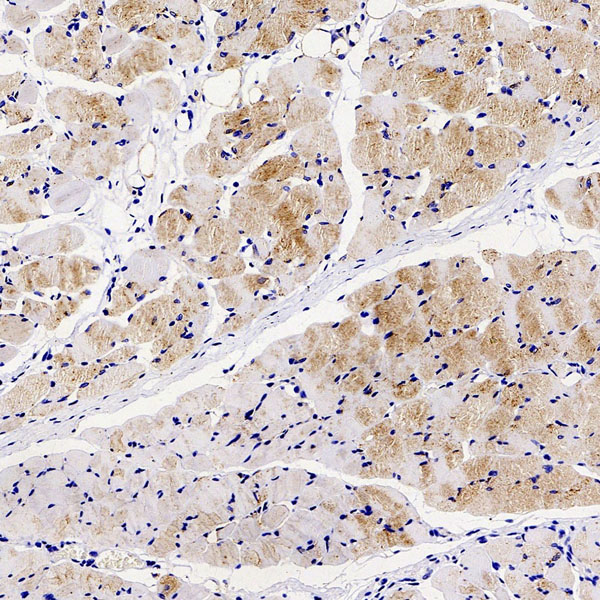
 IHC (Immunohistochemistry)
(Immunohistochemistry analysis of paraffin-embedded rat testis using CDK4 Polyclonal Antibody at dilution of 1:100.)
IHC (Immunohistochemistry)
(Immunohistochemistry analysis of paraffin-embedded rat testis using CDK4 Polyclonal Antibody at dilution of 1:100.)
CDK4, Polyclonal Antibody (Cat# AAA174413)
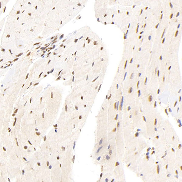
 IHC (Immunohiostchemistry)
(Immunohistochemistry analysis of paraffin-embedded rat skeletal muscle using GLUT-4 Polyclonal Antibody at dilution of 1:300.)
IHC (Immunohiostchemistry)
(Immunohistochemistry analysis of paraffin-embedded rat skeletal muscle using GLUT-4 Polyclonal Antibody at dilution of 1:300.)
GLUT-4, Polyclonal Antibody (Cat# AAA174416)
 IHC (Immunohistochemisry)
(Immunohistochemistry analysis of paraffin-embedded Rat kidney using Galectin 3 Polyclonal Antibody at dilution of 1:300.)
IHC (Immunohistochemisry)
(Immunohistochemistry analysis of paraffin-embedded Rat kidney using Galectin 3 Polyclonal Antibody at dilution of 1:300.)
Galectin 3, Polyclonal Antibody (Cat# AAA174417)
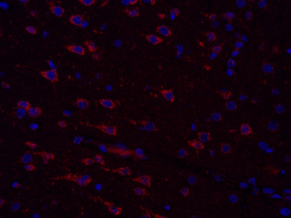 IF (Immunofluorescence)
(Immunofluorescence analysis of paraffin-embedded Rat brain using COX4I1 Polyclonal Antibody at dilution of 1:300.)
IF (Immunofluorescence)
(Immunofluorescence analysis of paraffin-embedded Rat brain using COX4I1 Polyclonal Antibody at dilution of 1:300.)
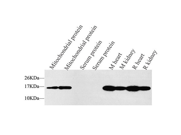
COX4I1, Polyclonal Antibody (Cat# AAA174420)
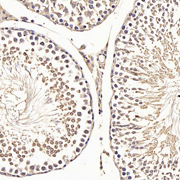
 IHC (Immunohistochemisry)
(Immunohistochemistry analysis of paraffin-embedded rat testis using AR Polyclonal Antibody at dilution of 1:300.)
IHC (Immunohistochemisry)
(Immunohistochemistry analysis of paraffin-embedded rat testis using AR Polyclonal Antibody at dilution of 1:300.)
AR, Polyclonal Antibody (Cat# AAA174422)
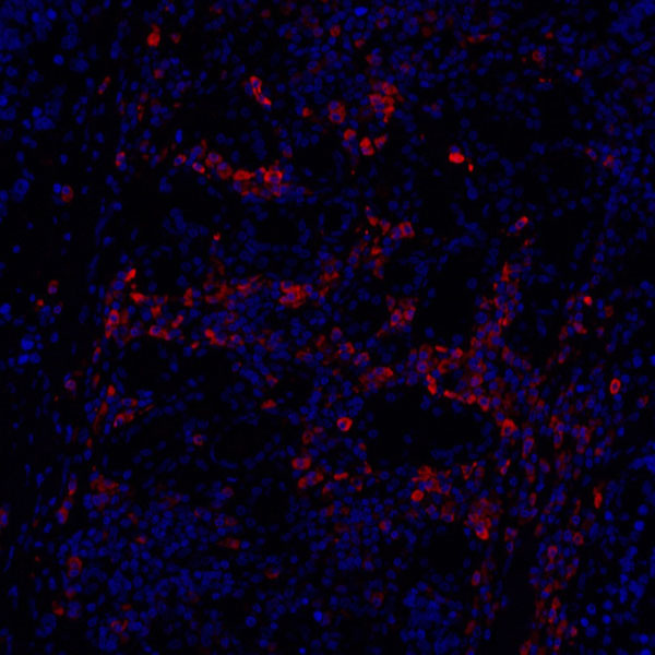 IF (Immunofluorescence)
(Immunofluorescence analysis of paraffin-embedded Human lung cancer using Cyclin B1 Polyclonal Antibody at dilution of 1:100.)
IF (Immunofluorescence)
(Immunofluorescence analysis of paraffin-embedded Human lung cancer using Cyclin B1 Polyclonal Antibody at dilution of 1:100.)
Cyclin B1, Polyclonal Antibody (Cat# AAA174424)
 IHC (Immunohistochemistry)
(Immunohistochemistry analysis of paraffin-embedded rat kidney using CXCR7 Polyclonal Antibody at dilution of 1:200.)
IHC (Immunohistochemistry)
(Immunohistochemistry analysis of paraffin-embedded rat kidney using CXCR7 Polyclonal Antibody at dilution of 1:200.)
CXCR7, Polyclonal Antibody (Cat# AAA174425)
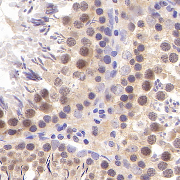
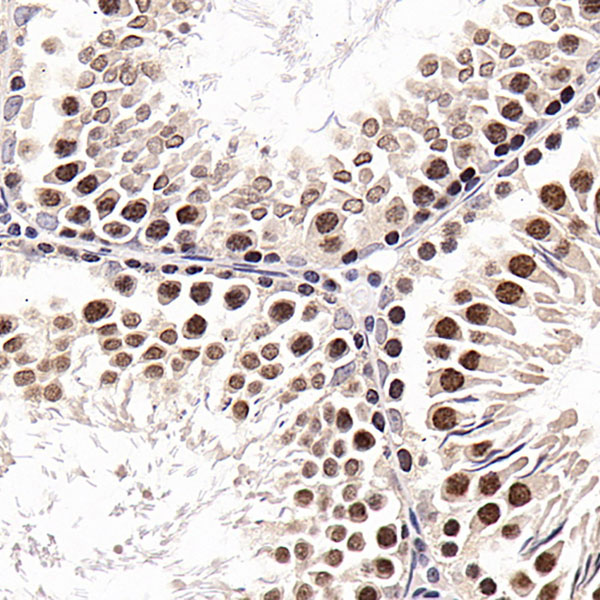 IHC (Immunohiostchemistry)
(Immunohistochemistry analysis of paraffin-embedded Rat testis using SNAI1 Polyclonal Antibody at dilution of 1:600.)
IHC (Immunohiostchemistry)
(Immunohistochemistry analysis of paraffin-embedded Rat testis using SNAI1 Polyclonal Antibody at dilution of 1:600.)
SNAI1, Polyclonal Antibody (Cat# AAA174427)
 IHC (Immunohiostchemistry)
(Immunohistochemistry analysis of paraffin-embedded mouse uterus using PGR Polyclonal Antibody at dilution of 1:400.)
IHC (Immunohiostchemistry)
(Immunohistochemistry analysis of paraffin-embedded mouse uterus using PGR Polyclonal Antibody at dilution of 1:400.)
PGR, Polyclonal Antibody (Cat# AAA174429)
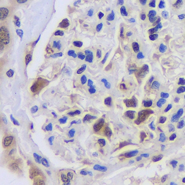
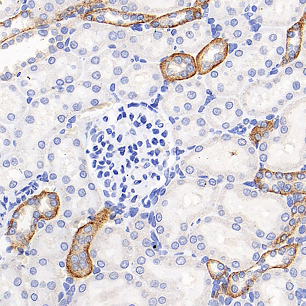
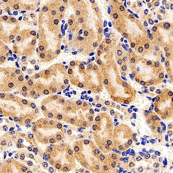
 IHC (Immunohiostchemistry)
(Immunohistochemistry analysis of paraffin-embedded Rat kidney using MYD88 Polyclonal Antibody at dilution of 1:200.)
IHC (Immunohiostchemistry)
(Immunohistochemistry analysis of paraffin-embedded Rat kidney using MYD88 Polyclonal Antibody at dilution of 1:200.)
MYD88, Polyclonal Antibody (Cat# AAA174432)
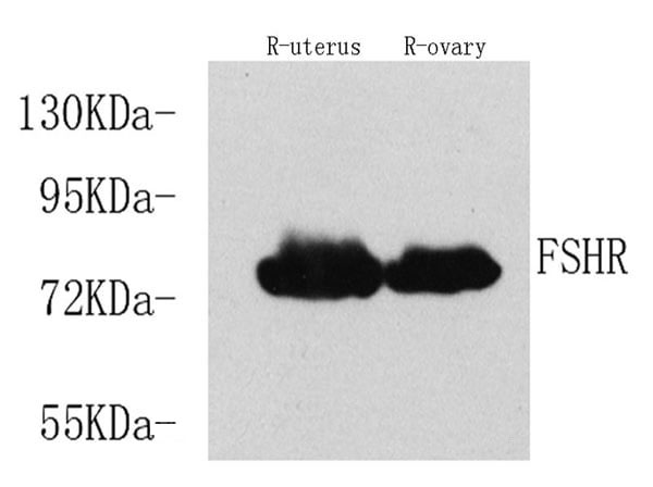
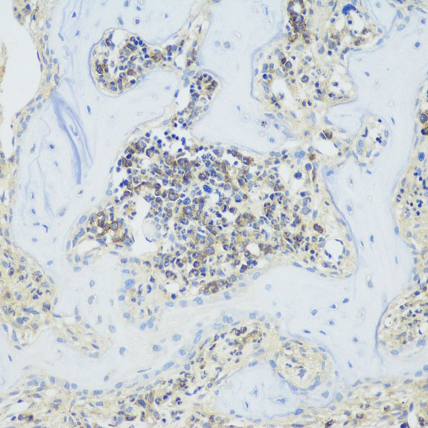
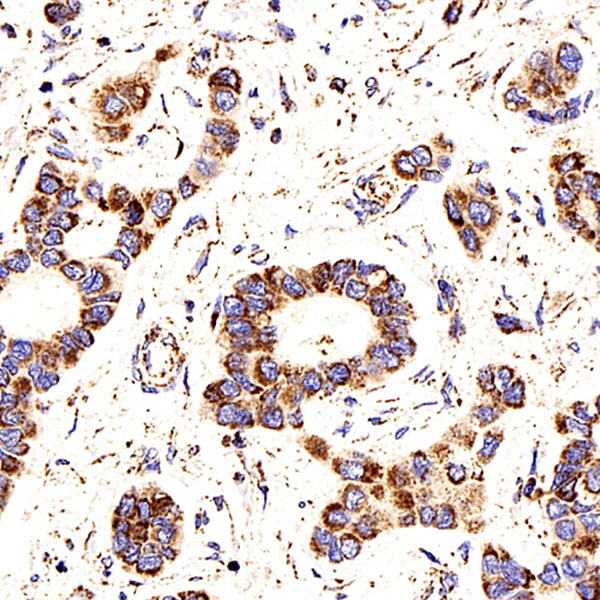
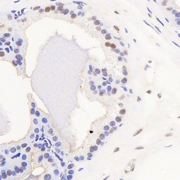
 IHC (Immunohistochemistry)
(Immunohistochemistry analysis of paraffin-embedded rat ovary using FSHR Polyclonal Antibody at dilution of 1:200.)
IHC (Immunohistochemistry)
(Immunohistochemistry analysis of paraffin-embedded rat ovary using FSHR Polyclonal Antibody at dilution of 1:200.)
FSHR, Polyclonal Antibody (Cat# AAA174434)


 IF (Immunofluorescence)
(Immunofluorescence analysis of paraffin-embedded rat colon using EpCAM Polyclonal Antibody at dilution of 1:400.)
IF (Immunofluorescence)
(Immunofluorescence analysis of paraffin-embedded rat colon using EpCAM Polyclonal Antibody at dilution of 1:400.)
FSHR, Polyclonal Antibody (Cat# AAA174435)
 IHC (Immunohistochemisry)
(Immunohistochemistry analysis of paraffin-embedded rat brain using FOXO1 Polyclonal Antibody at dilution of 1:300.)
IHC (Immunohistochemisry)
(Immunohistochemistry analysis of paraffin-embedded rat brain using FOXO1 Polyclonal Antibody at dilution of 1:300.)
FOXO1, Polyclonal Antibody (Cat# AAA174438)
 IHC (Immunohistochemistry)
(Immunohistochemistry analysis of paraffin-embedded rat lung using ATF6 Polyclonal Antibody at dilution of 1:400.)
IHC (Immunohistochemistry)
(Immunohistochemistry analysis of paraffin-embedded rat lung using ATF6 Polyclonal Antibody at dilution of 1:400.)
ATF6, Polyclonal Antibody (Cat# AAA174446)
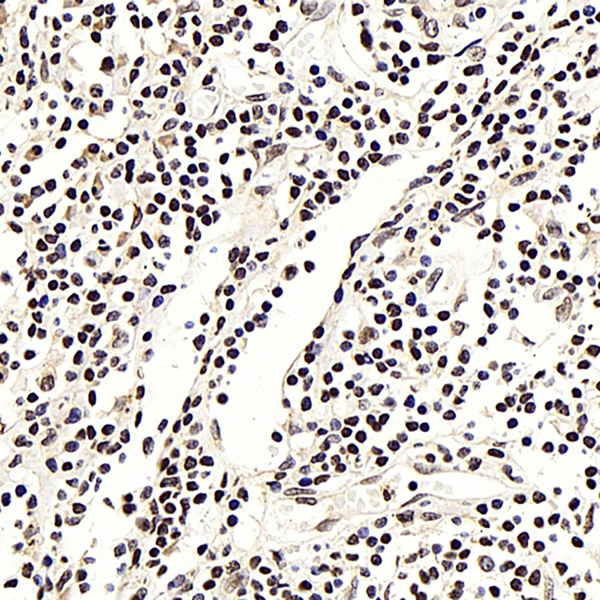
 IHC (Immunohistochemistry)
(Immunohistochemistry analysis of paraffin-embedded Rat testis using NFKBIB Polyclonal Antibody at dilution of 1:300.)
IHC (Immunohistochemistry)
(Immunohistochemistry analysis of paraffin-embedded Rat testis using NFKBIB Polyclonal Antibody at dilution of 1:300.)
NFKBIB, Polyclonal Antibody (Cat# AAA174452)
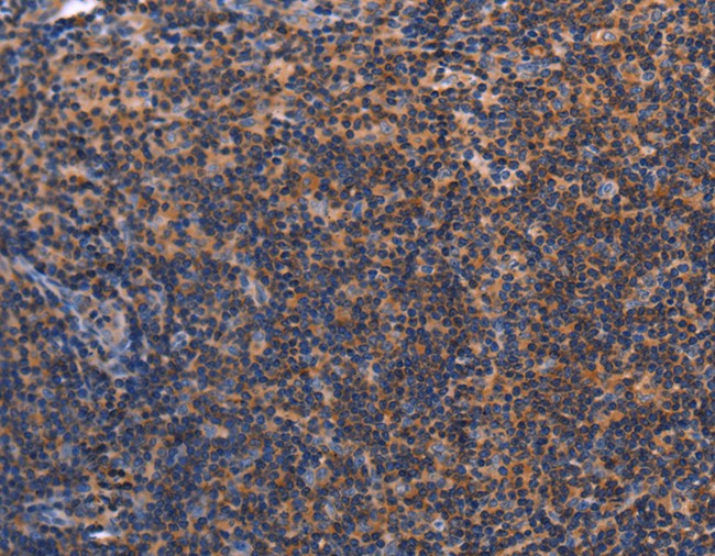
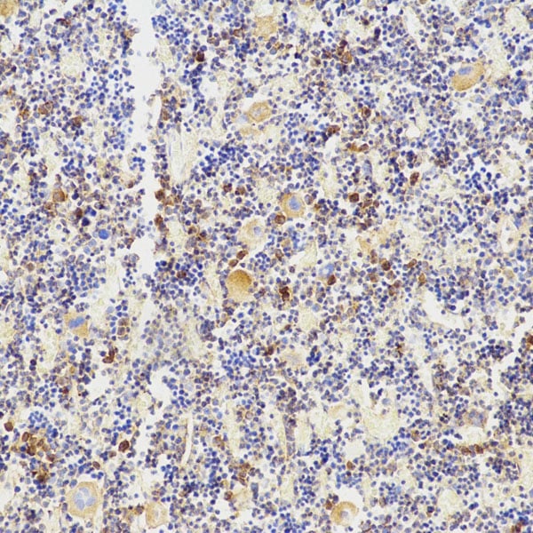
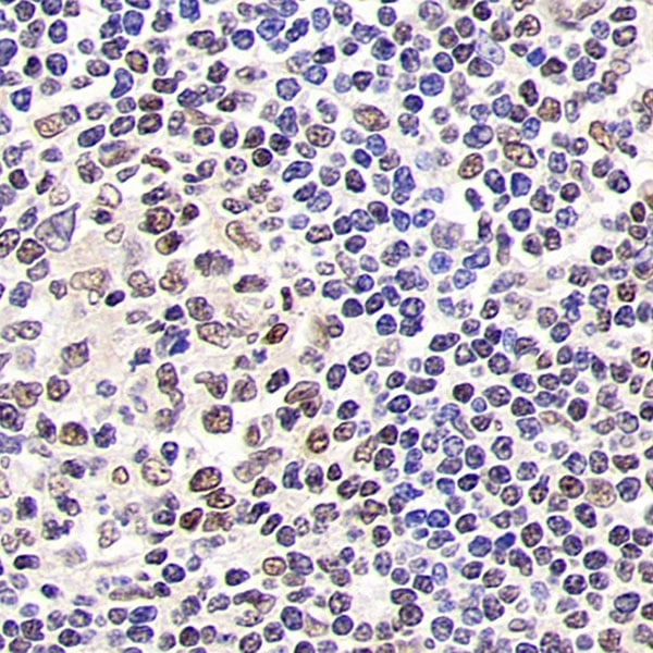
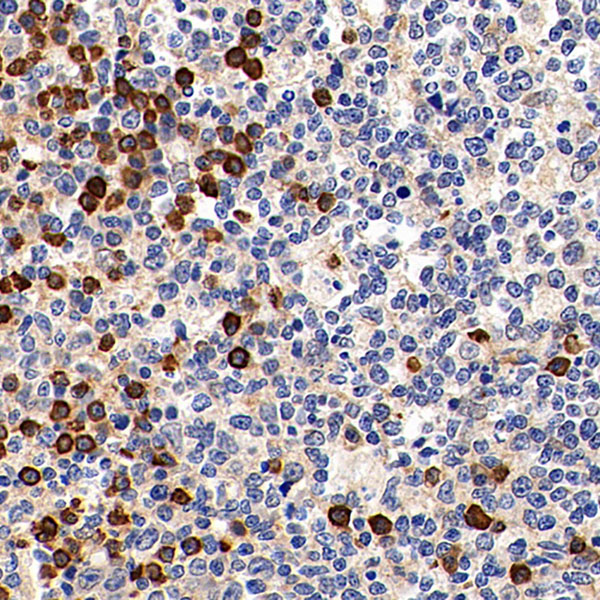
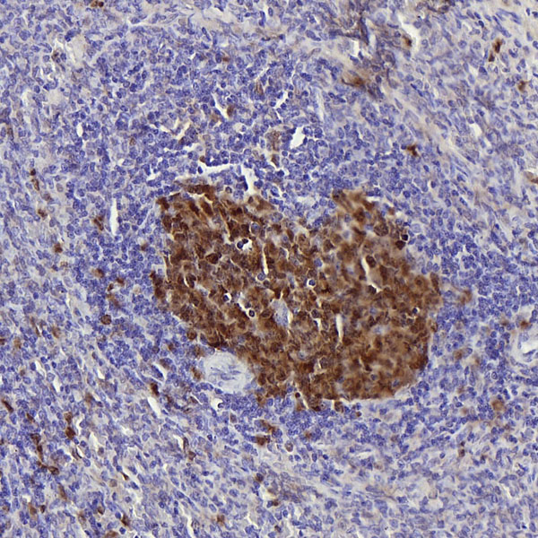
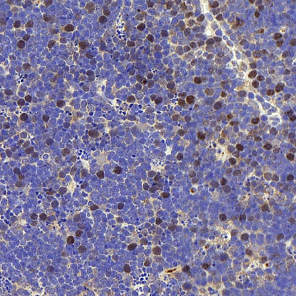 IHC (Immunohiostchemistry)
(Immunohistochemistry analysis of paraffin-embedded mouse thymus using RACGAP1 Polyclonal Antibody at dilution of 1:400.)
IHC (Immunohiostchemistry)
(Immunohistochemistry analysis of paraffin-embedded mouse thymus using RACGAP1 Polyclonal Antibody at dilution of 1:400.)
RACGAP1, Polyclonal Antibody (Cat# AAA174454)
 IHC (Immunohistochemistry)
(Immunohistochemistry analysis of paraffin-embedded Rat lung using LEP Polyclonal Antibody at dilution of 1:100.)
IHC (Immunohistochemistry)
(Immunohistochemistry analysis of paraffin-embedded Rat lung using LEP Polyclonal Antibody at dilution of 1:100.)
LEP, Polyclonal Antibody (Cat# AAA174455)
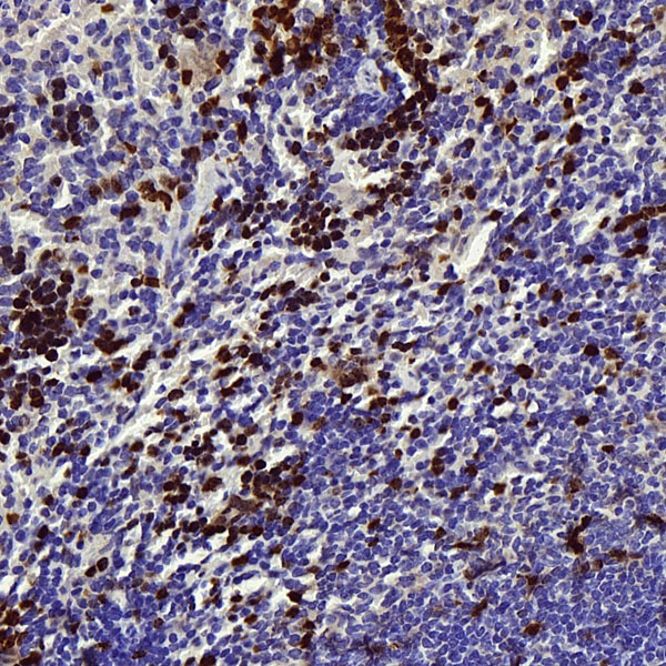
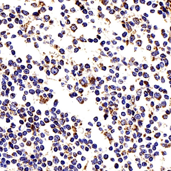
 IHC (Immunohistochemisry)
(Immunohistochemistry analysis of paraffin-embedded rat spleen using Survivin Polyclonal Antibody at dilution of 1:300.)
IHC (Immunohistochemisry)
(Immunohistochemistry analysis of paraffin-embedded rat spleen using Survivin Polyclonal Antibody at dilution of 1:300.)
Survivin, Polyclonal Antibody (Cat# AAA174547)

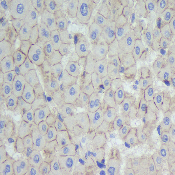
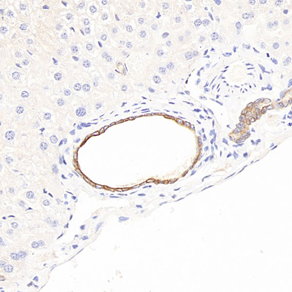
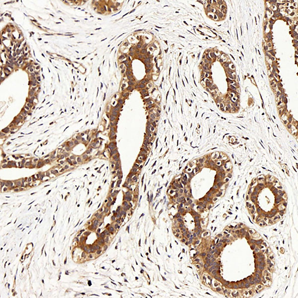
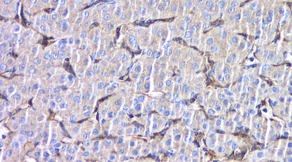
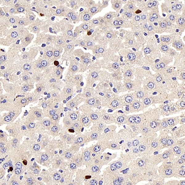
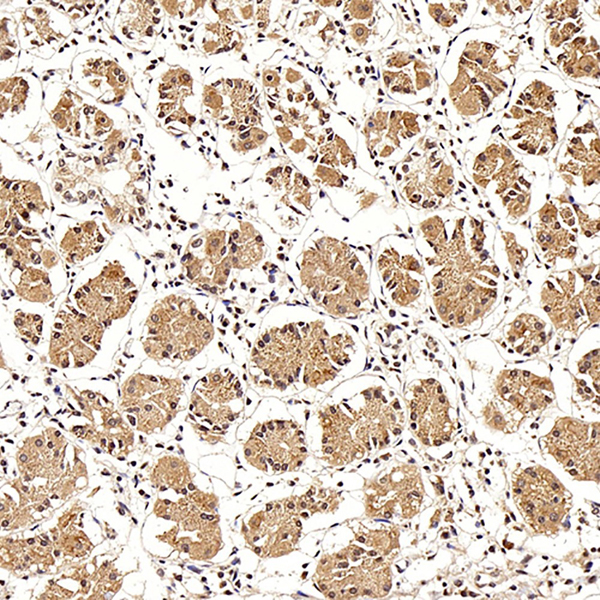
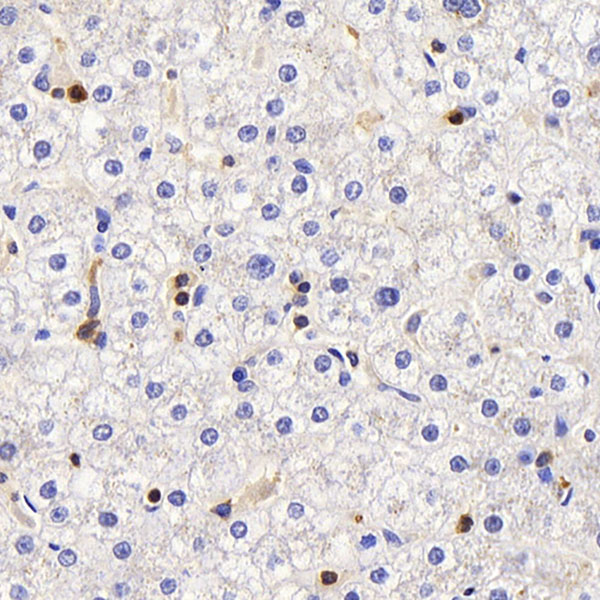
 IHC (Immunohistochemisry)
(Immunohistochemistry analysis of paraffin-embedded rat liver using CAV1 Polyclonal Antibody at dilution of 1:1000.)
IHC (Immunohistochemisry)
(Immunohistochemistry analysis of paraffin-embedded rat liver using CAV1 Polyclonal Antibody at dilution of 1:1000.)
CAV1, Polyclonal Antibody (Cat# AAA174550)
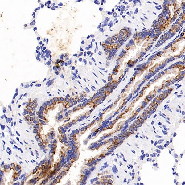
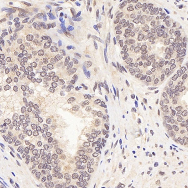
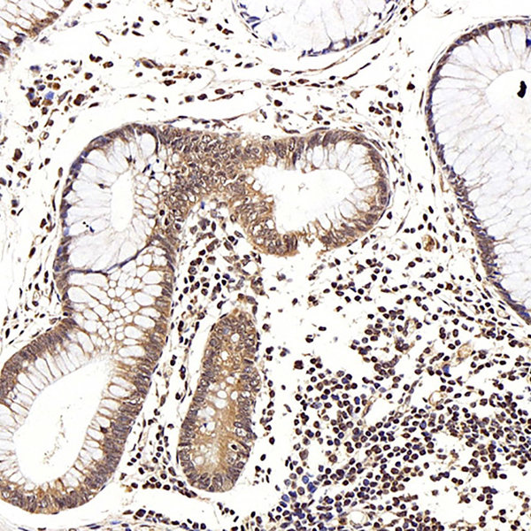
 IHC (Immunohistochemisry)
(Immunohistochemistry analysis of paraffin-embedded rat colon using c-Kit Polyclonal Antibody at dilution of 1:200.)
IHC (Immunohistochemisry)
(Immunohistochemistry analysis of paraffin-embedded rat colon using c-Kit Polyclonal Antibody at dilution of 1:200.)
c-Kit, Polyclonal Antibody (Cat# AAA174553)
What are Polyclonal Antibodies?
Polyclonal antibodies are antibodies that come from multiple B cell clones of a host animal. The typical hosts used for the majority of polyclonal antibody production are rabbits, goats, sheep, and donkeys. These polyclonal antibodies, once having identified their target, will bind to different epitopes located at different regions or sequences on the same protein/antigen. As a result, they are ideal at locating and binding to the target, even if the target is in very low concentrations (due to many different antibodies being able to bind to the same target molecule, which allows for significant amplification of a downstream signal).
Polyclonal antibodies are typically produced by injecting an antigen into a host animal, which causes the animal’s immune system to attack the foreign antigen by mass generating antibodies against it. After a period of time, serum is collected from the animal and purified using physicochemical fractionation, class-specific affinity purification, and/or antigen-affinity purification.
Key Uses of Polyclonal Antibodies
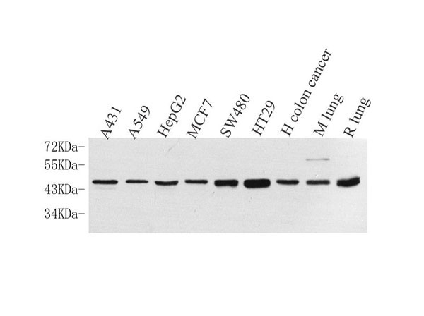
- Western Blotting: This method is used to find specific proteins in biological samples after separating them by size.
- Immunohistochemistry: IHC helps visualize the location of proteins in tissue sections using various staining techniques.
- ELISA: (Enzyme-Linked Immunosorbent Assay) is typically used to identify specific protein quantities in a sample. ELISAs can be either “Quantitative” or “Qualitative”.
- Flow Cytometry: technique that identifies and measures the specific protein on the surface or inside the cells in a fluid suspension.
- Immunoprecipitation: IP isolates and studies a specific protein from a complex mixture using antibodies.
Why Buy Polyclonal Antibodies from AAA Biotech?
1. Ideal for Various Applications
Our antibodies are generally going to be validated for use in multiple types of assays, including ELISA, Western Blotting, Immunohistochemistry, Immunoprecipitation, amongst others. They are ideal for a wide range of research applications.
2. Rigorous Quality Control
All of the antibodies in our catalog undergo strict quality testing to ensure specificity, sensitivity, and consistent performance. We are confident in the ability of our antibodies to provide you with accurate results.
3. Wide Assortment of Antibodies
Antibodies in are catalog can be found for both common and exotic species, and these antibodies are also available in both conjugated and recombinant forms to suit many diverse experimental needs.
4. Highly Purified
Our antibodies are available in purified forms with over 85% purity, as confirmed by SDS-PAGE. They are also available with tags such as His, Flag, GST, or MBP. We cater to customers worldwide.
FAQ
1. How are polyclonal antibodies produced?
Traditionally, polyclonal antibodies are produced by injecting an antigen into a host animal (such as a rabbit or goat), which then triggers an immune response from the host animal. The animal’s B cells produce antibodies that will recognize different parts of the injected antigen. These antibodies are then collected from the animal’s blood and purified for use.
2. How do polyclonal antibodies differ from monoclonal antibodies?
Polyclonal antibodies are a mix of antibodies that bind to different locations (epitopes) of the same antigen, while monoclonal antibodies are identical and bind to just one specific epitope. This makes polyclonal antibodies more versatile and better at detecting proteins that may be present in low quantities or in altered/modified forms.
3. How should I store polyclonal antibodies?
Polyclonal antibodies should be stored at 4°C for short-term use (up to a few weeks) and at -20°C or -80°C for long-term storage. Avoid repeated freeze-thaw cycles by dividing them into small aliquots. Always check the datasheet for specific storage instructions.