Filters
▼Clonality
▼Type
▼Reactivity
▼Gene Name
▼Isotype
▼Host
▼Application
▼Clone
▼Polyclonal Antibodies
At AAA Biotech also known as AAA Bio or AAABio, we provide a broad range of purified polyclonal antibodies (pAbs) that are able to all be browsed online through our website. Due to their high specificity and strong binding affinity, these antibodies are ideal for wide swathes of research and experimental applications.
Our polyclonal antibodies can easily support your work, whether you use them for Western Blotting, Immunocytochemistry (with or without Immunofluorescence used in conjunction), Immunohistochemistry, Immunoprecipitation, and ELISA tests. We highly encourage you to browse our range of pAbs and choose the one that best suits your experimental model.
Viewing 6350-6400 of 96805 product results
 SDS-PAGE
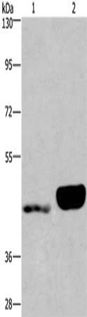
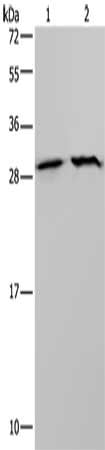
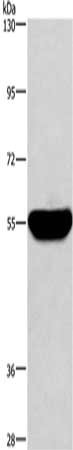
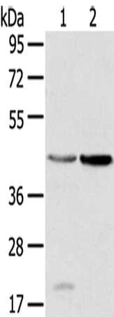
(Gel: 8%SDS-PAGE, Lysate: 40 ug, Lane 1-2: Hela cells, hepG2 cells, Primary antibody: AAA241072(AIMP1 Antibody) at dilution 1/400, Secondary antibody: Goat anti rabbit IgG at 1/8000 dilution, Exposure time: 5 second)
SDS-PAGE
(Gel: 8%SDS-PAGE, Lysate: 40 ug, Lane 1-2: Hela cells, hepG2 cells, Primary antibody: AAA241072(AIMP1 Antibody) at dilution 1/400, Secondary antibody: Goat anti rabbit IgG at 1/8000 dilution, Exposure time: 5 second)
AIMP1, Polyclonal Antibody (Cat# AAA241072)
 IHC (Immunohiostchemistry)
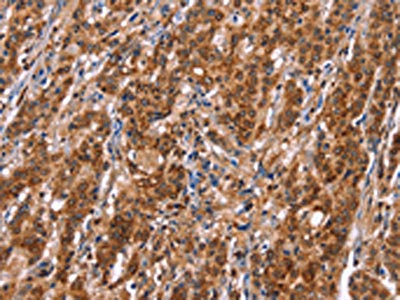
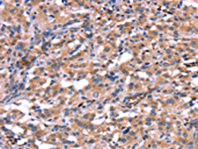
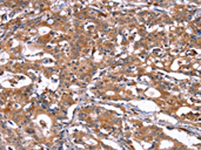
(The image on the left is immunohistochemistry of paraffin-embedded Human ovarian cancer tissue using AAA241075(EMP2 Antibody) at dilution 1/30, on the right is treated with synthetic peptide. (Original magnification: ×200))
IHC (Immunohiostchemistry)
(The image on the left is immunohistochemistry of paraffin-embedded Human ovarian cancer tissue using AAA241075(EMP2 Antibody) at dilution 1/30, on the right is treated with synthetic peptide. (Original magnification: ×200))
EMP2, Polyclonal Antibody (Cat# AAA241075)
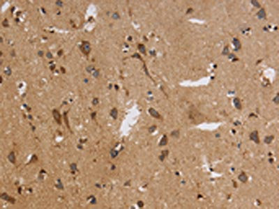
 IHC (Immunohiostchemistry)
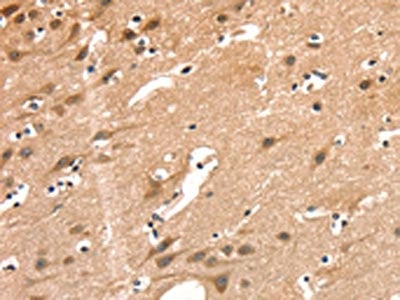
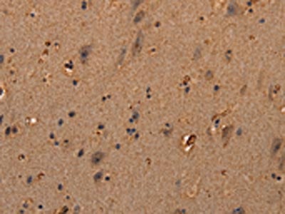
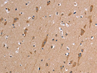
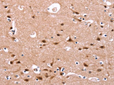
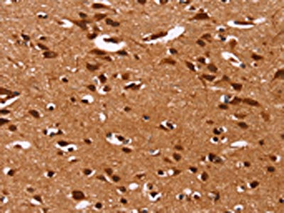
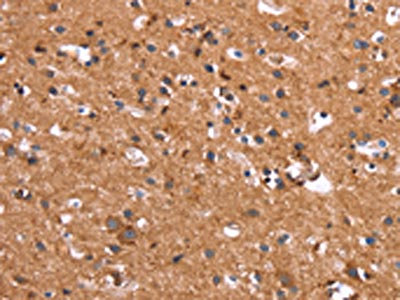
(The image on the left is immunohistochemistry of paraffin-embedded Human brain tissue using AAA241076(ENDOG Antibody) at dilution 1/15, on the right is treated with synthetic peptide. (Original magnification: ×200))
IHC (Immunohiostchemistry)
(The image on the left is immunohistochemistry of paraffin-embedded Human brain tissue using AAA241076(ENDOG Antibody) at dilution 1/15, on the right is treated with synthetic peptide. (Original magnification: ×200))
ENDOG, Polyclonal Antibody (Cat# AAA241076)
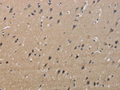
 IHC (Immunohiostchemistry)
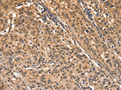
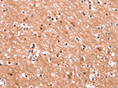
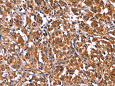
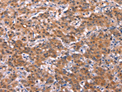
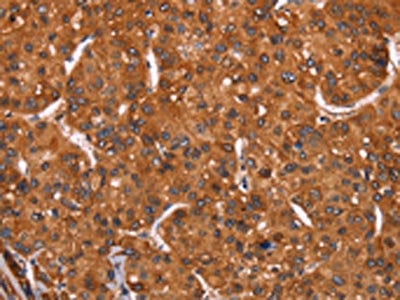
(The image on the left is immunohistochemistry of paraffin-embedded Human brain tissue using AAA241078(COL18A1 Antibody) at dilution 1/40, on the right is treated with synthetic peptide. (Original magnification: ×200))
IHC (Immunohiostchemistry)
(The image on the left is immunohistochemistry of paraffin-embedded Human brain tissue using AAA241078(COL18A1 Antibody) at dilution 1/40, on the right is treated with synthetic peptide. (Original magnification: ×200))
COL18A1, Polyclonal Antibody (Cat# AAA241078)
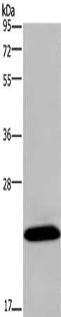
 SDS-PAGE
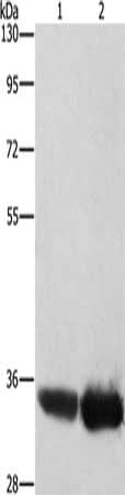
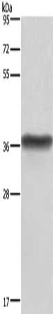
(Gel: 6%SDS-PAGE, Lysate: 40 ug, Lane 1-2: Human endometrial cancer tissue, Human placenta tissue, Primary antibody: AAA241079(ENPP4 Antibody) at dilution 1/200, Secondary antibody: Goat anti rabbit IgG at 1/8000 dilution, Exposure time: 40 seconds)
SDS-PAGE
(Gel: 6%SDS-PAGE, Lysate: 40 ug, Lane 1-2: Human endometrial cancer tissue, Human placenta tissue, Primary antibody: AAA241079(ENPP4 Antibody) at dilution 1/200, Secondary antibody: Goat anti rabbit IgG at 1/8000 dilution, Exposure time: 40 seconds)
ENPP4, Polyclonal Antibody (Cat# AAA241079)
 SDS-PAGE
(Gel: 10%SDS-PAGE, Lysate: 40 ug, Lane: Mouse heart tissue, Primary antibody: AAA241089(ERAS Antibody) at dilution 1/450, Secondary antibody: Goat anti rabbit IgG at 1/8000 dilution, Exposure time: 1 minute)
SDS-PAGE
(Gel: 10%SDS-PAGE, Lysate: 40 ug, Lane: Mouse heart tissue, Primary antibody: AAA241089(ERAS Antibody) at dilution 1/450, Secondary antibody: Goat anti rabbit IgG at 1/8000 dilution, Exposure time: 1 minute)
ERAS, Polyclonal Antibody (Cat# AAA241089)
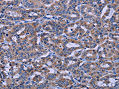
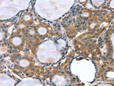
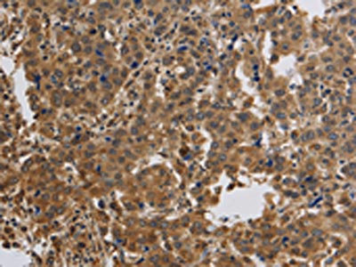
 IHC (Immunohiostchemistry)
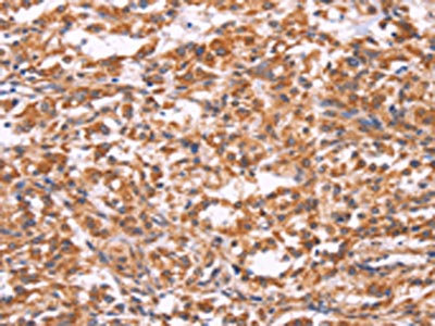
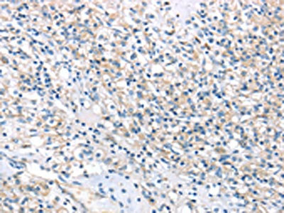
(The image on the left is immunohistochemistry of paraffin-embedded Human thyroid cancer tissue using AAA241092(ERN2 Antibody) at dilution 1/40, on the right is treated with synthetic peptide. (Original magnification: ×200))
IHC (Immunohiostchemistry)
(The image on the left is immunohistochemistry of paraffin-embedded Human thyroid cancer tissue using AAA241092(ERN2 Antibody) at dilution 1/40, on the right is treated with synthetic peptide. (Original magnification: ×200))
ERN2, Polyclonal Antibody (Cat# AAA241092)
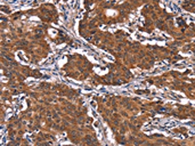
 IHC (Immunohiostchemistry)
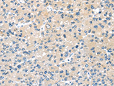
(The image on the left is immunohistochemistry of paraffin-embedded Human ovarian cancer tissue using AAA241095(ESRRB Antibody) at dilution 1/40, on the right is treated with synthetic peptide. (Original magnification: ×200))
IHC (Immunohiostchemistry)
(The image on the left is immunohistochemistry of paraffin-embedded Human ovarian cancer tissue using AAA241095(ESRRB Antibody) at dilution 1/40, on the right is treated with synthetic peptide. (Original magnification: ×200))
ESRRB, Polyclonal Antibody (Cat# AAA241095)
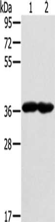
 SDS-PAGE
(Gel: 10%SDS-PAGE, Lysate: 40 ug, Lane 1-2: LoVo cells, Hela cells, Primary antibody: AAA241099(ETFB Antibody) at dilution 1/200, Secondary antibody: Goat anti rabbit IgG at 1/8000 dilution, Exposure time: 20 seconds)
SDS-PAGE
(Gel: 10%SDS-PAGE, Lysate: 40 ug, Lane 1-2: LoVo cells, Hela cells, Primary antibody: AAA241099(ETFB Antibody) at dilution 1/200, Secondary antibody: Goat anti rabbit IgG at 1/8000 dilution, Exposure time: 20 seconds)
ETFB, Polyclonal Antibody (Cat# AAA241099)
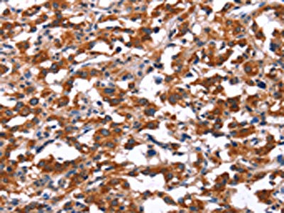
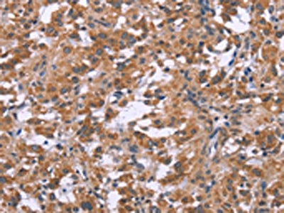
 IHC (Immunohiostchemistry)
(The image on the left is immunohistochemistry of paraffin-embedded Human thyroid cancer tissue using AAA241101(TDP2 Antibody) at dilution 1/40, on the right is treated with synthetic peptide. (Original magnification: ×200))
IHC (Immunohiostchemistry)
(The image on the left is immunohistochemistry of paraffin-embedded Human thyroid cancer tissue using AAA241101(TDP2 Antibody) at dilution 1/40, on the right is treated with synthetic peptide. (Original magnification: ×200))
TDP2, Polyclonal Antibody (Cat# AAA241101)
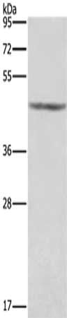
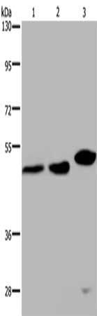 SDS-PAGE
(Gel: 8%SDS-PAGE, Lysate: 40 ug, Lane 1-3: Hela cells, 293T cells, mouse liver tissue, Primary antibody: AAA241102(TDP2 Antibody) at dilution 1/200, Secondary antibody: Goat anti rabbit IgG at 1/8000 dilution, Exposure time: 6 minutes)
SDS-PAGE
(Gel: 8%SDS-PAGE, Lysate: 40 ug, Lane 1-3: Hela cells, 293T cells, mouse liver tissue, Primary antibody: AAA241102(TDP2 Antibody) at dilution 1/200, Secondary antibody: Goat anti rabbit IgG at 1/8000 dilution, Exposure time: 6 minutes)
TDP2, Polyclonal Antibody (Cat# AAA241102)
 SDS-PAGE
(Gel: 6%SDS-PAGE, Lysate: 40 ug, Lane: Human placenta tissue, Primary antibody: AAA241104(EVC2 Antibody) at dilution 1/450, Secondary antibody: Goat anti rabbit IgG at 1/8000 dilution, Exposure time: 7 minutes)
SDS-PAGE
(Gel: 6%SDS-PAGE, Lysate: 40 ug, Lane: Human placenta tissue, Primary antibody: AAA241104(EVC2 Antibody) at dilution 1/450, Secondary antibody: Goat anti rabbit IgG at 1/8000 dilution, Exposure time: 7 minutes)
EVC2, Polyclonal Antibody (Cat# AAA241104)
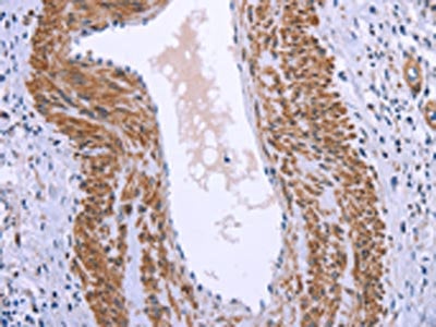
 IHC (Immunohiostchemistry)
(The image on the left is immunohistochemistry of paraffin-embedded Human ovarian cancer tissue using AAA241112(FAIM Antibody) at dilution 1/25, on the right is treated with synthetic peptide. (Original magnification: ×200))
IHC (Immunohiostchemistry)
(The image on the left is immunohistochemistry of paraffin-embedded Human ovarian cancer tissue using AAA241112(FAIM Antibody) at dilution 1/25, on the right is treated with synthetic peptide. (Original magnification: ×200))
FAIM, Polyclonal Antibody (Cat# AAA241112)
 IHC (Immunohiostchemistry)
(The image on the left is immunohistochemistry of paraffin-embedded Human gastric cancer tissue using AAA241115(FAM13B Antibody) at dilution 1/20, on the right is treated with synthetic peptide. (Original magnification: ×200))
IHC (Immunohiostchemistry)
(The image on the left is immunohistochemistry of paraffin-embedded Human gastric cancer tissue using AAA241115(FAM13B Antibody) at dilution 1/20, on the right is treated with synthetic peptide. (Original magnification: ×200))
FAM13B, Polyclonal Antibody (Cat# AAA241115)
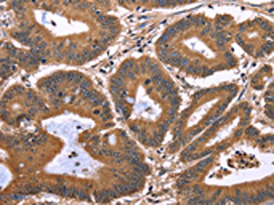
 IHC (Immunohiostchemistry)
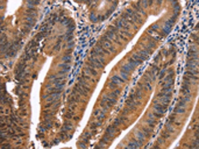
(The image on the left is immunohistochemistry of paraffin-embedded Human colon cancer tissue using AAA241119(FBXO31 Antibody) at dilution 1/40, on the right is treated with synthetic peptide. (Original magnification: ×200))
IHC (Immunohiostchemistry)
(The image on the left is immunohistochemistry of paraffin-embedded Human colon cancer tissue using AAA241119(FBXO31 Antibody) at dilution 1/40, on the right is treated with synthetic peptide. (Original magnification: ×200))
FBXO31, Polyclonal Antibody (Cat# AAA241119)
 IHC (Immunohiostchemistry)
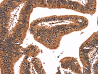
(The image on the left is immunohistochemistry of paraffin-embedded Human colon cancer tissue using AAA241128(FGF22 Antibody) at dilution 1/20, on the right is treated with synthetic peptide. (Original magnification: ×200))
IHC (Immunohiostchemistry)
(The image on the left is immunohistochemistry of paraffin-embedded Human colon cancer tissue using AAA241128(FGF22 Antibody) at dilution 1/20, on the right is treated with synthetic peptide. (Original magnification: ×200))
FGF22, Polyclonal Antibody (Cat# AAA241128)
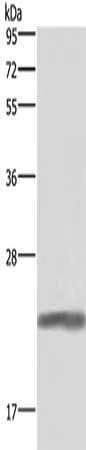 SDS-PAGE
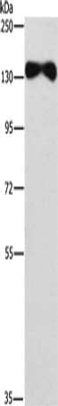
(Gel: 10%SDS-PAGE, Lysate: 40 ug, Lane: Human placenta tissue, Primary antibody: AAA241165(GH1 Antibody) at dilution 1/500, Secondary antibody: Goat anti rabbit IgG at 1/8000 dilution, Exposure time: 1 second)
SDS-PAGE
(Gel: 10%SDS-PAGE, Lysate: 40 ug, Lane: Human placenta tissue, Primary antibody: AAA241165(GH1 Antibody) at dilution 1/500, Secondary antibody: Goat anti rabbit IgG at 1/8000 dilution, Exposure time: 1 second)
GH1, Polyclonal Antibody (Cat# AAA241165)
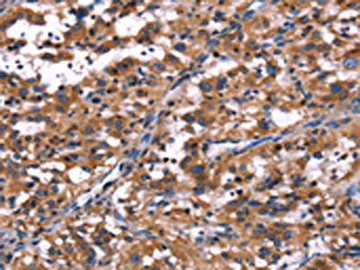
 IHC (Immunohiostchemistry)
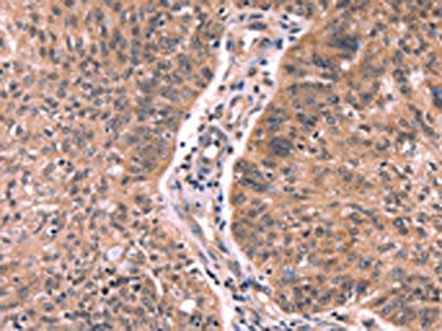
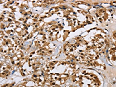
(The image on the left is immunohistochemistry of paraffin-embedded Human gastric cancer tissue using AAA241167(R3HCC1L Antibody) at dilution 1/40, on the right is treated with synthetic peptide. (Original magnification: ×200))
IHC (Immunohiostchemistry)
(The image on the left is immunohistochemistry of paraffin-embedded Human gastric cancer tissue using AAA241167(R3HCC1L Antibody) at dilution 1/40, on the right is treated with synthetic peptide. (Original magnification: ×200))
R3HCC1L, Polyclonal Antibody (Cat# AAA241167)
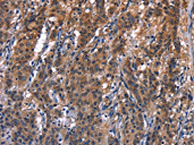
 IHC (Immunohiostchemistry)
(The image on the left is immunohistochemistry of paraffin-embedded Human thyroid cancer tissue using AAA241170(KCNJ9 Antibody) at dilution 1/30, on the right is treated with synthetic peptide. (Original magnification: ×200))
IHC (Immunohiostchemistry)
(The image on the left is immunohistochemistry of paraffin-embedded Human thyroid cancer tissue using AAA241170(KCNJ9 Antibody) at dilution 1/30, on the right is treated with synthetic peptide. (Original magnification: ×200))
KCNJ9, Polyclonal Antibody (Cat# AAA241170)
 SDS-PAGE
(Gel: 8%SDS-PAGE, Lysate: 40 ug, Lane: Human placenta tissue , Primary antibody: AAA241171(KCNJ9 Antibody) at dilution 1/350, Secondary antibody: Goat anti rabbit IgG at 1/8000 dilution, Exposure time: 1 second)
SDS-PAGE
(Gel: 8%SDS-PAGE, Lysate: 40 ug, Lane: Human placenta tissue , Primary antibody: AAA241171(KCNJ9 Antibody) at dilution 1/350, Secondary antibody: Goat anti rabbit IgG at 1/8000 dilution, Exposure time: 1 second)
KCNJ9, Polyclonal Antibody (Cat# AAA241171)
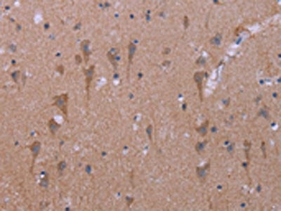
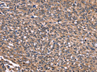
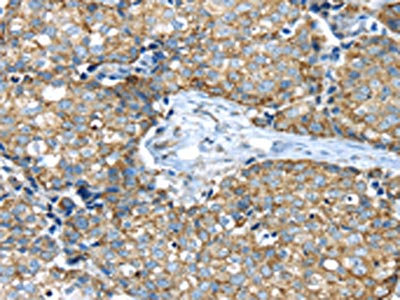
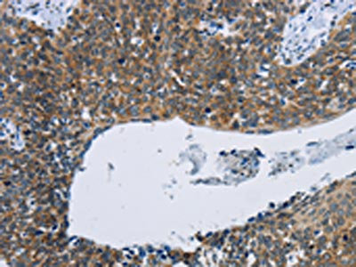
 IHC (Immunohiostchemistry)
(The image on the left is immunohistochemistry of paraffin-embedded Human brain tissue using AAA241175(GLMN Antibody) at dilution 1/40, on the right is treated with synthetic peptide. (Original magnification: ×200))
IHC (Immunohiostchemistry)
(The image on the left is immunohistochemistry of paraffin-embedded Human brain tissue using AAA241175(GLMN Antibody) at dilution 1/40, on the right is treated with synthetic peptide. (Original magnification: ×200))
GLMN, Polyclonal Antibody (Cat# AAA241175)
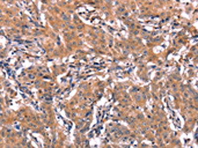
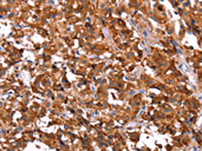
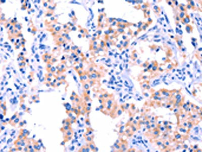
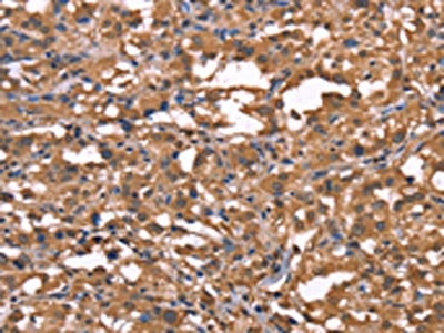
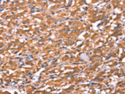
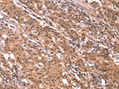
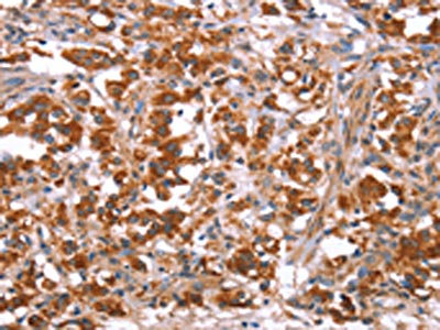
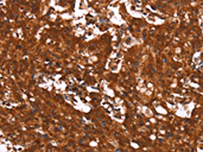
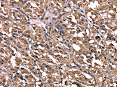
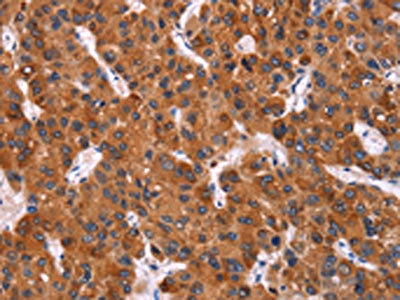
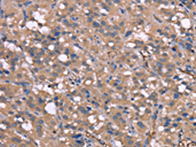
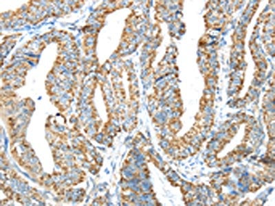
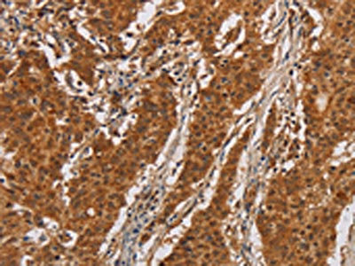
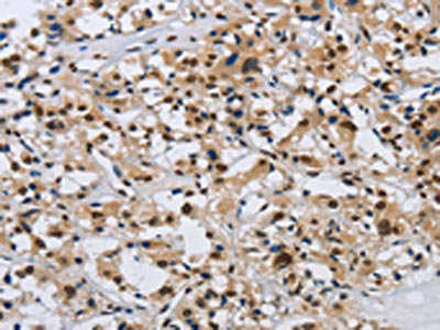
 IHC (Immunohiostchemistry)
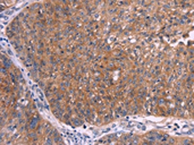
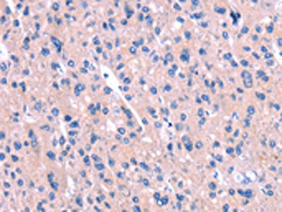
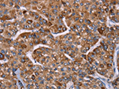
(The image on the left is immunohistochemistry of paraffin-embedded Human liver cancer tissue using AAA241182(CSF2RB Antibody) at dilution 1/25, on the right is treated with synthetic peptide. (Original magnification: ×200))
IHC (Immunohiostchemistry)
(The image on the left is immunohistochemistry of paraffin-embedded Human liver cancer tissue using AAA241182(CSF2RB Antibody) at dilution 1/25, on the right is treated with synthetic peptide. (Original magnification: ×200))
CSF2RB, Polyclonal Antibody (Cat# AAA241182)
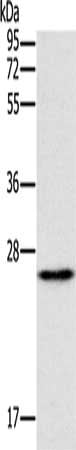
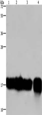
 SDS-PAGE
(Gel: 8%SDS-PAGE, Lysate: 40 ug, Lane 1-2: Mouse heart tissue, Mouse muscle tissue, Primary antibody: AAA241184(GNAT3 Antibody) at dilution 1/400, Secondary antibody: Goat anti rabbit IgG at 1/8000 dilution, Exposure time: 1 minute)
SDS-PAGE
(Gel: 8%SDS-PAGE, Lysate: 40 ug, Lane 1-2: Mouse heart tissue, Mouse muscle tissue, Primary antibody: AAA241184(GNAT3 Antibody) at dilution 1/400, Secondary antibody: Goat anti rabbit IgG at 1/8000 dilution, Exposure time: 1 minute)
GNAT3, Polyclonal Antibody (Cat# AAA241184)
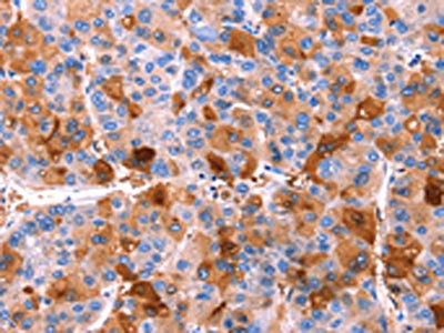
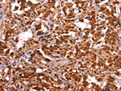
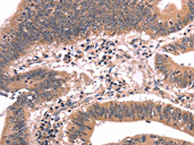
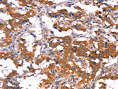
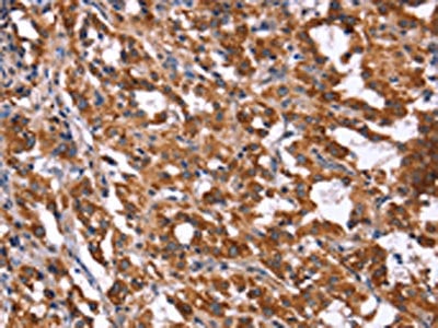
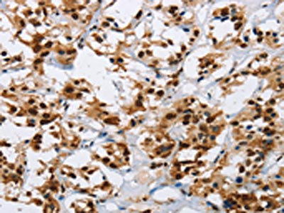
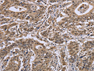
 IHC (Immunohiostchemistry)
(The image on the left is immunohistochemistry of paraffin-embedded Human cervical cancer tissue using AAA241185(GNPAT Antibody) at dilution 1/40, on the right is treated with synthetic peptide. (Original magnification: ×200))
IHC (Immunohiostchemistry)
(The image on the left is immunohistochemistry of paraffin-embedded Human cervical cancer tissue using AAA241185(GNPAT Antibody) at dilution 1/40, on the right is treated with synthetic peptide. (Original magnification: ×200))
GNPAT, Polyclonal Antibody (Cat# AAA241185)
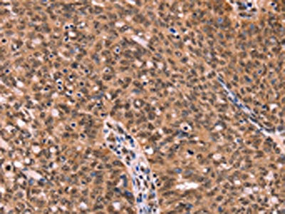
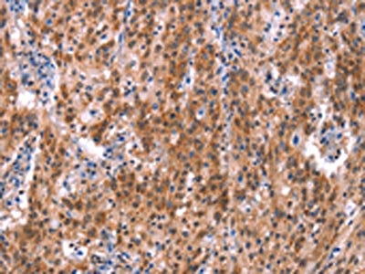
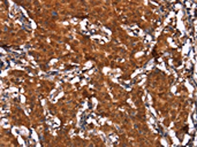
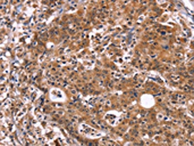
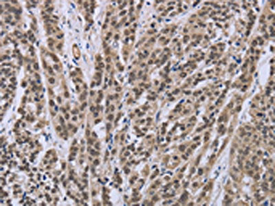
 IHC (Immunohiostchemistry)
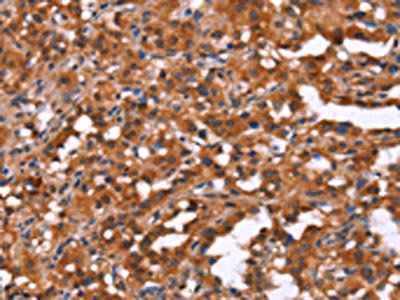
(The image on the left is immunohistochemistry of paraffin-embedded Human thyroid cancer tissue using AAA241187(HCAR2 Antibody) at dilution 1/40, on the right is treated with synthetic peptide. (Original magnification: ×200))
IHC (Immunohiostchemistry)
(The image on the left is immunohistochemistry of paraffin-embedded Human thyroid cancer tissue using AAA241187(HCAR2 Antibody) at dilution 1/40, on the right is treated with synthetic peptide. (Original magnification: ×200))
HCAR2, Polyclonal Antibody (Cat# AAA241187)
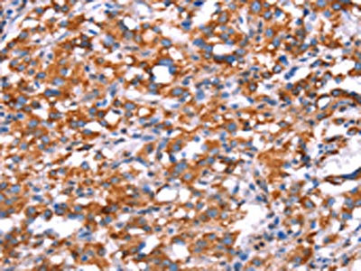
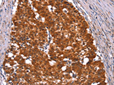
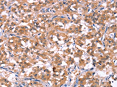 IHC (Immunohiostchemistry)
(The image on the left is immunohistochemistry of paraffin-embedded Human thyroid cancer tissue using AAA241188(HCAR2 Antibody) at dilution 1/40, on the right is treated with synthetic peptide. (Original magnification: ×200))
IHC (Immunohiostchemistry)
(The image on the left is immunohistochemistry of paraffin-embedded Human thyroid cancer tissue using AAA241188(HCAR2 Antibody) at dilution 1/40, on the right is treated with synthetic peptide. (Original magnification: ×200))
HCAR2, Polyclonal Antibody (Cat# AAA241188)
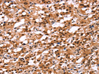
 IHC (Immunohiostchemistry)
(The image on the left is immunohistochemistry of paraffin-embedded Human thyroid cancer tissue using AAA241198(GPR65 Antibody) at dilution 1/20, on the right is treated with synthetic peptide. (Original magnification: ×200))
IHC (Immunohiostchemistry)
(The image on the left is immunohistochemistry of paraffin-embedded Human thyroid cancer tissue using AAA241198(GPR65 Antibody) at dilution 1/20, on the right is treated with synthetic peptide. (Original magnification: ×200))
GPR65, Polyclonal Antibody (Cat# AAA241198)
 IHC (Immunohiostchemistry)
(The image on the left is immunohistochemistry of paraffin-embedded Human gastric cancer tissue using AAA241203(HACE1 Antibody) at dilution 1/40, on the right is treated with synthetic peptide. (Original magnification: ×200))
IHC (Immunohiostchemistry)
(The image on the left is immunohistochemistry of paraffin-embedded Human gastric cancer tissue using AAA241203(HACE1 Antibody) at dilution 1/40, on the right is treated with synthetic peptide. (Original magnification: ×200))
HACE1, Polyclonal Antibody (Cat# AAA241203)
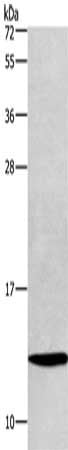 SDS-PAGE
(Gel: 10%SDS-PAGE, Lysate: 40 ug, Lane: LoVo cells, Primary antibody: AAA241211(HINT1 Antibody) at dilution 1/400, Secondary antibody: Goat anti rabbit IgG at 1/8000 dilution, Exposure time: 15 seconds)
SDS-PAGE
(Gel: 10%SDS-PAGE, Lysate: 40 ug, Lane: LoVo cells, Primary antibody: AAA241211(HINT1 Antibody) at dilution 1/400, Secondary antibody: Goat anti rabbit IgG at 1/8000 dilution, Exposure time: 15 seconds)
HINT1, Polyclonal Antibody (Cat# AAA241211)
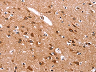
 IHC (Immunohiostchemistry)
(The image on the left is immunohistochemistry of paraffin-embedded Human brain tissue using AAA241212(HIPK1 Antibody) at dilution 1/25, on the right is treated with synthetic peptide. (Original magnification: ×200))
IHC (Immunohiostchemistry)
(The image on the left is immunohistochemistry of paraffin-embedded Human brain tissue using AAA241212(HIPK1 Antibody) at dilution 1/25, on the right is treated with synthetic peptide. (Original magnification: ×200))
HIPK1, Polyclonal Antibody (Cat# AAA241212)
 SDS-PAGE
(Gel: 6%SDS-PAGE, Lysate: 40 ug, Lane: Human fetal liver tissue, Primary antibody: AAA241218(HMGCS1 Antibody) at dilution 1/300, Secondary antibody: Goat anti rabbit IgG at 1/8000 dilution, Exposure time: 30 seconds)
SDS-PAGE
(Gel: 6%SDS-PAGE, Lysate: 40 ug, Lane: Human fetal liver tissue, Primary antibody: AAA241218(HMGCS1 Antibody) at dilution 1/300, Secondary antibody: Goat anti rabbit IgG at 1/8000 dilution, Exposure time: 30 seconds)
HMGCS1, Polyclonal Antibody (Cat# AAA241218)
 SDS-PAGE
(Gel: 8%SDS-PAGE, Lysate: 40 ug, Lane: Human fetal liver tissue, Primary antibody: AAA241220(HMGCS2 Antibody) at dilution 1/300, Secondary antibody: Goat anti rabbit IgG at 1/8000 dilution, Exposure time: 30 seconds)
SDS-PAGE
(Gel: 8%SDS-PAGE, Lysate: 40 ug, Lane: Human fetal liver tissue, Primary antibody: AAA241220(HMGCS2 Antibody) at dilution 1/300, Secondary antibody: Goat anti rabbit IgG at 1/8000 dilution, Exposure time: 30 seconds)
HMGCS2, Polyclonal Antibody (Cat# AAA241220)
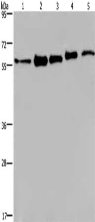 SDS-PAGE
(Gel: 6%SDS-PAGE, Lysate: 40 ug, Lane 1-5: MCF7 cells, 293T cells, A549 cells, Hela cells, HepG2 cells, Primary antibody: AAA241222(HNRNPL Antibody) at dilution 1/450, Secondary antibody: Goat anti rabbit IgG at 1/8000 dilution, Exposure time: 40 seconds)
SDS-PAGE
(Gel: 6%SDS-PAGE, Lysate: 40 ug, Lane 1-5: MCF7 cells, 293T cells, A549 cells, Hela cells, HepG2 cells, Primary antibody: AAA241222(HNRNPL Antibody) at dilution 1/450, Secondary antibody: Goat anti rabbit IgG at 1/8000 dilution, Exposure time: 40 seconds)
HNRNPL, Polyclonal Antibody (Cat# AAA241222)
 IHC (Immunohiostchemistry)
(The image on the left is immunohistochemistry of paraffin-embedded Human ovarian cancer tissue using AAA241223(HRG Antibody) at dilution 1/50, on the right is treated with synthetic peptide. (Original magnification: ×200))
IHC (Immunohiostchemistry)
(The image on the left is immunohistochemistry of paraffin-embedded Human ovarian cancer tissue using AAA241223(HRG Antibody) at dilution 1/50, on the right is treated with synthetic peptide. (Original magnification: ×200))
HRG, Polyclonal Antibody (Cat# AAA241223)
 IHC (Immunohiostchemistry)
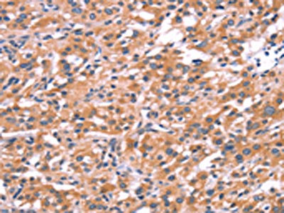
(The image on the left is immunohistochemistry of paraffin-embedded Human gastric cancer tissue using AAA241225(HP Antibody) at dilution 1/50, on the right is treated with synthetic peptide. (Original magnification: ×200))
IHC (Immunohiostchemistry)
(The image on the left is immunohistochemistry of paraffin-embedded Human gastric cancer tissue using AAA241225(HP Antibody) at dilution 1/50, on the right is treated with synthetic peptide. (Original magnification: ×200))
HP, Polyclonal Antibody (Cat# AAA241225)
 IHC (Immunohiostchemistry)
(The image on the left is immunohistochemistry of paraffin-embedded Human thyroid cancer tissue using AAA241231(HRH1 Antibody) at dilution 1/40, on the right is treated with synthetic peptide. (Original magnification: ×200))
IHC (Immunohiostchemistry)
(The image on the left is immunohistochemistry of paraffin-embedded Human thyroid cancer tissue using AAA241231(HRH1 Antibody) at dilution 1/40, on the right is treated with synthetic peptide. (Original magnification: ×200))
HRH1, Polyclonal Antibody (Cat# AAA241231)
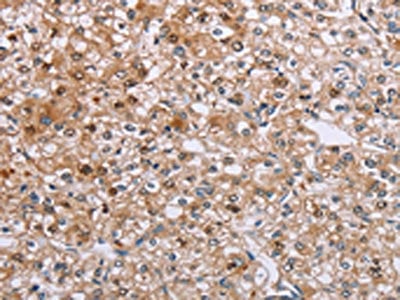
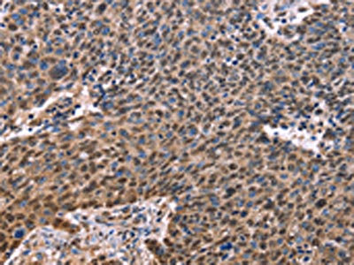
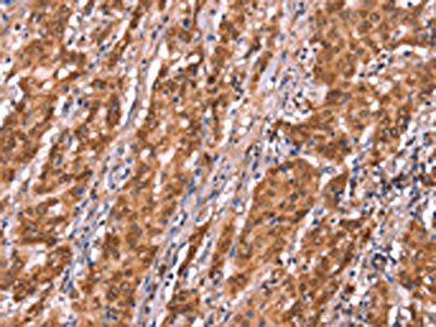
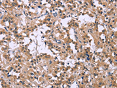
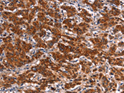
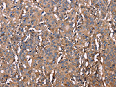
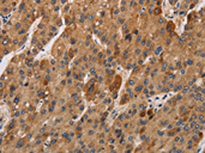
 IHC (Immunohiostchemistry)
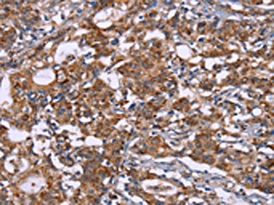
(The image on the left is immunohistochemistry of paraffin-embedded Human liver cancer tissue using AAA241235(HRK Antibody) at dilution 1/40, on the right is treated with synthetic peptide. (Original magnification: ×200))
IHC (Immunohiostchemistry)
(The image on the left is immunohistochemistry of paraffin-embedded Human liver cancer tissue using AAA241235(HRK Antibody) at dilution 1/40, on the right is treated with synthetic peptide. (Original magnification: ×200))
HRK, Polyclonal Antibody (Cat# AAA241235)
 SDS-PAGE
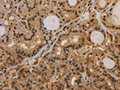
(Gel: 8%SDS-PAGE,Lysate: 40 ug,Lane 1-2: Human placenta tissue, Human normal kidney tissue,Primary antibody: AAA241236(HSD11B2 Antibody) at dilution 1/200 dilution,Secondary antibody: Goat anti rabbit IgG at 1/8000 dilution,Exposure time: 30 seconds)
SDS-PAGE
(Gel: 8%SDS-PAGE,Lysate: 40 ug,Lane 1-2: Human placenta tissue, Human normal kidney tissue,Primary antibody: AAA241236(HSD11B2 Antibody) at dilution 1/200 dilution,Secondary antibody: Goat anti rabbit IgG at 1/8000 dilution,Exposure time: 30 seconds)
HSD11B2, Polyclonal Antibody (Cat# AAA241236)
 SDS-PAGE
(Gel: 8%SDS-PAGE, Lysate: 40 ug, Lane: Human placenta tissue, Primary antibody: AAA241239(HSD17B1 Antibody) at dilution 1/600, Secondary antibody: Goat anti rabbit IgG at 1/8000 dilution, Exposure time: 1 second)
SDS-PAGE
(Gel: 8%SDS-PAGE, Lysate: 40 ug, Lane: Human placenta tissue, Primary antibody: AAA241239(HSD17B1 Antibody) at dilution 1/600, Secondary antibody: Goat anti rabbit IgG at 1/8000 dilution, Exposure time: 1 second)
HSD17B1, Polyclonal Antibody (Cat# AAA241239)
 IHC (Immunohiostchemistry)
(The image on the left is immunohistochemistry of paraffin-embedded Human gastric cancer tissue using AAA241241(HSPE1 Antibody) at dilution 1/40, on the right is treated with synthetic peptide. (Original magnification: ×200))
IHC (Immunohiostchemistry)
(The image on the left is immunohistochemistry of paraffin-embedded Human gastric cancer tissue using AAA241241(HSPE1 Antibody) at dilution 1/40, on the right is treated with synthetic peptide. (Original magnification: ×200))
HSPE1, Polyclonal Antibody (Cat# AAA241241)
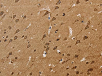
 IHC (Immunohiostchemistry)
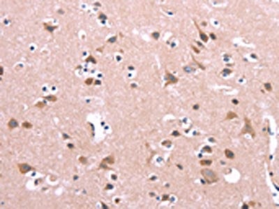
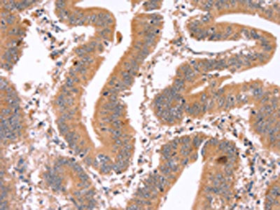
(The image on the left is immunohistochemistry of paraffin-embedded Human brain tissue using AAA241247(ICAM5 Antibody) at dilution 1/20, on the right is treated with synthetic peptide. (Original magnification: ×200))
IHC (Immunohiostchemistry)
(The image on the left is immunohistochemistry of paraffin-embedded Human brain tissue using AAA241247(ICAM5 Antibody) at dilution 1/20, on the right is treated with synthetic peptide. (Original magnification: ×200))
ICAM5, Polyclonal Antibody (Cat# AAA241247)
 IHC (Immunohiostchemistry)
(The image on the left is immunohistochemistry of paraffin-embedded Human colon cancer tissue using AAA241251(IFNA2 Antibody) at dilution 1/40, on the right is treated with synthetic peptide. (Original magnification: ×200))
IHC (Immunohiostchemistry)
(The image on the left is immunohistochemistry of paraffin-embedded Human colon cancer tissue using AAA241251(IFNA2 Antibody) at dilution 1/40, on the right is treated with synthetic peptide. (Original magnification: ×200))
IFNA2, Polyclonal Antibody (Cat# AAA241251)
 SDS-PAGE
(Gel: 10%SDS-PAGE, Lysate: 40 ug, Lane 1-4: A549 cells, K562 cells, Raji cells, human fetal intestine tissue, Primary antibody: AAA241258(IL17C Antibody) at dilution 1/300, Secondary antibody: Goat anti rabbit IgG at 1/8000 dilution, Exposure time: 20 seconds)
SDS-PAGE
(Gel: 10%SDS-PAGE, Lysate: 40 ug, Lane 1-4: A549 cells, K562 cells, Raji cells, human fetal intestine tissue, Primary antibody: AAA241258(IL17C Antibody) at dilution 1/300, Secondary antibody: Goat anti rabbit IgG at 1/8000 dilution, Exposure time: 20 seconds)
IL17C, Polyclonal Antibody (Cat# AAA241258)
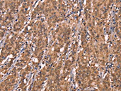
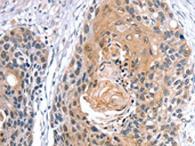
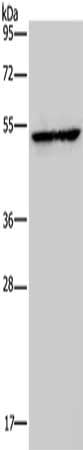
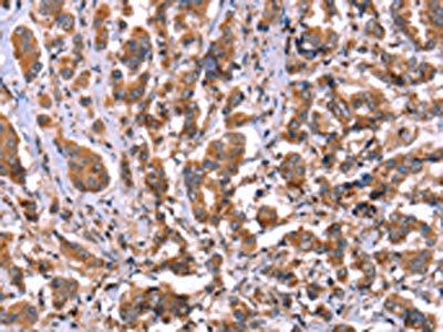
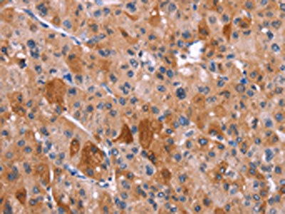
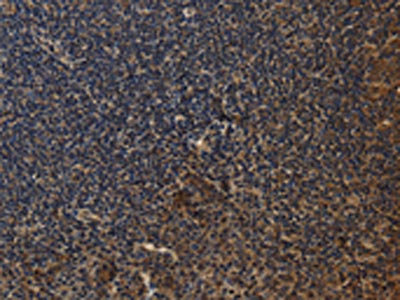
 IHC (Immunohiostchemistry)
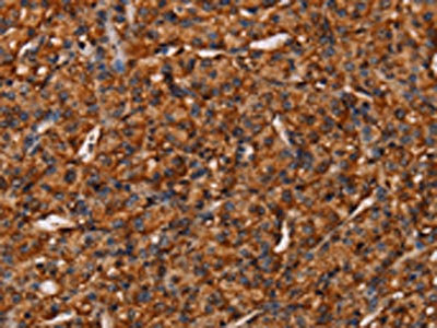
(The image on the left is immunohistochemistry of paraffin-embedded Human liver cancer tissue using AAA241260(IL17RA Antibody) at dilution 1/20, on the right is treated with synthetic peptide. (Original magnification: ×200))
IHC (Immunohiostchemistry)
(The image on the left is immunohistochemistry of paraffin-embedded Human liver cancer tissue using AAA241260(IL17RA Antibody) at dilution 1/20, on the right is treated with synthetic peptide. (Original magnification: ×200))
IL17RA, Polyclonal Antibody (Cat# AAA241260)
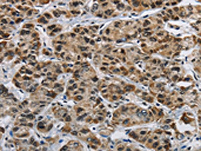
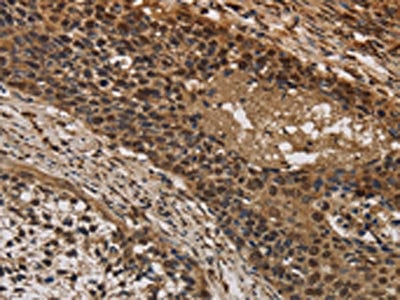 IHC (Immunohiostchemistry)
(The image on the left is immunohistochemistry of paraffin-embedded Human cervical cancer tissue using AAA241274(CYBA Antibody) at dilution 1/20, on the right is treated with synthetic peptide. (Original magnification: ×200))
IHC (Immunohiostchemistry)
(The image on the left is immunohistochemistry of paraffin-embedded Human cervical cancer tissue using AAA241274(CYBA Antibody) at dilution 1/20, on the right is treated with synthetic peptide. (Original magnification: ×200))
CYBA, Polyclonal Antibody (Cat# AAA241274)
 IHC (Immunohiostchemistry)
(The image on the left is immunohistochemistry of paraffin-embedded Human thyroid cancer tissue using AAA241278(KPNB1 Antibody) at dilution 1/40, on the right is treated with synthetic peptide. (Original magnification: ×200))
IHC (Immunohiostchemistry)
(The image on the left is immunohistochemistry of paraffin-embedded Human thyroid cancer tissue using AAA241278(KPNB1 Antibody) at dilution 1/40, on the right is treated with synthetic peptide. (Original magnification: ×200))
KPNB1, Polyclonal Antibody (Cat# AAA241278)
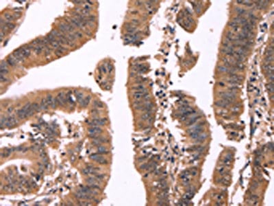
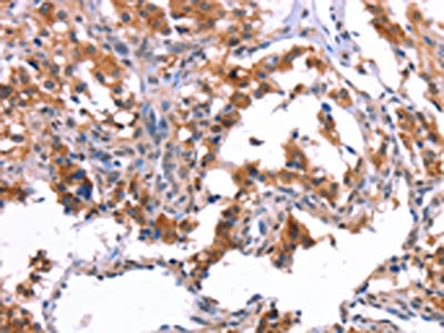
 IHC (Immunohiostchemistry)
(The image on the left is immunohistochemistry of paraffin-embedded Human esophagus cancer tissue using AAA241284(IPO8 Antibody) at dilution 1/50, on the right is treated with synthetic peptide. (Original magnification: ×200))
IHC (Immunohiostchemistry)
(The image on the left is immunohistochemistry of paraffin-embedded Human esophagus cancer tissue using AAA241284(IPO8 Antibody) at dilution 1/50, on the right is treated with synthetic peptide. (Original magnification: ×200))
IPO8, Polyclonal Antibody (Cat# AAA241284)
 IHC (Immunohiostchemistry)
(The image on the left is immunohistochemistry of paraffin-embedded Human esophagus cancer tissue using AAA241296(ITPR3 Antibody) at dilution 1/30, on the right is treated with synthetic peptide. (Original magnification: ×200))
IHC (Immunohiostchemistry)
(The image on the left is immunohistochemistry of paraffin-embedded Human esophagus cancer tissue using AAA241296(ITPR3 Antibody) at dilution 1/30, on the right is treated with synthetic peptide. (Original magnification: ×200))
ITPR3, Polyclonal Antibody (Cat# AAA241296)
 SDS-PAGE
(Gel: 10%SDS-PAGE, Lysate: 40 ug, Lane: Mouse brain tissue, Primary antibody: AAA241315(KCNMB4 Antibody) at dilution 1/500, Secondary antibody: Goat anti rabbit IgG at 1/8000 dilution, Exposure time: 1 minute)
SDS-PAGE
(Gel: 10%SDS-PAGE, Lysate: 40 ug, Lane: Mouse brain tissue, Primary antibody: AAA241315(KCNMB4 Antibody) at dilution 1/500, Secondary antibody: Goat anti rabbit IgG at 1/8000 dilution, Exposure time: 1 minute)
KCNMB4, Polyclonal Antibody (Cat# AAA241315)
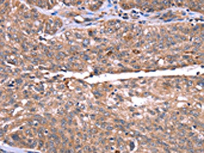
 IHC (Immunohiostchemistry)
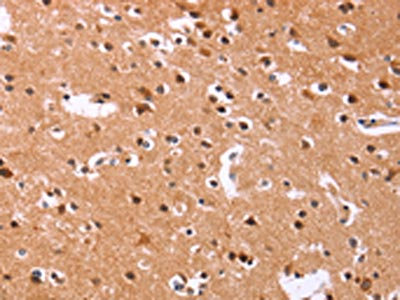
(The image on the left is immunohistochemistry of paraffin-embedded Human brain tissue using AAA241317(KCNN4 Antibody) at dilution 1/50, on the right is treated with synthetic peptide. (Original magnification: ×200))
IHC (Immunohiostchemistry)
(The image on the left is immunohistochemistry of paraffin-embedded Human brain tissue using AAA241317(KCNN4 Antibody) at dilution 1/50, on the right is treated with synthetic peptide. (Original magnification: ×200))
KCNN4, Polyclonal Antibody (Cat# AAA241317)
What are Polyclonal Antibodies?
Polyclonal antibodies are antibodies that come from multiple B cell clones of a host animal. The typical hosts used for the majority of polyclonal antibody production are rabbits, goats, sheep, and donkeys. These polyclonal antibodies, once having identified their target, will bind to different epitopes located at different regions or sequences on the same protein/antigen. As a result, they are ideal at locating and binding to the target, even if the target is in very low concentrations (due to many different antibodies being able to bind to the same target molecule, which allows for significant amplification of a downstream signal).
Polyclonal antibodies are typically produced by injecting an antigen into a host animal, which causes the animal’s immune system to attack the foreign antigen by mass generating antibodies against it. After a period of time, serum is collected from the animal and purified using physicochemical fractionation, class-specific affinity purification, and/or antigen-affinity purification.
Key Uses of Polyclonal Antibodies
- Western Blotting: This method is used to find specific proteins in biological samples after separating them by size.
- Immunohistochemistry: IHC helps visualize the location of proteins in tissue sections using various staining techniques.
- ELISA: (Enzyme-Linked Immunosorbent Assay) is typically used to identify specific protein quantities in a sample. ELISAs can be either “Quantitative” or “Qualitative”.
- Flow Cytometry: technique that identifies and measures the specific protein on the surface or inside the cells in a fluid suspension.
- Immunoprecipitation: IP isolates and studies a specific protein from a complex mixture using antibodies.
Why Buy Polyclonal Antibodies from AAA Biotech?
1. Ideal for Various Applications
Our antibodies are generally going to be validated for use in multiple types of assays, including ELISA, Western Blotting, Immunohistochemistry, Immunoprecipitation, amongst others. They are ideal for a wide range of research applications.
2. Rigorous Quality Control
All of the antibodies in our catalog undergo strict quality testing to ensure specificity, sensitivity, and consistent performance. We are confident in the ability of our antibodies to provide you with accurate results.
3. Wide Assortment of Antibodies
Antibodies in are catalog can be found for both common and exotic species, and these antibodies are also available in both conjugated and recombinant forms to suit many diverse experimental needs.
4. Highly Purified
Our antibodies are available in purified forms with over 85% purity, as confirmed by SDS-PAGE. They are also available with tags such as His, Flag, GST, or MBP. We cater to customers worldwide.
FAQ
1. How are polyclonal antibodies produced?
Traditionally, polyclonal antibodies are produced by injecting an antigen into a host animal (such as a rabbit or goat), which then triggers an immune response from the host animal. The animal’s B cells produce antibodies that will recognize different parts of the injected antigen. These antibodies are then collected from the animal’s blood and purified for use.
2. How do polyclonal antibodies differ from monoclonal antibodies?
Polyclonal antibodies are a mix of antibodies that bind to different locations (epitopes) of the same antigen, while monoclonal antibodies are identical and bind to just one specific epitope. This makes polyclonal antibodies more versatile and better at detecting proteins that may be present in low quantities or in altered/modified forms.
3. How should I store polyclonal antibodies?
Polyclonal antibodies should be stored at 4°C for short-term use (up to a few weeks) and at -20°C or -80°C for long-term storage. Avoid repeated freeze-thaw cycles by dividing them into small aliquots. Always check the datasheet for specific storage instructions.