Filters
▼Clonality
▼Type
▼Reactivity
▼Gene Name
▼Isotype
▼Host
▼Application
▼Clone
▼Polyclonal Antibodies
At AAA Biotech also known as AAA Bio or AAABio, we provide a broad range of purified polyclonal antibodies (pAbs) that are able to all be browsed online through our website. Due to their high specificity and strong binding affinity, these antibodies are ideal for wide swathes of research and experimental applications.
Our polyclonal antibodies can easily support your work, whether you use them for Western Blotting, Immunocytochemistry (with or without Immunofluorescence used in conjunction), Immunohistochemistry, Immunoprecipitation, and ELISA tests. We highly encourage you to browse our range of pAbs and choose the one that best suits your experimental model.
Viewing 6450-6500 of 96805 product results
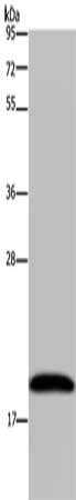 SDS-PAGE
(Gel: 10%SDS-PAGE, Lysate: 40 ug, Lane: 293T cells, Primary antibody: AAA241454(NDUFA8 Antibody) at dilution 1/300, Secondary antibody: Goat anti rabbit IgG at 1/8000 dilution, Exposure time: 1 minute)
SDS-PAGE
(Gel: 10%SDS-PAGE, Lysate: 40 ug, Lane: 293T cells, Primary antibody: AAA241454(NDUFA8 Antibody) at dilution 1/300, Secondary antibody: Goat anti rabbit IgG at 1/8000 dilution, Exposure time: 1 minute)
NDUFA8, Polyclonal Antibody (Cat# AAA241454)
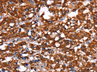
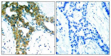
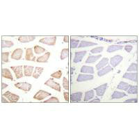
 IHC (Immunohiostchemistry)
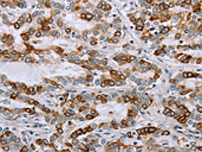
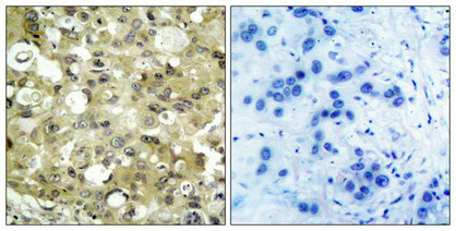
(The image on the left is immunohistochemistry of paraffin-embedded Human thyroid cancer tissue using AAA241461(NRP2 Antibody) at dilution 1/35, on the right is treated with synthetic peptide. (Original magnification: ×200))
IHC (Immunohiostchemistry)
(The image on the left is immunohistochemistry of paraffin-embedded Human thyroid cancer tissue using AAA241461(NRP2 Antibody) at dilution 1/35, on the right is treated with synthetic peptide. (Original magnification: ×200))
NRP2, Polyclonal Antibody (Cat# AAA241461)
 SDS-PAGE
(Gel: 8%SDS-PAGE,Lysate: 40 ug,,Primary antibody: AAA241470(ADRB3 Antibody) at dilution 1/200 dilution,Secondary antibody: Goat anti rabbit IgG at 1/8000 dilution,Exposure time: 2 minutes)
SDS-PAGE
(Gel: 8%SDS-PAGE,Lysate: 40 ug,,Primary antibody: AAA241470(ADRB3 Antibody) at dilution 1/200 dilution,Secondary antibody: Goat anti rabbit IgG at 1/8000 dilution,Exposure time: 2 minutes)
ADRB3, Polyclonal Antibody (Cat# AAA241470)
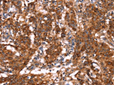
 IHC (Immunohiostchemistry)
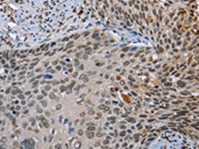
(The image on the left is immunohistochemistry of paraffin-embedded Human cervical cancer tissue using AAA241482(NMT1 Antibody) at dilution 1/45, on the right is treated with synthetic peptide. (Original magnification: ×200))
IHC (Immunohiostchemistry)
(The image on the left is immunohistochemistry of paraffin-embedded Human cervical cancer tissue using AAA241482(NMT1 Antibody) at dilution 1/45, on the right is treated with synthetic peptide. (Original magnification: ×200))
NMT1, Polyclonal Antibody (Cat# AAA241482)
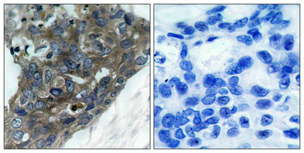
 IHC (Immunohiostchemistry)
(The image on the left is immunohistochemistry of paraffin-embedded Human colon cancer tissue using AAA241140(FOLH1B Antibody) at dilution 1/20, on the right is treated with synthetic peptide. (Original magnification: ×200))
IHC (Immunohiostchemistry)
(The image on the left is immunohistochemistry of paraffin-embedded Human colon cancer tissue using AAA241140(FOLH1B Antibody) at dilution 1/20, on the right is treated with synthetic peptide. (Original magnification: ×200))
FOLH1B, Polyclonal Antibody (Cat# AAA241140)
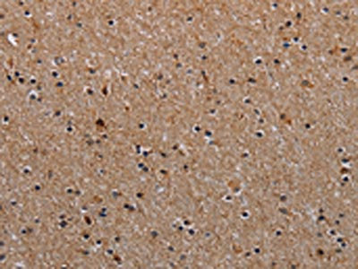
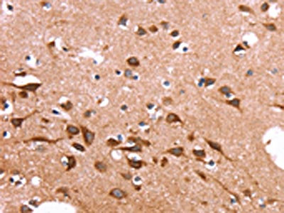 IHC (Immunohiostchemistry)
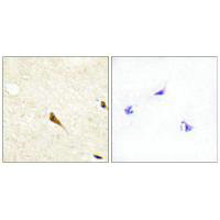
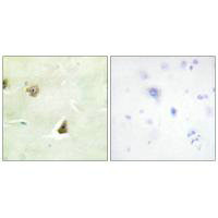
(The image on the left is immunohistochemistry of paraffin-embedded Human brain tissue using AAA241143(SPAST Antibody) at dilution 1/20, on the right is treated with synthetic peptide. (Original magnification: ×200))
IHC (Immunohiostchemistry)
(The image on the left is immunohistochemistry of paraffin-embedded Human brain tissue using AAA241143(SPAST Antibody) at dilution 1/20, on the right is treated with synthetic peptide. (Original magnification: ×200))
SPAST, Polyclonal Antibody (Cat# AAA241143)
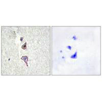
 IHC (Immunohiostchemistry)
(The image on the left is immunohistochemistry of paraffin-embedded Human gastric cancer tissue using AAA241149(FXR1 Antibody) at dilution 1/20, on the right is treated with synthetic peptide. (Original magnification: ×200))
IHC (Immunohiostchemistry)
(The image on the left is immunohistochemistry of paraffin-embedded Human gastric cancer tissue using AAA241149(FXR1 Antibody) at dilution 1/20, on the right is treated with synthetic peptide. (Original magnification: ×200))
FXR1, Polyclonal Antibody (Cat# AAA241149)
 IHC (Immunohiostchemistry)
(The image on the left is immunohistochemistry of paraffin-embedded Human gastric cancer tissue using AAA241151(SLC6A1 Antibody) at dilution 1/25, on the right is treated with synthetic peptide. (Original magnification: ×200))
IHC (Immunohiostchemistry)
(The image on the left is immunohistochemistry of paraffin-embedded Human gastric cancer tissue using AAA241151(SLC6A1 Antibody) at dilution 1/25, on the right is treated with synthetic peptide. (Original magnification: ×200))
SLC6A1, Polyclonal Antibody (Cat# AAA241151)
 SDS-PAGE
(Gel: 8%SDS-PAGE, Lysate: 40 ug, Lane: LoVo cells, Primary antibody: AAA241153(GABRA1 Antibody) at dilution 1/300, Secondary antibody: Goat anti rabbit IgG at 1/8000 dilution, Exposure time: 1 minute)
SDS-PAGE
(Gel: 8%SDS-PAGE, Lysate: 40 ug, Lane: LoVo cells, Primary antibody: AAA241153(GABRA1 Antibody) at dilution 1/300, Secondary antibody: Goat anti rabbit IgG at 1/8000 dilution, Exposure time: 1 minute)
GABRA1, Polyclonal Antibody (Cat# AAA241153)
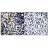
 IHC (Immunohiostchemistry)
(The image on the left is immunohistochemistry of paraffin-embedded Human gastric cancer tissue using AAA241156(GAGE12I Antibody) at dilution 1/30, on the right is treated with synthetic peptide. (Original magnification: ×200))
IHC (Immunohiostchemistry)
(The image on the left is immunohistochemistry of paraffin-embedded Human gastric cancer tissue using AAA241156(GAGE12I Antibody) at dilution 1/30, on the right is treated with synthetic peptide. (Original magnification: ×200))
GAGE12I, Polyclonal Antibody (Cat# AAA241156)
 IHC (Immunohiostchemistry)
(The image on the left is immunohistochemistry of paraffin-embedded Human gastric cancer tissue using AAA241162(TUBGCP2 Antibody) at dilution 1/15, on the right is treated with synthetic peptide. (Original magnification: ×200))
IHC (Immunohiostchemistry)
(The image on the left is immunohistochemistry of paraffin-embedded Human gastric cancer tissue using AAA241162(TUBGCP2 Antibody) at dilution 1/15, on the right is treated with synthetic peptide. (Original magnification: ×200))
TUBGCP2, Polyclonal Antibody (Cat# AAA241162)
 IHC (Immunohiostchemistry)
(The image on the left is immunohistochemistry of paraffin-embedded Human gastric cancer tissue using AAA241163(GPHN Antibody) at dilution 1/30, on the right is treated with synthetic peptide. (Original magnification: ×200))
IHC (Immunohiostchemistry)
(The image on the left is immunohistochemistry of paraffin-embedded Human gastric cancer tissue using AAA241163(GPHN Antibody) at dilution 1/30, on the right is treated with synthetic peptide. (Original magnification: ×200))
GPHN, Polyclonal Antibody (Cat# AAA241163)
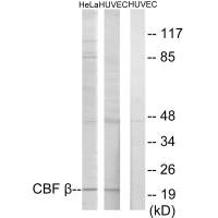
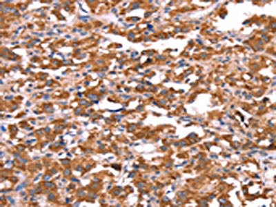
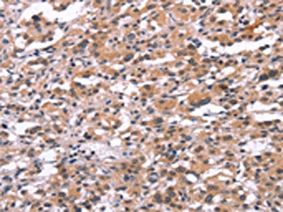
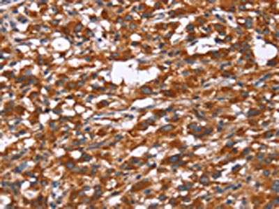
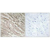
 IHC (Immunohiostchemistry)
(Immunohistochemistry analysis of paraffin-embedded human heart tissue using CBF beta antibody.)
IHC (Immunohiostchemistry)
(Immunohistochemistry analysis of paraffin-embedded human heart tissue using CBF beta antibody.)
CBFB, Polyclonal Antibody (Cat# AAA242439)
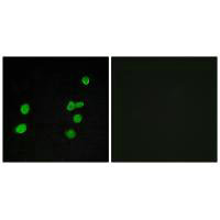
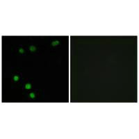
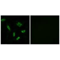
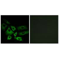
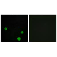 IF (Immunofluorescence)
(Immunofluorescence analysis of MCF-7 cells, using HLX1 antibody.)
IF (Immunofluorescence)
(Immunofluorescence analysis of MCF-7 cells, using HLX1 antibody.)
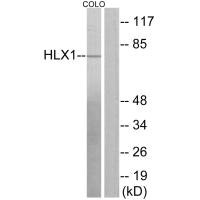
HLX, Polyclonal Antibody (Cat# AAA242440)
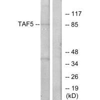
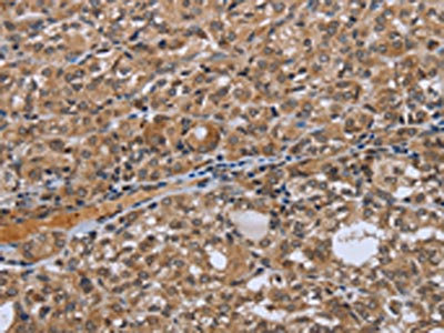
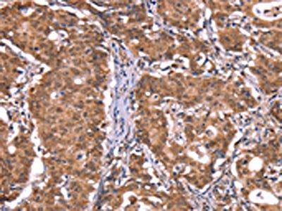
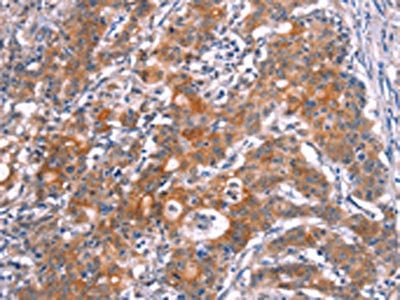
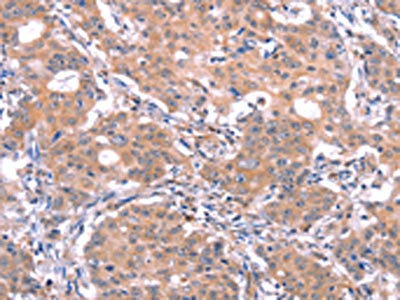
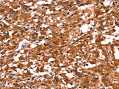
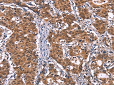
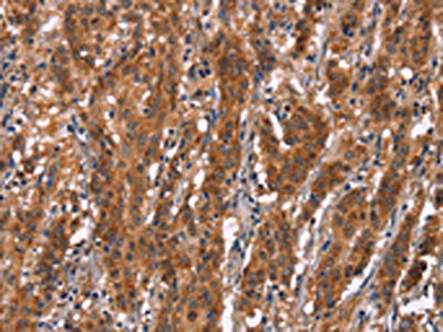
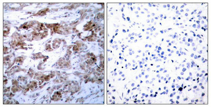
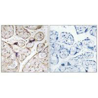
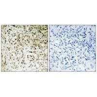 IHC (Immunohiostchemistry)
(Immunohistochemistry analysis of paraffin-embedded human liver carcinoma tissue using TAF5 antibody.)
IHC (Immunohiostchemistry)
(Immunohistochemistry analysis of paraffin-embedded human liver carcinoma tissue using TAF5 antibody.)
TAF5, Polyclonal Antibody (Cat# AAA242444)

 IF (Immunofluorescence)
(Immunofluorescence analysis of MCF-7 cells, using p97 MAPK antibody.)
IF (Immunofluorescence)
(Immunofluorescence analysis of MCF-7 cells, using p97 MAPK antibody.)
MAPK6, Polyclonal Antibody (Cat# AAA242448)
 IF (Immunofluorescence)
(Immunofluorescence analysis of MCF-7 cells, using TP53INP2 antibody.)
IF (Immunofluorescence)
(Immunofluorescence analysis of MCF-7 cells, using TP53INP2 antibody.)
TP53INP2, Polyclonal Antibody (Cat# AAA242458)
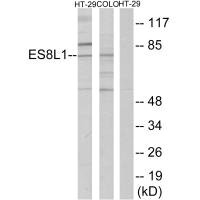
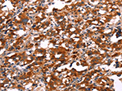
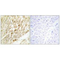
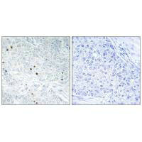
 IHC (Immunohiostchemistry)
(Immunohistochemistry analysis of paraffin-embedded human colon carcinoma tissue using ES8L1 antibody.)
IHC (Immunohiostchemistry)
(Immunohistochemistry analysis of paraffin-embedded human colon carcinoma tissue using ES8L1 antibody.)
EPS8L1, Polyclonal Antibody (Cat# AAA242465)
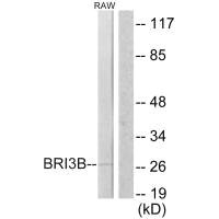
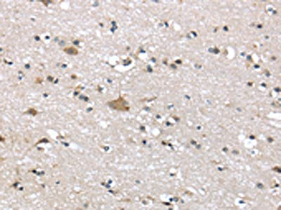
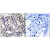
 IHC (Immunohiostchemistry)
(Immunohistochemistry analysis of paraffin-embedded human brain tissue using BRI3B antibody.)
IHC (Immunohiostchemistry)
(Immunohistochemistry analysis of paraffin-embedded human brain tissue using BRI3B antibody.)
BRI3BP, Polyclonal Antibody (Cat# AAA242468)
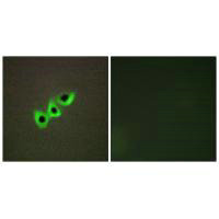 IF (Immunofluorescence)
(Immunofluorescence analysis of A549 cells, using RHG07 antibody.)
IF (Immunofluorescence)
(Immunofluorescence analysis of A549 cells, using RHG07 antibody.)
DLC1, Polyclonal Antibody (Cat# AAA242473)
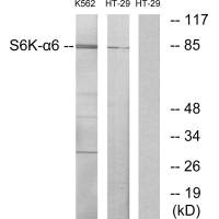
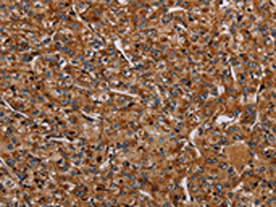
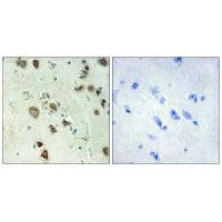 IHC (Immunohiostchemistry)
(Immunohistochemistry analysis of paraffin-embedded human brain tissue using S6K-alpha6 antibody.)
IHC (Immunohiostchemistry)
(Immunohistochemistry analysis of paraffin-embedded human brain tissue using S6K-alpha6 antibody.)
RPS6KA6, Polyclonal Antibody (Cat# AAA242487)
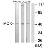
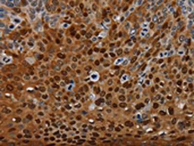
 IHC (Immunohiostchemistry)
(Immunohistochemistry analysis of paraffin-embedded human brain tissue using MOK antibody.)
IHC (Immunohiostchemistry)
(Immunohistochemistry analysis of paraffin-embedded human brain tissue using MOK antibody.)
MOK, Polyclonal Antibody (Cat# AAA242491)
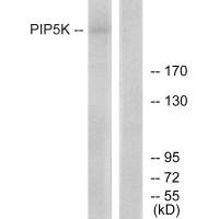
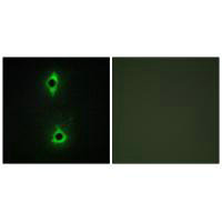 IF (Immunofluorescence)
(Immunofluorescence analysis of COS7 cells, using PIP5K antibody.)
IF (Immunofluorescence)
(Immunofluorescence analysis of COS7 cells, using PIP5K antibody.)
PIKFYVE, Polyclonal Antibody (Cat# AAA242492)
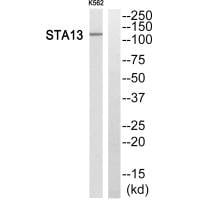
 IF (Immunofluorescence)
(Immunofluorescence analysis of A549 cells, using STA13 antibody.)
IF (Immunofluorescence)
(Immunofluorescence analysis of A549 cells, using STA13 antibody.)
STARD13, Polyclonal Antibody (Cat# AAA242493)
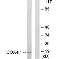
 IHC (Immunohiostchemistry)
(Immunohistochemistry analysis of paraffin-embedded human colon carcinoma tissue using COX41 antibody.)
IHC (Immunohiostchemistry)
(Immunohistochemistry analysis of paraffin-embedded human colon carcinoma tissue using COX41 antibody.)
COX4I1, Polyclonal Antibody (Cat# AAA242517)
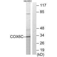
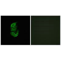
 IF (Immunofluorescence)
(Immunofluorescence analysis of HepG2 cells, using COX6C antibody.)
IF (Immunofluorescence)
(Immunofluorescence analysis of HepG2 cells, using COX6C antibody.)
COX6C, Polyclonal Antibody (Cat# AAA242518)
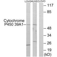
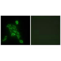
 IF (Immunofluorescence)
(Immunofluorescence analysis of A549 cells, using Cytochrome P450 39A1 antibody.)
IF (Immunofluorescence)
(Immunofluorescence analysis of A549 cells, using Cytochrome P450 39A1 antibody.)
CYP39A1, Polyclonal Antibody (Cat# AAA242521)
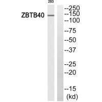
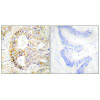
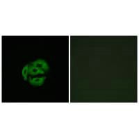
 IHC (Immunohiostchemistry)
(Immunohistochemistry analysis of paraffin-embedded human lung carcinoma tissue, using ZBTB40 antibody.)
IHC (Immunohiostchemistry)
(Immunohistochemistry analysis of paraffin-embedded human lung carcinoma tissue, using ZBTB40 antibody.)
ZBTB40, Polyclonal Antibody (Cat# AAA242692)
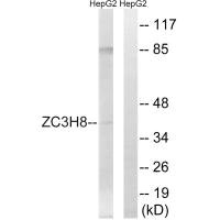
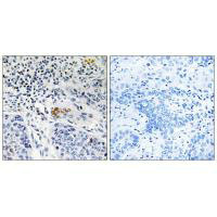
 IHC (Immunohiostchemistry)
(Immunohistochemistry analysis of paraffin-embedded human colon carcinoma tissue, using ZC3H8 antibody.)
IHC (Immunohiostchemistry)
(Immunohistochemistry analysis of paraffin-embedded human colon carcinoma tissue, using ZC3H8 antibody.)
ZC3H8, Polyclonal Antibody (Cat# AAA242694)
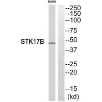
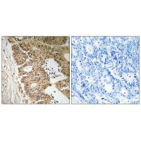
 IHC (Immunohiostchemistry)
(Immunohistochemistry analysis of paraffin-embedded human tonsil tissue, using STK17B antibody.)
IHC (Immunohiostchemistry)
(Immunohistochemistry analysis of paraffin-embedded human tonsil tissue, using STK17B antibody.)
STK17B, Polyclonal Antibody (Cat# AAA242702)
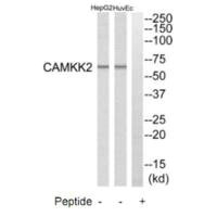
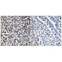
 IHC (Immunohiostchemistry)
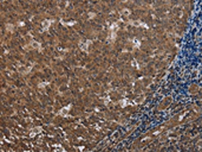
(Immunohistochemistry analysis of paraffin-embedded human prostate carcinoma tissue, using CAMKK2 antibody.)
IHC (Immunohiostchemistry)
(Immunohistochemistry analysis of paraffin-embedded human prostate carcinoma tissue, using CAMKK2 antibody.)
CAMKK2, Polyclonal Antibody (Cat# AAA242704)
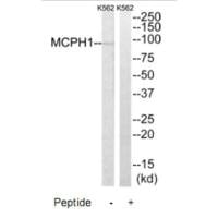
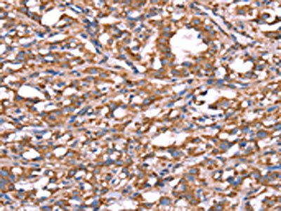
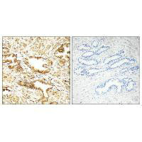
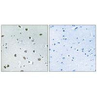
 IHC (Immunohiostchemistry)
(Immunohistochemistry analysis of paraffin-embedded human thymus gland tissue using MCPH1 antibody.)
IHC (Immunohiostchemistry)
(Immunohistochemistry analysis of paraffin-embedded human thymus gland tissue using MCPH1 antibody.)
MCPH1, Polyclonal Antibody (Cat# AAA242705)
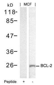
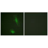
 IF (Immunofluorescence)
(Immunofluorescence staining of methanol-fixed Hela cells using BCL-2(Ab-70) Antibody.)
IF (Immunofluorescence)
(Immunofluorescence staining of methanol-fixed Hela cells using BCL-2(Ab-70) Antibody.)
BCL2, Polyclonal Antibody (Cat# AAA242738)
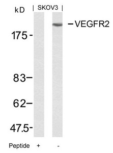
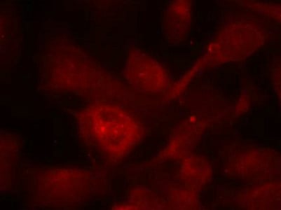
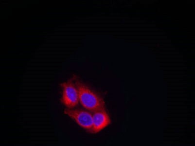 IF (Immunofluorescence)
(Immunofluorescence staining of methanol-fixed MCF cells using VEGFR2(Ab-951) Antibody.)
IF (Immunofluorescence)
(Immunofluorescence staining of methanol-fixed MCF cells using VEGFR2(Ab-951) Antibody.)
KDR, Polyclonal Antibody (Cat# AAA242744)
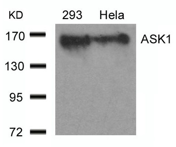
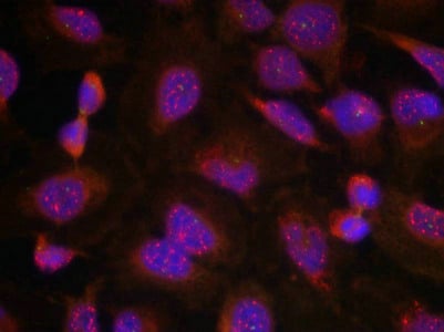
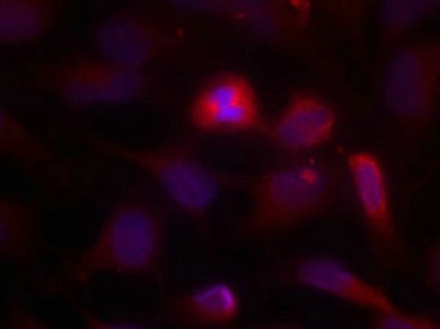 IF (Immunofluorescence)
(Immunofluorescence staining of methanol-fixed Hela cells using ASK1(Ab-966) Antibody.)
IF (Immunofluorescence)
(Immunofluorescence staining of methanol-fixed Hela cells using ASK1(Ab-966) Antibody.)
MAP3K5, Polyclonal Antibody (Cat# AAA242750)
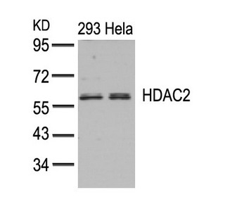
 IF (Immunofluorescence)
(Immunofluorescence staining of methanol-fixed Hela cells using HDAC2(Ab-394) Antibody.)
IF (Immunofluorescence)
(Immunofluorescence staining of methanol-fixed Hela cells using HDAC2(Ab-394) Antibody.)
HDAC2, Polyclonal Antibody (Cat# AAA242752)
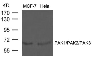
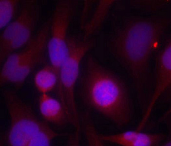
 IF (Immunofluorescence)
(Immunofluorescence staining of methanol-fixed Hela cells using PAK1/PAK2/PAK3(Ab-423/402/421) Antibody.)
IF (Immunofluorescence)
(Immunofluorescence staining of methanol-fixed Hela cells using PAK1/PAK2/PAK3(Ab-423/402/421) Antibody.)
PAK1/PAK2/PAK3, Polyclonal Antibody (Cat# AAA242757)

 IF (Immunofluorescence)
(Immunofluorescence analysis of HeLa cells, using DUSP6 antibody.)
IF (Immunofluorescence)
(Immunofluorescence analysis of HeLa cells, using DUSP6 antibody.)
DUSP6, Polyclonal Antibody (Cat# AAA242943)
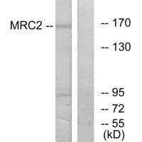
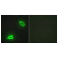
 IF (Immunofluorescence)
(Immunofluorescence analysis of HepG2 cells, using MRC2 antibody.)
IF (Immunofluorescence)
(Immunofluorescence analysis of HepG2 cells, using MRC2 antibody.)
MRC2, Polyclonal Antibody (Cat# AAA242963)
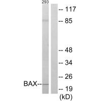
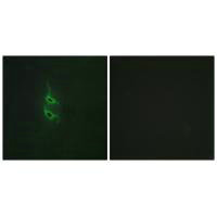
 IF (Immunofluorescence)
(Immunofluorescence analysis of HUVEC cells, using BAX antibody.)
IF (Immunofluorescence)
(Immunofluorescence analysis of HUVEC cells, using BAX antibody.)
BAX, Polyclonal Antibody (Cat# AAA242976)
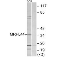
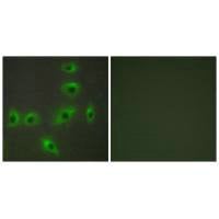
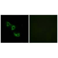 IF (Immunofluorescence)
(Immunofluorescence analysis of HuvEc cells, using MRPL44 antibody.)
IF (Immunofluorescence)
(Immunofluorescence analysis of HuvEc cells, using MRPL44 antibody.)
MRPL44, Polyclonal Antibody (Cat# AAA242984)
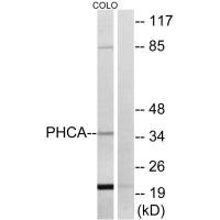
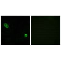 IF (Immunofluorescence)
(Immunofluorescence analysis of MCF-7 cells, using PHCA antibody.)
IF (Immunofluorescence)
(Immunofluorescence analysis of MCF-7 cells, using PHCA antibody.)
ACER3, Polyclonal Antibody (Cat# AAA242986)
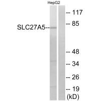
 IF (Immunofluorescence)
(Immunofluorescence analysis of A549 cells, using SLC27A5 antibody.)
IF (Immunofluorescence)
(Immunofluorescence analysis of A549 cells, using SLC27A5 antibody.)
SLC27A5, Polyclonal Antibody (Cat# AAA242990)
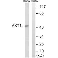
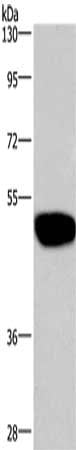
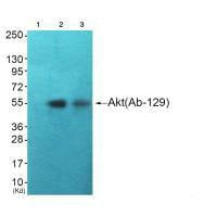 WB (Western Blot)
(Western blot analysis of extracts from HuvEc cells (Lane 2) and JK cells (Lane 3), using Akt (Ab-129) antiobdy. The lane on the left is treated with synthesized peptide.)
WB (Western Blot)
(Western blot analysis of extracts from HuvEc cells (Lane 2) and JK cells (Lane 3), using Akt (Ab-129) antiobdy. The lane on the left is treated with synthesized peptide.)
AKT1, Polyclonal Antibody (Cat# AAA243003)
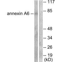
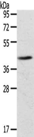
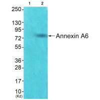 WB (Western Blot)
(Western blot analysis of extracts from HepG2 cells using Annexin A6 antibody. The lane on the left is treated with synthesized peptide.)
WB (Western Blot)
(Western blot analysis of extracts from HepG2 cells using Annexin A6 antibody. The lane on the left is treated with synthesized peptide.)
ANXA6, Polyclonal Antibody (Cat# AAA243007)
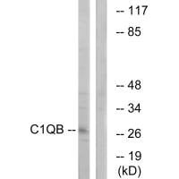
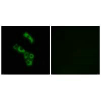 IF (Immunofluorescence)
(Immunofluorescence analysis of A549 cells, using C1QB antibody.)
IF (Immunofluorescence)
(Immunofluorescence analysis of A549 cells, using C1QB antibody.)
C1QB, Polyclonal Antibody (Cat# AAA242614)
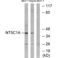
 IF (Immunofluorescence)
(Immunofluorescence analysis of A549 cells, using NT5C1A antibody.)
IF (Immunofluorescence)
(Immunofluorescence analysis of A549 cells, using NT5C1A antibody.)
NT5C1A, Polyclonal Antibody (Cat# AAA242619)
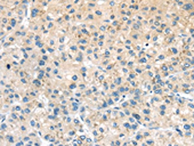
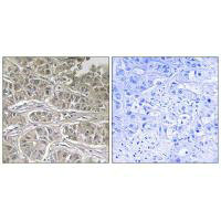 IHC (Immunohiostchemistry)
(Immunohistochemistry analysis of paraffin-embedded human liver carcinoma tissue using SLC25A21 antibody.)
IHC (Immunohiostchemistry)
(Immunohistochemistry analysis of paraffin-embedded human liver carcinoma tissue using SLC25A21 antibody.)
SLC25A21, Polyclonal Antibody (Cat# AAA242643)
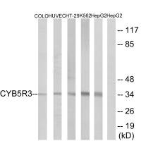
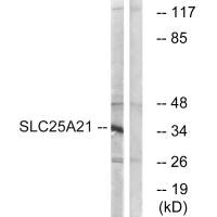
 IHC (Immunohiostchemistry)
(Immunohistochemistry analysis of paraffin-embedded human placenta tissue using CYB5R3 antibody.)
IHC (Immunohiostchemistry)
(Immunohistochemistry analysis of paraffin-embedded human placenta tissue using CYB5R3 antibody.)
CYB5R3, Polyclonal Antibody (Cat# AAA242646)
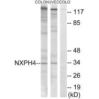
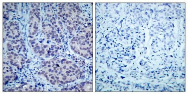
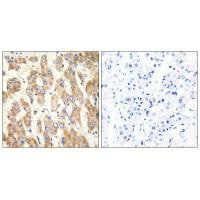
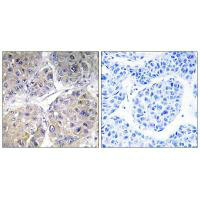
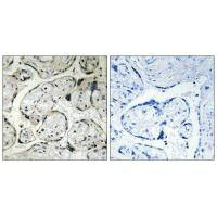
 IHC (Immunohiostchemistry)
(Immunohistochemistry analysis of paraffin-embedded human brain tissue using NXPH4 antibody.)
IHC (Immunohiostchemistry)
(Immunohistochemistry analysis of paraffin-embedded human brain tissue using NXPH4 antibody.)
NXPH4, Polyclonal Antibody (Cat# AAA242648)
What are Polyclonal Antibodies?
Polyclonal antibodies are antibodies that come from multiple B cell clones of a host animal. The typical hosts used for the majority of polyclonal antibody production are rabbits, goats, sheep, and donkeys. These polyclonal antibodies, once having identified their target, will bind to different epitopes located at different regions or sequences on the same protein/antigen. As a result, they are ideal at locating and binding to the target, even if the target is in very low concentrations (due to many different antibodies being able to bind to the same target molecule, which allows for significant amplification of a downstream signal).
Polyclonal antibodies are typically produced by injecting an antigen into a host animal, which causes the animal’s immune system to attack the foreign antigen by mass generating antibodies against it. After a period of time, serum is collected from the animal and purified using physicochemical fractionation, class-specific affinity purification, and/or antigen-affinity purification.
Key Uses of Polyclonal Antibodies
- Western Blotting: This method is used to find specific proteins in biological samples after separating them by size.
- Immunohistochemistry: IHC helps visualize the location of proteins in tissue sections using various staining techniques.
- ELISA: (Enzyme-Linked Immunosorbent Assay) is typically used to identify specific protein quantities in a sample. ELISAs can be either “Quantitative” or “Qualitative”.
- Flow Cytometry: technique that identifies and measures the specific protein on the surface or inside the cells in a fluid suspension.
- Immunoprecipitation: IP isolates and studies a specific protein from a complex mixture using antibodies.
Why Buy Polyclonal Antibodies from AAA Biotech?
1. Ideal for Various Applications
Our antibodies are generally going to be validated for use in multiple types of assays, including ELISA, Western Blotting, Immunohistochemistry, Immunoprecipitation, amongst others. They are ideal for a wide range of research applications.
2. Rigorous Quality Control
All of the antibodies in our catalog undergo strict quality testing to ensure specificity, sensitivity, and consistent performance. We are confident in the ability of our antibodies to provide you with accurate results.
3. Wide Assortment of Antibodies
Antibodies in are catalog can be found for both common and exotic species, and these antibodies are also available in both conjugated and recombinant forms to suit many diverse experimental needs.
4. Highly Purified
Our antibodies are available in purified forms with over 85% purity, as confirmed by SDS-PAGE. They are also available with tags such as His, Flag, GST, or MBP. We cater to customers worldwide.
FAQ
1. How are polyclonal antibodies produced?
Traditionally, polyclonal antibodies are produced by injecting an antigen into a host animal (such as a rabbit or goat), which then triggers an immune response from the host animal. The animal’s B cells produce antibodies that will recognize different parts of the injected antigen. These antibodies are then collected from the animal’s blood and purified for use.
2. How do polyclonal antibodies differ from monoclonal antibodies?
Polyclonal antibodies are a mix of antibodies that bind to different locations (epitopes) of the same antigen, while monoclonal antibodies are identical and bind to just one specific epitope. This makes polyclonal antibodies more versatile and better at detecting proteins that may be present in low quantities or in altered/modified forms.
3. How should I store polyclonal antibodies?
Polyclonal antibodies should be stored at 4°C for short-term use (up to a few weeks) and at -20°C or -80°C for long-term storage. Avoid repeated freeze-thaw cycles by dividing them into small aliquots. Always check the datasheet for specific storage instructions.