Filters
▼Clonality
▼Type
▼Reactivity
▼Gene Name
▼Isotype
▼Host
▼Application
▼Clone
▼Polyclonal Antibodies
At AAA Biotech also known as AAA Bio or AAABio, we provide a broad range of purified polyclonal antibodies (pAbs) that are able to all be browsed online through our website. Due to their high specificity and strong binding affinity, these antibodies are ideal for wide swathes of research and experimental applications.
Our polyclonal antibodies can easily support your work, whether you use them for Western Blotting, Immunocytochemistry (with or without Immunofluorescence used in conjunction), Immunohistochemistry, Immunoprecipitation, and ELISA tests. We highly encourage you to browse our range of pAbs and choose the one that best suits your experimental model.
Viewing 6250-6300 of 96805 product results
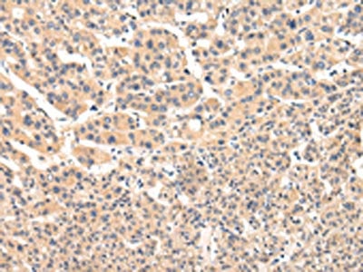
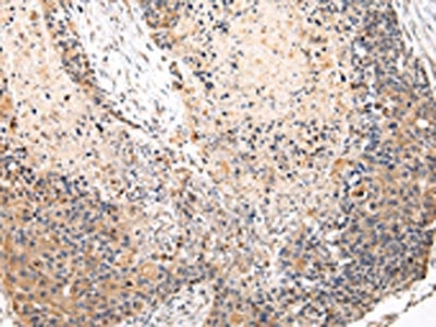
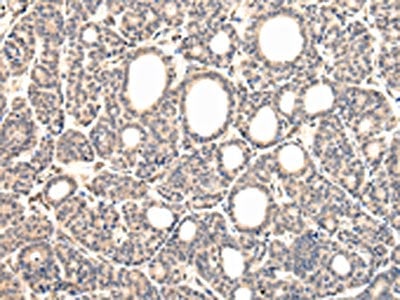
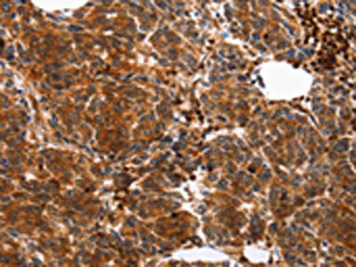
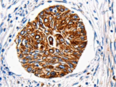
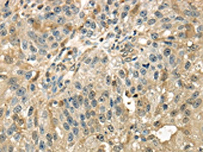
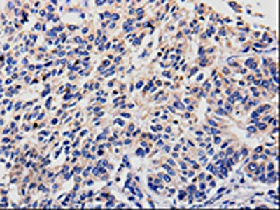
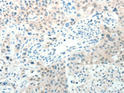
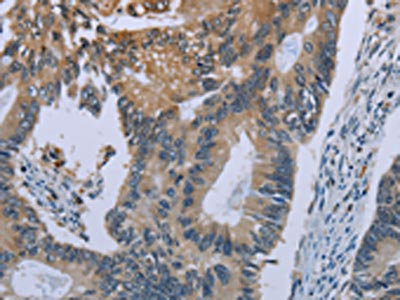
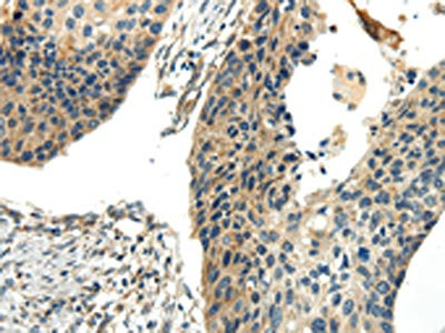
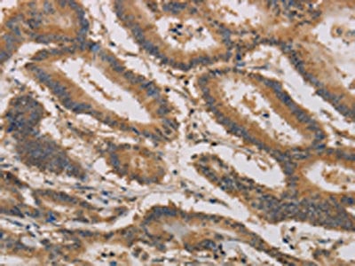
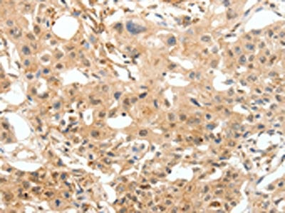
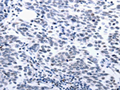
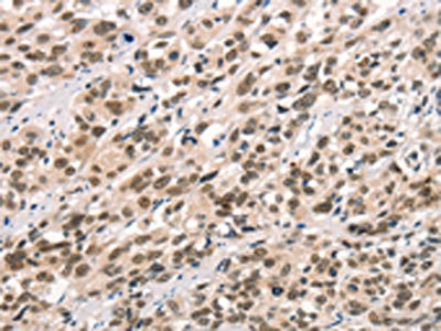
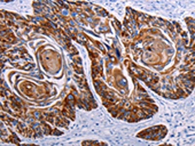
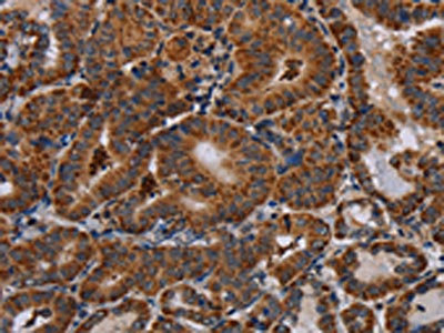
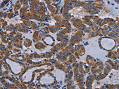
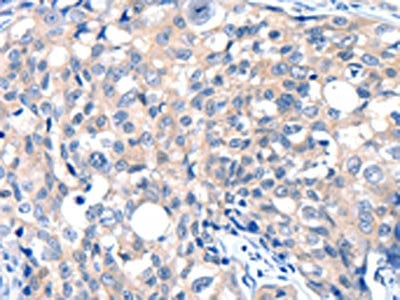
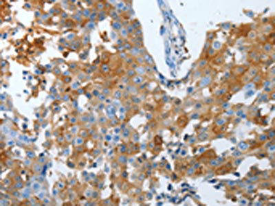
 IHC (Immunohiostchemistry)
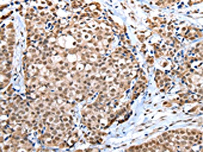
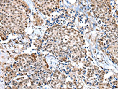
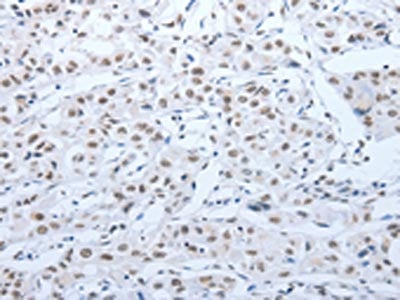
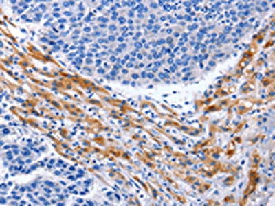
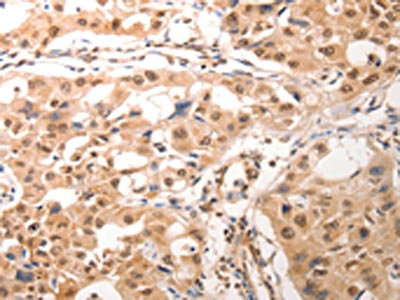
(The image on the left is immunohistochemistry of paraffin-embedded Human thyroid cancer tissue using AAA239626(VPS33B Antibody) at dilution 1/25, on the right is treated with fusion protein. (Original magnification: ×200))
IHC (Immunohiostchemistry)
(The image on the left is immunohistochemistry of paraffin-embedded Human thyroid cancer tissue using AAA239626(VPS33B Antibody) at dilution 1/25, on the right is treated with fusion protein. (Original magnification: ×200))
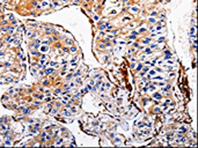
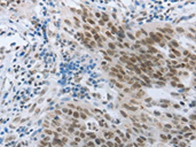
VPS33B, Polyclonal Antibody (Cat# AAA239626)
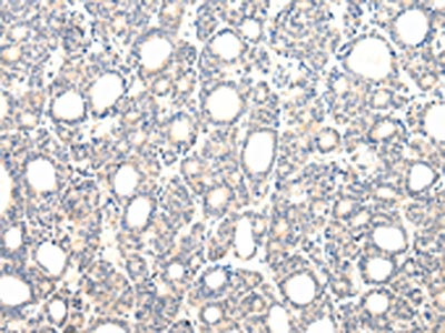
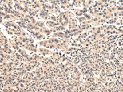
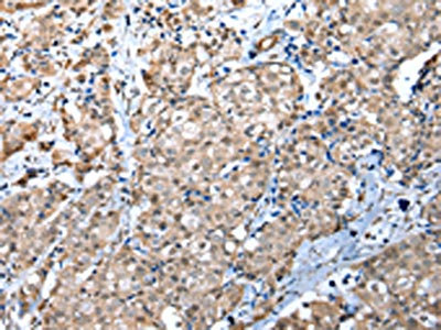
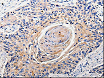
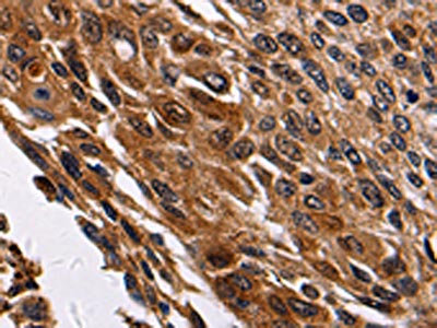
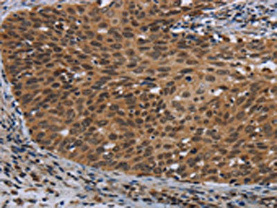
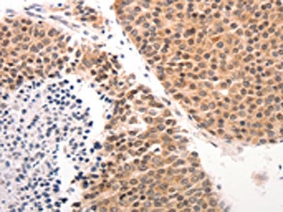
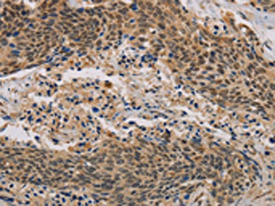
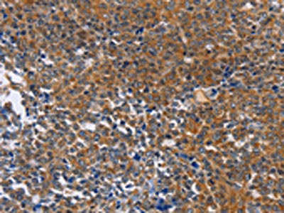
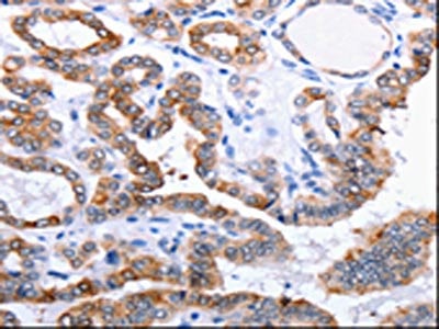
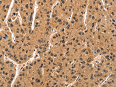
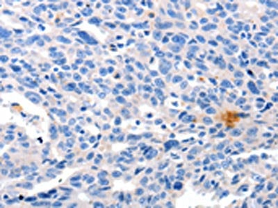
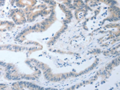
 IHC (Immunohiostchemistry)
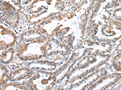
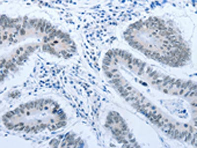
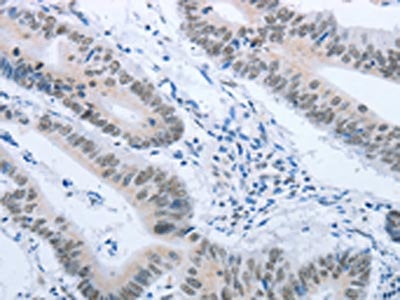
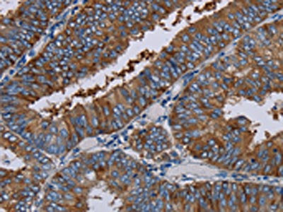
(The image on the left is immunohistochemistry of paraffin-embedded Human prostate cancer tissue using AAA239627(VPS33B Antibody) at dilution 1/40, on the right is treated with fusion protein. (Original magnification: ×200))
IHC (Immunohiostchemistry)
(The image on the left is immunohistochemistry of paraffin-embedded Human prostate cancer tissue using AAA239627(VPS33B Antibody) at dilution 1/40, on the right is treated with fusion protein. (Original magnification: ×200))
VPS33B, Polyclonal Antibody (Cat# AAA239627)
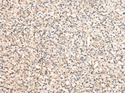
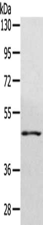
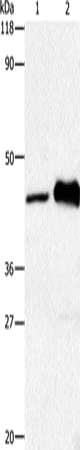
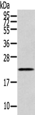
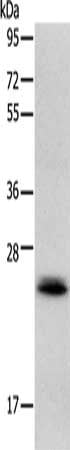
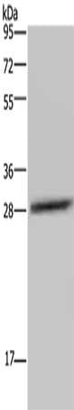
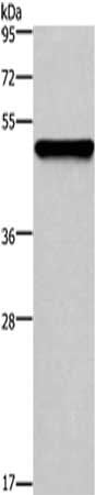
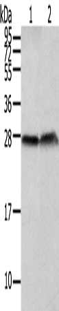
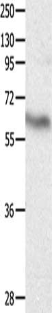
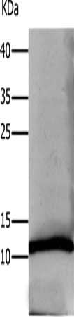
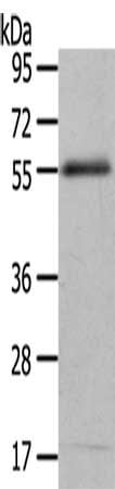
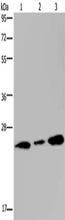
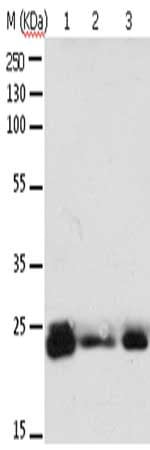
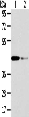 SDS-PAGE
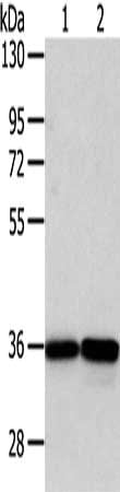
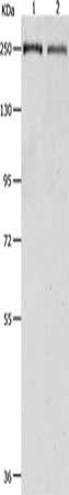
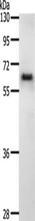
(Gel: 8%SDS-PAGE, Lysate: 40 ug, Lane 1-2: Human fetal liver tissue and hela cell, Primary antibody: AAA239628(VPS37A Antibody) at dilution 1/200 dilution, Secondary antibody: Goat anti rabbit IgG at 1/8000 dilution, Exposure time: 15 seconds)
SDS-PAGE
(Gel: 8%SDS-PAGE, Lysate: 40 ug, Lane 1-2: Human fetal liver tissue and hela cell, Primary antibody: AAA239628(VPS37A Antibody) at dilution 1/200 dilution, Secondary antibody: Goat anti rabbit IgG at 1/8000 dilution, Exposure time: 15 seconds)
VPS37A, Polyclonal Antibody (Cat# AAA239628)
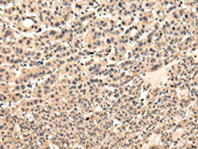
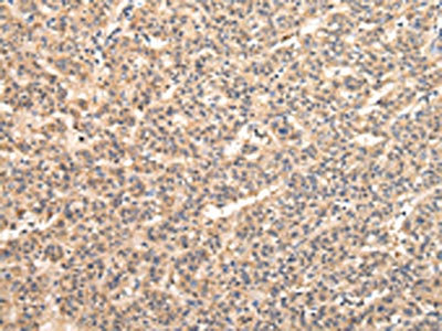
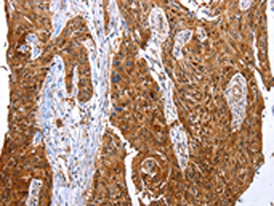
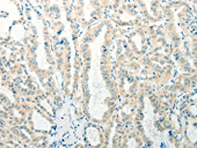
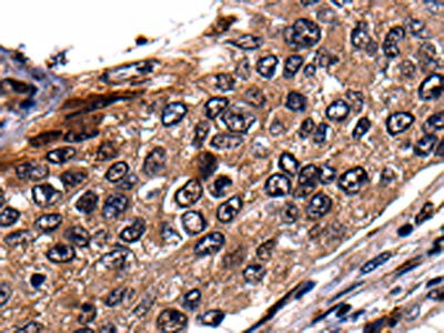
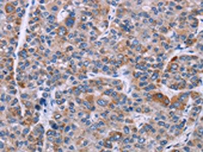
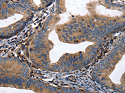
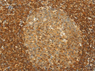
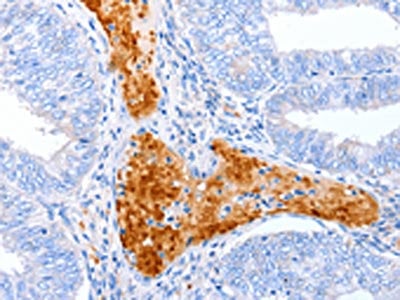
 IHC (Immunohiostchemistry)
(The image on the left is immunohistochemistry of paraffin-embedded Human thyroid cancer tissue using AAA239630(VPS37D Antibody) at dilution 1/30, on the right is treated with fusion protein. (Original magnification: ×200))
IHC (Immunohiostchemistry)
(The image on the left is immunohistochemistry of paraffin-embedded Human thyroid cancer tissue using AAA239630(VPS37D Antibody) at dilution 1/30, on the right is treated with fusion protein. (Original magnification: ×200))
VPS37D, Polyclonal Antibody (Cat# AAA239630)
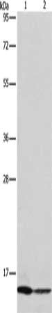
 SDS-PAGE
(Gel: 8%SDS-PAGE, Lysate: 40 ug, Lane: NIH/3T3 cell, Primary antibody: AAA239632(WDR4 Antibody) at dilution 1/450 dilution, Secondary antibody: Goat anti rabbit IgG at 1/8000 dilution, Exposure time: 30 seconds)
SDS-PAGE
(Gel: 8%SDS-PAGE, Lysate: 40 ug, Lane: NIH/3T3 cell, Primary antibody: AAA239632(WDR4 Antibody) at dilution 1/450 dilution, Secondary antibody: Goat anti rabbit IgG at 1/8000 dilution, Exposure time: 30 seconds)
WDR4, Polyclonal Antibody (Cat# AAA239632)
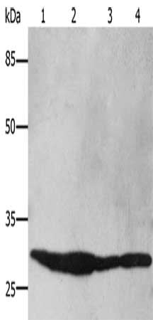
 SDS-PAGE
(Gel: 8%SDS-PAGE, Lysate: 40 ug, Lane 1-2: PC3 and A375 cell, Primary antibody: AAA239633(WDR5 Antibody) at dilution 1/350 dilution, Secondary antibody: Goat anti rabbit IgG at 1/8000 dilution, Exposure time: 5 seconds)
SDS-PAGE
(Gel: 8%SDS-PAGE, Lysate: 40 ug, Lane 1-2: PC3 and A375 cell, Primary antibody: AAA239633(WDR5 Antibody) at dilution 1/350 dilution, Secondary antibody: Goat anti rabbit IgG at 1/8000 dilution, Exposure time: 5 seconds)
WDR5, Polyclonal Antibody (Cat# AAA239633)
 SDS-PAGE
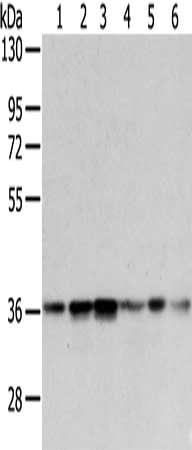
(Gel: 8%SDS-PAGE, Lysate: 40 ug, Lane 1-6: A375, 231 and PC3 cell, human lung cancer tissue, hela and NIH/3T3 cell, Primary antibody: AAA239634(WDR5 Antibody) at dilution 1/300 dilution, Secondary antibody: Goat anti rabbit IgG at 1/8000 dilution, Exposure time: 5 seconds)
SDS-PAGE
(Gel: 8%SDS-PAGE, Lysate: 40 ug, Lane 1-6: A375, 231 and PC3 cell, human lung cancer tissue, hela and NIH/3T3 cell, Primary antibody: AAA239634(WDR5 Antibody) at dilution 1/300 dilution, Secondary antibody: Goat anti rabbit IgG at 1/8000 dilution, Exposure time: 5 seconds)
WDR5, Polyclonal Antibody (Cat# AAA239634)
 SDS-PAGE
(Gel: 6%SDS-PAGE,Lysate: 40 ug,Lane 1-3: Hela cells, A549 cells, HUVEC cells,Primary antibody: AAA239637(APAF1 Antibody) at dilution 1/350 dilution,Secondary antibody: Goat anti rabbit IgG at 1/8000 dilution,Exposure time: 20 seconds)
SDS-PAGE
(Gel: 6%SDS-PAGE,Lysate: 40 ug,Lane 1-3: Hela cells, A549 cells, HUVEC cells,Primary antibody: AAA239637(APAF1 Antibody) at dilution 1/350 dilution,Secondary antibody: Goat anti rabbit IgG at 1/8000 dilution,Exposure time: 20 seconds)
APAF1, Polyclonal Antibody (Cat# AAA239637)
 SDS-PAGE
(Gel: 12%SDS-PAGE, Lysate: 40 ug, Lane 1-2: Jurkat cells, MCF7 cells, Primary antibody: AAA239639(CASP3 (active) Antibody) at dilution 1/120, Secondary antibody: Goat anti rabbit IgG at 1/8000 dilution, Exposure time: 20 seconds)
SDS-PAGE
(Gel: 12%SDS-PAGE, Lysate: 40 ug, Lane 1-2: Jurkat cells, MCF7 cells, Primary antibody: AAA239639(CASP3 (active) Antibody) at dilution 1/120, Secondary antibody: Goat anti rabbit IgG at 1/8000 dilution, Exposure time: 20 seconds)
CASP3, Polyclonal Antibody (Cat# AAA239639)
 SDS-PAGE
(Gel: 10%SDS-PAGE, Lysate: 40 ug, Lane 1-4: HT29 cells, 293T cells, Hela cells, Mouse brain tissue, Primary antibody: AAA239641(YWHAB Antibody) at dilution 1/250, Secondary antibody: Goat anti rabbit IgG at 1/8000 dilution, Exposure time: 1 second)
SDS-PAGE
(Gel: 10%SDS-PAGE, Lysate: 40 ug, Lane 1-4: HT29 cells, 293T cells, Hela cells, Mouse brain tissue, Primary antibody: AAA239641(YWHAB Antibody) at dilution 1/250, Secondary antibody: Goat anti rabbit IgG at 1/8000 dilution, Exposure time: 1 second)
YWHAB, Polyclonal Antibody (Cat# AAA239641)
 SDS-PAGE
(Gel: 8%SDS-PAGE, Lysate: 30 ug, Lane 1-2: HT29 cells, Mouse brain tissue, Primary antibody: AAA239642(HTR1A Antibody) at dilution 1/1500, Secondary antibody: Goat anti rabbit IgG at 1/8000 dilution, Exposure time: 2 minutes)
SDS-PAGE
(Gel: 8%SDS-PAGE, Lysate: 30 ug, Lane 1-2: HT29 cells, Mouse brain tissue, Primary antibody: AAA239642(HTR1A Antibody) at dilution 1/1500, Secondary antibody: Goat anti rabbit IgG at 1/8000 dilution, Exposure time: 2 minutes)
HTR1A, Polyclonal Antibody (Cat# AAA239642)
 SDS-PAGE
(Gel: 10%SDS-PAGE, Lysate: 20 ug, Lane: HepG2 cells, Primary antibody: AAA239648(ABCG2 Antibody) at dilution 1/1000, Secondary antibody: Goat anti rabbit IgG at 1/8000 dilution, Exposure time: 10 seconds)
SDS-PAGE
(Gel: 10%SDS-PAGE, Lysate: 20 ug, Lane: HepG2 cells, Primary antibody: AAA239648(ABCG2 Antibody) at dilution 1/1000, Secondary antibody: Goat anti rabbit IgG at 1/8000 dilution, Exposure time: 10 seconds)
ABCG2, Polyclonal Antibody (Cat# AAA239648)
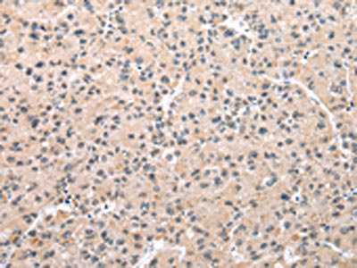
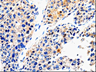
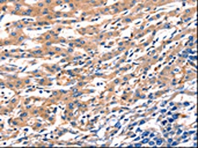
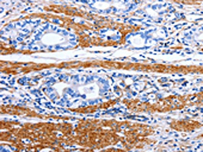
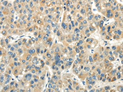
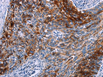
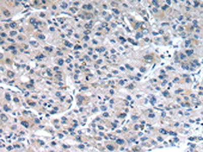
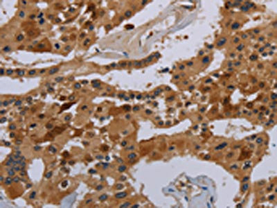
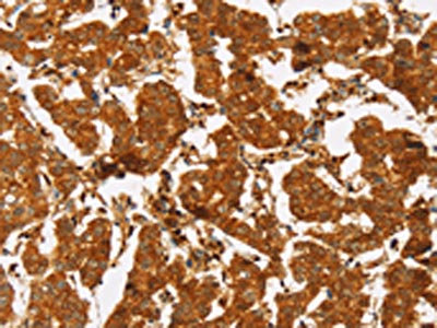
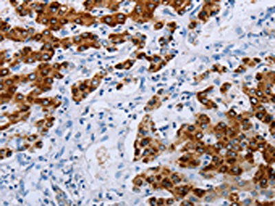
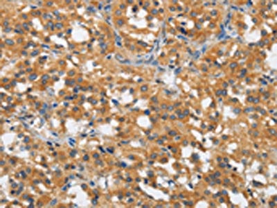
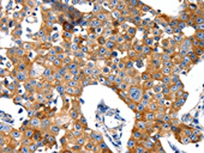
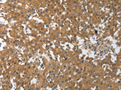
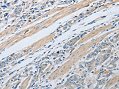
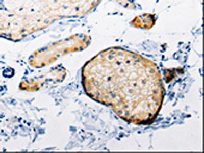
 IHC (Immunohiostchemistry)
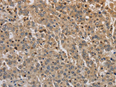
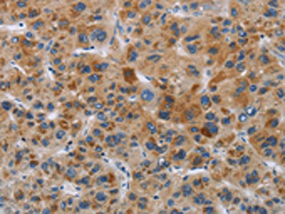
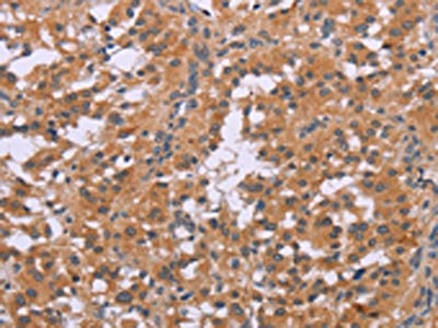
(The image is immunohistochemistry of paraffin-embedded Human liver cancer tissue using AAA239652(ACE2 Antibody) at dilution 1/40. (Original magnification: ×200))
IHC (Immunohiostchemistry)
(The image is immunohistochemistry of paraffin-embedded Human liver cancer tissue using AAA239652(ACE2 Antibody) at dilution 1/40. (Original magnification: ×200))
ACE2, Polyclonal Antibody (Cat# AAA239652)
 SDS-PAGE
(Gel: 8%SDS-PAGE, Lysate: 40 ug, Lane: 231 cells, Primary antibody: AAA239654(ACIN1 Antibody) at dilution 1/550, Secondary antibody: Goat anti rabbit IgG at 1/8000 dilution, Exposure time: 1 minute)
SDS-PAGE
(Gel: 8%SDS-PAGE, Lysate: 40 ug, Lane: 231 cells, Primary antibody: AAA239654(ACIN1 Antibody) at dilution 1/550, Secondary antibody: Goat anti rabbit IgG at 1/8000 dilution, Exposure time: 1 minute)
ACIN1, Polyclonal Antibody (Cat# AAA239654)
 SDS-PAGE
(Gel: 8%SDS-PAGE, Lysate: 40 ug, Lane 1-2: K562 cells, 231 cells, Primary antibody: AAA239655(ACIN1 Antibody) at dilution 1/500, Secondary antibody: Goat anti rabbit IgG at 1/8000 dilution, Exposure time: 10 seconds)
SDS-PAGE
(Gel: 8%SDS-PAGE, Lysate: 40 ug, Lane 1-2: K562 cells, 231 cells, Primary antibody: AAA239655(ACIN1 Antibody) at dilution 1/500, Secondary antibody: Goat anti rabbit IgG at 1/8000 dilution, Exposure time: 10 seconds)
ACIN1, Polyclonal Antibody (Cat# AAA239655)
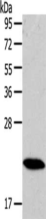
 SDS-PAGE
(Gel: 10%SDS-PAGE, Lysate: 30 ug, Lane: Mouse testis tissue, Primary antibody: AAA239661(ADO Antibody) at dilution 1/1200, Secondary antibody: Goat anti rabbit IgG at 1/8000 dilution, Exposure time: 30 minutes)
SDS-PAGE
(Gel: 10%SDS-PAGE, Lysate: 30 ug, Lane: Mouse testis tissue, Primary antibody: AAA239661(ADO Antibody) at dilution 1/1200, Secondary antibody: Goat anti rabbit IgG at 1/8000 dilution, Exposure time: 30 minutes)
ADO, Polyclonal Antibody (Cat# AAA239661)
 SDS-PAGE
(Gel: 12%SDS-PAGE,Lysate: 40 ug,,Primary antibody: AAA239665(RAB17 Antibody) at dilution 1/200 dilution,Secondary antibody: Goat anti rabbit IgG at 1/8000 dilution,Exposure time: 20 seconds)
SDS-PAGE
(Gel: 12%SDS-PAGE,Lysate: 40 ug,,Primary antibody: AAA239665(RAB17 Antibody) at dilution 1/200 dilution,Secondary antibody: Goat anti rabbit IgG at 1/8000 dilution,Exposure time: 20 seconds)
RAB17, Polyclonal Antibody (Cat# AAA239665)
 SDS-PAGE
(Gel: 10%SDS-PAGE, Lysate: 40 ug, Lane: Mouse heart tissue, Primary antibody: AAA239666(RAB18 Antibody) at dilution 1/400, Secondary antibody: Goat anti rabbit IgG at 1/8000 dilution, Exposure time: 1 second)
SDS-PAGE
(Gel: 10%SDS-PAGE, Lysate: 40 ug, Lane: Mouse heart tissue, Primary antibody: AAA239666(RAB18 Antibody) at dilution 1/400, Secondary antibody: Goat anti rabbit IgG at 1/8000 dilution, Exposure time: 1 second)
RAB18, Polyclonal Antibody (Cat# AAA239666)
 SDS-PAGE
(Gel: 12%SDS-PAGE, Lysate: 40 ug, Lane: Mouse heart tissue, Primary antibody: AAA239668(RAB22A Antibody) at dilution 1/300, Secondary antibody: Goat anti rabbit IgG at 1/8000 dilution, Exposure time: 10 seconds)
SDS-PAGE
(Gel: 12%SDS-PAGE, Lysate: 40 ug, Lane: Mouse heart tissue, Primary antibody: AAA239668(RAB22A Antibody) at dilution 1/300, Secondary antibody: Goat anti rabbit IgG at 1/8000 dilution, Exposure time: 10 seconds)
RAB22A, Polyclonal Antibody (Cat# AAA239668)
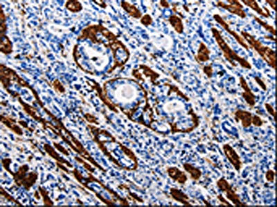
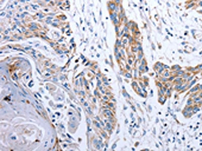
 IHC (Immunohiostchemistry)
(The image on the left is immunohistochemistry of paraffin-embedded Human lung cancer tissue using AAA239669(RAB25 Antibody) at dilution 1/50, on the right is treated with synthetic peptide. (Original magnification: ×200))
IHC (Immunohiostchemistry)
(The image on the left is immunohistochemistry of paraffin-embedded Human lung cancer tissue using AAA239669(RAB25 Antibody) at dilution 1/50, on the right is treated with synthetic peptide. (Original magnification: ×200))
RAB25, Polyclonal Antibody (Cat# AAA239669)
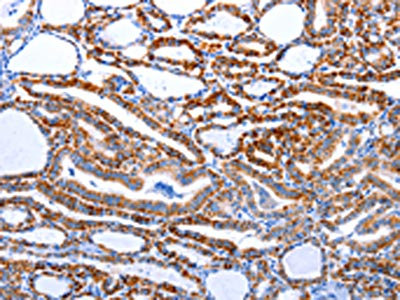
 IHC (Immunohiostchemistry)
(The image is immunohistochemistry of paraffin-embedded Human renal cancer tissue using AAA239674(RAB41 Antibody) at dilution 1/100. (Original magnification: ×200))
IHC (Immunohiostchemistry)
(The image is immunohistochemistry of paraffin-embedded Human renal cancer tissue using AAA239674(RAB41 Antibody) at dilution 1/100. (Original magnification: ×200))
RAB41, Polyclonal Antibody (Cat# AAA239674)
 IHC (Immunohiostchemistry)
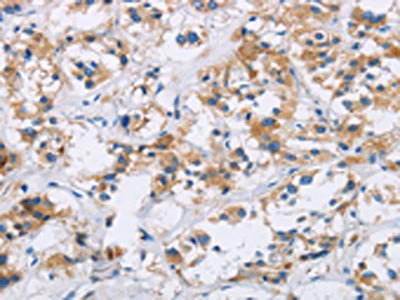
(The image on the left is immunohistochemistry of paraffin-embedded Human liver cancer tissue using AAA239678(PRKAB1 Antibody) at dilution 1/15, on the right is treated with synthetic peptide. (Original magnification: ×200))
IHC (Immunohiostchemistry)
(The image on the left is immunohistochemistry of paraffin-embedded Human liver cancer tissue using AAA239678(PRKAB1 Antibody) at dilution 1/15, on the right is treated with synthetic peptide. (Original magnification: ×200))
PRKAB1, Polyclonal Antibody (Cat# AAA239678)
 IHC (Immunohiostchemistry)
(The image on the left is immunohistochemistry of paraffin-embedded Human thyroid cancer tissue using AAA239679(IAPP Antibody) at dilution 1/40, on the right is treated with synthetic peptide. (Original magnification: ×200))
IHC (Immunohiostchemistry)
(The image on the left is immunohistochemistry of paraffin-embedded Human thyroid cancer tissue using AAA239679(IAPP Antibody) at dilution 1/40, on the right is treated with synthetic peptide. (Original magnification: ×200))
IAPP, Polyclonal Antibody (Cat# AAA239679)
 SDS-PAGE
(Gel: 10%SDS-PAGE, Lysate: 40 ug, Lane: K562 cells, Primary antibody: AAA239766(CDC6 Antibody) at dilution 1/200, Secondary antibody: Goat anti rabbit IgG at 1/8000 dilution, Exposure time: 2 minutes)
SDS-PAGE
(Gel: 10%SDS-PAGE, Lysate: 40 ug, Lane: K562 cells, Primary antibody: AAA239766(CDC6 Antibody) at dilution 1/200, Secondary antibody: Goat anti rabbit IgG at 1/8000 dilution, Exposure time: 2 minutes)
CDC6, Polyclonal Antibody (Cat# AAA239766)

 SDS-PAGE
(Gel: 8%SDS-PAGE,Lysate: 40 ug,Lane 1-3: Jurkat cells, Hela cells, HT-29 cells,Primary antibody: AAA239767(CDK1 Antibody) at dilution 1/300 dilution,Secondary antibody: Goat anti rabbit IgG at 1/8000 dilution,Exposure time: 5 minutes)
SDS-PAGE
(Gel: 8%SDS-PAGE,Lysate: 40 ug,Lane 1-3: Jurkat cells, Hela cells, HT-29 cells,Primary antibody: AAA239767(CDK1 Antibody) at dilution 1/300 dilution,Secondary antibody: Goat anti rabbit IgG at 1/8000 dilution,Exposure time: 5 minutes)
CDK1, Polyclonal Antibody (Cat# AAA239767)
 SDS-PAGE
(Gel: 10%SDS-PAGE, Lysate: 40 ug, Lane: LO2 cells, Primary antibody: AAA239771(CEBPD Antibody) at dilution 1/300, Secondary antibody: Goat anti rabbit IgG at 1/8000 dilution, Exposure time: 5 minutes)
SDS-PAGE
(Gel: 10%SDS-PAGE, Lysate: 40 ug, Lane: LO2 cells, Primary antibody: AAA239771(CEBPD Antibody) at dilution 1/300, Secondary antibody: Goat anti rabbit IgG at 1/8000 dilution, Exposure time: 5 minutes)
CEBPD, Polyclonal Antibody (Cat# AAA239771)
 IHC (Immunohiostchemistry)
(The image on the left is immunohistochemistry of paraffin-embedded Human esophagus cancer tissue using AAA239772(CHGB Antibody) at dilution 1/30, on the right is treated with synthetic peptide. (Original magnification: ×200))
IHC (Immunohiostchemistry)
(The image on the left is immunohistochemistry of paraffin-embedded Human esophagus cancer tissue using AAA239772(CHGB Antibody) at dilution 1/30, on the right is treated with synthetic peptide. (Original magnification: ×200))
CHGB, Polyclonal Antibody (Cat# AAA239772)

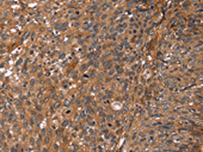
 IHC (Immunohiostchemistry)
(The image on the left is immunohistochemistry of paraffin-embedded Human ovarian cancer tissue using AAA239778(KRT6A/KRT6B/KRT6C Antibody) at dilution 1/50, on the right is treated with synthetic peptide. (Original magnification: ×200))
IHC (Immunohiostchemistry)
(The image on the left is immunohistochemistry of paraffin-embedded Human ovarian cancer tissue using AAA239778(KRT6A/KRT6B/KRT6C Antibody) at dilution 1/50, on the right is treated with synthetic peptide. (Original magnification: ×200))
KRT6A/KRT6B/KRT6C, Polyclonal Antibody (Cat# AAA239778)
 SDS-PAGE
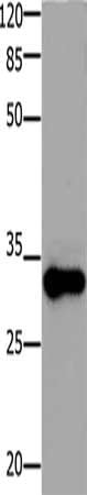
(Gel: 8%SDS-PAGE,Lysate: 20 ug,,Primary antibody: AAA239779(KRT14 Antibody) at dilution 1/1000 dilution,Secondary antibody: Goat anti rabbit IgG at 1/8000 dilution,Exposure time: 20 seconds)
SDS-PAGE
(Gel: 8%SDS-PAGE,Lysate: 20 ug,,Primary antibody: AAA239779(KRT14 Antibody) at dilution 1/1000 dilution,Secondary antibody: Goat anti rabbit IgG at 1/8000 dilution,Exposure time: 20 seconds)
KRT14, Polyclonal Antibody (Cat# AAA239779)
 IHC (Immunohiostchemistry)
(The image on the left is immunohistochemistry of paraffin-embedded Human cervical cancer tissue using AAA239783(CLEC5A Antibody) at dilution 1/20, on the right is treated with synthetic peptide. (Original magnification: ×200))
IHC (Immunohiostchemistry)
(The image on the left is immunohistochemistry of paraffin-embedded Human cervical cancer tissue using AAA239783(CLEC5A Antibody) at dilution 1/20, on the right is treated with synthetic peptide. (Original magnification: ×200))
CLEC5A, Polyclonal Antibody (Cat# AAA239783)
 SDS-PAGE
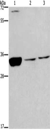
(Gel: 12%SDS-PAGE, Lysate: 40 ug, Lane 1-2: Hepg2 and 293T cell, Primary antibody: AAA239785(CLDN19 Antibody) at dilution 1/200 dilution, Secondary antibody: Goat anti rabbit IgG at 1/8000 dilution, Exposure time: 5 seconds)
SDS-PAGE
(Gel: 12%SDS-PAGE, Lysate: 40 ug, Lane 1-2: Hepg2 and 293T cell, Primary antibody: AAA239785(CLDN19 Antibody) at dilution 1/200 dilution, Secondary antibody: Goat anti rabbit IgG at 1/8000 dilution, Exposure time: 5 seconds)
CLDN19, Polyclonal Antibody (Cat# AAA239785)
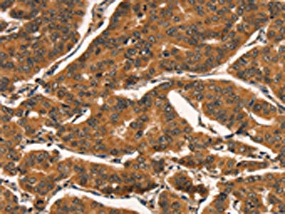
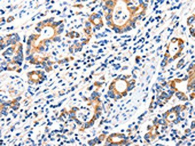
 IHC (Immunohiostchemistry)
(The image on the left is immunohistochemistry of paraffin-embedded Human liver cancer tissue using AAA239786(COX18 Antibody) at dilution 1/20, on the right is treated with synthetic peptide. (Original magnification: ×200))
IHC (Immunohiostchemistry)
(The image on the left is immunohistochemistry of paraffin-embedded Human liver cancer tissue using AAA239786(COX18 Antibody) at dilution 1/20, on the right is treated with synthetic peptide. (Original magnification: ×200))
COX18, Polyclonal Antibody (Cat# AAA239786)
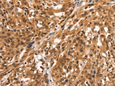
 IHC (Immunohiostchemistry)
(The image on the left is immunohistochemistry of paraffin-embedded Human breast cancer tissue using AAA239788(COL4A1 Antibody) at dilution 1/30, on the right is treated with synthetic peptide. (Original magnification: ×200))
IHC (Immunohiostchemistry)
(The image on the left is immunohistochemistry of paraffin-embedded Human breast cancer tissue using AAA239788(COL4A1 Antibody) at dilution 1/30, on the right is treated with synthetic peptide. (Original magnification: ×200))
COL4A1, Polyclonal Antibody (Cat# AAA239788)
 SDS-PAGE
(Gel: 8%SDS-PAGE, Lysate: 30 ug, Lane: Human fetal brain tissue, Primary antibody: AAA239792(DPYSL4 Antibody) at dilution 1/500, Secondary antibody: Goat anti rabbit IgG at 1/8000 dilution, Exposure time: 20 seconds)
SDS-PAGE
(Gel: 8%SDS-PAGE, Lysate: 30 ug, Lane: Human fetal brain tissue, Primary antibody: AAA239792(DPYSL4 Antibody) at dilution 1/500, Secondary antibody: Goat anti rabbit IgG at 1/8000 dilution, Exposure time: 20 seconds)
DPYSL4, Polyclonal Antibody (Cat# AAA239792)
 IHC (Immunohiostchemistry)
(The image on the left is immunohistochemistry of paraffin-embedded Human thyroid cancer tissue using AAA239807(CCND1 Antibody) at dilution 1/25, on the right is treated with synthetic peptide. (Original magnification: ×200))
IHC (Immunohiostchemistry)
(The image on the left is immunohistochemistry of paraffin-embedded Human thyroid cancer tissue using AAA239807(CCND1 Antibody) at dilution 1/25, on the right is treated with synthetic peptide. (Original magnification: ×200))
CCND1, Polyclonal Antibody (Cat# AAA239807)
 IHC (Immunohiostchemistry)
(The image on the left is immunohistochemistry of paraffin-embedded Human breast cancer tissue using AAA239810(CCNE2 Antibody) at dilution 1/25, on the right is treated with synthetic peptide. (Original magnification: ×200))
IHC (Immunohiostchemistry)
(The image on the left is immunohistochemistry of paraffin-embedded Human breast cancer tissue using AAA239810(CCNE2 Antibody) at dilution 1/25, on the right is treated with synthetic peptide. (Original magnification: ×200))
CCNE2, Polyclonal Antibody (Cat# AAA239810)
 IHC (Immunohiostchemistry)
(The image on the left is immunohistochemistry of paraffin-embedded Human liver cancer tissue using AAA239811(CCNT1 Antibody) at dilution 1/20, on the right is treated with synthetic peptide. (Original magnification: ×200))
IHC (Immunohiostchemistry)
(The image on the left is immunohistochemistry of paraffin-embedded Human liver cancer tissue using AAA239811(CCNT1 Antibody) at dilution 1/20, on the right is treated with synthetic peptide. (Original magnification: ×200))
CCNT1, Polyclonal Antibody (Cat# AAA239811)
 IHC (Immunohiostchemistry)
(The image on the left is immunohistochemistry of paraffin-embedded Human breast cancer tissue using AAA239816(SLC6A3 Antibody) at dilution 1/20, on the right is treated with synthetic peptide. (Original magnification: ×200))
IHC (Immunohiostchemistry)
(The image on the left is immunohistochemistry of paraffin-embedded Human breast cancer tissue using AAA239816(SLC6A3 Antibody) at dilution 1/20, on the right is treated with synthetic peptide. (Original magnification: ×200))
SLC6A3, Polyclonal Antibody (Cat# AAA239816)
 SDS-PAGE
(Gel: 12%SDS-PAGE, Lysate: 30 ug, Lane: Human lung tissue, Primary antibody: AAA239819(DEFA1 Antibody) at dilution 1/550, Secondary antibody: Goat anti rabbit IgG at 1/8000 dilution, Exposure time: 20 seconds)
SDS-PAGE
(Gel: 12%SDS-PAGE, Lysate: 30 ug, Lane: Human lung tissue, Primary antibody: AAA239819(DEFA1 Antibody) at dilution 1/550, Secondary antibody: Goat anti rabbit IgG at 1/8000 dilution, Exposure time: 20 seconds)
DEFA1, Polyclonal Antibody (Cat# AAA239819)
 IHC (Immunohiostchemistry)
(The image on the left is immunohistochemistry of paraffin-embedded Human esophagus cancer tissue using AAA239821(NDUFA12 Antibody) at dilution 1/50, on the right is treated with synthetic peptide. (Original magnification: ×200))
IHC (Immunohiostchemistry)
(The image on the left is immunohistochemistry of paraffin-embedded Human esophagus cancer tissue using AAA239821(NDUFA12 Antibody) at dilution 1/50, on the right is treated with synthetic peptide. (Original magnification: ×200))
NDUFA12, Polyclonal Antibody (Cat# AAA239821)
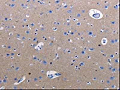
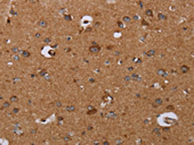 IHC (Immunohiostchemistry)
(The image on the left is immunohistochemistry of paraffin-embedded Human brain tissue using AAA239822(DISC1 Antibody) at dilution 1/30, on the right is treated with synthetic peptide. (Original magnification: ×200))
IHC (Immunohiostchemistry)
(The image on the left is immunohistochemistry of paraffin-embedded Human brain tissue using AAA239822(DISC1 Antibody) at dilution 1/30, on the right is treated with synthetic peptide. (Original magnification: ×200))
DISC1, Polyclonal Antibody (Cat# AAA239822)
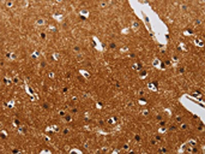
 IHC (Immunohiostchemistry)
(The image on the left is immunohistochemistry of paraffin-embedded Human thyroid cancer tissue using AAA239825(DTNBP1 Antibody) at dilution 1/20, on the right is treated with synthetic peptide. (Original magnification: ×200))
IHC (Immunohiostchemistry)
(The image on the left is immunohistochemistry of paraffin-embedded Human thyroid cancer tissue using AAA239825(DTNBP1 Antibody) at dilution 1/20, on the right is treated with synthetic peptide. (Original magnification: ×200))
DTNBP1, Polyclonal Antibody (Cat# AAA239825)
 IHC (Immunohiostchemistry)
(The image on the left is immunohistochemistry of paraffin-embedded Human colon cancer tissue using AAA239843(EDNRB Antibody) at dilution 1/30, on the right is treated with synthetic peptide. (Original magnification: ×200))
IHC (Immunohiostchemistry)
(The image on the left is immunohistochemistry of paraffin-embedded Human colon cancer tissue using AAA239843(EDNRB Antibody) at dilution 1/30, on the right is treated with synthetic peptide. (Original magnification: ×200))
EDNRB, Polyclonal Antibody (Cat# AAA239843)
 SDS-PAGE
(Gel: 10%SDS-PAGE, Lysate: 30 ug, Lane: 293T cells, Primary antibody: AAA239849(ENPP7 Antibody) at dilution 1/700, Secondary antibody: Goat anti rabbit IgG at 1/8000 dilution, Exposure time: 1 minute)
SDS-PAGE
(Gel: 10%SDS-PAGE, Lysate: 30 ug, Lane: 293T cells, Primary antibody: AAA239849(ENPP7 Antibody) at dilution 1/700, Secondary antibody: Goat anti rabbit IgG at 1/8000 dilution, Exposure time: 1 minute)
ENPP7, Polyclonal Antibody (Cat# AAA239849)
 IHC (Immunohiostchemistry)
(The image on the left is immunohistochemistry of paraffin-embedded Human ovarian cancer tissue using AAA240123(GRIN2D Antibody) at dilution 1/30, on the right is treated with synthetic peptide. (Original magnification: ×200))
IHC (Immunohiostchemistry)
(The image on the left is immunohistochemistry of paraffin-embedded Human ovarian cancer tissue using AAA240123(GRIN2D Antibody) at dilution 1/30, on the right is treated with synthetic peptide. (Original magnification: ×200))
GRIN2D, Polyclonal Antibody (Cat# AAA240123)
 IHC (Immunohiostchemistry)
(The image on the left is immunohistochemistry of paraffin-embedded Human gastic cancer tissue using AAA240124(GRIA2 Antibody) at dilution 1/40, on the right is treated with synthetic peptide. (Original magnification: ×200))
IHC (Immunohiostchemistry)
(The image on the left is immunohistochemistry of paraffin-embedded Human gastic cancer tissue using AAA240124(GRIA2 Antibody) at dilution 1/40, on the right is treated with synthetic peptide. (Original magnification: ×200))
GRIA2, Polyclonal Antibody (Cat# AAA240124)
 IHC (Immunohiostchemistry)
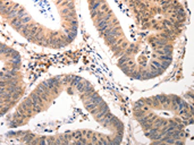
(The image on the left is immunohistochemistry of paraffin-embedded Human gastric cancer tissue using AAA240143(PXN Antibody) at dilution 1/30, on the right is treated with synthetic peptide. (Original magnification: ×200))
IHC (Immunohiostchemistry)
(The image on the left is immunohistochemistry of paraffin-embedded Human gastric cancer tissue using AAA240143(PXN Antibody) at dilution 1/30, on the right is treated with synthetic peptide. (Original magnification: ×200))
PXN, Polyclonal Antibody (Cat# AAA240143)
 SDS-PAGE
(Gel: 10%SDS-PAGE, Lysate: 40 ug, Lane 1-2: Human fetal brain tissue, 293T cells, Primary antibody: AAA239688(RAB3c Antibody) at dilution 1/400, Secondary antibody: Goat anti rabbit IgG at 1/8000 dilution, Exposure time: 1 minute)
SDS-PAGE
(Gel: 10%SDS-PAGE, Lysate: 40 ug, Lane 1-2: Human fetal brain tissue, 293T cells, Primary antibody: AAA239688(RAB3c Antibody) at dilution 1/400, Secondary antibody: Goat anti rabbit IgG at 1/8000 dilution, Exposure time: 1 minute)
RAB3C, Polyclonal Antibody (Cat# AAA239688)
 IHC (Immunohiostchemistry)
(The image on the left is immunohistochemistry of paraffin-embedded Human colon cancer tissue using AAA239691(LOX Antibody) at dilution 1/25, on the right is treated with synthetic peptide. (Original magnification: ×200))
IHC (Immunohiostchemistry)
(The image on the left is immunohistochemistry of paraffin-embedded Human colon cancer tissue using AAA239691(LOX Antibody) at dilution 1/25, on the right is treated with synthetic peptide. (Original magnification: ×200))
LOX, Polyclonal Antibody (Cat# AAA239691)
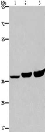
 SDS-PAGE
(Gel: 10%SDS-PAGE, Lysate: 30 ug, Lane 1-6: 293T cells, mouse brain tissue, A549 cells, Hela cells, A172 cells, HT29 cells, Primary antibody: AAA239698(RAB6A Antibody) at dilution 1/650, Secondary antibody: Goat anti rabbit IgG at 1/8000 dilution, Exposure time: 1 minute)
SDS-PAGE
(Gel: 10%SDS-PAGE, Lysate: 30 ug, Lane 1-6: 293T cells, mouse brain tissue, A549 cells, Hela cells, A172 cells, HT29 cells, Primary antibody: AAA239698(RAB6A Antibody) at dilution 1/650, Secondary antibody: Goat anti rabbit IgG at 1/8000 dilution, Exposure time: 1 minute)
RAB6A, Polyclonal Antibody (Cat# AAA239698)
What are Polyclonal Antibodies?
Polyclonal antibodies are antibodies that come from multiple B cell clones of a host animal. The typical hosts used for the majority of polyclonal antibody production are rabbits, goats, sheep, and donkeys. These polyclonal antibodies, once having identified their target, will bind to different epitopes located at different regions or sequences on the same protein/antigen. As a result, they are ideal at locating and binding to the target, even if the target is in very low concentrations (due to many different antibodies being able to bind to the same target molecule, which allows for significant amplification of a downstream signal).
Polyclonal antibodies are typically produced by injecting an antigen into a host animal, which causes the animal’s immune system to attack the foreign antigen by mass generating antibodies against it. After a period of time, serum is collected from the animal and purified using physicochemical fractionation, class-specific affinity purification, and/or antigen-affinity purification.
Key Uses of Polyclonal Antibodies
- Western Blotting: This method is used to find specific proteins in biological samples after separating them by size.
- Immunohistochemistry: IHC helps visualize the location of proteins in tissue sections using various staining techniques.
- ELISA: (Enzyme-Linked Immunosorbent Assay) is typically used to identify specific protein quantities in a sample. ELISAs can be either “Quantitative” or “Qualitative”.
- Flow Cytometry: technique that identifies and measures the specific protein on the surface or inside the cells in a fluid suspension.
- Immunoprecipitation: IP isolates and studies a specific protein from a complex mixture using antibodies.
Why Buy Polyclonal Antibodies from AAA Biotech?
1. Ideal for Various Applications
Our antibodies are generally going to be validated for use in multiple types of assays, including ELISA, Western Blotting, Immunohistochemistry, Immunoprecipitation, amongst others. They are ideal for a wide range of research applications.
2. Rigorous Quality Control
All of the antibodies in our catalog undergo strict quality testing to ensure specificity, sensitivity, and consistent performance. We are confident in the ability of our antibodies to provide you with accurate results.
3. Wide Assortment of Antibodies
Antibodies in are catalog can be found for both common and exotic species, and these antibodies are also available in both conjugated and recombinant forms to suit many diverse experimental needs.
4. Highly Purified
Our antibodies are available in purified forms with over 85% purity, as confirmed by SDS-PAGE. They are also available with tags such as His, Flag, GST, or MBP. We cater to customers worldwide.
FAQ
1. How are polyclonal antibodies produced?
Traditionally, polyclonal antibodies are produced by injecting an antigen into a host animal (such as a rabbit or goat), which then triggers an immune response from the host animal. The animal’s B cells produce antibodies that will recognize different parts of the injected antigen. These antibodies are then collected from the animal’s blood and purified for use.
2. How do polyclonal antibodies differ from monoclonal antibodies?
Polyclonal antibodies are a mix of antibodies that bind to different locations (epitopes) of the same antigen, while monoclonal antibodies are identical and bind to just one specific epitope. This makes polyclonal antibodies more versatile and better at detecting proteins that may be present in low quantities or in altered/modified forms.
3. How should I store polyclonal antibodies?
Polyclonal antibodies should be stored at 4°C for short-term use (up to a few weeks) and at -20°C or -80°C for long-term storage. Avoid repeated freeze-thaw cycles by dividing them into small aliquots. Always check the datasheet for specific storage instructions.