Filters
▼Clonality
▼Type
▼Reactivity
▼Gene Name
▼Isotype
▼Host
▼Application
▼Clone
▼Polyclonal Antibodies
At AAA Biotech also known as AAA Bio or AAABio, we provide a broad range of purified polyclonal antibodies (pAbs) that are able to all be browsed online through our website. Due to their high specificity and strong binding affinity, these antibodies are ideal for wide swathes of research and experimental applications.
Our polyclonal antibodies can easily support your work, whether you use them for Western Blotting, Immunocytochemistry (with or without Immunofluorescence used in conjunction), Immunohistochemistry, Immunoprecipitation, and ELISA tests. We highly encourage you to browse our range of pAbs and choose the one that best suits your experimental model.
Viewing 5550-5600 of 96812 product results
 IHC (Immunohiostchemistry)
(DABstainingonIHC-P.Samples:MouseTissue))
IHC (Immunohiostchemistry)
(DABstainingonIHC-P.Samples:MouseTissue))
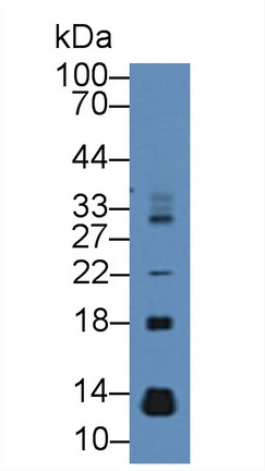
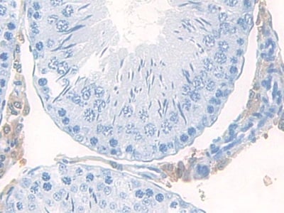
Tyrosine Kinase With Immunoglobulin Like And EGF Like Domains Protein 1, Polyclonal Antibody (Cat# AAA142802)
 IHC (Immunohistochemistry)
(DAB staining on IHC-P; Samples: Mouse Testis Tissue))
IHC (Immunohistochemistry)
(DAB staining on IHC-P; Samples: Mouse Testis Tissue))
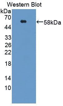
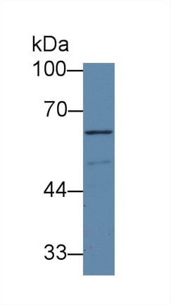
Poliovirus Receptor Related Protein 3, Polyclonal Antibody (Cat# AAA142807)
 IHC (Immunohiostchemistry)
(DABstainingonIHC-P.Samples:HumanTissue))
IHC (Immunohiostchemistry)
(DABstainingonIHC-P.Samples:HumanTissue))
Cyclin A1, Polyclonal Antibody (Cat# AAA142826)

 IHC (Immunohiostchemistry)
(DABstainingonIHC-P.Samples:RatTissue))
IHC (Immunohiostchemistry)
(DABstainingonIHC-P.Samples:RatTissue))
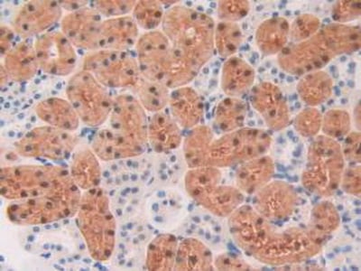
Chymase 1, Polyclonal Antibody (Cat# AAA142834)
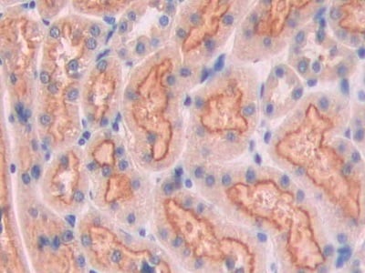
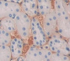
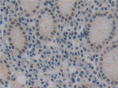
 IHC (Immunohiostchemistry)
(DAB staining on fromalin fixed paraffin-embedded kidney tissue))
IHC (Immunohiostchemistry)
(DAB staining on fromalin fixed paraffin-embedded kidney tissue))
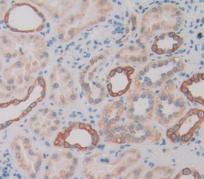
Plasmalemma Vesicle Associated Protein, Polyclonal Antibody (Cat# AAA142589)
 IHC (Immunohistochemisry)
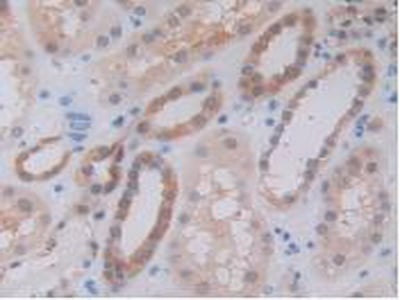
(DAB staining on IHC-P; Samples: Rat Kidney Tissue))
IHC (Immunohistochemisry)
(DAB staining on IHC-P; Samples: Rat Kidney Tissue))
Cubilin, Polyclonal Antibody (Cat# AAA142590)
 IHC (Immunohistochemistry)
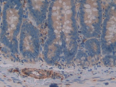
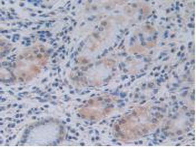
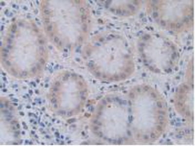
(DAB staining on IHC-P;Samples: Mouse Stomach Tissue;Primary Ab: 10ug/ml Rabbit Anti-Mouse ASGR1 AntibodySecond Ab: 2ug/mL HRP-Linked Caprine Anti-Rabbit IgG Polyclonal Antibody)
IHC (Immunohistochemistry)
(DAB staining on IHC-P;Samples: Mouse Stomach Tissue;Primary Ab: 10ug/ml Rabbit Anti-Mouse ASGR1 AntibodySecond Ab: 2ug/mL HRP-Linked Caprine Anti-Rabbit IgG Polyclonal Antibody)
Asialoglycoprotein Receptor 1, Polyclonal Antibody (Cat# AAA142596)
Interferon Beta, Polyclonal Antibody (Cat# AAA142604)
 IHC (Immunohistochemistry)
(DAB staining on IHC-P; Samples: Mouse Ovary Tissue))
IHC (Immunohistochemistry)
(DAB staining on IHC-P; Samples: Mouse Ovary Tissue))
Hermansky Pudlak Syndrome Protein 4, Polyclonal Antibody (Cat# AAA142605)
 IHC (Immunohiostchemistry)
(DABstainingonIHC-P.Samples:RatTissue))
IHC (Immunohiostchemistry)
(DABstainingonIHC-P.Samples:RatTissue))
Period Circadian Protein 2, Polyclonal Antibody (Cat# AAA142606)

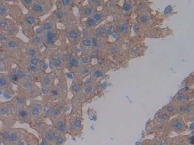
 IHC (Immunohistochemisry)
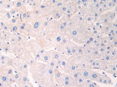
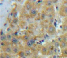
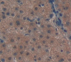
(DAB staining on IHC-P;Samples: Human Liver Tissue;Primary Ab: 20ug/ml Rabbit Anti-Human PCK2 AntibodySecond Ab: 2ug/mL HRP-Linked Caprine Anti-Rabbit IgG Polyclonal Antibody)
IHC (Immunohistochemisry)
(DAB staining on IHC-P;Samples: Human Liver Tissue;Primary Ab: 20ug/ml Rabbit Anti-Human PCK2 AntibodySecond Ab: 2ug/mL HRP-Linked Caprine Anti-Rabbit IgG Polyclonal Antibody)
Phosphoenolpyruvate Carboxykinase 2, Polyclonal Antibody (Cat# AAA142608)
 IHC (Immunohiostchemistry)
(DAB staining on fromalin fixed paraffin- embedded Kidney tissue))
IHC (Immunohiostchemistry)
(DAB staining on fromalin fixed paraffin- embedded Kidney tissue))
Semaphorin 4D, Polyclonal Antibody (Cat# AAA142610)
 IHC (Immunohistochemisry)
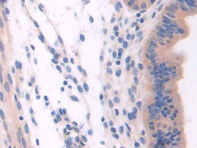
(DAB staining on IHC-P; Samples: Human Breast cancer Tissue))
IHC (Immunohistochemisry)
(DAB staining on IHC-P; Samples: Human Breast cancer Tissue))
Adipose Differentiation, Polyclonal Antibody (Cat# AAA142611)
 IHC (Immunohistochemisry)
(DAB staining on IHC-P; Samples: Rat Testis Tissue))
IHC (Immunohistochemisry)
(DAB staining on IHC-P; Samples: Rat Testis Tissue))
Mucosae Associated Epithelia Chemokine, Polyclonal Antibody (Cat# AAA142614)
 IHC (Immunohistochemisry)
(DAB staining on fromalin fixed paraffin- embedded Kidney tissue))
IHC (Immunohistochemisry)
(DAB staining on fromalin fixed paraffin- embedded Kidney tissue))
Activin A Receptor Type I C, Polyclonal Antibody (Cat# AAA142616)
 IHC (Immunohistochemisry)
(DAB staining on fromalin fixed paraffin- embedded liver tissue))
IHC (Immunohistochemisry)
(DAB staining on fromalin fixed paraffin- embedded liver tissue))
Cellular Repressor Of E1A Stimulated Genes 1, Polyclonal Antibody (Cat# AAA142617)
 IHC (Immunohiostchemistry)
(DABstainingonIHC-P.Samples:HumanTissue))
IHC (Immunohiostchemistry)
(DABstainingonIHC-P.Samples:HumanTissue))
Histone Deacetylase 9, Polyclonal Antibody (Cat# AAA142618)
 IHC (Immunohiostchemistry)
(DABstainingonIHC-P.Samples:HumanTissue))
IHC (Immunohiostchemistry)
(DABstainingonIHC-P.Samples:HumanTissue))
Melanotransferrin, Polyclonal Antibody (Cat# AAA142620)
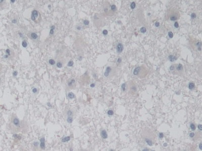
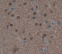
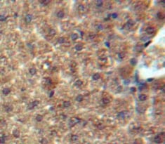
 IHC (Immunohiostchemistry)
(DAB staining on fromalin fixed paraffin-embedded Brain tissue))
IHC (Immunohiostchemistry)
(DAB staining on fromalin fixed paraffin-embedded Brain tissue))
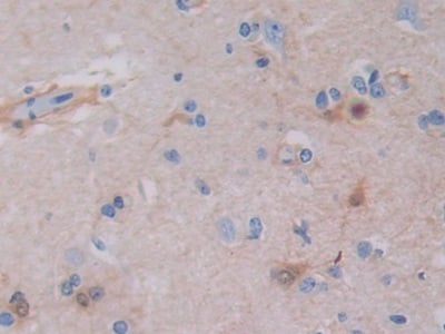
Interleukin 1 Delta, Polyclonal Antibody (Cat# AAA142626)
 IHC (Immunohistochemisry)
(DAB staining on fromalin fixed paraffin-embedded testis tissue))
IHC (Immunohistochemisry)
(DAB staining on fromalin fixed paraffin-embedded testis tissue))
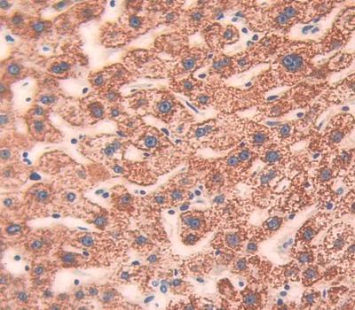
Epiphycan, Polyclonal Antibody (Cat# AAA142628)
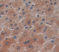
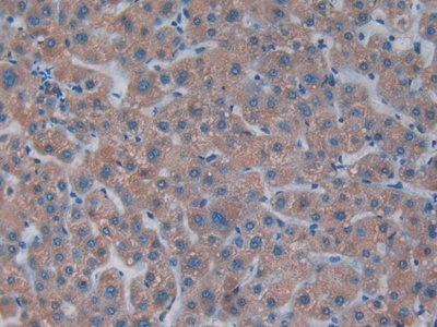
 IHC (Immunohistochemistry)
(DAB staining on fromalin fixed paraffin-embedded Liver tissue))
IHC (Immunohistochemistry)
(DAB staining on fromalin fixed paraffin-embedded Liver tissue))
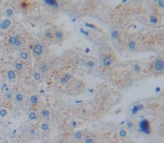
Apolipoprotein E, Polyclonal Antibody (Cat# AAA142635)
 IHC (Immunohistochemistry)
(DAB staining on IHC-P; Samples: Mouse Uterus Tissue))
IHC (Immunohistochemistry)
(DAB staining on IHC-P; Samples: Mouse Uterus Tissue))
Cytospin B, Polyclonal Antibody (Cat# AAA142697)

 IHC (Immunohiostchemistry)
(DABstainingonIHC-P.Samples:HumanTissue))
IHC (Immunohiostchemistry)
(DABstainingonIHC-P.Samples:HumanTissue))
Acetylcholinesterase, Polyclonal Antibody (Cat# AAA142700)
 IHC (Immunohistochemisry)
(DAB staining on IHC-P. Samples: Mouse Tissue))
IHC (Immunohistochemisry)
(DAB staining on IHC-P. Samples: Mouse Tissue))
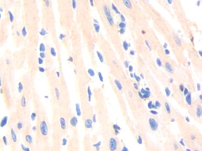
Alpha-Tocopherol, Polyclonal Antibody (Cat# AAA142702)
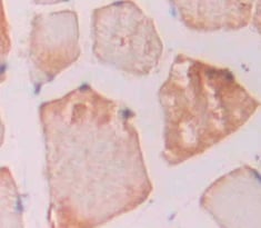 IHC (Immunohistochemisry)
(DAB staining on fromalin fixed paraffin- embedded skeletal muscle tissue))
IHC (Immunohistochemisry)
(DAB staining on fromalin fixed paraffin- embedded skeletal muscle tissue))
Iron Responsive Element Binding Protein 2, Polyclonal Antibody (Cat# AAA142711)
 IHC (Immunohiostchemistry)
(DAB staining on fromalin fixed paraffin- embedded Kidney tissue))
IHC (Immunohiostchemistry)
(DAB staining on fromalin fixed paraffin- embedded Kidney tissue))
Lymphocyte Activation Gene 3, Polyclonal Antibody (Cat# AAA142725)
 IHC (Immunohiostchemistry)
(DABstainingonIHC-P.Samples:HumanTissue))
IHC (Immunohiostchemistry)
(DABstainingonIHC-P.Samples:HumanTissue))
SH3 Domain Binding Glutamic Acid Rich Protein Like Protein, Polyclonal Antibody (Cat# AAA142726)
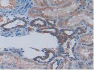
 IHC (Immunohistochemistry)
(DAB staining on IHC-P; Samples: Mouse Kidney Tissue.)
IHC (Immunohistochemistry)
(DAB staining on IHC-P; Samples: Mouse Kidney Tissue.)
Apolipoprotein C4, Polyclonal Antibody (Cat# AAA142741)
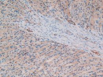
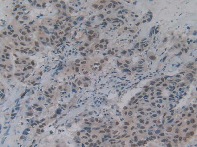 IHC (Immunohistochemistry)
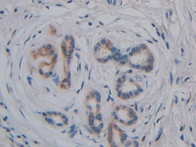
(DAB staining on IHC-P; Samples: Human Breast Cancer Tissue.)
IHC (Immunohistochemistry)
(DAB staining on IHC-P; Samples: Human Breast Cancer Tissue.)
DNA Fragmentation Factor Subunit Alpha, Polyclonal Antibody (Cat# AAA142744)
 IHC (Immunohistochemisry)
(DAB staining on fromalin fixed paraffin-embedded Kidney tissue))
IHC (Immunohistochemisry)
(DAB staining on fromalin fixed paraffin-embedded Kidney tissue))
Defensin Beta 103A, Polyclonal Antibody (Cat# AAA142745)
 IHC (Immunohiostchemistry)
(DAB staining on fromalin fixed paraffin-embedded Testis tissue))
IHC (Immunohiostchemistry)
(DAB staining on fromalin fixed paraffin-embedded Testis tissue))
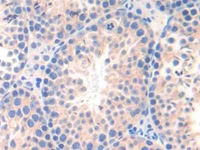
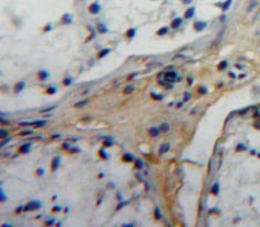
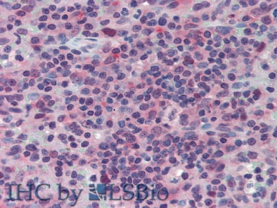
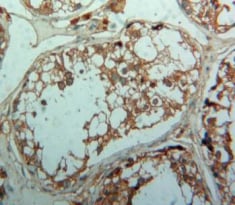
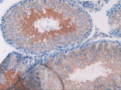
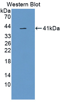
Transforming Growth Factor Beta Receptor I, Polyclonal Antibody (Cat# AAA142936)
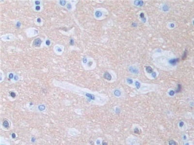

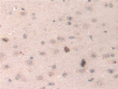
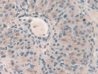
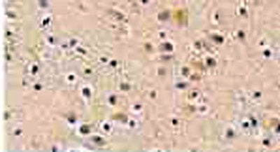
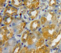
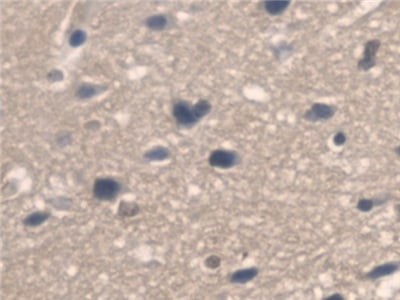
 IHC (Immunohistochemistry)
(DAB staining on IHC-P; Samples: Human Cerebrum Tissue))
IHC (Immunohistochemistry)
(DAB staining on IHC-P; Samples: Human Cerebrum Tissue))
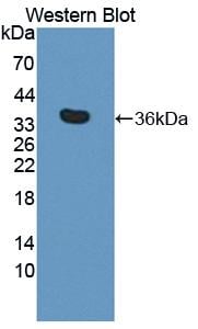
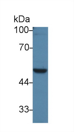
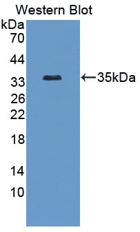
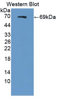
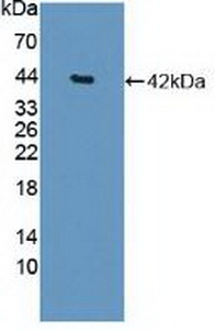
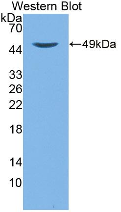
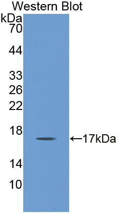
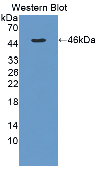
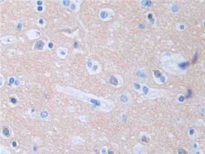
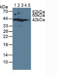
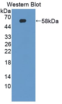
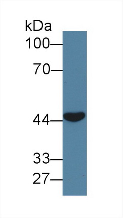
Protein Phosphatase 3, Polyclonal Antibody (Cat# AAA143000)
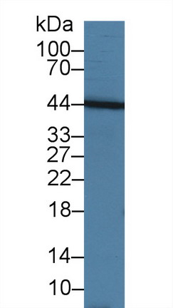
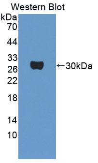
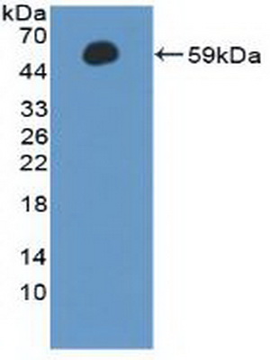
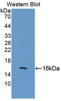
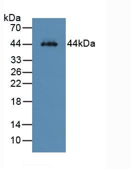
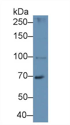
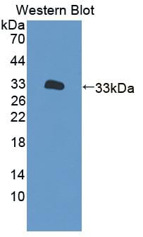
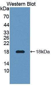
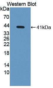
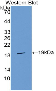
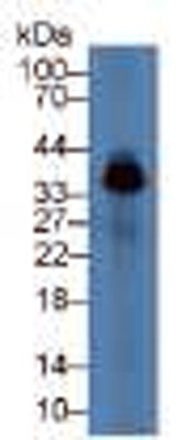
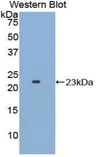
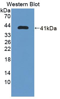
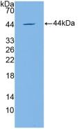
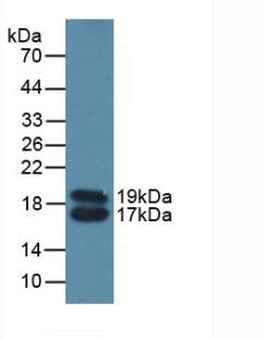
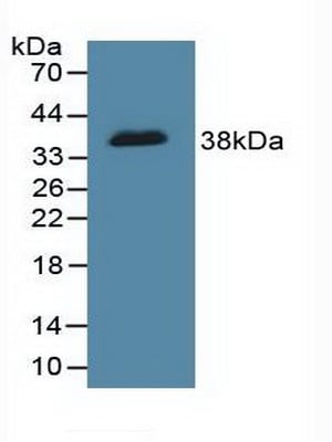
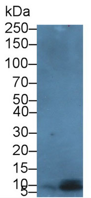
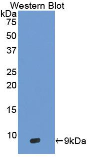
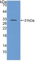
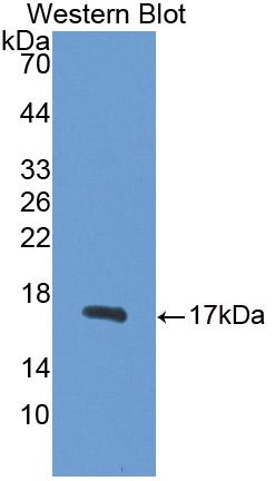
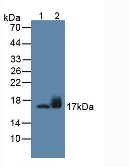
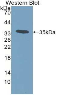
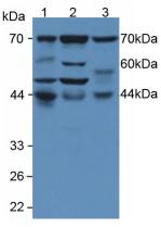
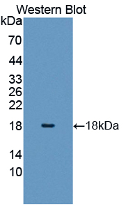
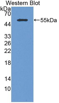
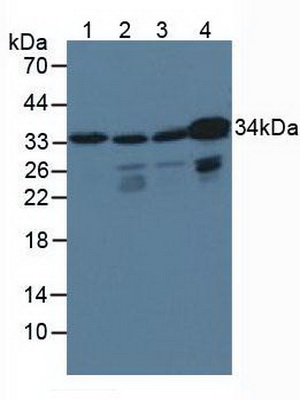
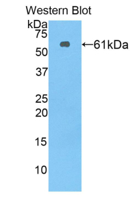
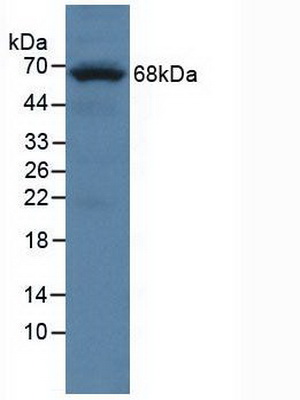
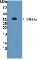
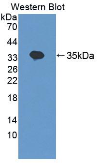
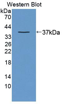
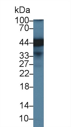
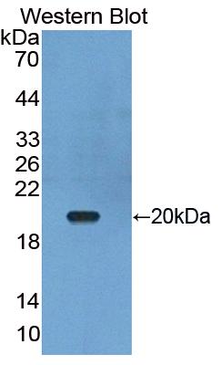 WB (Western Blot)
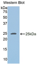
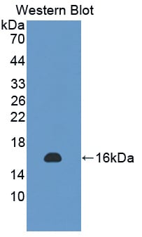
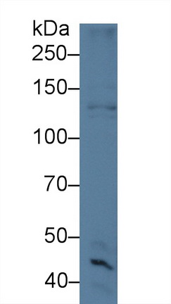
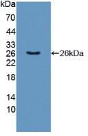
(Western Blot: Sample: Recombinant protein.)
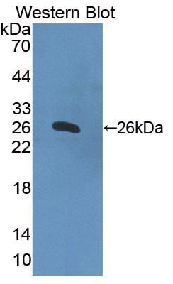
WB (Western Blot)
(Western Blot: Sample: Recombinant protein.)
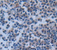
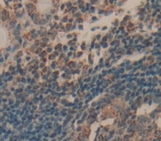
Tumor Necrosis Factor Ligand Superfamily, Polyclonal Antibody (Cat# AAA143002)
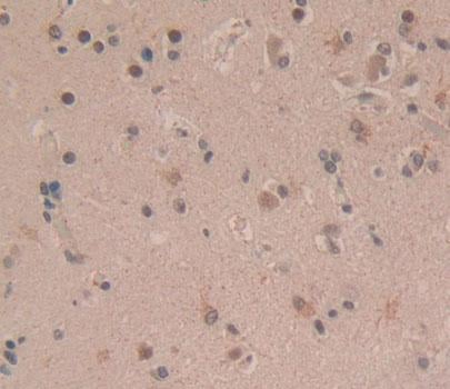
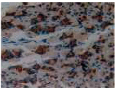
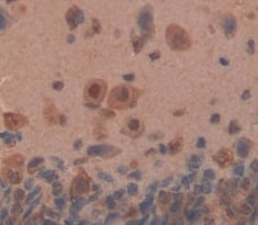
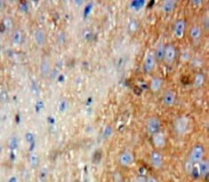
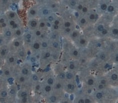
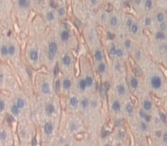
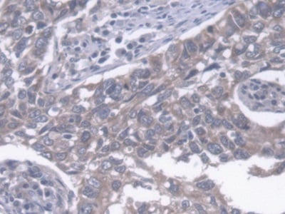
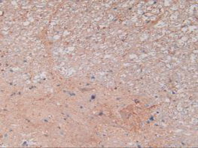 IHC (Immunohistochemistry)
(DAB staining on IHC-P; Samples: Rat Spinal Cord Tissue)
IHC (Immunohistochemistry)
(DAB staining on IHC-P; Samples: Rat Spinal Cord Tissue)
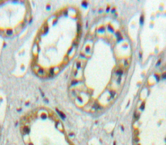
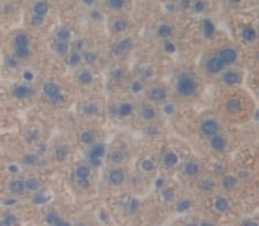
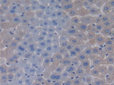
Synuclein Gamma, Polyclonal Antibody (Cat# AAA143004)
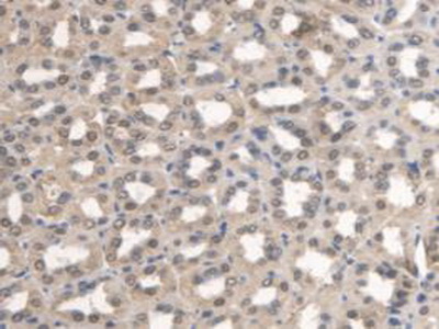
 IHC (Immunohiostchemistry)
(DAB staining on fromalin fixed paraffin-embedded Liver tissue))
IHC (Immunohiostchemistry)
(DAB staining on fromalin fixed paraffin-embedded Liver tissue))
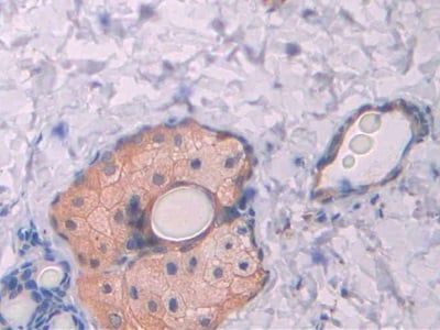
Monocyte Chemotactic Protein 3, Polyclonal Antibody (Cat# AAA143013)


 IHC (Immunohistochemisry)
(DAB staining on IHC-P. Samples: Human Tissue))
IHC (Immunohistochemisry)
(DAB staining on IHC-P. Samples: Human Tissue))
Phospholipase C Gamma 2, Polyclonal Antibody (Cat# AAA143022)

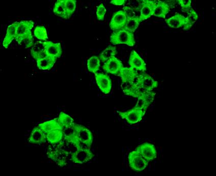 IHC (Immunohistochemistry)
(FITC staining on IHC-P Simple: Mcf7 cells))
IHC (Immunohistochemistry)
(FITC staining on IHC-P Simple: Mcf7 cells))
Protein Kinase R, Polyclonal Antibody (Cat# AAA143024)
 IHC (Immunohiostchemistry)
((Used in DAB staining on formalin fixed paraffin- embedded Kidney tissue))
IHC (Immunohiostchemistry)
((Used in DAB staining on formalin fixed paraffin- embedded Kidney tissue))
Histone Cluster 2, Polyclonal Antibody (Cat# AAA143030)


 IHC (Immunohistochemistry)
(DAB staining on IHC-P; Samples: Rat Testis Tissue.)
IHC (Immunohistochemistry)
(DAB staining on IHC-P; Samples: Rat Testis Tissue.)
Sorbitol Dehydrogenase, Polyclonal Antibody (Cat# AAA143041)
 IHC (Immunohistochemistry)
(DAB staining on IHC-P; Samples: Human Kidney Tissue.)
IHC (Immunohistochemistry)
(DAB staining on IHC-P; Samples: Human Kidney Tissue.)
Tumor Necrosis Factor Ligand Superfamily, Polyclonal Antibody (Cat# AAA143047)
 IHC (Immunohistochemisry)
(DAB staining on fromalin fixed paraffin-embedded Lung tissue))
IHC (Immunohistochemisry)
(DAB staining on fromalin fixed paraffin-embedded Lung tissue))
Keratin 14, Polyclonal Antibody (Cat# AAA143053)

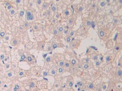 IHC (Immunohistochemistry)
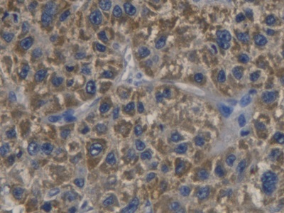
(DAB staining on IHC-P; Samples: Human Liver Tissue))
IHC (Immunohistochemistry)
(DAB staining on IHC-P; Samples: Human Liver Tissue))
Crk Like Protein, Polyclonal Antibody (Cat# AAA143058)
 IHC (Immunohiostchemistry)
(DABstainingonIHC-P.Samples:RatTissue))
IHC (Immunohiostchemistry)
(DABstainingonIHC-P.Samples:RatTissue))
Noggin, Polyclonal Antibody (Cat# AAA143062)
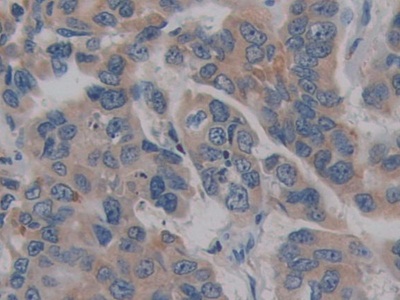
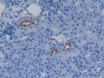
 IHC (Immunohistochemisry)
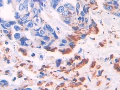
(DAB staining on IHC-P; Samples: Human Pancreatic cancer Tissue))
IHC (Immunohistochemisry)
(DAB staining on IHC-P; Samples: Human Pancreatic cancer Tissue))
Glycosylphosphatidylinositol Specific Phospholipase D1, Polyclonal Antibody (Cat# AAA142646)
 IHC (Immunohiostchemistry)
(DAB staining on IHC-P; Samples: Human Liver cancer Tissue))
IHC (Immunohiostchemistry)
(DAB staining on IHC-P; Samples: Human Liver cancer Tissue))
Cyclin Dependent Kinase Inhibitor 3, Polyclonal Antibody (Cat# AAA142688)

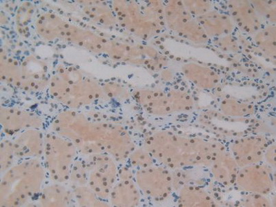
 IHC (Immunohistochemisry)
(DAB staining on fromalin fixed paraffin- embedded Kidney tissue))
IHC (Immunohistochemisry)
(DAB staining on fromalin fixed paraffin- embedded Kidney tissue))
Protein Tyrosine Phosphatase Receptor Type O, Polyclonal Antibody (Cat# AAA142695)
 IHC (Immunohistochemistry)
(DAB staining on IHC-P; Samples: Mouse Kidney Tissue)
IHC (Immunohistochemistry)
(DAB staining on IHC-P; Samples: Mouse Kidney Tissue)
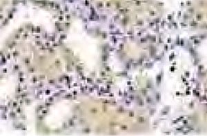
Pyridoxamine-5'-Phosphate Oxidase, Polyclonal Antibody (Cat# AAA145344)
 IHC (Immunohistochemisry)
(DAB staining on IHC-P. Samples: Human Tissue))
IHC (Immunohistochemisry)
(DAB staining on IHC-P. Samples: Human Tissue))
C-Type Lectin Domain Family 11, Polyclonal Antibody (Cat# AAA145354)
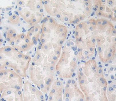
 IHC (Immunohiostchemistry)
(DAB staining on fromalin fixed paraffin- embedded Kidney tissue))
IHC (Immunohiostchemistry)
(DAB staining on fromalin fixed paraffin- embedded Kidney tissue))
Trinucleotide Repeat Containing Protein 6A, Polyclonal Antibody (Cat# AAA145364)
 IHC (Immunohistochemisry)
(DAB staining on IHC-P; Samples: Human Lung cancer Tissue))
IHC (Immunohistochemisry)
(DAB staining on IHC-P; Samples: Human Lung cancer Tissue))
Endoplasmic Reticulum Lipid Raft Associated Protein 2, Polyclonal Antibody (Cat# AAA145372)
What are Polyclonal Antibodies?
Polyclonal antibodies are antibodies that come from multiple B cell clones of a host animal. The typical hosts used for the majority of polyclonal antibody production are rabbits, goats, sheep, and donkeys. These polyclonal antibodies, once having identified their target, will bind to different epitopes located at different regions or sequences on the same protein/antigen. As a result, they are ideal at locating and binding to the target, even if the target is in very low concentrations (due to many different antibodies being able to bind to the same target molecule, which allows for significant amplification of a downstream signal).
Polyclonal antibodies are typically produced by injecting an antigen into a host animal, which causes the animal’s immune system to attack the foreign antigen by mass generating antibodies against it. After a period of time, serum is collected from the animal and purified using physicochemical fractionation, class-specific affinity purification, and/or antigen-affinity purification.
Key Uses of Polyclonal Antibodies
- Western Blotting: This method is used to find specific proteins in biological samples after separating them by size.
- Immunohistochemistry: IHC helps visualize the location of proteins in tissue sections using various staining techniques.
- ELISA: (Enzyme-Linked Immunosorbent Assay) is typically used to identify specific protein quantities in a sample. ELISAs can be either “Quantitative” or “Qualitative”.
- Flow Cytometry: technique that identifies and measures the specific protein on the surface or inside the cells in a fluid suspension.
- Immunoprecipitation: IP isolates and studies a specific protein from a complex mixture using antibodies.
Why Buy Polyclonal Antibodies from AAA Biotech?
1. Ideal for Various Applications
Our antibodies are generally going to be validated for use in multiple types of assays, including ELISA, Western Blotting, Immunohistochemistry, Immunoprecipitation, amongst others. They are ideal for a wide range of research applications.
2. Rigorous Quality Control
All of the antibodies in our catalog undergo strict quality testing to ensure specificity, sensitivity, and consistent performance. We are confident in the ability of our antibodies to provide you with accurate results.
3. Wide Assortment of Antibodies
Antibodies in are catalog can be found for both common and exotic species, and these antibodies are also available in both conjugated and recombinant forms to suit many diverse experimental needs.
4. Highly Purified
Our antibodies are available in purified forms with over 85% purity, as confirmed by SDS-PAGE. They are also available with tags such as His, Flag, GST, or MBP. We cater to customers worldwide.
FAQ
1. How are polyclonal antibodies produced?
Traditionally, polyclonal antibodies are produced by injecting an antigen into a host animal (such as a rabbit or goat), which then triggers an immune response from the host animal. The animal’s B cells produce antibodies that will recognize different parts of the injected antigen. These antibodies are then collected from the animal’s blood and purified for use.
2. How do polyclonal antibodies differ from monoclonal antibodies?
Polyclonal antibodies are a mix of antibodies that bind to different locations (epitopes) of the same antigen, while monoclonal antibodies are identical and bind to just one specific epitope. This makes polyclonal antibodies more versatile and better at detecting proteins that may be present in low quantities or in altered/modified forms.
3. How should I store polyclonal antibodies?
Polyclonal antibodies should be stored at 4°C for short-term use (up to a few weeks) and at -20°C or -80°C for long-term storage. Avoid repeated freeze-thaw cycles by dividing them into small aliquots. Always check the datasheet for specific storage instructions.