Filters
▼Clonality
▼Type
▼Reactivity
▼Gene Name
▼Isotype
▼Host
▼Application
▼Clone
▼Polyclonal Antibodies
At AAA Biotech also known as AAA Bio or AAABio, we provide a broad range of purified polyclonal antibodies (pAbs) that are able to all be browsed online through our website. Due to their high specificity and strong binding affinity, these antibodies are ideal for wide swathes of research and experimental applications.
Our polyclonal antibodies can easily support your work, whether you use them for Western Blotting, Immunocytochemistry (with or without Immunofluorescence used in conjunction), Immunohistochemistry, Immunoprecipitation, and ELISA tests. We highly encourage you to browse our range of pAbs and choose the one that best suits your experimental model.
Viewing 5500-5550 of 96805 product results
 IF (Immunofluorescence)
(Immunofluorescence analysis of Rat spleen tissue with Phospho-I?B-? (Ser32/S36) Polyclonal Antibody at dilution of 1:200)
IF (Immunofluorescence)
(Immunofluorescence analysis of Rat spleen tissue with Phospho-I?B-? (Ser32/S36) Polyclonal Antibody at dilution of 1:200)
IkappaB-alpha, Polyclonal Antibody (Cat# AAA171690)
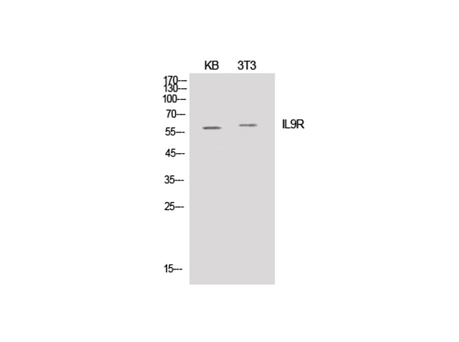
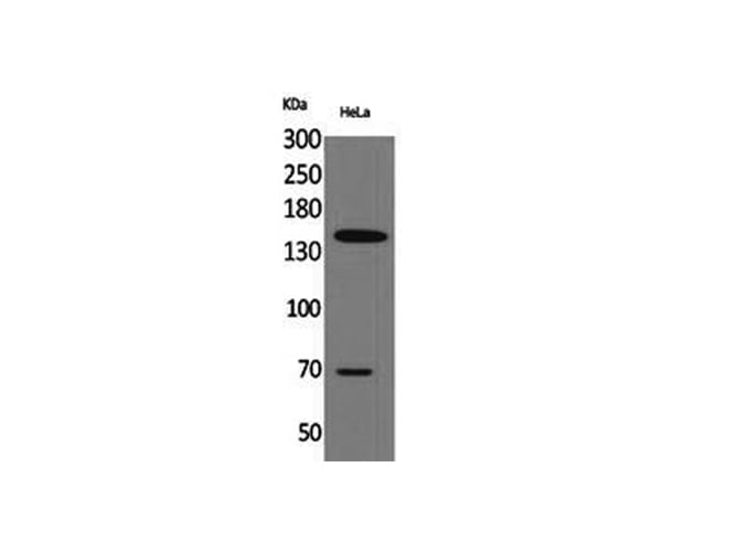
 WB (Western Blot)
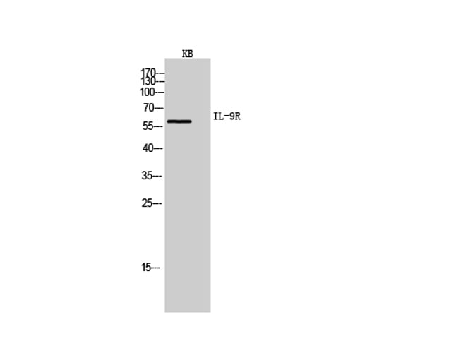
(Western Blot analysis of KB cells using IL-9R Polyclonal Antibody at dilution of 1:20000.)
WB (Western Blot)
(Western Blot analysis of KB cells using IL-9R Polyclonal Antibody at dilution of 1:20000.)
IL-9R, Polyclonal Antibody (Cat# AAA171691)
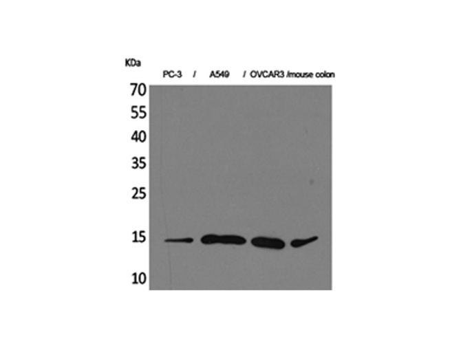
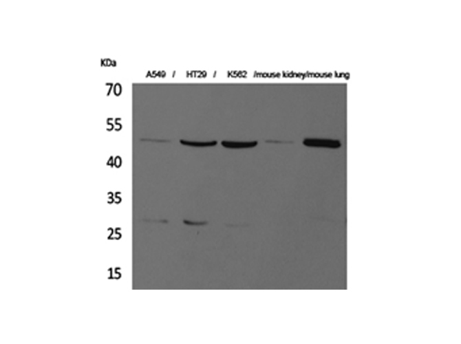
 WB (Western Blot)
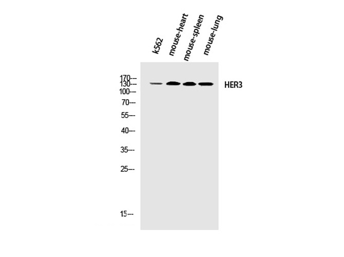
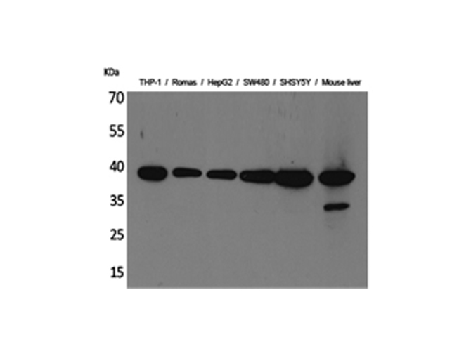
(Western Blot analysis of K562, Mouse heart, Mouse spleen, Mouse lung using ErbB-3 Polyclonal Antibody at dilution of 1:2000.)
WB (Western Blot)
(Western Blot analysis of K562, Mouse heart, Mouse spleen, Mouse lung using ErbB-3 Polyclonal Antibody at dilution of 1:2000.)
ErbB-3, Polyclonal Antibody (Cat# AAA171692)
 IF (Immunofluorescence)
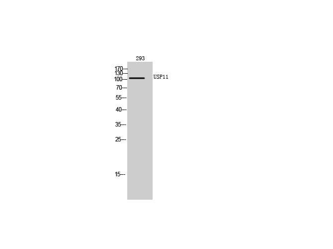
(Immunofluorescence analysis of Human lung tissue using USP11 Polyclonal Antibody at dilution of 1:200.)
IF (Immunofluorescence)
(Immunofluorescence analysis of Human lung tissue using USP11 Polyclonal Antibody at dilution of 1:200.)
USP11, Polyclonal Antibody (Cat# AAA171694)
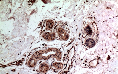 IHC (Immunohiostchemistry)
(Immunohistochemistry of paraffin-embedded Human breast cancer tissue using IL-10 Polyclonal Antibody at dilution of 1:100.)
IHC (Immunohiostchemistry)
(Immunohistochemistry of paraffin-embedded Human breast cancer tissue using IL-10 Polyclonal Antibody at dilution of 1:100.)
IL-10, Polyclonal Antibody (Cat# AAA171697)
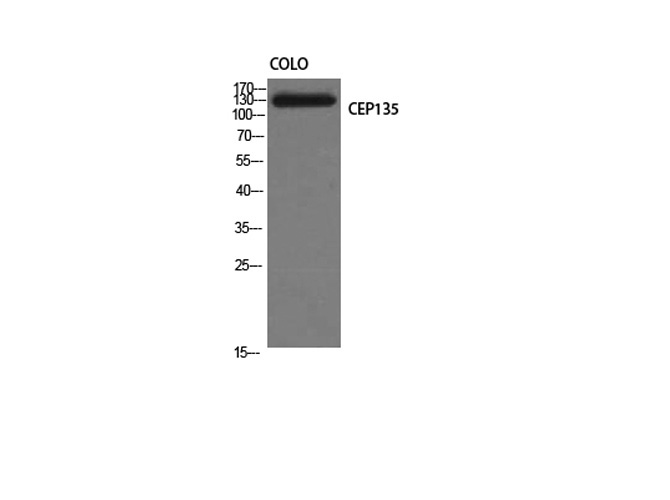
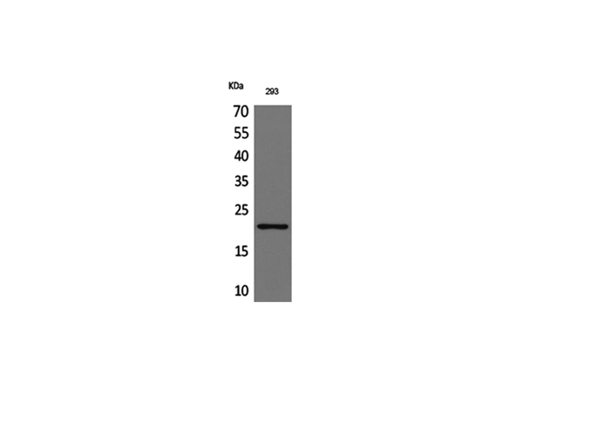
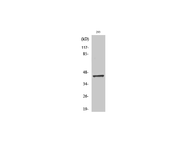
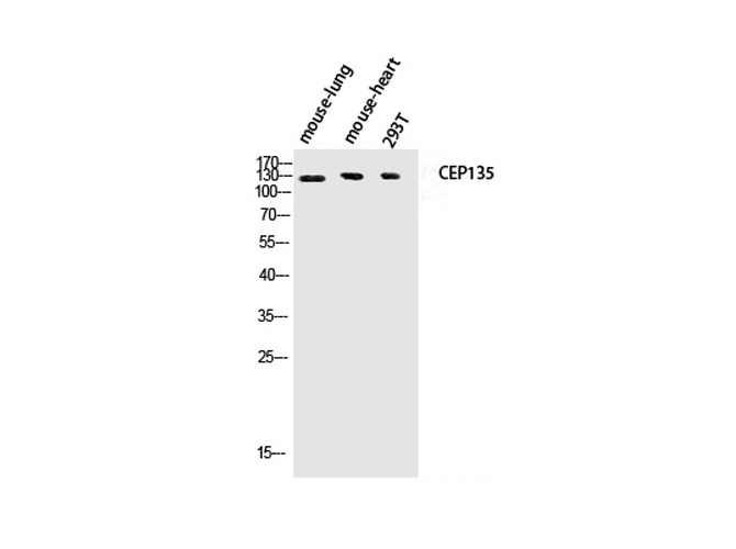 WB (Western Blot)
(Western Blot analysis of Mouse lung, Mouse heart, 293T using CEP135 Polyclonal Antibody at dilution of 1:1000.)
WB (Western Blot)
(Western Blot analysis of Mouse lung, Mouse heart, 293T using CEP135 Polyclonal Antibody at dilution of 1:1000.)
CEP135, Polyclonal Antibody (Cat# AAA171699)
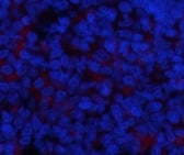
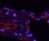
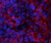
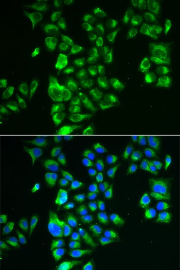 IF (Immunofluorescence)
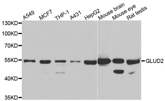
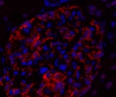
(Immunofluorescence analysis of A549 cell using GLUD2 antibody. Blue: DAPI for nuclear staining.)
IF (Immunofluorescence)
(Immunofluorescence analysis of A549 cell using GLUD2 antibody. Blue: DAPI for nuclear staining.)
GLUD2 , Polyclonal Antibody (Cat# AAA171700)
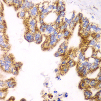
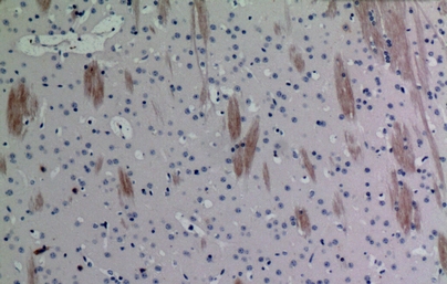
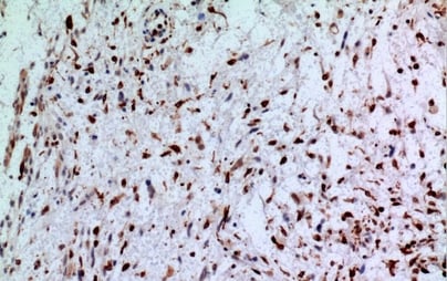
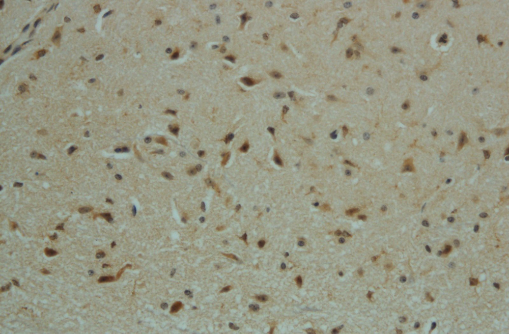
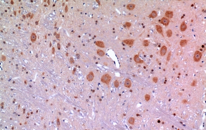 IHC (Immunohiostchemistry)
(Immunohistochemistry of paraffin-embedded Rat brain tissue using ARP Polyclonal Antibody at dilution of 1:100.)
IHC (Immunohiostchemistry)
(Immunohistochemistry of paraffin-embedded Rat brain tissue using ARP Polyclonal Antibody at dilution of 1:100.)
ARP, Polyclonal Antibody (Cat# AAA171703)
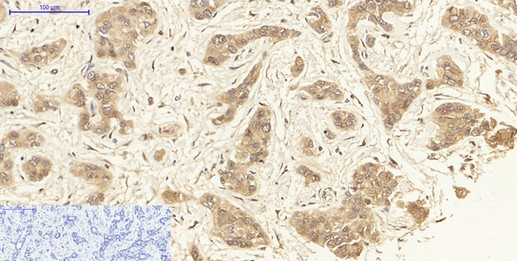
 IHC (Immunohiostchemistry)
(Immunohistochemistry of paraffin-embedded Human ovary cancer tissue using SNAP 23 Polyclonal Antibody at dilution of 1:100.)
IHC (Immunohiostchemistry)
(Immunohistochemistry of paraffin-embedded Human ovary cancer tissue using SNAP 23 Polyclonal Antibody at dilution of 1:100.)
SNAP 23, Polyclonal Antibody (Cat# AAA171707)
 IHC (Immunohiostchemistry)
(Immunohistochemistry of paraffin-embedded Human colon cancer tissue using Histone H2A Polyclonal Antibody at dilution of 1:100.)
IHC (Immunohiostchemistry)
(Immunohistochemistry of paraffin-embedded Human colon cancer tissue using Histone H2A Polyclonal Antibody at dilution of 1:100.)
Histone H2A, Polyclonal Antibody (Cat# AAA171721)
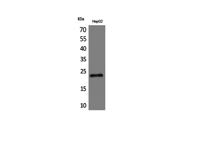
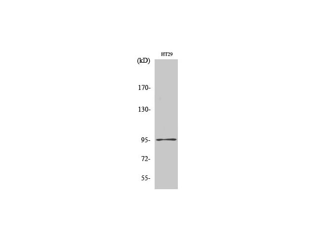
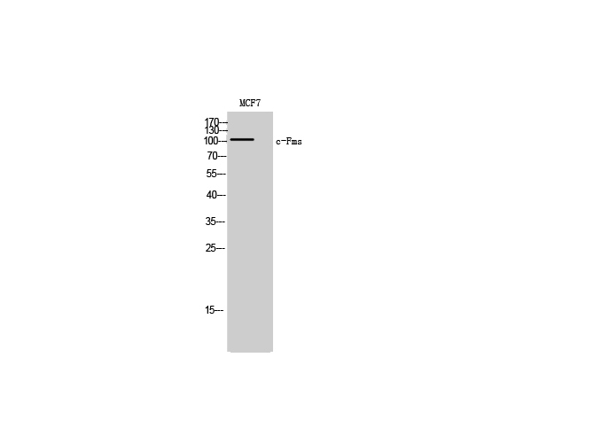 WB (Western Blot)
(Western Blot analysis of MCF7 cells with c-Fms Polyclonal Antibody.)
WB (Western Blot)
(Western Blot analysis of MCF7 cells with c-Fms Polyclonal Antibody.)
c-Fms, Polyclonal Antibody (Cat# AAA171725)
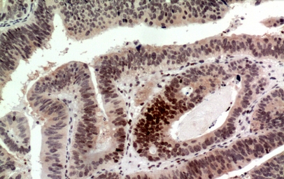
 IHC (Immunohistochemisry)
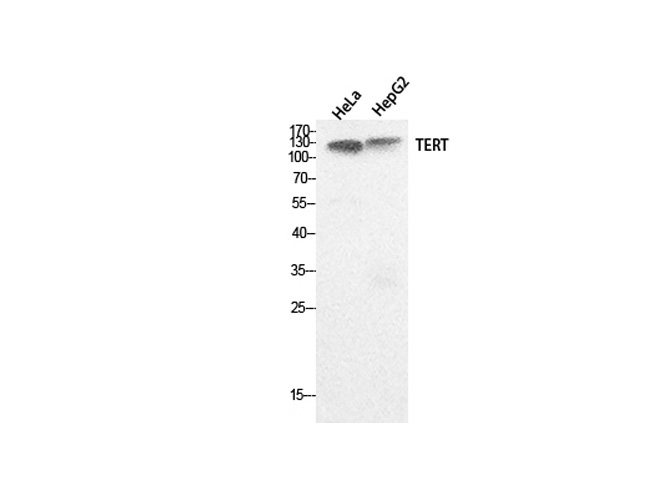
(Immunohistochemistry of paraffin-embedded Human stomach tissue using TERT Polyclonal Antibody at dilution of 1:100.)
IHC (Immunohistochemisry)
(Immunohistochemistry of paraffin-embedded Human stomach tissue using TERT Polyclonal Antibody at dilution of 1:100.)
TERT, Polyclonal Antibody (Cat# AAA171726)
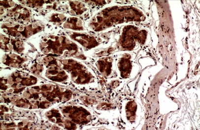
 IHC (Immunohistochemisry)
(Immunohistochemistry of paraffin-embedded Human lung tissue using CD42b Polyclonal Antibody at dilution of 1:100.)
IHC (Immunohistochemisry)
(Immunohistochemistry of paraffin-embedded Human lung tissue using CD42b Polyclonal Antibody at dilution of 1:100.)
CD42b, Polyclonal Antibody (Cat# AAA171728)
 IHC (Immunohiostchemistry)
(Immunohistochemistry of paraffin-embedded Mouse brain tissue using Eotaxin-3 Polyclonal Antibody at dilution of 1:100.)
IHC (Immunohiostchemistry)
(Immunohistochemistry of paraffin-embedded Mouse brain tissue using Eotaxin-3 Polyclonal Antibody at dilution of 1:100.)
Eotaxin-3, Polyclonal Antibody (Cat# AAA171731)
 IF (Immunofluorescence)
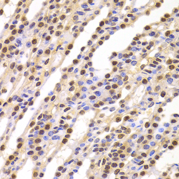
(Immunofluorescence analysis of Human kidney tissue using Dnmt3b Polyclonal Antibody at dilution of 1:200.)
IF (Immunofluorescence)
(Immunofluorescence analysis of Human kidney tissue using Dnmt3b Polyclonal Antibody at dilution of 1:200.)
Dnmt3b, Polyclonal Antibody (Cat# AAA171744)
 IF (Immunofluorescence)
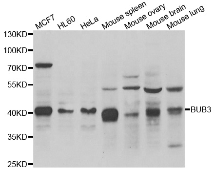
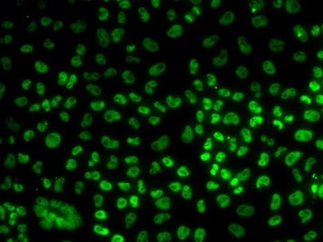
(Immunofluorescence analysis of U20S cell using BUB3 antibody.)
IF (Immunofluorescence)
(Immunofluorescence analysis of U20S cell using BUB3 antibody.)
BUB3, Polyclonal Antibody (Cat# AAA171745)
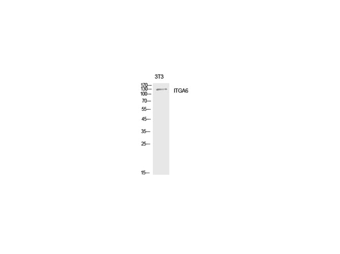
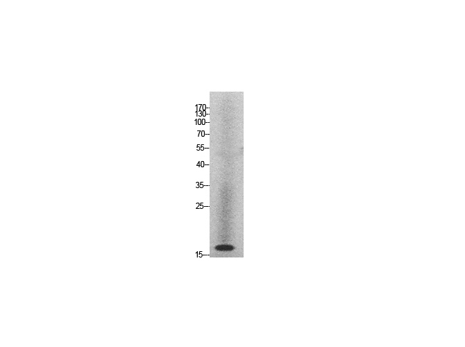
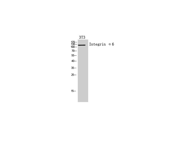 WB (Western Blot)
(Western Blot analysis of 3T3 cells with Integrin ?6 Polyclonal Antibody.)
WB (Western Blot)
(Western Blot analysis of 3T3 cells with Integrin ?6 Polyclonal Antibody.)
Integrin alpha6, Polyclonal Antibody (Cat# AAA171748)
 IHC (Immunohistochemisry)
(Immunohistochemistry of paraffin-embedded Human brain tissue using CD74 Polyclonal Antibody at dilution of 1:100.)
IHC (Immunohistochemisry)
(Immunohistochemistry of paraffin-embedded Human brain tissue using CD74 Polyclonal Antibody at dilution of 1:100.)
CD74, Polyclonal Antibody (Cat# AAA171751)
 IHC (Immunohiostchemistry)
(Immunohistochemistry of paraffin-embedded Rat brain tissue using GDI-2 Polyclonal Antibody at dilution of 1:100.)
IHC (Immunohiostchemistry)
(Immunohistochemistry of paraffin-embedded Rat brain tissue using GDI-2 Polyclonal Antibody at dilution of 1:100.)
GDI-2, Polyclonal Antibody (Cat# AAA171625)
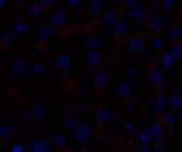 IF (Immunofluorescence)
(Immunofluorescence analysis of Human liver tissue using Caspase-9 Polyclonal Antibody at dilution of 1:200.)
IF (Immunofluorescence)
(Immunofluorescence analysis of Human liver tissue using Caspase-9 Polyclonal Antibody at dilution of 1:200.)
Caspase-9, Polyclonal Antibody (Cat# AAA171629)
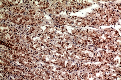
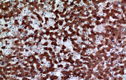 IHC (Immunohiostchemistry)
(Immunohistochemistry of paraffin-embedded Human liver tissue using FMO3 Polyclonal Antibody at dilution of 1:100.)
IHC (Immunohiostchemistry)
(Immunohistochemistry of paraffin-embedded Human liver tissue using FMO3 Polyclonal Antibody at dilution of 1:100.)
FMO3, Polyclonal Antibody (Cat# AAA171633)
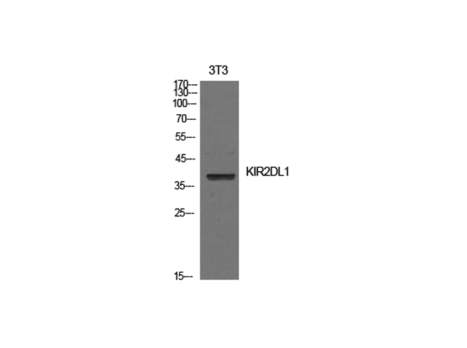
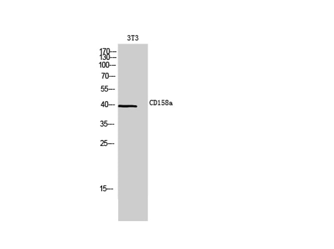 WB (Western Blot)
(Western Blot analysis of 3T3 cells using CD158a Polyclonal Antibody at dilution of 1:20000.)
WB (Western Blot)
(Western Blot analysis of 3T3 cells using CD158a Polyclonal Antibody at dilution of 1:20000.)
CD158a, Polyclonal Antibody (Cat# AAA171634)
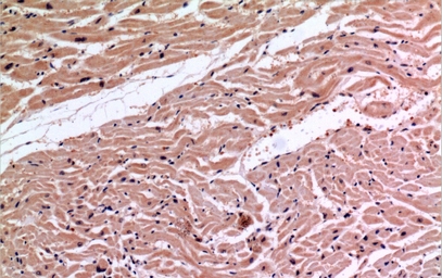 IHC (Immunohiostchemistry)
(Immunohistochemistry of paraffin-embedded Human heart tissue using Ku-70 Polyclonal Antibody at dilution of 1:100.)
IHC (Immunohiostchemistry)
(Immunohistochemistry of paraffin-embedded Human heart tissue using Ku-70 Polyclonal Antibody at dilution of 1:100.)
Ku-70, Polyclonal Antibody (Cat# AAA171636)
 IF (Immunofluorescence)
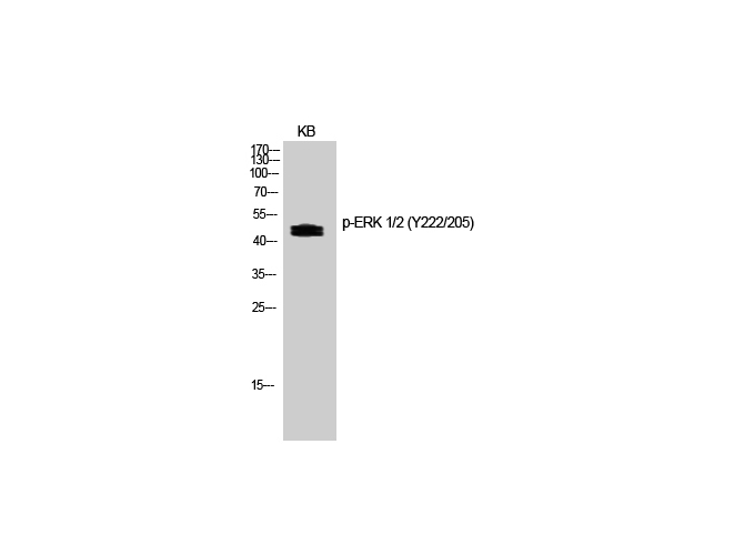
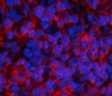
(Immunofluorescence analysis of Rat spleen tissue with Phospho-ERK 1/2 (Tyr222/205) Polyclonal Antibody at dilution of 1:200)
IF (Immunofluorescence)
(Immunofluorescence analysis of Rat spleen tissue with Phospho-ERK 1/2 (Tyr222/205) Polyclonal Antibody at dilution of 1:200)
ERK 1/2, Polyclonal Antibody (Cat# AAA171643)
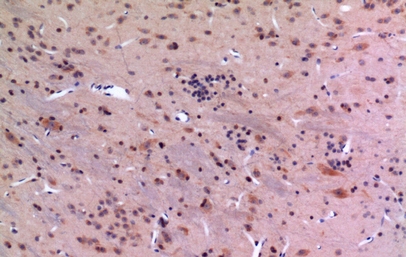
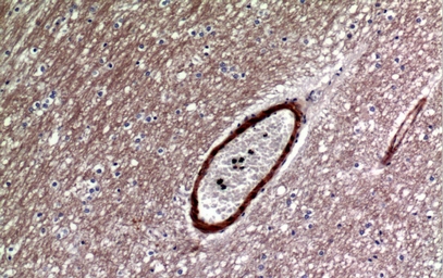 IHC (Immunohiostchemistry)
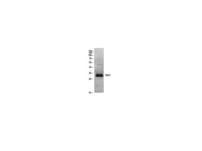
(Immunohistochemistry of paraffin-embedded Human brain tissue using Dkk-1 Polyclonal Antibody at dilution of 1:100.)
IHC (Immunohiostchemistry)
(Immunohistochemistry of paraffin-embedded Human brain tissue using Dkk-1 Polyclonal Antibody at dilution of 1:100.)
Dkk-1, Polyclonal Antibody (Cat# AAA171645)
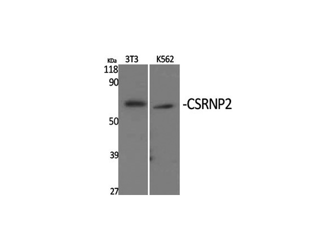
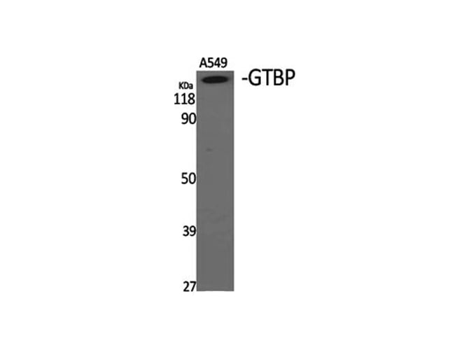
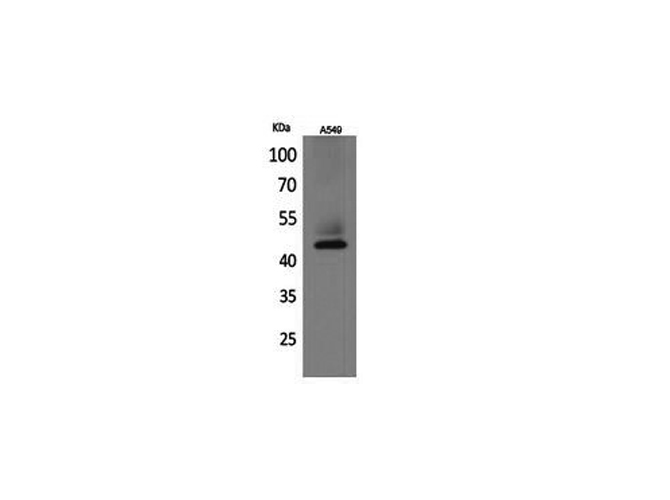
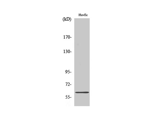 WB (Western Blot)
(Western Blot analysis of A549 cells with CSRNP2 Polyclonal Antibody.)
WB (Western Blot)
(Western Blot analysis of A549 cells with CSRNP2 Polyclonal Antibody.)
CSRNP2, Polyclonal Antibody (Cat# AAA171646)
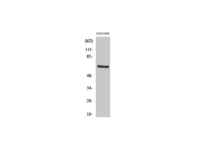
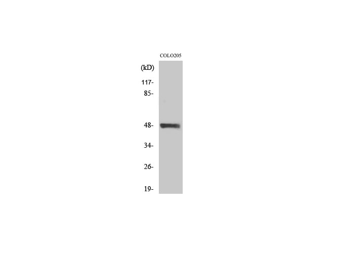 WB (Western Blot)
(Western Blot analysis of COLO205 cells using STK24 Polyclonal Antibody at dilution of 1:1000.)
WB (Western Blot)
(Western Blot analysis of COLO205 cells using STK24 Polyclonal Antibody at dilution of 1:1000.)
MST-3, Polyclonal Antibody (Cat# AAA171648)
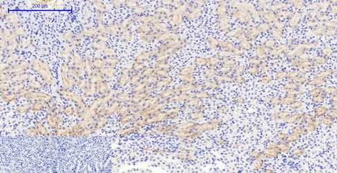
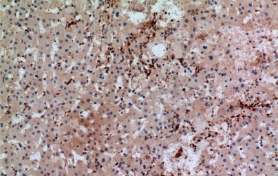
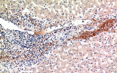 IHC (Immunohiostchemistry)
(Immunohistochemistry of paraffin-embedded Human liver tissue using ALR Polyclonal Antibody at dilution of 1:100.)
IHC (Immunohiostchemistry)
(Immunohistochemistry of paraffin-embedded Human liver tissue using ALR Polyclonal Antibody at dilution of 1:100.)
ALR, Polyclonal Antibody (Cat# AAA171655)
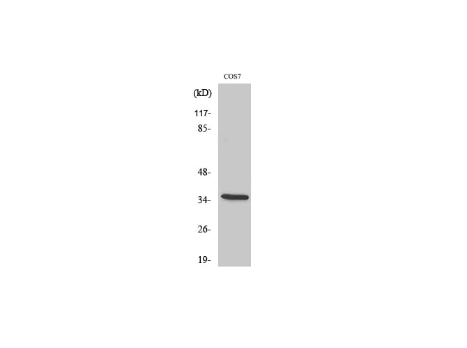
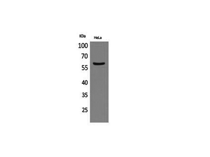
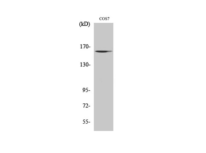 WB (Western Blot)
(Western Blot analysis of COS7 cells using GTBP Polyclonal Antibody at dilution of 1:1000.)
WB (Western Blot)
(Western Blot analysis of COS7 cells using GTBP Polyclonal Antibody at dilution of 1:1000.)
GTBP, Polyclonal Antibody (Cat# AAA171657)
 IF (Immunofluorescence)
(Immunofluorescence analysis of Rat lung tissue using PDK1 Polyclonal Antibody at dilution of 1:200.)
IF (Immunofluorescence)
(Immunofluorescence analysis of Rat lung tissue using PDK1 Polyclonal Antibody at dilution of 1:200.)
PDK1, Polyclonal Antibody (Cat# AAA171658)
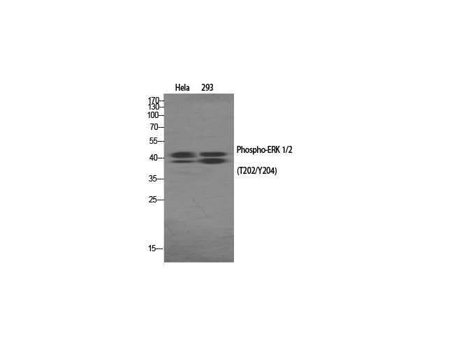
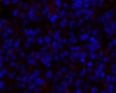
 IF (Immunofluorescence)
(Immunofluorescence analysis of Rat spleen tissue with Phospho-ERK 1/2 (Thr202/Tyr204) Polyclonal Antibody at dilution of 1:200)
IF (Immunofluorescence)
(Immunofluorescence analysis of Rat spleen tissue with Phospho-ERK 1/2 (Thr202/Tyr204) Polyclonal Antibody at dilution of 1:200)
ERK 1/2, Polyclonal Antibody (Cat# AAA171662)
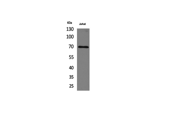
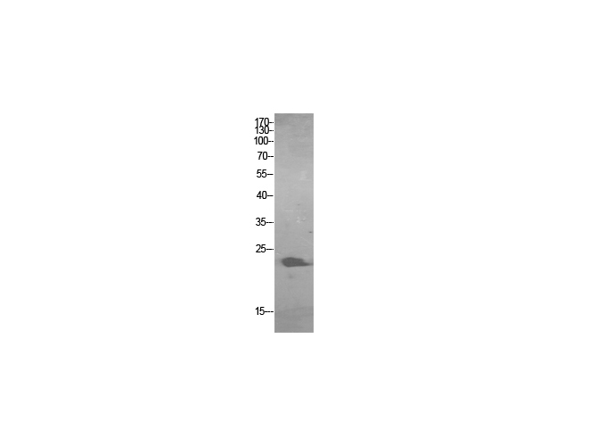
 WB (Western Blot)
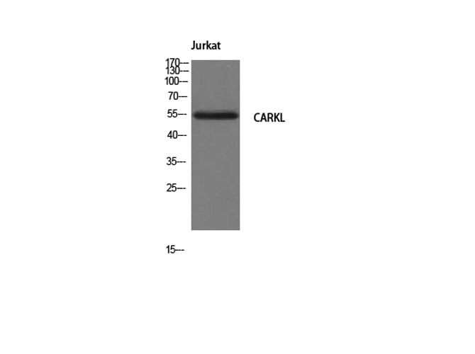
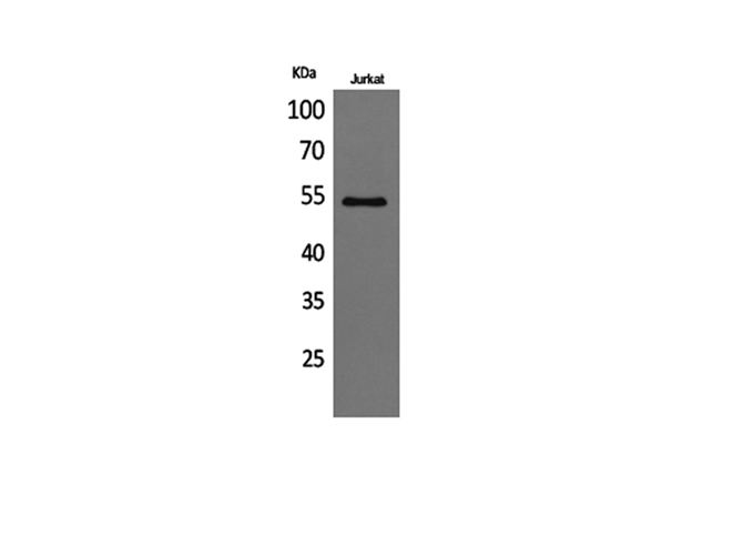
(Western Blot analysis of Jurkat cells with CARKL Polyclonal Antibody.)
WB (Western Blot)
(Western Blot analysis of Jurkat cells with CARKL Polyclonal Antibody.)
CARKL, Polyclonal Antibody (Cat# AAA171663)
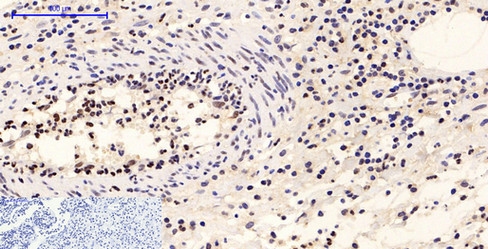
 IHC (Immunohiostchemistry)
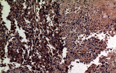
(Immunohistochemistry of paraffin-embedded Human lung cancer tissue using MMP-12 Polyclonal Antibody at dilution of 1:100.)
IHC (Immunohiostchemistry)
(Immunohistochemistry of paraffin-embedded Human lung cancer tissue using MMP-12 Polyclonal Antibody at dilution of 1:100.)
MMP-12, Polyclonal Antibody (Cat# AAA171665)
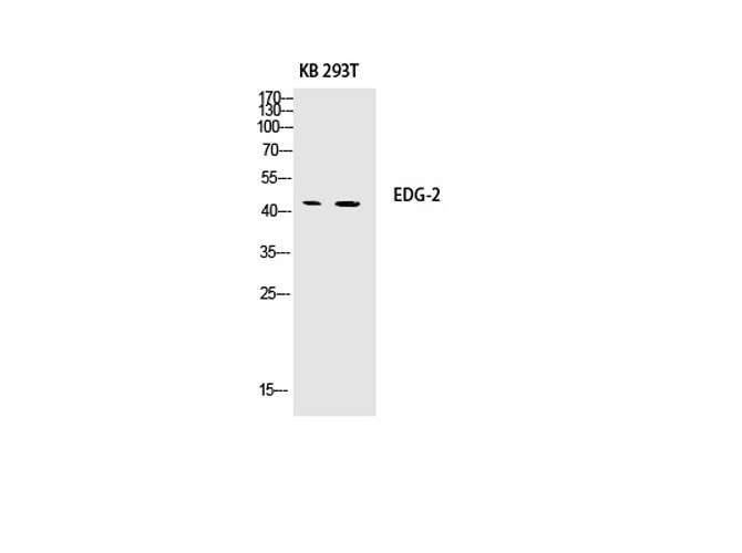 WB (Western Blot)
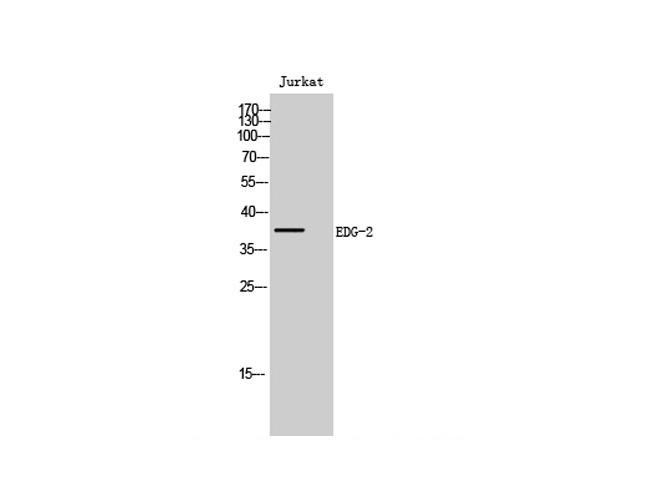
(Western Blot analysis of KB, 293T cells using EDG-2 Polyclonal Antibody at dilution of 1:500.)
WB (Western Blot)
(Western Blot analysis of KB, 293T cells using EDG-2 Polyclonal Antibody at dilution of 1:500.)
EDG-2, Polyclonal Antibody (Cat# AAA171666)
 IHC (Immunohistochemisry)
(Immunohistochemistry of paraffin-embedded Human lymph gland tissue using Cytokeratin 19 Polyclonal Antibody at dilution of 1:100.)
IHC (Immunohistochemisry)
(Immunohistochemistry of paraffin-embedded Human lymph gland tissue using Cytokeratin 19 Polyclonal Antibody at dilution of 1:100.)
Cytokeratin 19, Polyclonal Antibody (Cat# AAA171667)
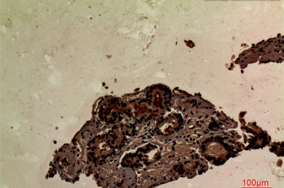 IHC (Immunohiostchemistry)
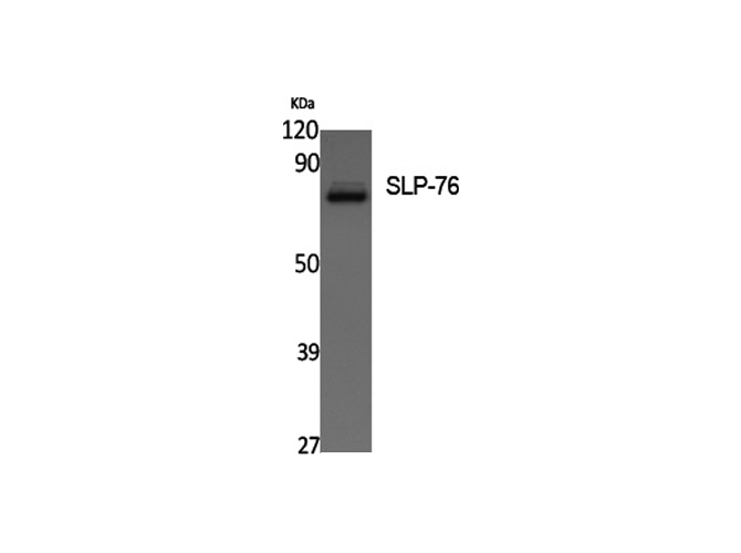
(Immunohistochemistry of paraffin-embedded Human prostate cancer tissue using SLP-76 Polyclonal Antibody at dilution of 1:100.)
IHC (Immunohiostchemistry)
(Immunohistochemistry of paraffin-embedded Human prostate cancer tissue using SLP-76 Polyclonal Antibody at dilution of 1:100.)
SLP-76, Polyclonal Antibody (Cat# AAA171668)
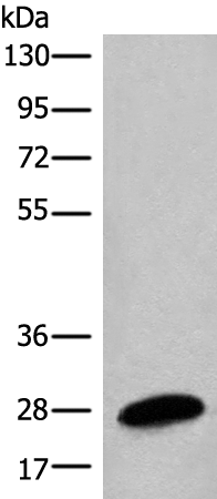
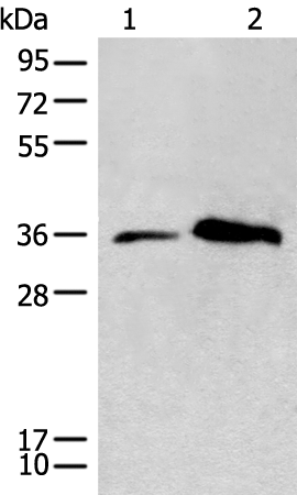
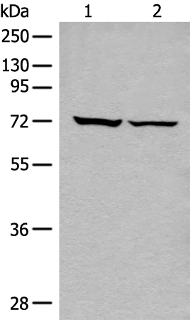
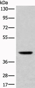 WB (Western Blot)
(Western blot analysis of Mouse liver tissue lysate using USP12 Polyclonal Antibody at dilution of 1:700)
WB (Western Blot)
(Western blot analysis of Mouse liver tissue lysate using USP12 Polyclonal Antibody at dilution of 1:700)
USP12, Polyclonal Antibody (Cat# AAA175219)
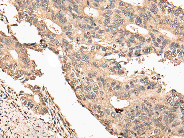
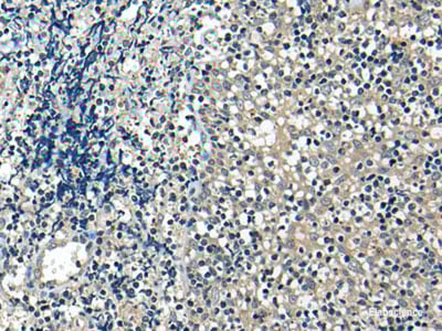
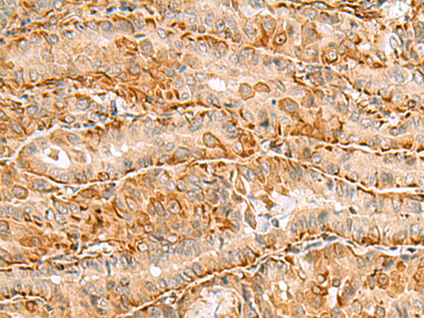
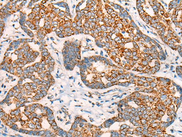
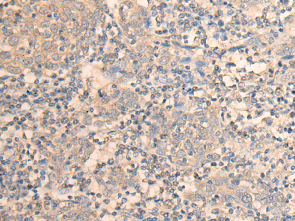
 IHC (Immunohiostchemistry)
(Immunohistochemistry of paraffin-embedded Human tonsil tissue using USP21 Polyclonal Antibody at dilution of 1:35(×200))
IHC (Immunohiostchemistry)
(Immunohistochemistry of paraffin-embedded Human tonsil tissue using USP21 Polyclonal Antibody at dilution of 1:35(×200))
USP21, Polyclonal Antibody (Cat# AAA175220)
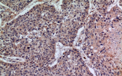
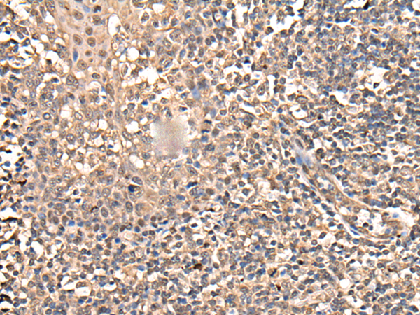
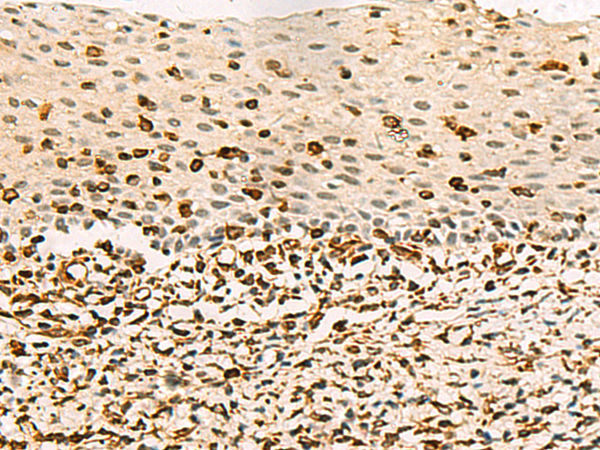
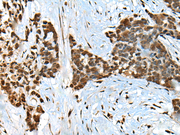
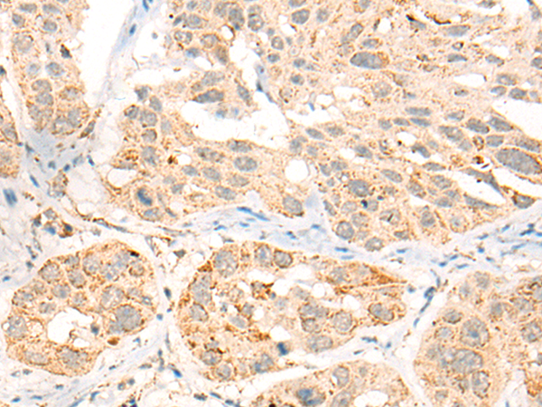
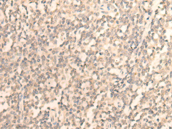
 IHC (Immunohiostchemistry)
(Immunohistochemistry of paraffin-embedded Human prost ate cancer tissue using BATF Polyclonal Antibody at dilution of 1:45(×200))
IHC (Immunohiostchemistry)
(Immunohistochemistry of paraffin-embedded Human prost ate cancer tissue using BATF Polyclonal Antibody at dilution of 1:45(×200))
BATF, Polyclonal Antibody (Cat# AAA175233)

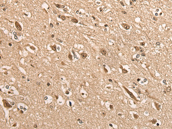
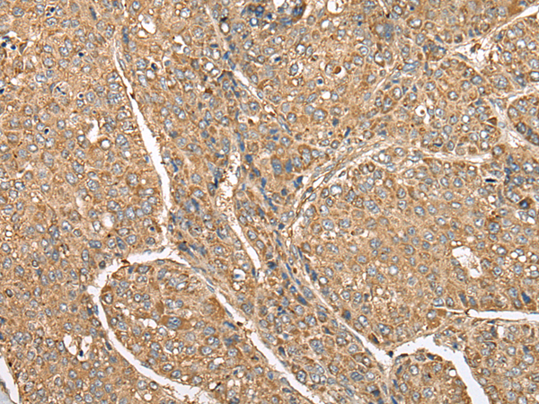
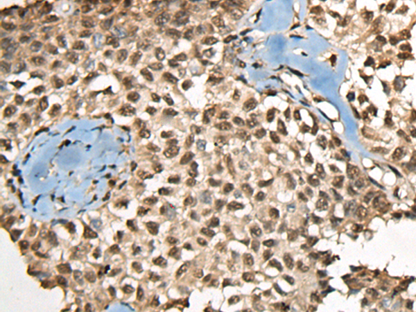
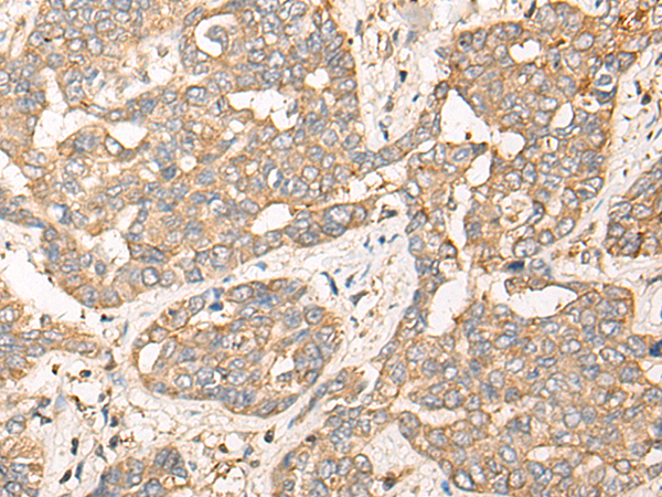
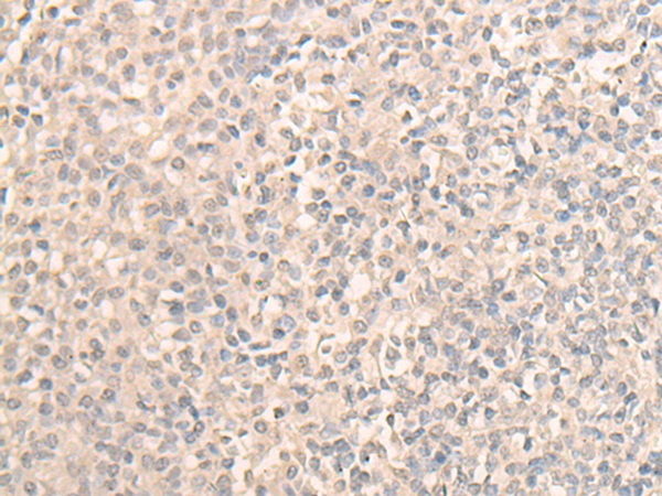
 IHC (Immunohiostchemistry)
(Immunohistochemistry of paraffin-embedded Human liver cancer tissue using BBOX1 Polyclonal Antibody at dilution of 1:45(×200))
IHC (Immunohiostchemistry)
(Immunohistochemistry of paraffin-embedded Human liver cancer tissue using BBOX1 Polyclonal Antibody at dilution of 1:45(×200))
BBOX1, Polyclonal Antibody (Cat# AAA175234)

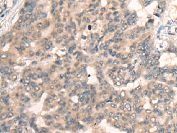
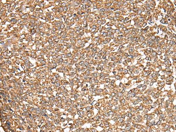
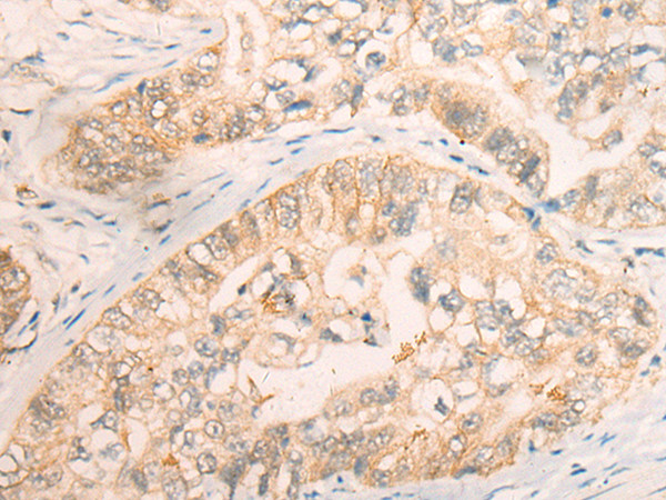
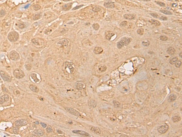
 IHC (Immunohistochemisry)
(Immunohistochemistry of paraffin-embedded Human gastric cancer tissue using BLNK Polyclonal Antibody at dilution of 1:40(×200))
IHC (Immunohistochemisry)
(Immunohistochemistry of paraffin-embedded Human gastric cancer tissue using BLNK Polyclonal Antibody at dilution of 1:40(×200))
BLNK, Polyclonal Antibody (Cat# AAA175240)

 IHC (Immunohistochemisry)
(Immunohistochemistry of paraffin-embedded Human esophagus cancer tissue using BPGM Polyclonal Antibody at dilution of 1:70(×200))
IHC (Immunohistochemisry)
(Immunohistochemistry of paraffin-embedded Human esophagus cancer tissue using BPGM Polyclonal Antibody at dilution of 1:70(×200))
BPGM, Polyclonal Antibody (Cat# AAA175242)
 IHC (Immunohiostchemistry)
(Immunohistochemistry of paraffin-embedded Human cervical cancer tissue using C3orf38 Polyclonal Antibody at dilution of 1:80(×200))
IHC (Immunohiostchemistry)
(Immunohistochemistry of paraffin-embedded Human cervical cancer tissue using C3orf38 Polyclonal Antibody at dilution of 1:80(×200))
C3orf38, Polyclonal Antibody (Cat# AAA175259)
 IHC (Immunohiostchemistry)
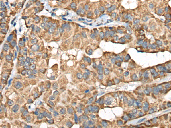
(Immunohistochemistry of paraffin-embedded Human esophagus cancer tissue using C5AR1 Polyclonal Antibody at dilution of 1:50(×200))
IHC (Immunohiostchemistry)
(Immunohistochemistry of paraffin-embedded Human esophagus cancer tissue using C5AR1 Polyclonal Antibody at dilution of 1:50(×200))
C5AR1, Polyclonal Antibody (Cat# AAA175261)
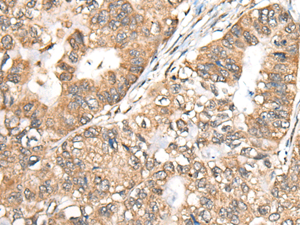
 IHC (Immunohiostchemistry)
(Immunohistochemistry of paraffin-embedded Human ovarian cancer tissue using CCDC12 Polyclonal Antibody at dilution of 1:60(×200))
IHC (Immunohiostchemistry)
(Immunohistochemistry of paraffin-embedded Human ovarian cancer tissue using CCDC12 Polyclonal Antibody at dilution of 1:60(×200))
CCDC12, Polyclonal Antibody (Cat# AAA175270)
 IHC (Immunohiostchemistry)
(Immunohistochemistry of paraffin-embedded Human gastric cancer tissue using CD86 Polyclonal Antibody at dilution of 1:30(×200))
IHC (Immunohiostchemistry)
(Immunohistochemistry of paraffin-embedded Human gastric cancer tissue using CD86 Polyclonal Antibody at dilution of 1:30(×200))
CD86, Polyclonal Antibody (Cat# AAA175271)
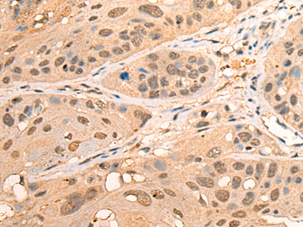
 IHC (Immunohiostchemistry)
(Immunohistochemistry of paraffin-embedded Human esophagus cancer tissue using CCT8L2 Polyclonal Antibody at dilution of 1:45(×200))
IHC (Immunohiostchemistry)
(Immunohistochemistry of paraffin-embedded Human esophagus cancer tissue using CCT8L2 Polyclonal Antibody at dilution of 1:45(×200))
CCT8L2, Polyclonal Antibody (Cat# AAA175275)
 IHC (Immunohiostchemistry)
(Immunohistochemistry of paraffin-embedded Human esophagus cancer tissue using CPNE3 Polyclonal Antibody at dilution of 1:40(×200))
IHC (Immunohiostchemistry)
(Immunohistochemistry of paraffin-embedded Human esophagus cancer tissue using CPNE3 Polyclonal Antibody at dilution of 1:40(×200))
CPNE3, Polyclonal Antibody (Cat# AAA175286)
 IHC (Immunohiostchemistry)
(Immunohistochemistry of paraffin-embedded Human tonsil tissue using DAZ1 Polyclonal Antibody at dilution of 1:55(×200))
IHC (Immunohiostchemistry)
(Immunohistochemistry of paraffin-embedded Human tonsil tissue using DAZ1 Polyclonal Antibody at dilution of 1:55(×200))
DAZ1, Polyclonal Antibody (Cat# AAA175293)
 IHC (Immunohiostchemistry)
(Immunohistochemistry of paraffin-embedded Human tonsil tissue using DDX59 Polyclonal Antibody at dilution of 1:30(×200))
IHC (Immunohiostchemistry)
(Immunohistochemistry of paraffin-embedded Human tonsil tissue using DDX59 Polyclonal Antibody at dilution of 1:30(×200))
DDX59, Polyclonal Antibody (Cat# AAA175300)
What are Polyclonal Antibodies?
Polyclonal antibodies are antibodies that come from multiple B cell clones of a host animal. The typical hosts used for the majority of polyclonal antibody production are rabbits, goats, sheep, and donkeys. These polyclonal antibodies, once having identified their target, will bind to different epitopes located at different regions or sequences on the same protein/antigen. As a result, they are ideal at locating and binding to the target, even if the target is in very low concentrations (due to many different antibodies being able to bind to the same target molecule, which allows for significant amplification of a downstream signal).
Polyclonal antibodies are typically produced by injecting an antigen into a host animal, which causes the animal’s immune system to attack the foreign antigen by mass generating antibodies against it. After a period of time, serum is collected from the animal and purified using physicochemical fractionation, class-specific affinity purification, and/or antigen-affinity purification.
Key Uses of Polyclonal Antibodies
- Western Blotting: This method is used to find specific proteins in biological samples after separating them by size.
- Immunohistochemistry: IHC helps visualize the location of proteins in tissue sections using various staining techniques.
- ELISA: (Enzyme-Linked Immunosorbent Assay) is typically used to identify specific protein quantities in a sample. ELISAs can be either “Quantitative” or “Qualitative”.
- Flow Cytometry: technique that identifies and measures the specific protein on the surface or inside the cells in a fluid suspension.
- Immunoprecipitation: IP isolates and studies a specific protein from a complex mixture using antibodies.
Why Buy Polyclonal Antibodies from AAA Biotech?
1. Ideal for Various Applications
Our antibodies are generally going to be validated for use in multiple types of assays, including ELISA, Western Blotting, Immunohistochemistry, Immunoprecipitation, amongst others. They are ideal for a wide range of research applications.
2. Rigorous Quality Control
All of the antibodies in our catalog undergo strict quality testing to ensure specificity, sensitivity, and consistent performance. We are confident in the ability of our antibodies to provide you with accurate results.
3. Wide Assortment of Antibodies
Antibodies in are catalog can be found for both common and exotic species, and these antibodies are also available in both conjugated and recombinant forms to suit many diverse experimental needs.
4. Highly Purified
Our antibodies are available in purified forms with over 85% purity, as confirmed by SDS-PAGE. They are also available with tags such as His, Flag, GST, or MBP. We cater to customers worldwide.
FAQ
1. How are polyclonal antibodies produced?
Traditionally, polyclonal antibodies are produced by injecting an antigen into a host animal (such as a rabbit or goat), which then triggers an immune response from the host animal. The animal’s B cells produce antibodies that will recognize different parts of the injected antigen. These antibodies are then collected from the animal’s blood and purified for use.
2. How do polyclonal antibodies differ from monoclonal antibodies?
Polyclonal antibodies are a mix of antibodies that bind to different locations (epitopes) of the same antigen, while monoclonal antibodies are identical and bind to just one specific epitope. This makes polyclonal antibodies more versatile and better at detecting proteins that may be present in low quantities or in altered/modified forms.
3. How should I store polyclonal antibodies?
Polyclonal antibodies should be stored at 4°C for short-term use (up to a few weeks) and at -20°C or -80°C for long-term storage. Avoid repeated freeze-thaw cycles by dividing them into small aliquots. Always check the datasheet for specific storage instructions.