Filters
▼Clonality
▼Type
▼Reactivity
▼Gene Name
▼Isotype
▼Host
▼Application
▼Clone
▼Polyclonal Antibodies
At AAA Biotech also known as AAA Bio or AAABio, we provide a broad range of purified polyclonal antibodies (pAbs) that are able to all be browsed online through our website. Due to their high specificity and strong binding affinity, these antibodies are ideal for wide swathes of research and experimental applications.
Our polyclonal antibodies can easily support your work, whether you use them for Western Blotting, Immunocytochemistry (with or without Immunofluorescence used in conjunction), Immunohistochemistry, Immunoprecipitation, and ELISA tests. We highly encourage you to browse our range of pAbs and choose the one that best suits your experimental model.
Viewing 5700-5750 of 96812 product results
 IHC (Immunohiostchemistry)
(DABstainingonIHC-P.Samples:RatTissue))
IHC (Immunohiostchemistry)
(DABstainingonIHC-P.Samples:RatTissue))
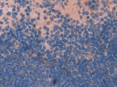
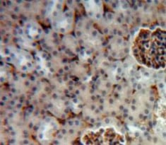
Monokine Induced By Interferon Gamma, Polyclonal Antibody (Cat# AAA141877)
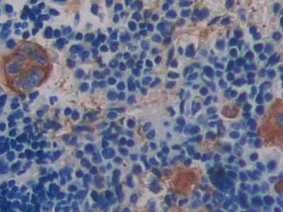 IHC (Immunohistochemisry)
(DAB staining on IHC-P; Samples: Mouse Spleen Tissue))
IHC (Immunohistochemisry)
(DAB staining on IHC-P; Samples: Mouse Spleen Tissue))
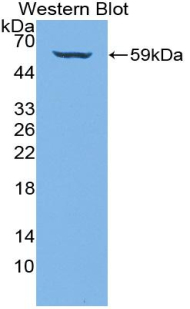
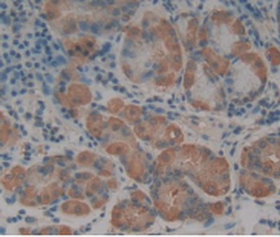
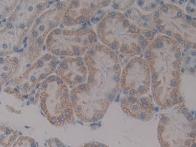
Integrin Beta 3, Polyclonal Antibody (Cat# AAA141888)
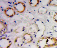
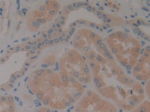
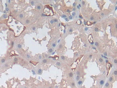
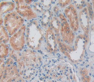 IHC (Immunohiostchemistry)
(DAB staining on fromalin fixed paraffin- embedded Kidney tissue))
IHC (Immunohiostchemistry)
(DAB staining on fromalin fixed paraffin- embedded Kidney tissue))
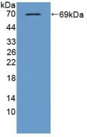
Interleukin 1 Alpha, Polyclonal Antibody (Cat# AAA141889)
 IHC (Immunohiostchemistry)
(DABstainingonIHC-P.Samples:MouseTissue))
IHC (Immunohiostchemistry)
(DABstainingonIHC-P.Samples:MouseTissue))
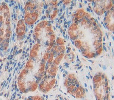
Doublecortin, Polyclonal Antibody (Cat# AAA141890)
 IHC (Immunohistochemistry)
(DAB staining on IHC-P; Samples: Human Stomach Tissue.)
IHC (Immunohistochemistry)
(DAB staining on IHC-P; Samples: Human Stomach Tissue.)
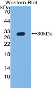
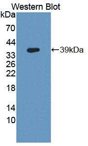
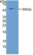
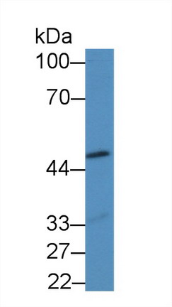
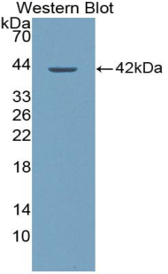
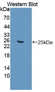
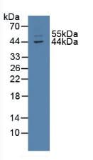
ATP Binding Cassette Transporter C2, Polyclonal Antibody (Cat# AAA141891)
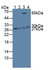
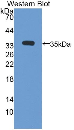
 WB (Western Blot)
(Sample:Lane 1: Bovine Serum;Lane 2: Human Placenta lysatePrimary Ab: 0.8ug/ml Rabbit Anti-Bovine PRLR AntibodySecond Ab: 0.2ug/mL HRP-Linked Caprine Anti-Rabbit IgG Polyclonal Antibody)
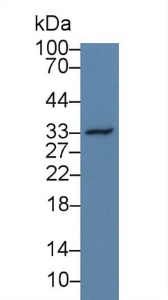
WB (Western Blot)
(Sample:Lane 1: Bovine Serum;Lane 2: Human Placenta lysatePrimary Ab: 0.8ug/ml Rabbit Anti-Bovine PRLR AntibodySecond Ab: 0.2ug/mL HRP-Linked Caprine Anti-Rabbit IgG Polyclonal Antibody)
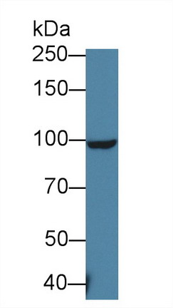
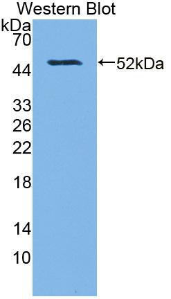
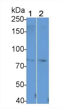
Prolactin Receptor, Polyclonal Antibody (Cat# AAA141894)
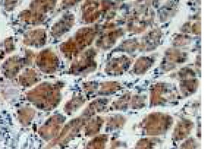
 IHC (Immunohistochemisry)
(DAB staining on fromalin fixed paraffin- embedded Kidney tissue))
IHC (Immunohistochemisry)
(DAB staining on fromalin fixed paraffin- embedded Kidney tissue))
G Protein Beta 2, Polyclonal Antibody (Cat# AAA141895)
 IHC (Immunohistochemistry)
(DAB staining on IHC-P; Samples: Rat Adrenal Gland Tissue)
IHC (Immunohistochemistry)
(DAB staining on IHC-P; Samples: Rat Adrenal Gland Tissue)
Glutathione S Transferase Theta 2, Polyclonal Antibody (Cat# AAA141897)

 IHC (Immunohiostchemistry)
(DABstainingonIHC-P.Samples:HumanTissue))
IHC (Immunohiostchemistry)
(DABstainingonIHC-P.Samples:HumanTissue))
Cytoglobin, Polyclonal Antibody (Cat# AAA141898)
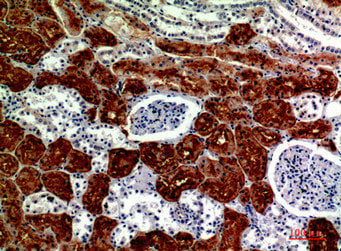
 IHC (Immunohistochemistry)
(DAB staining on IHC-P;Samples: Human Kidney Tissue;Primary Ab: 10ug/ml Rabbit Anti-Human COL4a3 AntibodySecond Ab: 2ug/mL HRP-Linked Caprine Anti-Rabbit IgG Polyclonal Antibody)
IHC (Immunohistochemistry)
(DAB staining on IHC-P;Samples: Human Kidney Tissue;Primary Ab: 10ug/ml Rabbit Anti-Human COL4a3 AntibodySecond Ab: 2ug/mL HRP-Linked Caprine Anti-Rabbit IgG Polyclonal Antibody)
Collagen Type IV Alpha 3, Polyclonal Antibody (Cat# AAA141899)
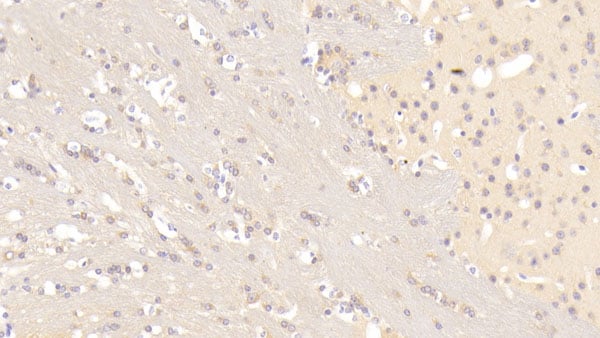
 IHC (Immunohiostchemistry)
(DAB staining on fromalin fixed paraffin-embedded Brain tissue))
IHC (Immunohiostchemistry)
(DAB staining on fromalin fixed paraffin-embedded Brain tissue))
TATA Binding Protein, Polyclonal Antibody (Cat# AAA141902)
 IHC (Immunohiostchemistry)
(DABstainingonIHC-P.Samples:MouseTissue))
IHC (Immunohiostchemistry)
(DABstainingonIHC-P.Samples:MouseTissue))
Upregulator Of Cell Proliferation, Polyclonal Antibody (Cat# AAA141907)
 IHC (Immunohistochemisry)
(DAB staining on IHC-P. Samples: Human Tissue))
IHC (Immunohistochemisry)
(DAB staining on IHC-P. Samples: Human Tissue))
Transcobalamin II, Polyclonal Antibody (Cat# AAA141916)
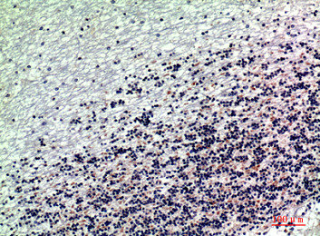
 IHC (Immunohistochemisry)
(DAB staining on IHC-P;Samples: Human Glioma Tissue;Primary Ab: 10ug/ml Rabbit Anti-Simian OPN AntibodySecond Ab: 2ug/mL HRP-Linked Caprine Anti-Rabbit IgG Polyclonal Antibody)
IHC (Immunohistochemisry)
(DAB staining on IHC-P;Samples: Human Glioma Tissue;Primary Ab: 10ug/ml Rabbit Anti-Simian OPN AntibodySecond Ab: 2ug/mL HRP-Linked Caprine Anti-Rabbit IgG Polyclonal Antibody)
Osteopontin, Polyclonal Antibody (Cat# AAA141922)
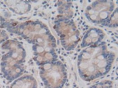
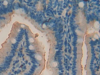 IHC (Immunohistochemisry)
(DAB staining on IHC-P; Samples: Mouse Intestine Tissue))
IHC (Immunohistochemisry)
(DAB staining on IHC-P; Samples: Mouse Intestine Tissue))
Acid Phosphatase 5, Polyclonal Antibody (Cat# AAA141926)
 IHC (Immunohiostchemistry)
(DABstainingonIHC-P.Samples:HumanTissue))
IHC (Immunohiostchemistry)
(DABstainingonIHC-P.Samples:HumanTissue))
Ring Finger Protein 39, Polyclonal Antibody (Cat# AAA141927)
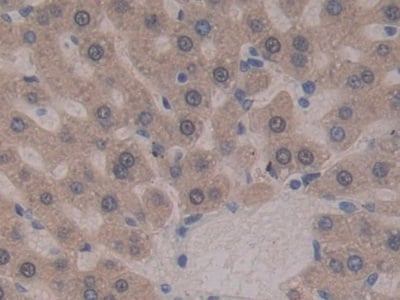
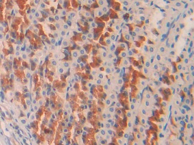
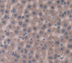
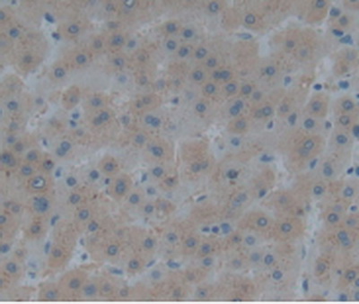
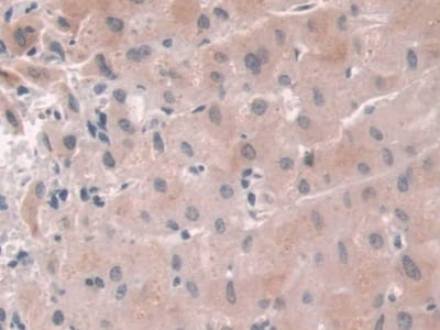
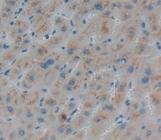
 IHC (Immunohistochemisry)
(DAB staining on fromalin fixed paraffin- embedded liver tissue))
IHC (Immunohistochemisry)
(DAB staining on fromalin fixed paraffin- embedded liver tissue))
Glutaminase 2, Polyclonal Antibody (Cat# AAA141936)
 IHC (Immunohiostchemistry)
(DAB staining on IHC-P; Samples: Rat Stomach Tissue))
IHC (Immunohiostchemistry)
(DAB staining on IHC-P; Samples: Rat Stomach Tissue))
Collagen Type II Alpha 1, Polyclonal Antibody (Cat# AAA141937)
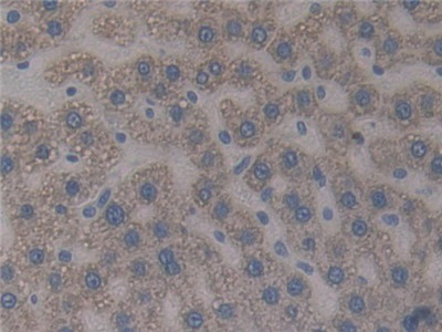
 IHC (Immunohistochemisry)
(DAB staining on fromalin fixed paraffin-embedded Liver tissue))
IHC (Immunohistochemisry)
(DAB staining on fromalin fixed paraffin-embedded Liver tissue))
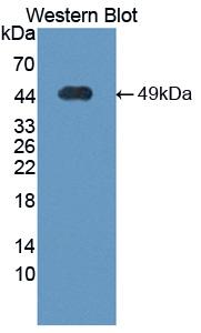
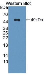
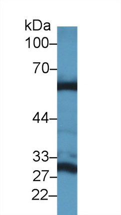
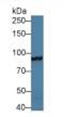
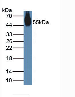
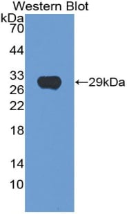
Hemojuvelin, Polyclonal Antibody (Cat# AAA141938)
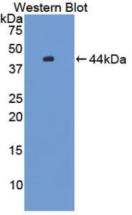
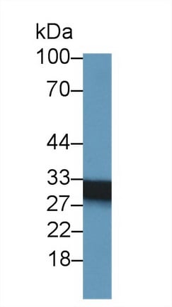
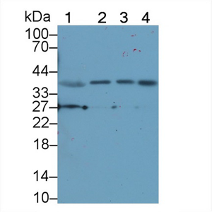 WB (Western Blot)
(Sample: Lane1: Porcine Stomach lysate;Lane2: Porcine Pancreas lysate;Lane3: PC3 cell lysate;Lane4: A549 cell lysatePrimary Ab: 1ug/ml Rabbit Anti-Human NT4 AntibodySecond Ab: 0.2ug/mL HRP-LinkedCaprine Anti-Rabbit IgG Polyclonal Antibody (Catalog: ))
WB (Western Blot)
(Sample: Lane1: Porcine Stomach lysate;Lane2: Porcine Pancreas lysate;Lane3: PC3 cell lysate;Lane4: A549 cell lysatePrimary Ab: 1ug/ml Rabbit Anti-Human NT4 AntibodySecond Ab: 0.2ug/mL HRP-LinkedCaprine Anti-Rabbit IgG Polyclonal Antibody (Catalog: ))
Neurotrophin 4, Polyclonal Antibody (Cat# AAA141941)
 IHC (Immunohiostchemistry)
(DAB staining on IHC-P. Samples: Mouse Tissue)
IHC (Immunohiostchemistry)
(DAB staining on IHC-P. Samples: Mouse Tissue)
Fibulin 2, Polyclonal Antibody (Cat# AAA141944)
 IHC (Immunohistochemisry)
(DAB staining on IHC-P. Samples: Human Tissue))
IHC (Immunohistochemisry)
(DAB staining on IHC-P. Samples: Human Tissue))
ATP Binding Cassette Transporter C3, Polyclonal Antibody (Cat# AAA141945)
 IHC (Immunohistochemistry)
(DAB staining on IHC-P. Samples: Mouse Tissue))
IHC (Immunohistochemistry)
(DAB staining on IHC-P. Samples: Mouse Tissue))
Microsomal Triglyceride Transfer Protein, Polyclonal Antibody (Cat# AAA141948)
 IHC (Immunohistochemisry)
(DAB staining on fromalin fixed paraffin-embedded Pancreas tissue))
IHC (Immunohistochemisry)
(DAB staining on fromalin fixed paraffin-embedded Pancreas tissue))
Neutrophil Cytosolic Factor 2, Polyclonal Antibody (Cat# AAA141958)
 IHC (Immunohistochemistry)
(DAB staining on IHC-P; Samples: Rat Spleen Tissue.)
IHC (Immunohistochemistry)
(DAB staining on IHC-P; Samples: Rat Spleen Tissue.)
Integrin Alpha D, Polyclonal Antibody (Cat# AAA141962)
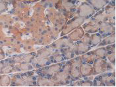
 IHC (Immunohiostchemistry)
(DAB staining on fromalin fixed paraffin-embedded Liver tissue))
IHC (Immunohiostchemistry)
(DAB staining on fromalin fixed paraffin-embedded Liver tissue))
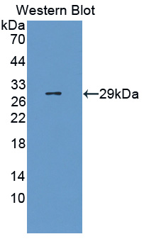
Growth Differentiation Factor 2, Polyclonal Antibody (Cat# AAA141966)
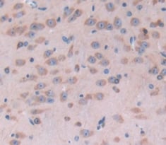
 IHC (Immunohiostchemistry)
(DABstainingonIHC-P.Samples:RatTissue))
IHC (Immunohiostchemistry)
(DABstainingonIHC-P.Samples:RatTissue))
Glucokinase Regulatory Protein, Polyclonal Antibody (Cat# AAA141967)
 IHC (Immunohistochemistry)
(DAB staining on IHC-P; Samples: Mouse Uterus Tissue)
IHC (Immunohistochemistry)
(DAB staining on IHC-P; Samples: Mouse Uterus Tissue)
Nucleobindin 2, Polyclonal Antibody (Cat# AAA141968)
 IHC (Immunohistochemisry)
(DAB staining on fromalin fixed paraffin-embedded Liver tissue))
IHC (Immunohistochemisry)
(DAB staining on fromalin fixed paraffin-embedded Liver tissue))
Growth Differentiation Factor 7, Polyclonal Antibody (Cat# AAA141970)
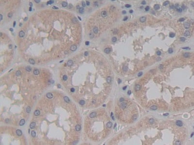
 IHC (Immunohiostchemistry)
(DAB staining on fromalin fixed paraffin- embedded Kidney tissue))
IHC (Immunohiostchemistry)
(DAB staining on fromalin fixed paraffin- embedded Kidney tissue))
Ribonuclease A9, Polyclonal Antibody (Cat# AAA141971)

 IHC (Immunohistochemistry)
(DAB staining on IHC-P; Samples: Mouse Stomach Tissue.)
IHC (Immunohistochemistry)
(DAB staining on IHC-P; Samples: Mouse Stomach Tissue.)
Dynamin 1, Polyclonal Antibody (Cat# AAA141974)

 IHC (Immunohiostchemistry)
(DAB staining on fromalin fixed paraffin- embedded Kidney tissue))
IHC (Immunohiostchemistry)
(DAB staining on fromalin fixed paraffin- embedded Kidney tissue))
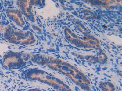
Forkhead Box Protein A1, Polyclonal Antibody (Cat# AAA141980)
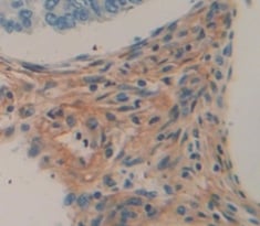
 IHC (Immunohistochemisry)
(Vector Red staining on IHC-P;Samples: Human Small Intestine Tissue;Primary Ab: 10ug/ml Rabbit Anti-Human IL4R AntibodySecond Ab: 2ug/mL HRP-Linked Caprine Anti-Rabbit IgG Polyclonal Antibody)
IHC (Immunohistochemisry)
(Vector Red staining on IHC-P;Samples: Human Small Intestine Tissue;Primary Ab: 10ug/ml Rabbit Anti-Human IL4R AntibodySecond Ab: 2ug/mL HRP-Linked Caprine Anti-Rabbit IgG Polyclonal Antibody)
Interleukin 4 Receptor, Polyclonal Antibody (Cat# AAA141981)
 IHC (Immunohiostchemistry)
(DABstainingonIHC-P.Samples:HumanTissue))
IHC (Immunohiostchemistry)
(DABstainingonIHC-P.Samples:HumanTissue))
AF4/FMR2, Polyclonal Antibody (Cat# AAA141985)
 IHC (Immunohiostchemistry)
(DAB staining on fromalin fixed paraffin-embedded Lung tissue))
IHC (Immunohiostchemistry)
(DAB staining on fromalin fixed paraffin-embedded Lung tissue))
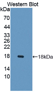
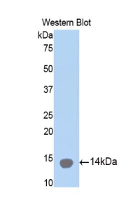
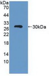
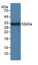
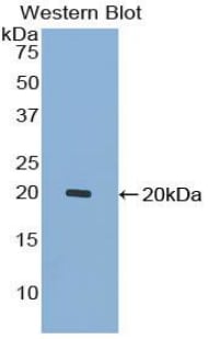
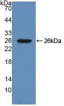
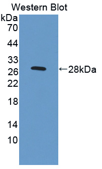
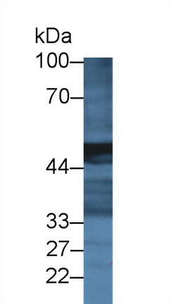
Lymphotactin, Polyclonal Antibody (Cat# AAA141987)
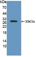
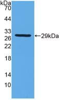
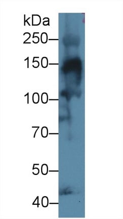
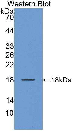
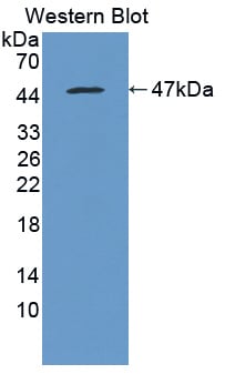
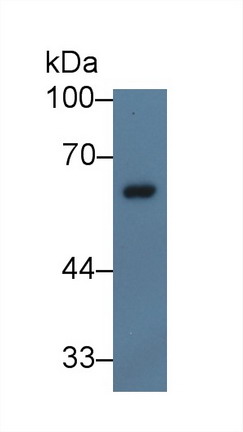
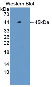
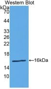
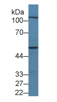
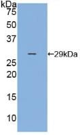
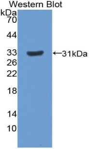
 WB (Western Blot)
(Figure. Western Blot; Sample: Recombinant MGMT, Rat.)
WB (Western Blot)
(Figure. Western Blot; Sample: Recombinant MGMT, Rat.)
O-6-Methylguanine DNA Methyltransferase, Polyclonal Antibody (Cat# AAA141992)
 IHC (Immunohistochemisry)
(DAB staining on fromalin fixed paraffin- embedded pancreas tissue))
IHC (Immunohistochemisry)
(DAB staining on fromalin fixed paraffin- embedded pancreas tissue))
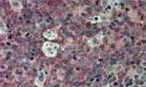
Carboxypeptidase A2, Polyclonal Antibody (Cat# AAA141993)
 IHC (Immunohistochemisry)
(DAB staining on fromalin fixed paraffin-embedded Kidney tissue))
IHC (Immunohistochemisry)
(DAB staining on fromalin fixed paraffin-embedded Kidney tissue))
Hexosaminidase A Alpha, Polyclonal Antibody (Cat# AAA142413)
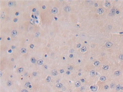
 IHC (Immunohistochemisry)
(DAB staining on fromalin fixed paraffin- embedded brain tissue))
IHC (Immunohistochemisry)
(DAB staining on fromalin fixed paraffin- embedded brain tissue))
Succinate Dehydrogenase Complex Subunit B, Polyclonal Antibody (Cat# AAA142416)
 IHC (Immunohistochemistry)
(DAB staining on IHC-P; Samples: Human Stomach.)
IHC (Immunohistochemistry)
(DAB staining on IHC-P; Samples: Human Stomach.)
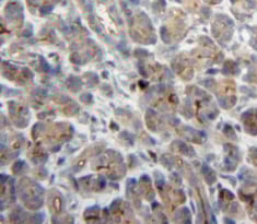
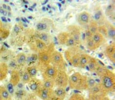
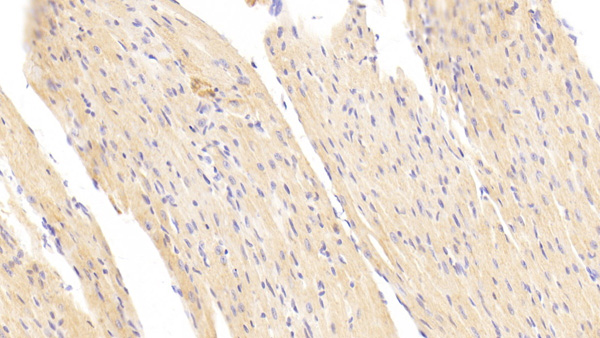
Renin, Polyclonal Antibody (Cat# AAA142440)

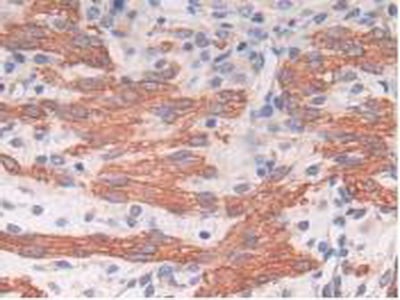
 IHC (Immunohistochemisry)
(DAB staining on fromalin fixed paraffin- embedded skeletal muscle tissue))
IHC (Immunohistochemisry)
(DAB staining on fromalin fixed paraffin- embedded skeletal muscle tissue))
Myopalladin, Polyclonal Antibody (Cat# AAA142449)
 IHC (Immunohistochemistry)
(DAB staining on IHC-P; Samples: Human Stomach Tissue.)
IHC (Immunohistochemistry)
(DAB staining on IHC-P; Samples: Human Stomach Tissue.)
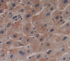
Selectin, Polyclonal Antibody (Cat# AAA142464)
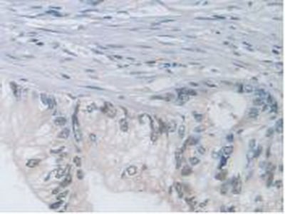
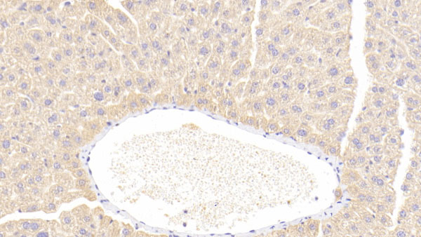
 IHC (Immunohistochemisry)
(DAB staining on IHC-P; Samples: Human Liver Tissue))
IHC (Immunohistochemisry)
(DAB staining on IHC-P; Samples: Human Liver Tissue))
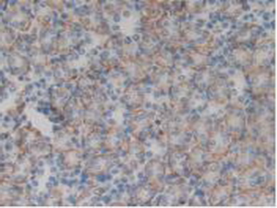
USP6 N-Terminal Like Protein, Polyclonal Antibody (Cat# AAA142333)
 IHC (Immunohistochemistry)
(DAB staining on IHC-P; Samples: Mouse Skeletal Muscle Tissue)
IHC (Immunohistochemistry)
(DAB staining on IHC-P; Samples: Mouse Skeletal Muscle Tissue)
Thioredoxin Binding Protein 2, Polyclonal Antibody (Cat# AAA142349)
 IHC (Immunohiostchemistry)
(DABstainingonIHC-P.Samples:HumanTissue))
IHC (Immunohiostchemistry)
(DABstainingonIHC-P.Samples:HumanTissue))
N-Acetylgalactosaminidase Alpha, Polyclonal Antibody (Cat# AAA142379)


 IHC (Immunohistochemisry)
(DAB staining on IHC-P. Samples: Mouse Tissue))
IHC (Immunohistochemisry)
(DAB staining on IHC-P. Samples: Mouse Tissue))
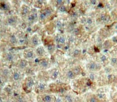
Phosphatidylethanolamine Binding Protein 1, Polyclonal Antibody (Cat# AAA142388)
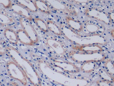
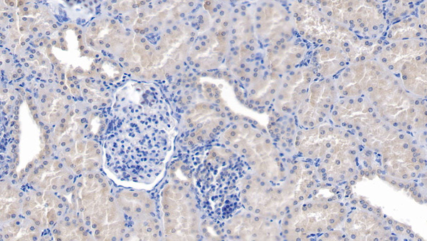
 IHC (Immunohistochemistry)
(DAB staining on IHC-P; Samples: Rat Kidney Tissue))
IHC (Immunohistochemistry)
(DAB staining on IHC-P; Samples: Rat Kidney Tissue))
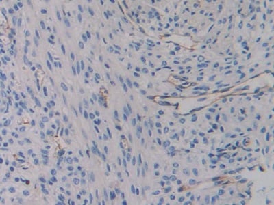
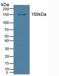
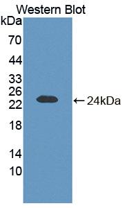
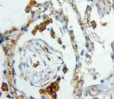
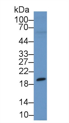
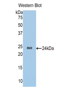
Paxillin, Polyclonal Antibody (Cat# AAA142393)
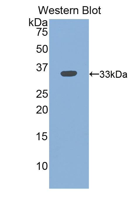
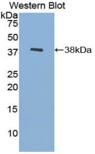
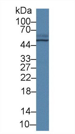
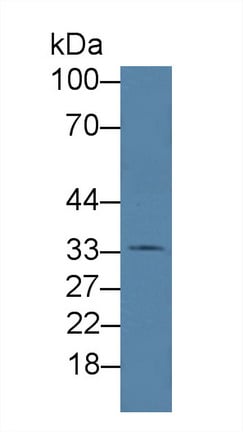
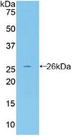
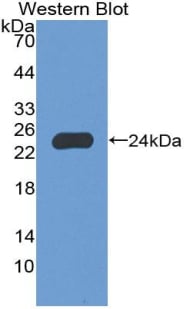
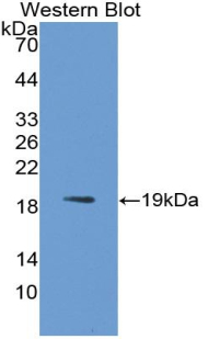
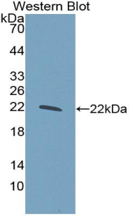
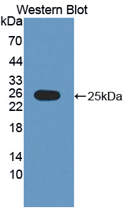
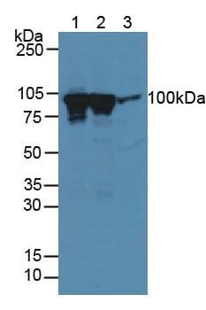
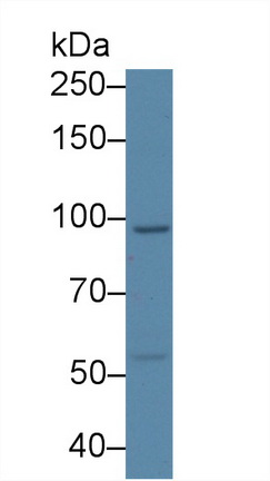
Cross Linked C-Telopeptide Of Type I Collagen (CTXI), Polyclonal Antibody (Cat# AAA152553)
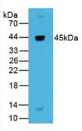
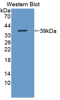
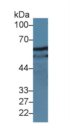
 WB (Western Blot)
(Western Blot; Sample: Lane1: Mouse Testis lysate; Lane2: Rat Testis lysate Primary Ab: 1 ug/ml Rabbit Anti-Mouse TAGAP Antibody Second Ab: 0.2ug/mL HRP-Linked Caprine Anti-Rabbit IgG Polyclonal Antibody)
WB (Western Blot)
(Western Blot; Sample: Lane1: Mouse Testis lysate; Lane2: Rat Testis lysate Primary Ab: 1 ug/ml Rabbit Anti-Mouse TAGAP Antibody Second Ab: 0.2ug/mL HRP-Linked Caprine Anti-Rabbit IgG Polyclonal Antibody)
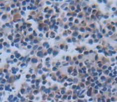
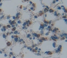
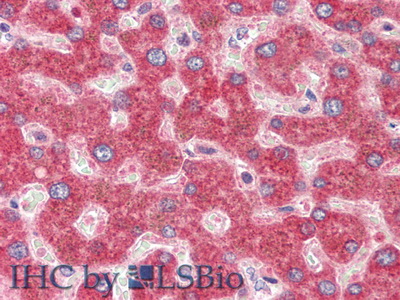
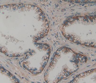
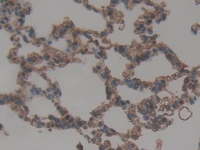
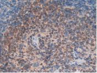
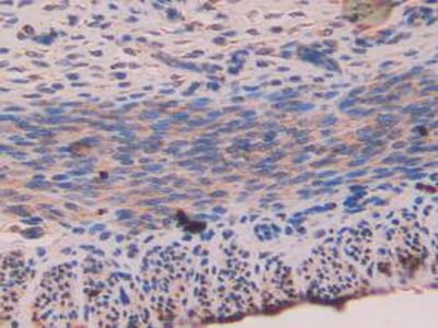
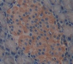
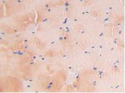
T-Cell Activation Rho GTPase Activating Protein (TAGAP), Polyclonal Antibody (Cat# AAA152615)
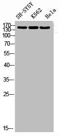
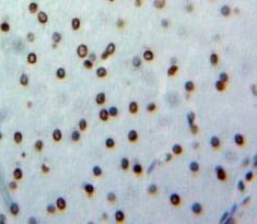
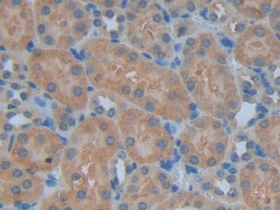
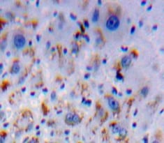
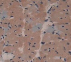
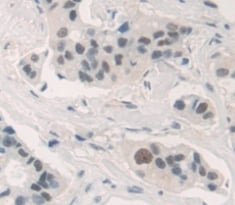
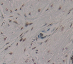
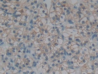
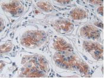
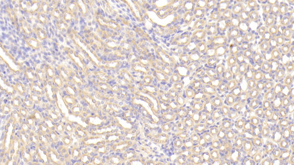
 IHC (Immunohistochemistry)
(Immunohistochemical analysis of paraffin-embedded human-kidney, antibody was diluted at 1:200)
IHC (Immunohistochemistry)
(Immunohistochemical analysis of paraffin-embedded human-kidney, antibody was diluted at 1:200)
DROSHA, Polyclonal Antibody (Cat# AAA236838)
What are Polyclonal Antibodies?
Polyclonal antibodies are antibodies that come from multiple B cell clones of a host animal. The typical hosts used for the majority of polyclonal antibody production are rabbits, goats, sheep, and donkeys. These polyclonal antibodies, once having identified their target, will bind to different epitopes located at different regions or sequences on the same protein/antigen. As a result, they are ideal at locating and binding to the target, even if the target is in very low concentrations (due to many different antibodies being able to bind to the same target molecule, which allows for significant amplification of a downstream signal).
Polyclonal antibodies are typically produced by injecting an antigen into a host animal, which causes the animal’s immune system to attack the foreign antigen by mass generating antibodies against it. After a period of time, serum is collected from the animal and purified using physicochemical fractionation, class-specific affinity purification, and/or antigen-affinity purification.
Key Uses of Polyclonal Antibodies
- Western Blotting: This method is used to find specific proteins in biological samples after separating them by size.
- Immunohistochemistry: IHC helps visualize the location of proteins in tissue sections using various staining techniques.
- ELISA: (Enzyme-Linked Immunosorbent Assay) is typically used to identify specific protein quantities in a sample. ELISAs can be either “Quantitative” or “Qualitative”.
- Flow Cytometry: technique that identifies and measures the specific protein on the surface or inside the cells in a fluid suspension.
- Immunoprecipitation: IP isolates and studies a specific protein from a complex mixture using antibodies.
Why Buy Polyclonal Antibodies from AAA Biotech?
1. Ideal for Various Applications
Our antibodies are generally going to be validated for use in multiple types of assays, including ELISA, Western Blotting, Immunohistochemistry, Immunoprecipitation, amongst others. They are ideal for a wide range of research applications.
2. Rigorous Quality Control
All of the antibodies in our catalog undergo strict quality testing to ensure specificity, sensitivity, and consistent performance. We are confident in the ability of our antibodies to provide you with accurate results.
3. Wide Assortment of Antibodies
Antibodies in are catalog can be found for both common and exotic species, and these antibodies are also available in both conjugated and recombinant forms to suit many diverse experimental needs.
4. Highly Purified
Our antibodies are available in purified forms with over 85% purity, as confirmed by SDS-PAGE. They are also available with tags such as His, Flag, GST, or MBP. We cater to customers worldwide.
FAQ
1. How are polyclonal antibodies produced?
Traditionally, polyclonal antibodies are produced by injecting an antigen into a host animal (such as a rabbit or goat), which then triggers an immune response from the host animal. The animal’s B cells produce antibodies that will recognize different parts of the injected antigen. These antibodies are then collected from the animal’s blood and purified for use.
2. How do polyclonal antibodies differ from monoclonal antibodies?
Polyclonal antibodies are a mix of antibodies that bind to different locations (epitopes) of the same antigen, while monoclonal antibodies are identical and bind to just one specific epitope. This makes polyclonal antibodies more versatile and better at detecting proteins that may be present in low quantities or in altered/modified forms.
3. How should I store polyclonal antibodies?
Polyclonal antibodies should be stored at 4°C for short-term use (up to a few weeks) and at -20°C or -80°C for long-term storage. Avoid repeated freeze-thaw cycles by dividing them into small aliquots. Always check the datasheet for specific storage instructions.