Filters
▼Clonality
▼Type
▼Reactivity
▼Gene Name
▼Isotype
▼Host
▼Application
▼Clone
▼Polyclonal Antibodies
At AAA Biotech also known as AAA Bio or AAABio, we provide a broad range of purified polyclonal antibodies (pAbs) that are able to all be browsed online through our website. Due to their high specificity and strong binding affinity, these antibodies are ideal for wide swathes of research and experimental applications.
Our polyclonal antibodies can easily support your work, whether you use them for Western Blotting, Immunocytochemistry (with or without Immunofluorescence used in conjunction), Immunohistochemistry, Immunoprecipitation, and ELISA tests. We highly encourage you to browse our range of pAbs and choose the one that best suits your experimental model.
Viewing 5800-5850 of 96812 product results

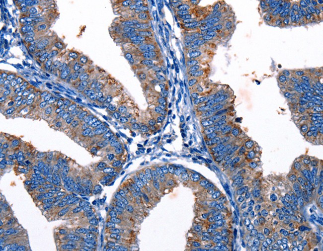
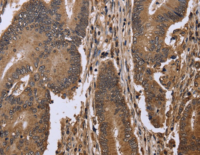
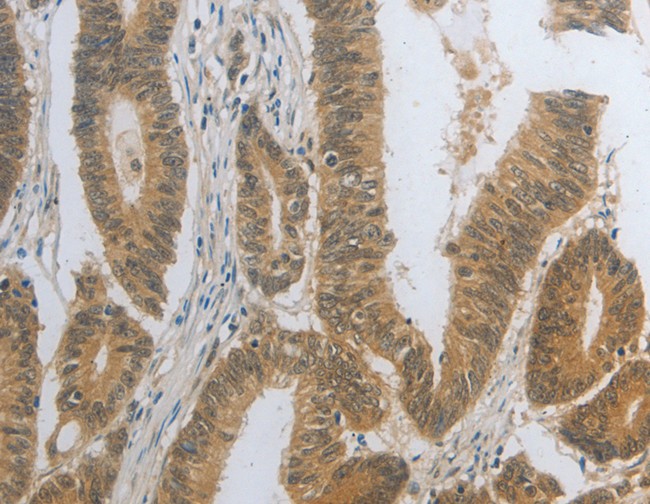
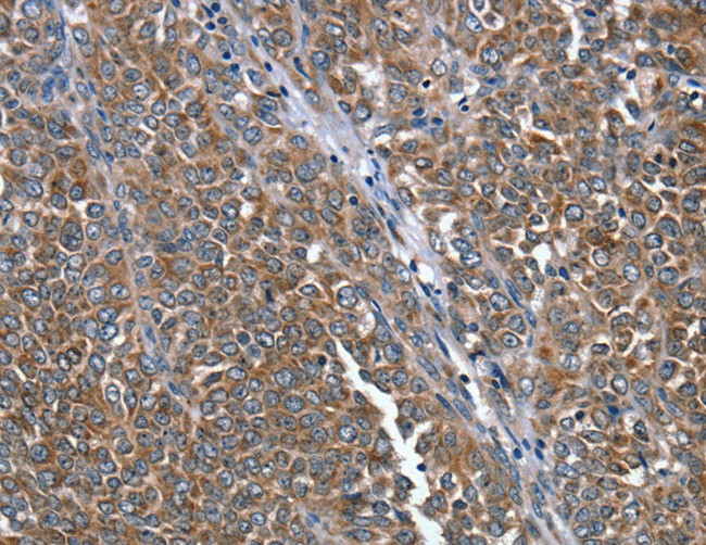
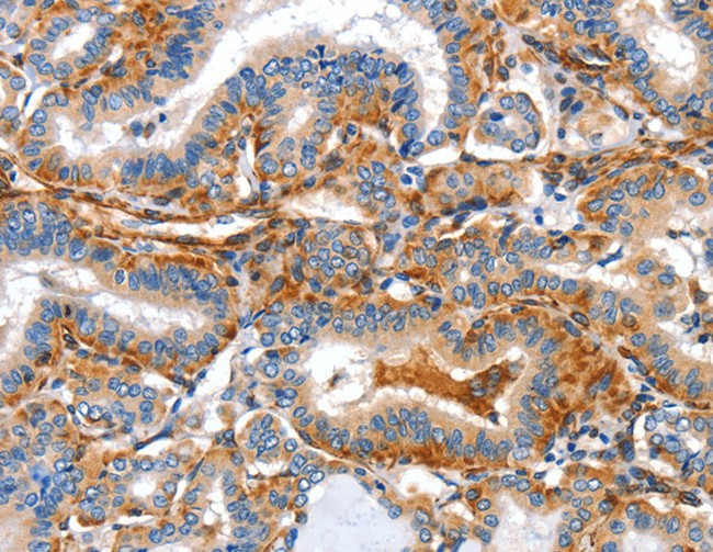
 IHC (Immunohiostchemistry)
(Immunohistochemistry of paraffin-embedded Human colon cancer tissue using SEPT9 Polyclonal Antibody at dilution 1:25)
IHC (Immunohiostchemistry)
(Immunohistochemistry of paraffin-embedded Human colon cancer tissue using SEPT9 Polyclonal Antibody at dilution 1:25)
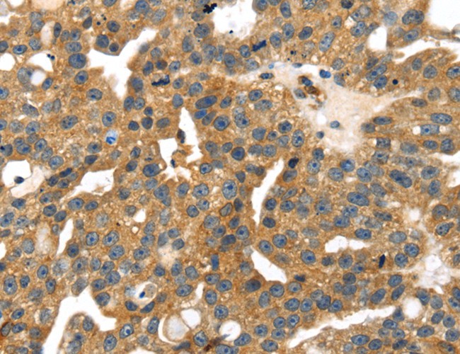
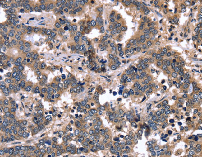
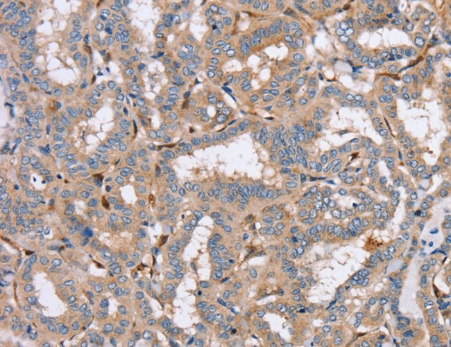
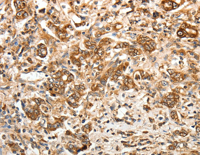
SEPT9, Polyclonal Antibody (Cat# AAA169158)
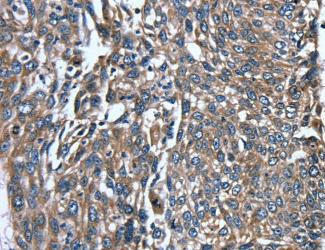
 IHC (Immunohiostchemistry)
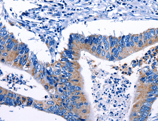
(Immunohistochemistry of paraffin-embedded Human esophagus cancer tissue using IRAK3 Polyclonal Antibody at dilution 1:50)
IHC (Immunohiostchemistry)
(Immunohistochemistry of paraffin-embedded Human esophagus cancer tissue using IRAK3 Polyclonal Antibody at dilution 1:50)
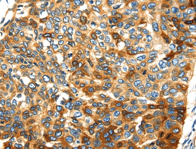
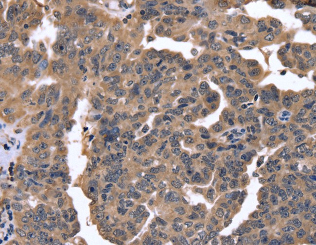
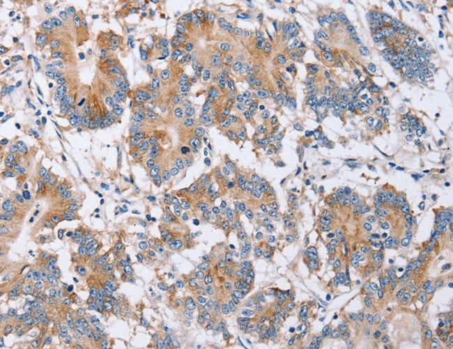
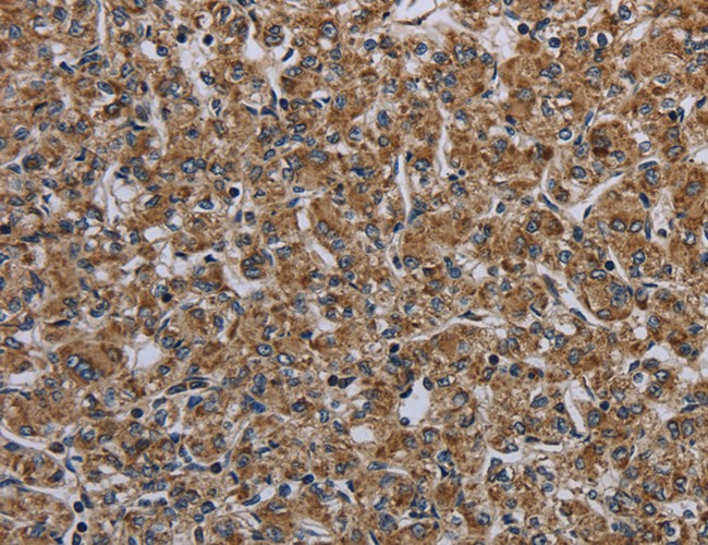
IRAK3, Polyclonal Antibody (Cat# AAA169159)

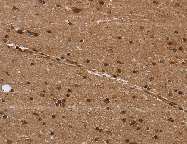
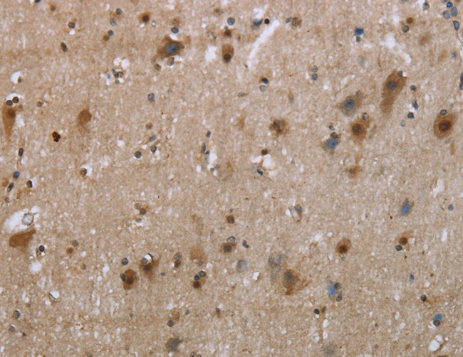
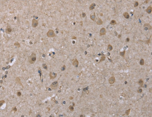
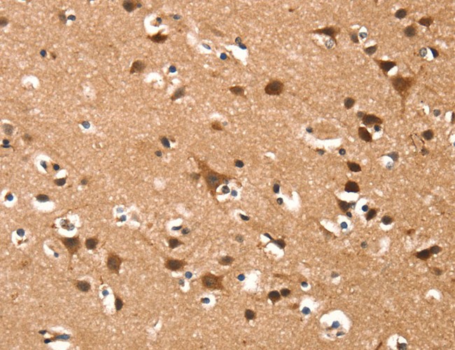
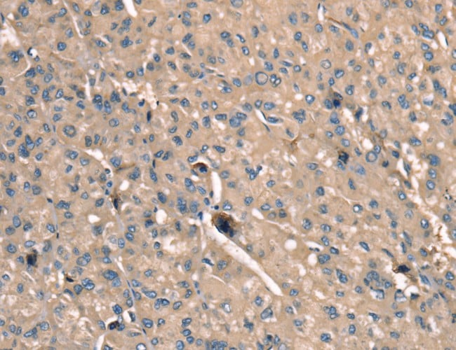
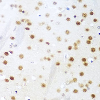
 IHC (Immunohistochemisry)
(Immunohistochemistry of paraffin-embedded Human brain using PSMC1 Polyclonal Antibody at dilution of 1:40)
IHC (Immunohistochemisry)
(Immunohistochemistry of paraffin-embedded Human brain using PSMC1 Polyclonal Antibody at dilution of 1:40)
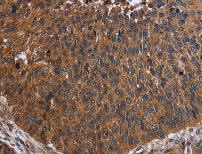
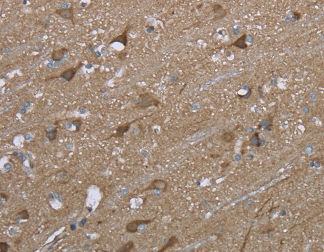
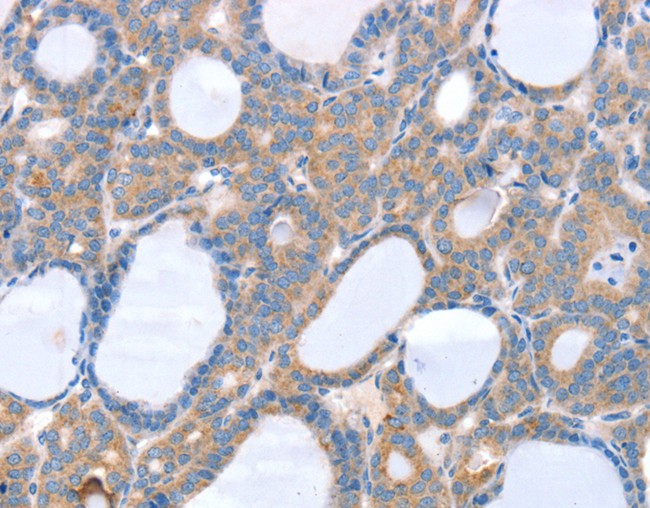
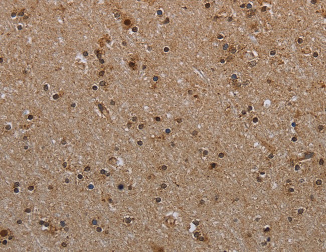
PSMC1, Polyclonal Antibody (Cat# AAA169161)
 IHC (Immunohiostchemistry)
(Immunohistochemistry of paraffin-embedded Human esophagus cancer tissue using SMYD4 Polyclonal Antibody at dilution 1:30)
IHC (Immunohiostchemistry)
(Immunohistochemistry of paraffin-embedded Human esophagus cancer tissue using SMYD4 Polyclonal Antibody at dilution 1:30)
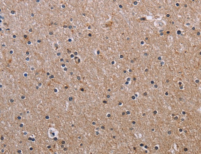
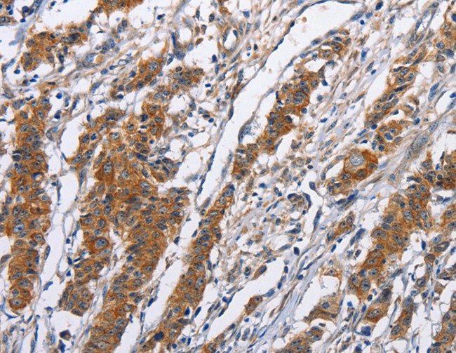
SMYD4, Polyclonal Antibody (Cat# AAA169166)
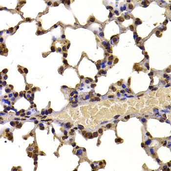
 IHC (Immunohiostchemistry)
(Immunohistochemistry of paraffin-embedded Human brain tissue using FCGR3A Polyclonal Antibody at dilution 1:60)
IHC (Immunohiostchemistry)
(Immunohistochemistry of paraffin-embedded Human brain tissue using FCGR3A Polyclonal Antibody at dilution 1:60)
FCGR3A, Polyclonal Antibody (Cat# AAA169177)
 IHC (Immunohiostchemistry)
(Immunohistochemistry of paraffin-embedded Human ovarian cancer using NDUFAF4 Polyclonal Antibody at dilution of 1:50)
IHC (Immunohiostchemistry)
(Immunohistochemistry of paraffin-embedded Human ovarian cancer using NDUFAF4 Polyclonal Antibody at dilution of 1:50)
NDUFAF4, Polyclonal Antibody (Cat# AAA169249)
 IHC (Immunohiostchemistry)
(Immunohistochemistry of paraffin-embedded Human colon cancer tissue using Gjc3 Polyclonal Antibody at dilution of 1:10)
IHC (Immunohiostchemistry)
(Immunohistochemistry of paraffin-embedded Human colon cancer tissue using Gjc3 Polyclonal Antibody at dilution of 1:10)
Gjc3, Polyclonal Antibody (Cat# AAA169290)
 IHC (Immunohiostchemistry)
(Immunohistochemistry of paraffin-embedded Human ovarian cancer tissue using FGFR1OP2 Polyclonal Antibody at dilution 1:40)
IHC (Immunohiostchemistry)
(Immunohistochemistry of paraffin-embedded Human ovarian cancer tissue using FGFR1OP2 Polyclonal Antibody at dilution 1:40)
FGFR1OP2, Polyclonal Antibody (Cat# AAA169298)
 IHC (Immunohiostchemistry)
(Immunohistochemistry of paraffin-embedded Human thyroid cancer tissue using NCAPD3 Polyclonal Antibody at dilution 1:50)
IHC (Immunohiostchemistry)
(Immunohistochemistry of paraffin-embedded Human thyroid cancer tissue using NCAPD3 Polyclonal Antibody at dilution 1:50)
NCAPD3, Polyclonal Antibody (Cat# AAA169305)
 IHC (Immunohistochemisry)
(Immunohistochemistry of paraffin-embedded Human cervical cancer using PTK2B Polyclonal Antibody at dilution of 1:50)
IHC (Immunohistochemisry)
(Immunohistochemistry of paraffin-embedded Human cervical cancer using PTK2B Polyclonal Antibody at dilution of 1:50)
PTK2B, Polyclonal Antibody (Cat# AAA169310)
 IHC (Immunohistochemisry)
(Immunohistochemistry of paraffin-embedded Human brain using EIF3H Polyclonal Antibody at dilution of 1:50)
IHC (Immunohistochemisry)
(Immunohistochemistry of paraffin-embedded Human brain using EIF3H Polyclonal Antibody at dilution of 1:50)
EIF3H, Polyclonal Antibody (Cat# AAA169321)
 IHC (Immunohistochemisry)
(Immunohistochemistry of paraffin-embedded Human lung cancer using DNAJC7 Polyclonal Antibody at dilution of 1:25)
IHC (Immunohistochemisry)
(Immunohistochemistry of paraffin-embedded Human lung cancer using DNAJC7 Polyclonal Antibody at dilution of 1:25)
DNAJC7, Polyclonal Antibody (Cat# AAA169329)
 IHC (Immunohiostchemistry)
(Immunohistochemistry of paraffin-embedded Human lung cancer tissue using SERP1 Polyclonal Antibody at dilution 1:30)
IHC (Immunohiostchemistry)
(Immunohistochemistry of paraffin-embedded Human lung cancer tissue using SERP1 Polyclonal Antibody at dilution 1:30)
SERP1, Polyclonal Antibody (Cat# AAA169330)
 IHC (Immunohistochemisry)
(Immunohistochemistry of paraffin-embedded Human colon cancer using PPM1F Polyclonal Antibody at dilution of 1:40)
IHC (Immunohistochemisry)
(Immunohistochemistry of paraffin-embedded Human colon cancer using PPM1F Polyclonal Antibody at dilution of 1:40)
PPM1F, Polyclonal Antibody (Cat# AAA169331)
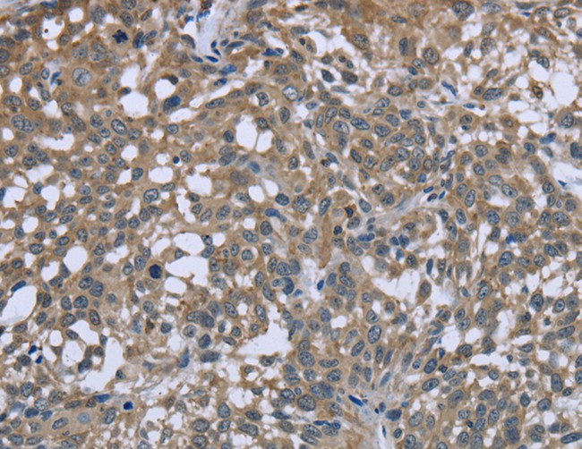
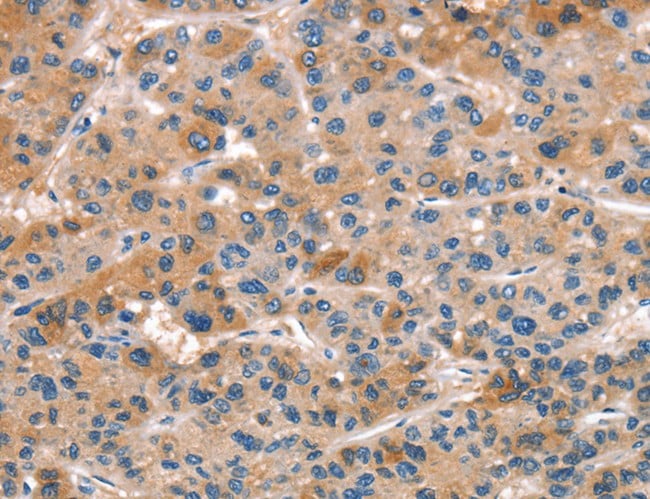
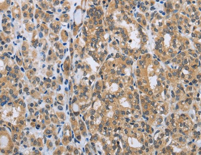
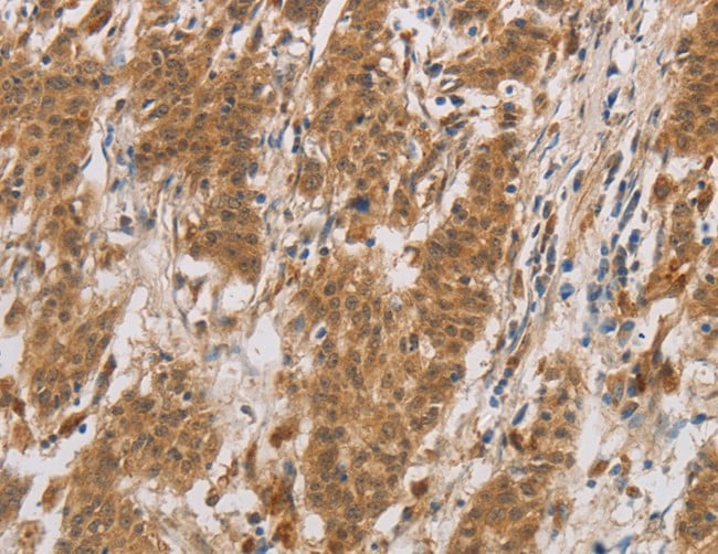
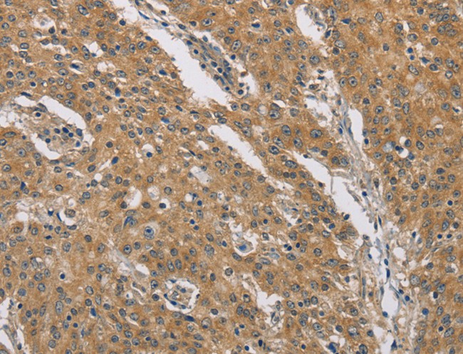
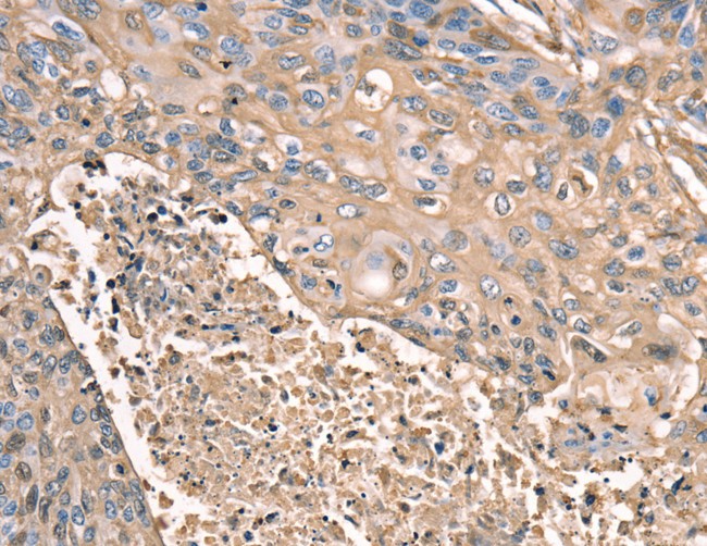
 IHC (Immunohistochemisry)
(Immunohistochemistry of paraffin-embedded Human liver cancer using SLC25A13 Polyclonal Antibody at dilution of 1:35)
IHC (Immunohistochemisry)
(Immunohistochemistry of paraffin-embedded Human liver cancer using SLC25A13 Polyclonal Antibody at dilution of 1:35)
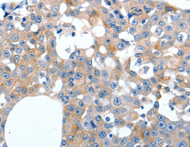
SLC25A13, Polyclonal Antibody (Cat# AAA169340)
 IHC (Immunohistochemisry)
(Immunohistochemistry of paraffin-embedded Human gastric cancer using CALR Polyclonal Antibody at dilution of 1:30)
IHC (Immunohistochemisry)
(Immunohistochemistry of paraffin-embedded Human gastric cancer using CALR Polyclonal Antibody at dilution of 1:30)
CALR, Polyclonal Antibody (Cat# AAA169345)
 IHC (Immunohiostchemistry)
(Immunohistochemistry of paraffin-embedded Human brain tissue using MYO7A Polyclonal Antibody at dilution 1:30)
IHC (Immunohiostchemistry)
(Immunohistochemistry of paraffin-embedded Human brain tissue using MYO7A Polyclonal Antibody at dilution 1:30)
MYO7A, Polyclonal Antibody (Cat# AAA169349)
 IHC (Immunohistochemistry)
(Immunohistochemistry of paraffin-embedded Human breast cancer tissue using LILRB3 Polyclonal Antibody at dilution 1:20)
IHC (Immunohistochemistry)
(Immunohistochemistry of paraffin-embedded Human breast cancer tissue using LILRB3 Polyclonal Antibody at dilution 1:20)
LILRB3, Polyclonal Antibody (Cat# AAA169351)
 IHC (Immunohistochemisry)
(Immunohistochemistry of paraffin-embedded Human colon cancer using NDUFA5 Polyclonal Antibody at dilution of 1:60)
IHC (Immunohistochemisry)
(Immunohistochemistry of paraffin-embedded Human colon cancer using NDUFA5 Polyclonal Antibody at dilution of 1:60)
NDUFA5, Polyclonal Antibody (Cat# AAA169358)
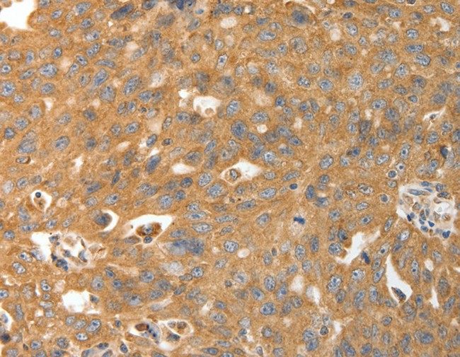
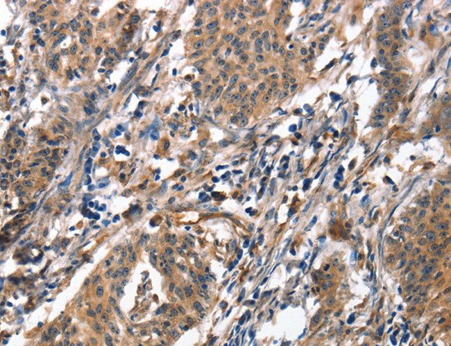
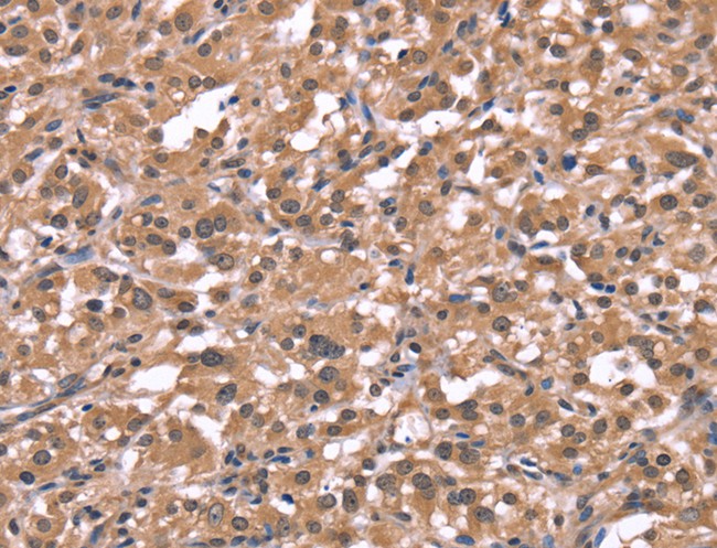
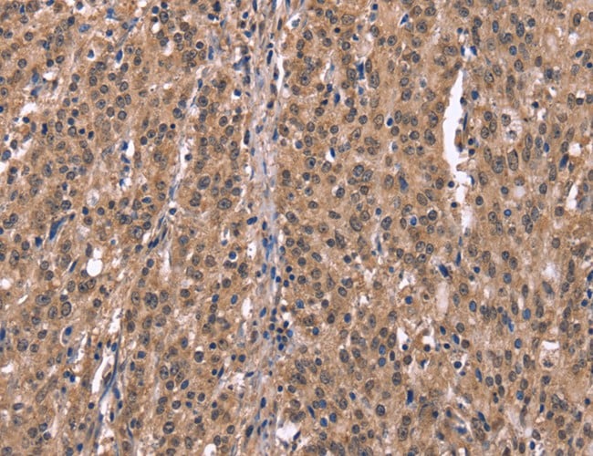
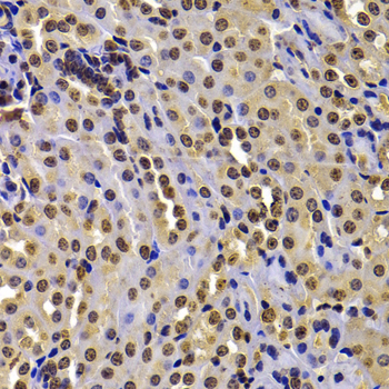
 IHC (Immunohiostchemistry)
(Immunohistochemistry of paraffin-embedded Human liver cancer tissue using NR4A2 Polyclonal Antibody at dilution 1:20)
IHC (Immunohiostchemistry)
(Immunohistochemistry of paraffin-embedded Human liver cancer tissue using NR4A2 Polyclonal Antibody at dilution 1:20)
NR4A2, Polyclonal Antibody (Cat# AAA169360)
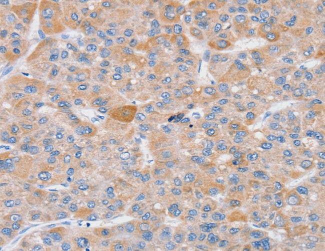
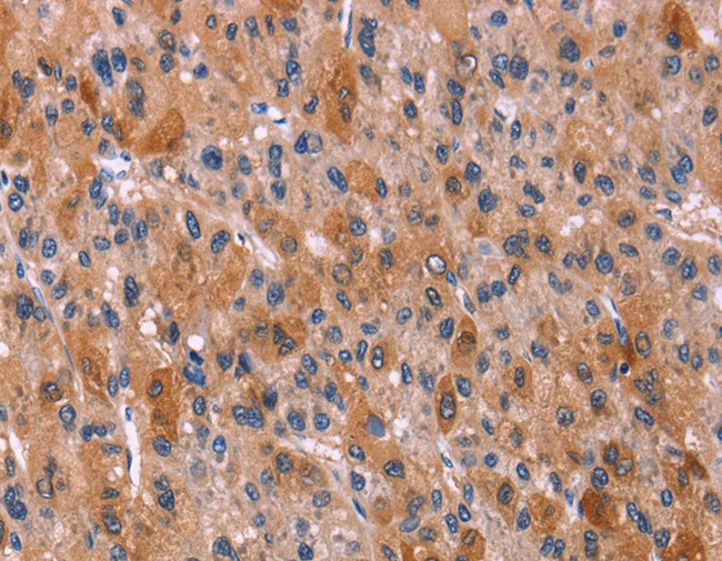
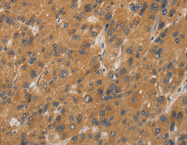
 IHC (Immunohiostchemistry)
(Immunohistochemistry of paraffin-embedded Human liver cancer tissue using ALOX15 Polyclonal Antibody at dilution 1:50)
IHC (Immunohiostchemistry)
(Immunohistochemistry of paraffin-embedded Human liver cancer tissue using ALOX15 Polyclonal Antibody at dilution 1:50)
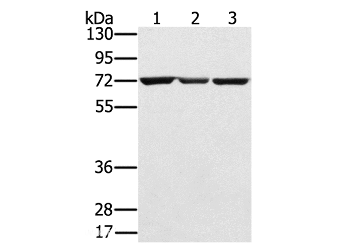
ALOX15, Polyclonal Antibody (Cat# AAA169182)
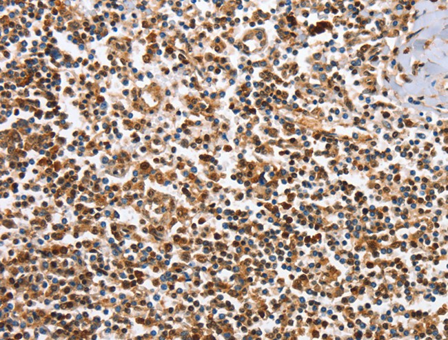
 WB (Western Blot)
(Western Blot analysis of Human lymphoma tissue using CCL24 Polyclonal Antibody at dilution of 1:400)
WB (Western Blot)
(Western Blot analysis of Human lymphoma tissue using CCL24 Polyclonal Antibody at dilution of 1:400)
CCL24, Polyclonal Antibody (Cat# AAA169185)
 IHC (Immunohiostchemistry)
(Immunohistochemistry of paraffin-embedded Human thyroid cancer tissue using EGFL8 Polyclonal Antibody at dilution 1:30)
IHC (Immunohiostchemistry)
(Immunohistochemistry of paraffin-embedded Human thyroid cancer tissue using EGFL8 Polyclonal Antibody at dilution 1:30)
EGFL8, Polyclonal Antibody (Cat# AAA169188)
 IHC (Immunohistochemisry)
(Immunohistochemistry of paraffin-embedded Human gastric cancer using HRH1 Polyclonal Antibody at dilution of 1:40)
IHC (Immunohistochemisry)
(Immunohistochemistry of paraffin-embedded Human gastric cancer using HRH1 Polyclonal Antibody at dilution of 1:40)
HRH1, Polyclonal Antibody (Cat# AAA169194)
 IHC (Immunohiostchemistry)
(Immunohistochemistry of paraffin-embedded Human gastric cancer tissue using CYP17A1 Polyclonal Antibody at dilution 1:20)
IHC (Immunohiostchemistry)
(Immunohistochemistry of paraffin-embedded Human gastric cancer tissue using CYP17A1 Polyclonal Antibody at dilution 1:20)
CYP17A1, Polyclonal Antibody (Cat# AAA169196)
 IHC (Immunohistochemisry)
(Immunohistochemistry of paraffin-embedded Human gastric cancer using DCTN1 Polyclonal Antibody at dilution of 1:60)
IHC (Immunohistochemisry)
(Immunohistochemistry of paraffin-embedded Human gastric cancer using DCTN1 Polyclonal Antibody at dilution of 1:60)
DCTN1, Polyclonal Antibody (Cat# AAA169198)
 IHC (Immunohistochemisry)
(Immunohistochemistry of paraffin-embedded Human brain using DYNLL1 Polyclonal Antibody at dilution of 1:25)
IHC (Immunohistochemisry)
(Immunohistochemistry of paraffin-embedded Human brain using DYNLL1 Polyclonal Antibody at dilution of 1:25)
DYNLL1, Polyclonal Antibody (Cat# AAA169205)
 IHC (Immunohistochemisry)
(Immunohistochemistry of paraffin-embedded Human lung cancer using PTRH2 Polyclonal Antibody at dilution of 1:20)
IHC (Immunohistochemisry)
(Immunohistochemistry of paraffin-embedded Human lung cancer using PTRH2 Polyclonal Antibody at dilution of 1:20)
PTRH2, Polyclonal Antibody (Cat# AAA169209)
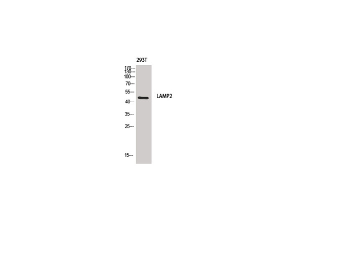
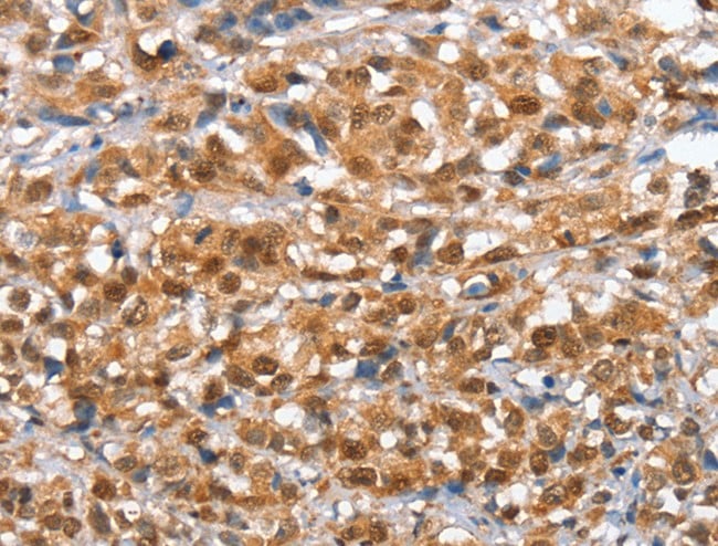
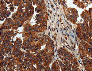
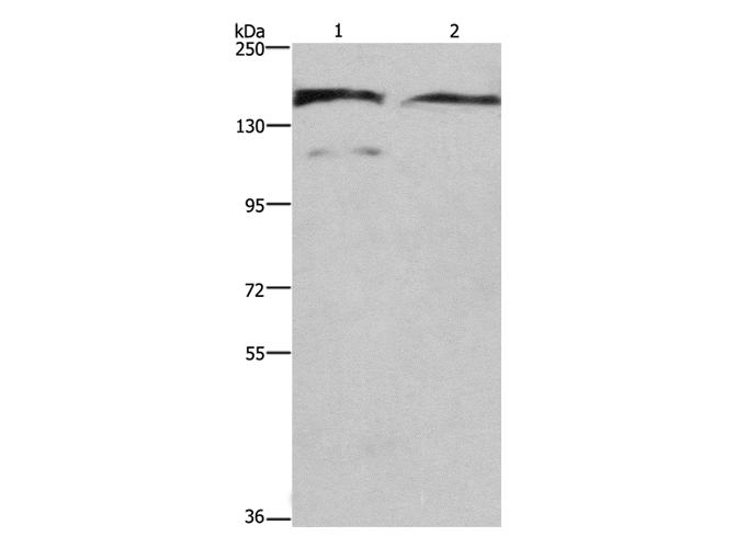
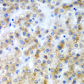
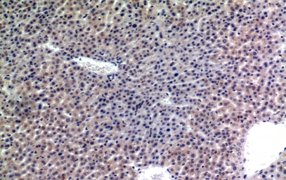 IHC (Immunohiostchemistry)
(Immunohistochemistry of paraffin-embedded Mouse liver tissue using CD107b Polyclonal Antibody at dilution of 1:100.)
IHC (Immunohiostchemistry)
(Immunohistochemistry of paraffin-embedded Mouse liver tissue using CD107b Polyclonal Antibody at dilution of 1:100.)
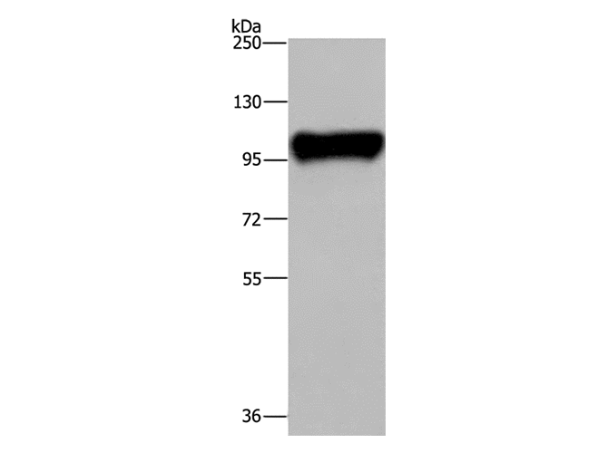
LAMP2, Polyclonal Antibody (Cat# AAA169212)
 IHC (Immunohistochemisry)
(Immunohistochemistry of paraffin-embedded Human brain using DAAM1 Polyclonal Antibody at dilution of 1:50)
IHC (Immunohistochemisry)
(Immunohistochemistry of paraffin-embedded Human brain using DAAM1 Polyclonal Antibody at dilution of 1:50)
DAAM1, Polyclonal Antibody (Cat# AAA169215)
 IHC (Immunohistochemisry)
(Immunohistochemistry of paraffin-embedded Human thyroid cancer using AIFM3 Polyclonal Antibody at dilution of 1:50)
IHC (Immunohistochemisry)
(Immunohistochemistry of paraffin-embedded Human thyroid cancer using AIFM3 Polyclonal Antibody at dilution of 1:50)
AIFM3, Polyclonal Antibody (Cat# AAA169222)
 IHC (Immunohiostchemistry)
(Immunohistochemistry of paraffin-embedded Human esophagus cancer tissue using SCN1B Polyclonal Antibody at dilution 1:35)
IHC (Immunohiostchemistry)
(Immunohistochemistry of paraffin-embedded Human esophagus cancer tissue using SCN1B Polyclonal Antibody at dilution 1:35)
SCN1B, Polyclonal Antibody (Cat# AAA169225)
 IHC (Immunohiostchemistry)
(Immunohistochemistry of paraffin-embedded Human cervical cancer using RGS22 Polyclonal Antibody at dilution of 1:40)
IHC (Immunohiostchemistry)
(Immunohistochemistry of paraffin-embedded Human cervical cancer using RGS22 Polyclonal Antibody at dilution of 1:40)
RGS22, Polyclonal Antibody (Cat# AAA169226)
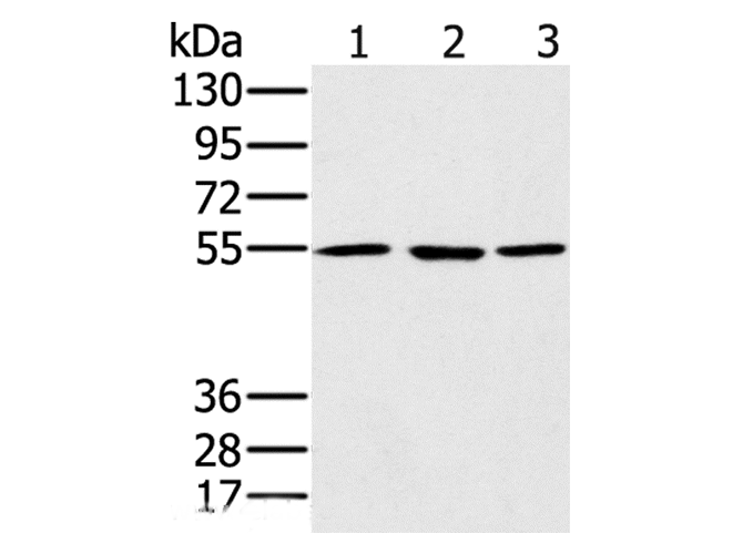
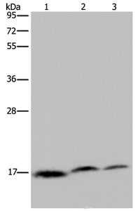
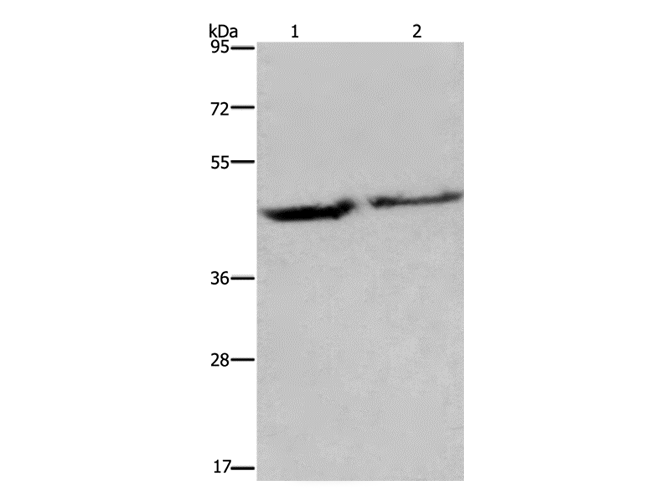
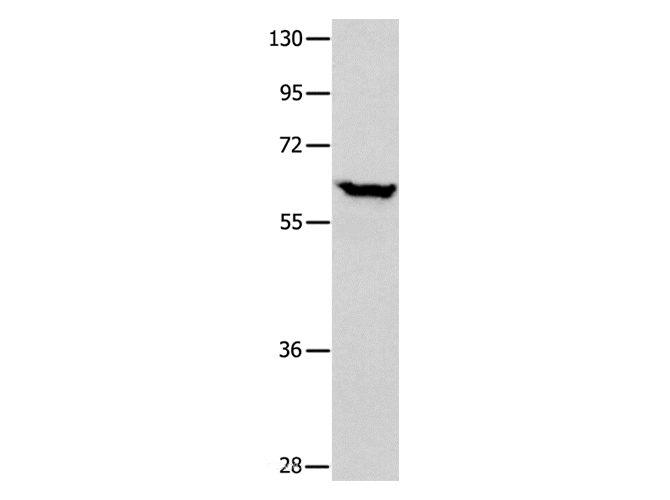
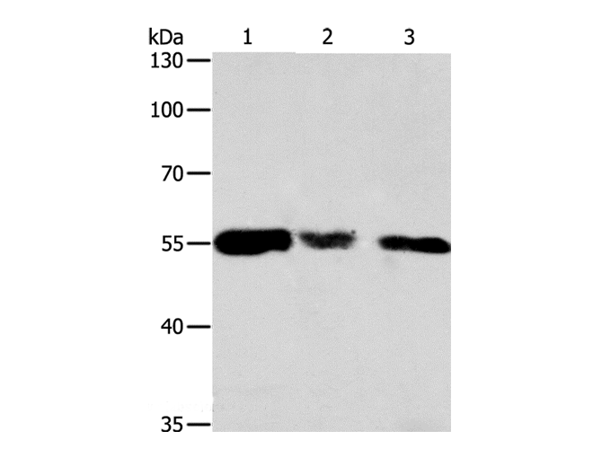
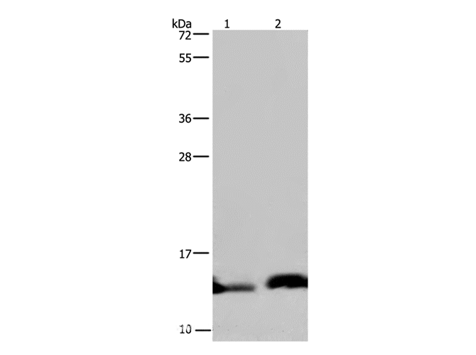
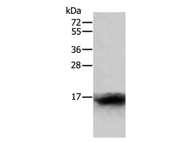
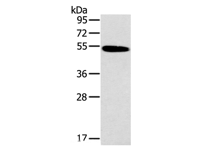
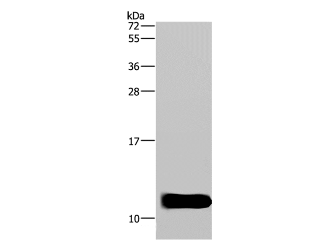
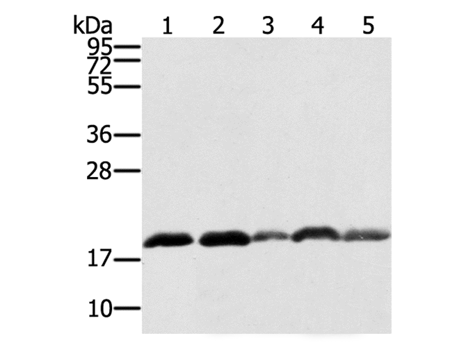
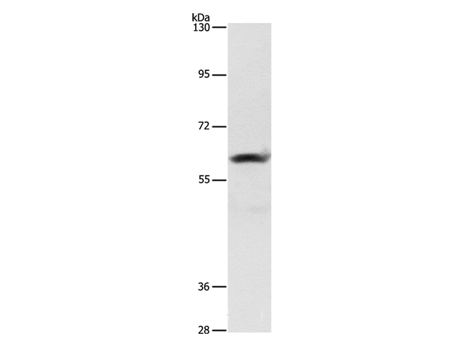
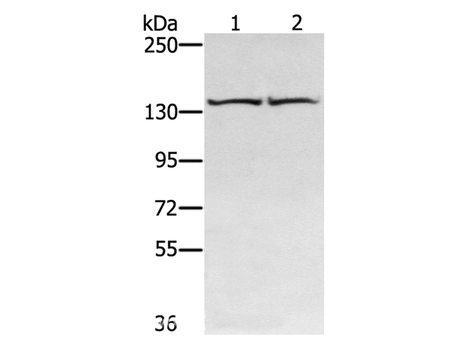
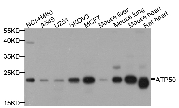 WB (Western Blot)
(Western blot analysis of extract of various cells, using ATP5O antibody.)
WB (Western Blot)
(Western blot analysis of extract of various cells, using ATP5O antibody.)
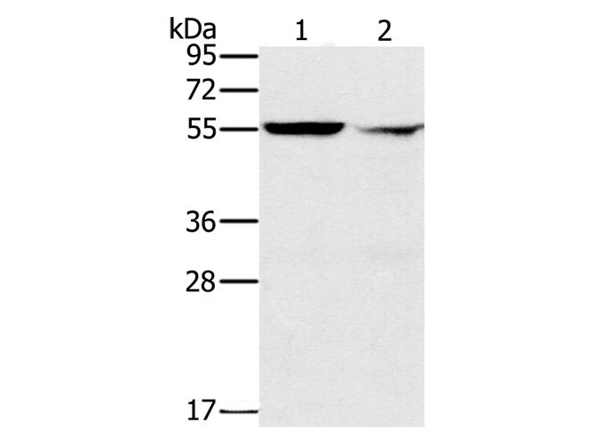
ATP5O, Polyclonal Antibody (Cat# AAA172740)
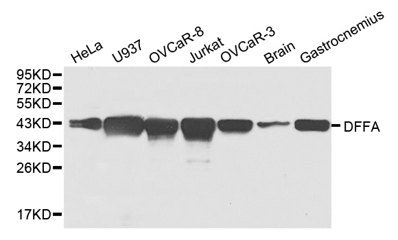
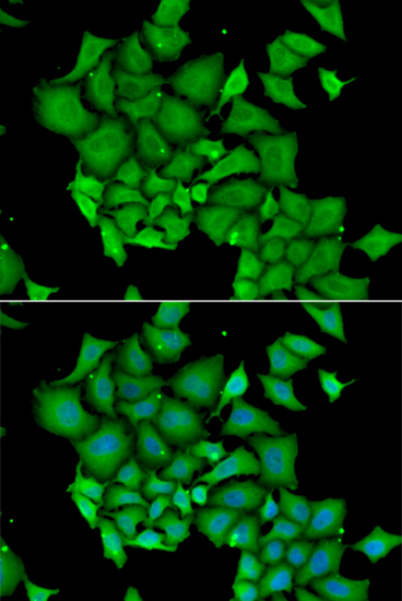 IF (Immunofluorescence)
(Immunofluorescence analysis of HeLa cell using DFFA antibody. Blue: DAPI for nuclear staining.)
IF (Immunofluorescence)
(Immunofluorescence analysis of HeLa cell using DFFA antibody. Blue: DAPI for nuclear staining.)
DFFA, Polyclonal Antibody (Cat# AAA172748)
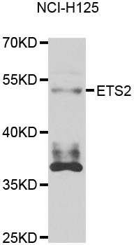
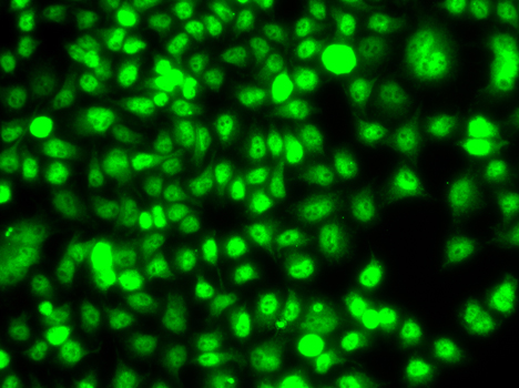 IF (Immunofluorescence)
(Immunofluorescence analysis of A549 cell using ETS2 antibody.)
IF (Immunofluorescence)
(Immunofluorescence analysis of A549 cell using ETS2 antibody.)
ETS2, Polyclonal Antibody (Cat# AAA172753)
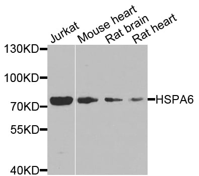
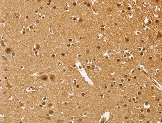
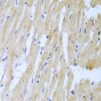
 IHC (Immunohiostchemistry)
(Immunohistochemistry of paraffin-embedded rat brain using HSPA6 antibody at dilution of1:100 (x40 lens).)
IHC (Immunohiostchemistry)
(Immunohistochemistry of paraffin-embedded rat brain using HSPA6 antibody at dilution of1:100 (x40 lens).)
HSPA6, Polyclonal Antibody (Cat# AAA172756)
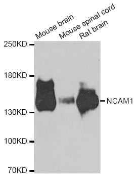
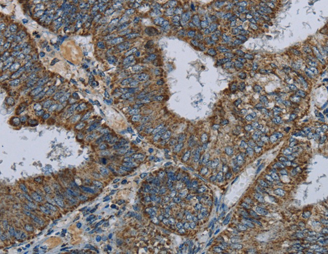
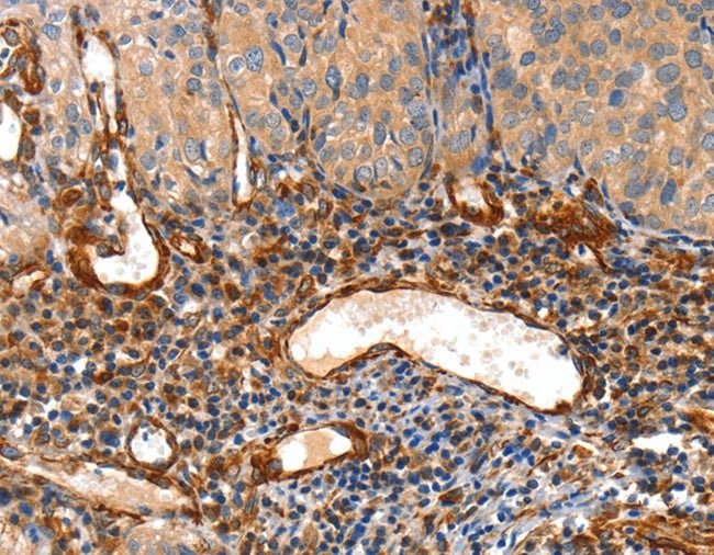
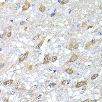
 IHC (Immunohiostchemistry)
(Immunohistochemistry of paraffin-embedded human stomach using NCAM1 antibody at dilution of1:100 (40x lens).)
IHC (Immunohiostchemistry)
(Immunohistochemistry of paraffin-embedded human stomach using NCAM1 antibody at dilution of1:100 (40x lens).)
NCAM1, Polyclonal Antibody (Cat# AAA172759)
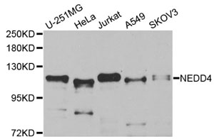
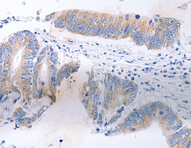
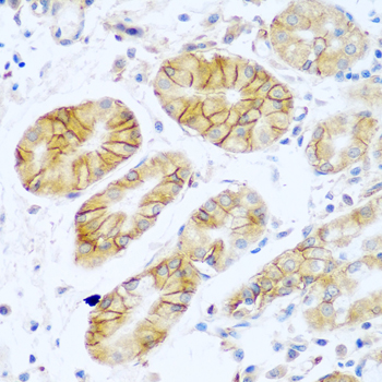
 IHC (Immunohistochemisry)
(Immunohistochemistry of paraffinembedded human gastric with NEDD4 Polyclonal Antibody)
IHC (Immunohistochemisry)
(Immunohistochemistry of paraffinembedded human gastric with NEDD4 Polyclonal Antibody)
NEDD4, Polyclonal Antibody (Cat# AAA172760)
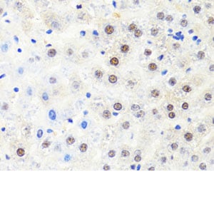
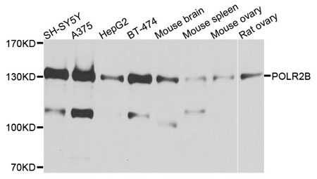
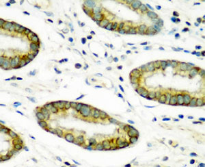
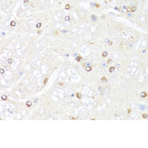 IHC (Immunohistochemisry)
(Immunohistochemistry of paraffin-embedded mouse brain with POLR2B Polyclonal Antibody)
IHC (Immunohistochemisry)
(Immunohistochemistry of paraffin-embedded mouse brain with POLR2B Polyclonal Antibody)
POLR2B, Polyclonal Antibody (Cat# AAA172763)
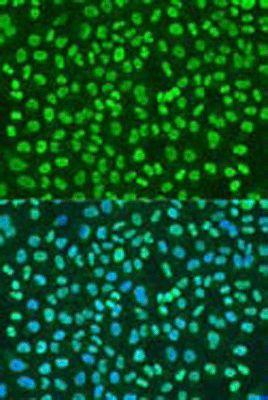 IF (Immunofluorescence)
(Immunofluorescence analysis of U2OS cells using PTBP1 Polyclonal Antibody at dilution of 1:100. Blue: DAPI for nuclear staining.)
IF (Immunofluorescence)
(Immunofluorescence analysis of U2OS cells using PTBP1 Polyclonal Antibody at dilution of 1:100. Blue: DAPI for nuclear staining.)
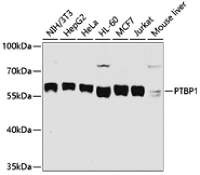
PTBP1, Polyclonal Antibody (Cat# AAA172770)
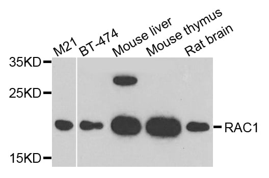
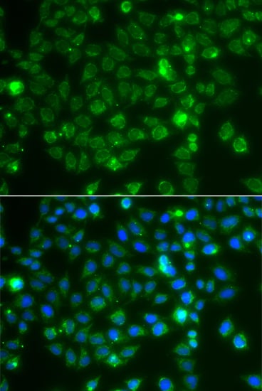 IF (Immunofluorescence)
(Immunofluorescence analysis of A549 cell using RAC1 antibody. Blue: DAPI for nuclear staining.)
IF (Immunofluorescence)
(Immunofluorescence analysis of A549 cell using RAC1 antibody. Blue: DAPI for nuclear staining.)
RAC1, Polyclonal Antibody (Cat# AAA172771)
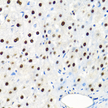
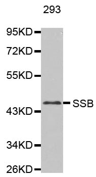
 IHC (Immunohistochemisry)
(Immunohistochemistry of paraffin-embedded human prostate cancer using SSB antibody at dilution of1:100 (40x lens).)
IHC (Immunohistochemisry)
(Immunohistochemistry of paraffin-embedded human prostate cancer using SSB antibody at dilution of1:100 (40x lens).)
SSB, Polyclonal Antibody (Cat# AAA172776)
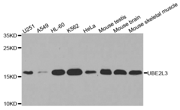
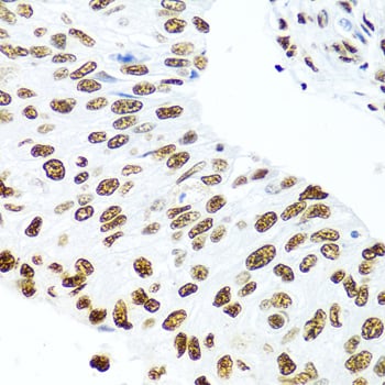
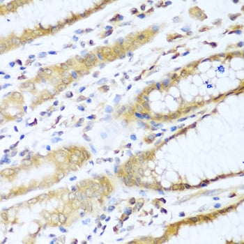 IHC (Immunohistochemisry)
(Immunohistochemistry of paraffin-embedded human stomach using UBE2L3 antibody at dilution of1:100 (40x lens).)
IHC (Immunohistochemisry)
(Immunohistochemistry of paraffin-embedded human stomach using UBE2L3 antibody at dilution of1:100 (40x lens).)
UBE2L3, Polyclonal Antibody (Cat# AAA172780)
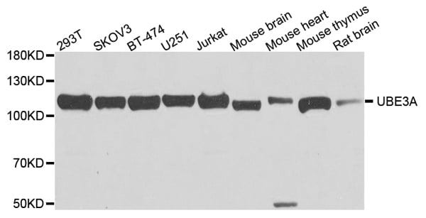
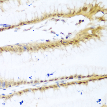
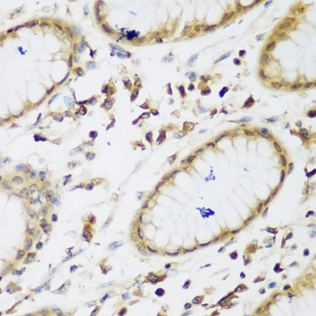 IHC (Immunohiostchemistry)
(Immunohistochemistry of paraffin-embedded human gastric using UBE3A antibody at dilution of1:200 (40x lens).)
IHC (Immunohiostchemistry)
(Immunohistochemistry of paraffin-embedded human gastric using UBE3A antibody at dilution of1:200 (40x lens).)
UBE3A, Polyclonal Antibody (Cat# AAA172781)
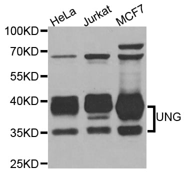
 IHC (Immunohistochemisry)
(Immunohistochemistry of paraffin-embedded mouse brain using UNG antibody at dilution of1:100 (40x lens).)
IHC (Immunohistochemisry)
(Immunohistochemistry of paraffin-embedded mouse brain using UNG antibody at dilution of1:100 (40x lens).)
UNG, Polyclonal Antibody (Cat# AAA172782)

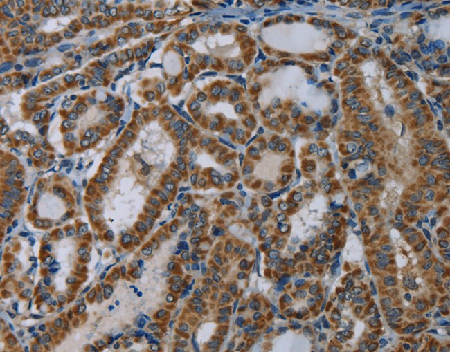
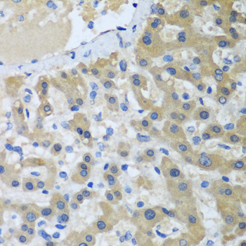
 IHC (Immunohistochemisry)
(Immunohistochemistry of paraffin-embedded mouse kidney using WHSC1 antibody at dilution of1:100 (40x lens).)
IHC (Immunohistochemisry)
(Immunohistochemistry of paraffin-embedded mouse kidney using WHSC1 antibody at dilution of1:100 (40x lens).)
WHSC1, Polyclonal Antibody (Cat# AAA172784)

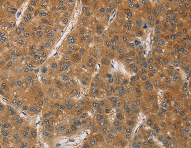
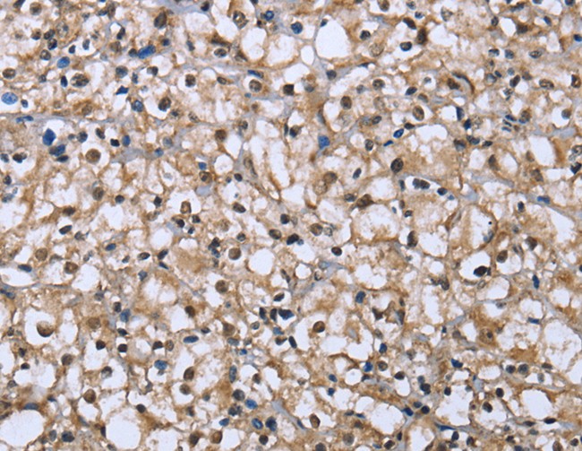
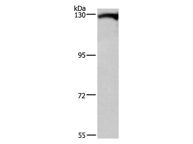
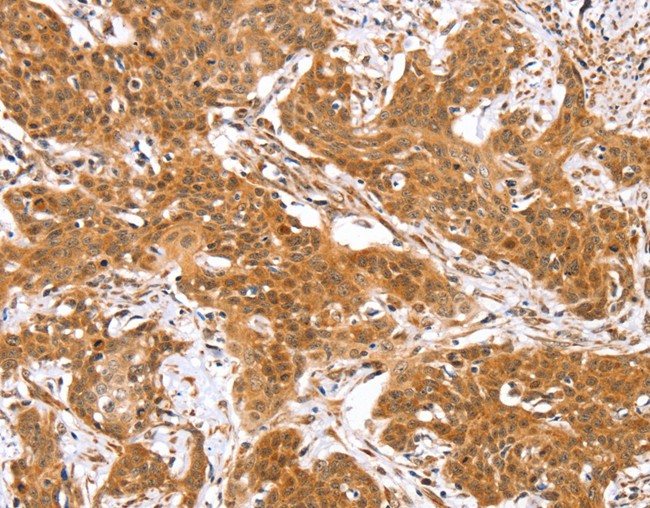
 IHC (Immunohiostchemistry)
(Immunohistochemistry of paraffin-embedded human liver cancer using XPC antibody at dilution of1:100 (40x lens).)
IHC (Immunohiostchemistry)
(Immunohistochemistry of paraffin-embedded human liver cancer using XPC antibody at dilution of1:100 (40x lens).)
XPC, Polyclonal Antibody (Cat# AAA172786)
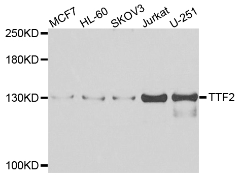
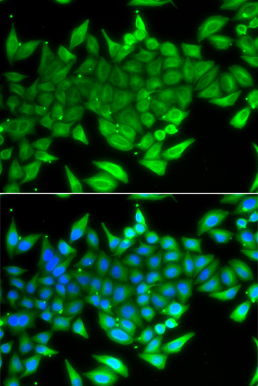 IF (Immunofluorescence)
(Immunofluorescence analysis of A549 cell using TTF2 antibody. Blue: DAPI for nuclear staining.)
IF (Immunofluorescence)
(Immunofluorescence analysis of A549 cell using TTF2 antibody. Blue: DAPI for nuclear staining.)
TTF2, Polyclonal Antibody (Cat# AAA172788)
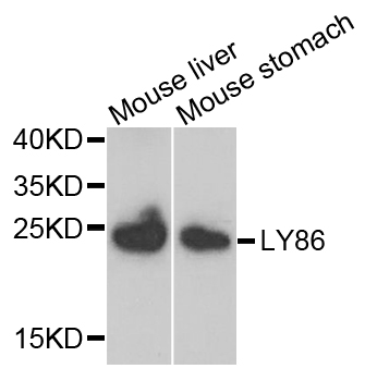
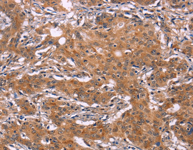
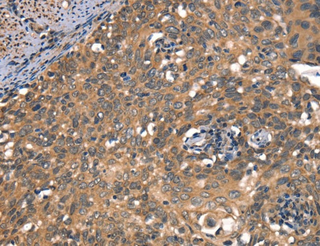
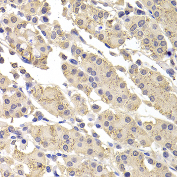
 IHC (Immunohiostchemistry)
(Immunohistochemistry of paraffin-embedded human liver cancer using LY86 antibody at dilution of 1:200 (40x lens).)
IHC (Immunohiostchemistry)
(Immunohistochemistry of paraffin-embedded human liver cancer using LY86 antibody at dilution of 1:200 (40x lens).)
LY86, Polyclonal Antibody (Cat# AAA172792)
What are Polyclonal Antibodies?
Polyclonal antibodies are antibodies that come from multiple B cell clones of a host animal. The typical hosts used for the majority of polyclonal antibody production are rabbits, goats, sheep, and donkeys. These polyclonal antibodies, once having identified their target, will bind to different epitopes located at different regions or sequences on the same protein/antigen. As a result, they are ideal at locating and binding to the target, even if the target is in very low concentrations (due to many different antibodies being able to bind to the same target molecule, which allows for significant amplification of a downstream signal).
Polyclonal antibodies are typically produced by injecting an antigen into a host animal, which causes the animal’s immune system to attack the foreign antigen by mass generating antibodies against it. After a period of time, serum is collected from the animal and purified using physicochemical fractionation, class-specific affinity purification, and/or antigen-affinity purification.
Key Uses of Polyclonal Antibodies
- Western Blotting: This method is used to find specific proteins in biological samples after separating them by size.
- Immunohistochemistry: IHC helps visualize the location of proteins in tissue sections using various staining techniques.
- ELISA: (Enzyme-Linked Immunosorbent Assay) is typically used to identify specific protein quantities in a sample. ELISAs can be either “Quantitative” or “Qualitative”.
- Flow Cytometry: technique that identifies and measures the specific protein on the surface or inside the cells in a fluid suspension.
- Immunoprecipitation: IP isolates and studies a specific protein from a complex mixture using antibodies.
Why Buy Polyclonal Antibodies from AAA Biotech?
1. Ideal for Various Applications
Our antibodies are generally going to be validated for use in multiple types of assays, including ELISA, Western Blotting, Immunohistochemistry, Immunoprecipitation, amongst others. They are ideal for a wide range of research applications.
2. Rigorous Quality Control
All of the antibodies in our catalog undergo strict quality testing to ensure specificity, sensitivity, and consistent performance. We are confident in the ability of our antibodies to provide you with accurate results.
3. Wide Assortment of Antibodies
Antibodies in are catalog can be found for both common and exotic species, and these antibodies are also available in both conjugated and recombinant forms to suit many diverse experimental needs.
4. Highly Purified
Our antibodies are available in purified forms with over 85% purity, as confirmed by SDS-PAGE. They are also available with tags such as His, Flag, GST, or MBP. We cater to customers worldwide.
FAQ
1. How are polyclonal antibodies produced?
Traditionally, polyclonal antibodies are produced by injecting an antigen into a host animal (such as a rabbit or goat), which then triggers an immune response from the host animal. The animal’s B cells produce antibodies that will recognize different parts of the injected antigen. These antibodies are then collected from the animal’s blood and purified for use.
2. How do polyclonal antibodies differ from monoclonal antibodies?
Polyclonal antibodies are a mix of antibodies that bind to different locations (epitopes) of the same antigen, while monoclonal antibodies are identical and bind to just one specific epitope. This makes polyclonal antibodies more versatile and better at detecting proteins that may be present in low quantities or in altered/modified forms.
3. How should I store polyclonal antibodies?
Polyclonal antibodies should be stored at 4°C for short-term use (up to a few weeks) and at -20°C or -80°C for long-term storage. Avoid repeated freeze-thaw cycles by dividing them into small aliquots. Always check the datasheet for specific storage instructions.