Filters
▼Clonality
▼Type
▼Reactivity
▼Gene Name
▼Isotype
▼Host
▼Application
▼Clone
▼Polyclonal Antibodies
At AAA Biotech also known as AAA Bio or AAABio, we provide a broad range of purified polyclonal antibodies (pAbs) that are able to all be browsed online through our website. Due to their high specificity and strong binding affinity, these antibodies are ideal for wide swathes of research and experimental applications.
Our polyclonal antibodies can easily support your work, whether you use them for Western Blotting, Immunocytochemistry (with or without Immunofluorescence used in conjunction), Immunohistochemistry, Immunoprecipitation, and ELISA tests. We highly encourage you to browse our range of pAbs and choose the one that best suits your experimental model.
Viewing 5750-5800 of 96812 product results
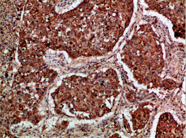
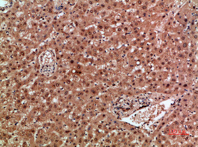
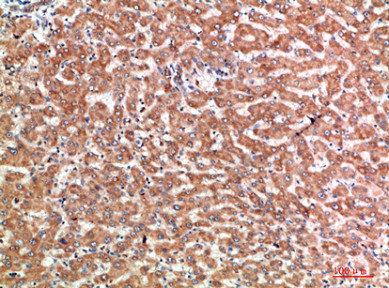
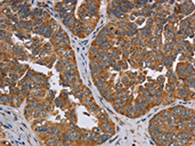
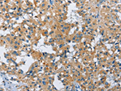
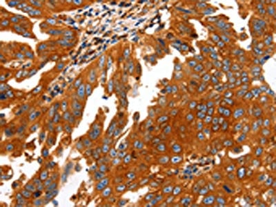
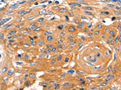
 IHC (Immunohistochemistry)
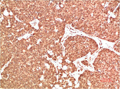
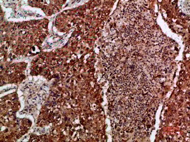
(Immunohistochemical analysis of paraffin-embedded human-liver, antibody was diluted at 1:200)
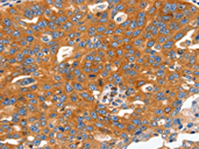
IHC (Immunohistochemistry)
(Immunohistochemical analysis of paraffin-embedded human-liver, antibody was diluted at 1:200)
ACER2, Polyclonal Antibody (Cat# AAA236840)
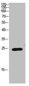
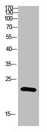
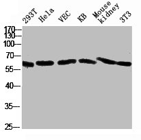
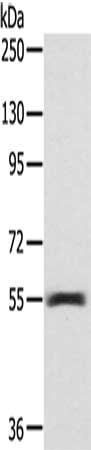
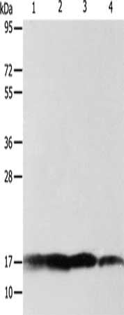
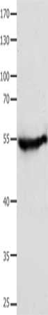
 WB (Western Blot)
(Western Blot analysis of Hela cells using Antibody diluted at 1:1000. Secondary antibody was diluted at 1:20000)
WB (Western Blot)
(Western Blot analysis of Hela cells using Antibody diluted at 1:1000. Secondary antibody was diluted at 1:20000)
NCAM1, Polyclonal Antibody (Cat# AAA236842)
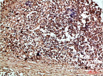
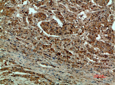
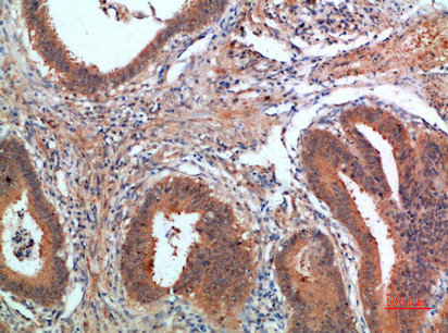
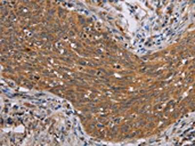
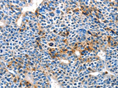
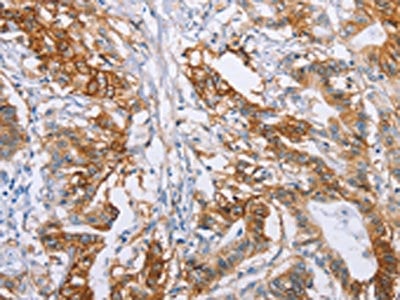
 IHC (Immunohistochemisry)
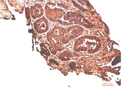
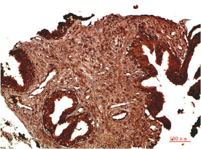
(Immunohistochemical analysis of paraffin-embedded Human Prostate Carcinoma Tissue using ATM Rabbit pAb diluted at 1:500.)
IHC (Immunohistochemisry)
(Immunohistochemical analysis of paraffin-embedded Human Prostate Carcinoma Tissue using ATM Rabbit pAb diluted at 1:500.)
ATM, Polyclonal Antibody (Cat# AAA236846)
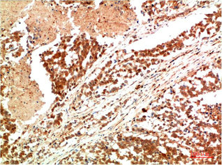
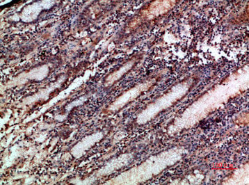
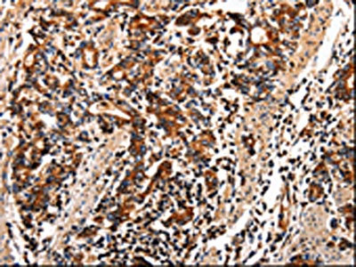
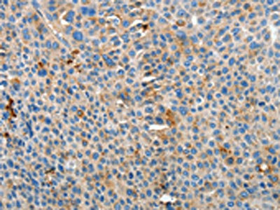
 IHC (Immunohistochemisry)
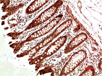
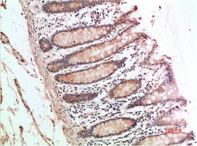
(Immunohistochemical analysis of paraffin-embedded Human Colon Carcinoma Tissue using ATM Rabbit pAb diluted at 1:200.)
IHC (Immunohistochemisry)
(Immunohistochemical analysis of paraffin-embedded Human Colon Carcinoma Tissue using ATM Rabbit pAb diluted at 1:200.)
ATM, Polyclonal Antibody (Cat# AAA236847)
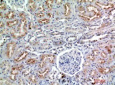
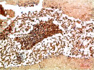
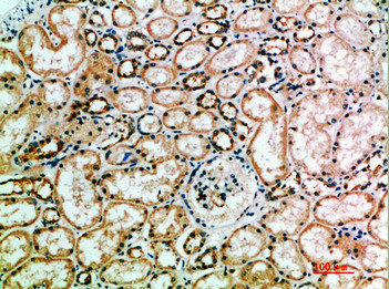
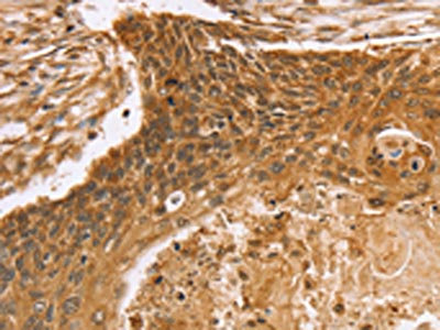
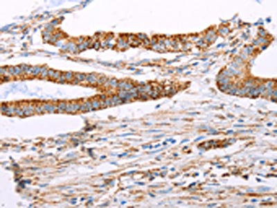
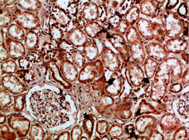 IHC (Immunohistochemistry)
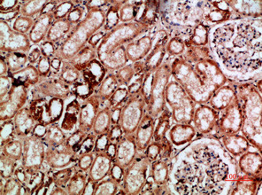
(Immunohistochemical analysis of paraffin-embedded human-kidney, antibody was diluted at 1:200)
IHC (Immunohistochemistry)
(Immunohistochemical analysis of paraffin-embedded human-kidney, antibody was diluted at 1:200)
IMP3, Polyclonal Antibody (Cat# AAA236858)

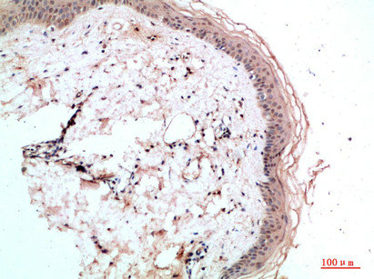 IHC (Immunohistochemisry)
(Immunohistochemical analysis of paraffin-embedded human-skin, antibody was diluted at 1:200)
IHC (Immunohistochemisry)
(Immunohistochemical analysis of paraffin-embedded human-skin, antibody was diluted at 1:200)
RCC1, Polyclonal Antibody (Cat# AAA236859)
 IHC (Immunohiostchemistry)
(Immunohistochemical analysis of paraffin-embedded Human Lung Carcinoma Tissue using JAK1 Rabbit pAb diluted at 1:200)
IHC (Immunohiostchemistry)
(Immunohistochemical analysis of paraffin-embedded Human Lung Carcinoma Tissue using JAK1 Rabbit pAb diluted at 1:200)
JAK1, Polyclonal Antibody (Cat# AAA236868)
 WB (Western Blot)
(Western blot analysis of 1) Hela Cell Lysate, 2)293t Cell Lysate using MEK2 Rabbit pAb diluted at 1:2000.)
WB (Western Blot)
(Western blot analysis of 1) Hela Cell Lysate, 2)293t Cell Lysate using MEK2 Rabbit pAb diluted at 1:2000.)
MAP2K2, Polyclonal Antibody (Cat# AAA236869)
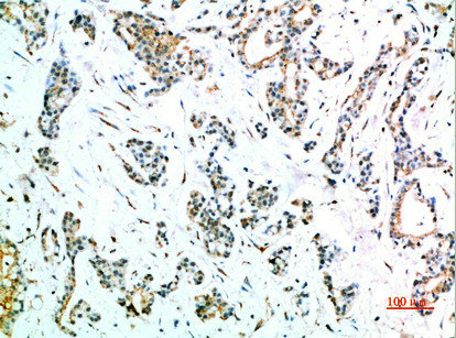
 IHC (Immunohiostchemistry)
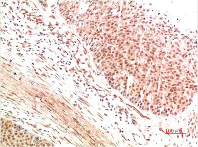
(Immunohistochemical analysis of paraffin-embedded Human Breast Carcinoma Tissue using FAK Rabbit pAb diluted at 1:500)
IHC (Immunohiostchemistry)
(Immunohistochemical analysis of paraffin-embedded Human Breast Carcinoma Tissue using FAK Rabbit pAb diluted at 1:500)
PTK2, Polyclonal Antibody (Cat# AAA236872)
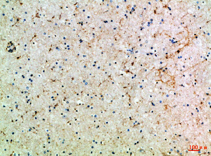
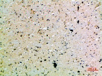
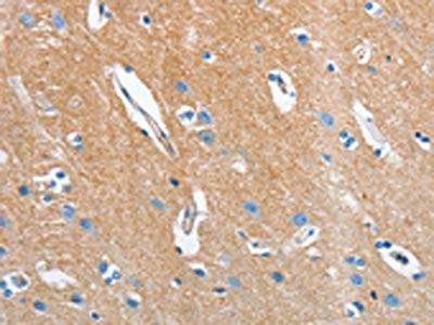
 IHC (Immunohistochemisry)
(Immunohistochemical analysis of paraffin-embedded Rat-brain, antibody was diluted at 1:100)
IHC (Immunohistochemisry)
(Immunohistochemical analysis of paraffin-embedded Rat-brain, antibody was diluted at 1:100)
SLC6A1, Polyclonal Antibody (Cat# AAA236873)
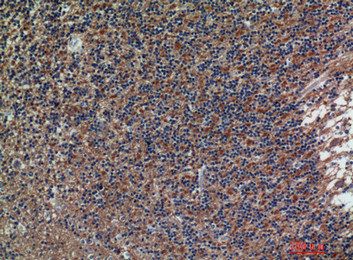
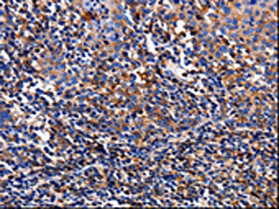
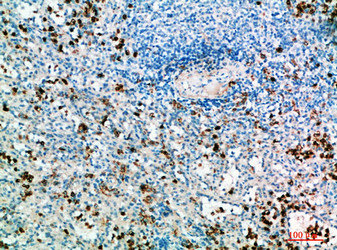 IHC (Immunohistochemistry)
(Immunohistochemical analysis of paraffin-embedded human-spleen, antibody was diluted at 1:200)
IHC (Immunohistochemistry)
(Immunohistochemical analysis of paraffin-embedded human-spleen, antibody was diluted at 1:200)
IL31, Polyclonal Antibody (Cat# AAA236875)
 IHC (Immunohiostchemistry)
(Immunohistochemical analysis of paraffin-embedded human-breast-cancer, antibody was diluted at 1:200)
IHC (Immunohiostchemistry)
(Immunohistochemical analysis of paraffin-embedded human-breast-cancer, antibody was diluted at 1:200)
DCT, Polyclonal Antibody (Cat# AAA236876)

 IHC (Immunohistochemistry)
(Immunohistochemical analysis of paraffin-embedded human-stomach, antibody was diluted at 1:200)
IHC (Immunohistochemistry)
(Immunohistochemical analysis of paraffin-embedded human-stomach, antibody was diluted at 1:200)
NOX1, Polyclonal Antibody (Cat# AAA236880)
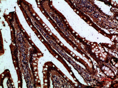
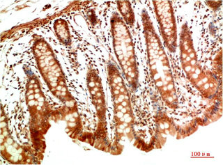
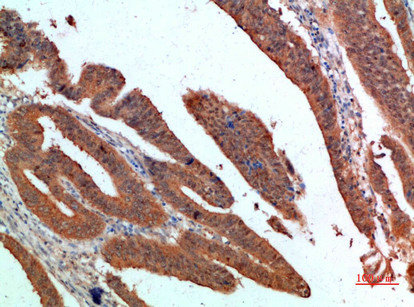
 IHC (Immunohistochemisry)
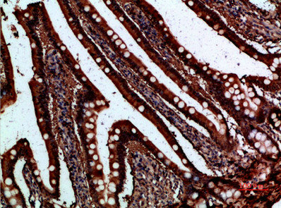
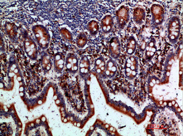
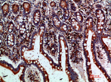
(Immunohistochemical analysis of paraffin-embedded human-small-intestine, antibody was diluted at 1:200)
IHC (Immunohistochemisry)
(Immunohistochemical analysis of paraffin-embedded human-small-intestine, antibody was diluted at 1:200)
FABP2, Polyclonal Antibody (Cat# AAA236884)
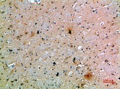
 IHC (Immunohiostchemistry)
(Immunohistochemical analysis of paraffin-embedded Human-brain, antibody was diluted at 1:100)
IHC (Immunohiostchemistry)
(Immunohistochemical analysis of paraffin-embedded Human-brain, antibody was diluted at 1:100)
ASIC1, Polyclonal Antibody (Cat# AAA236886)
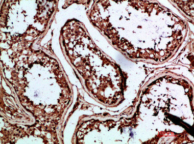
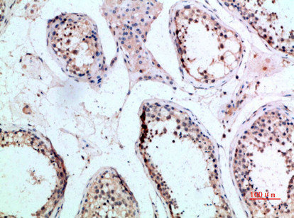
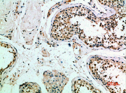
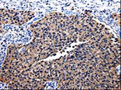
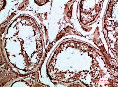 IHC (Immunohiostchemistry)
(Immunohistochemical analysis of paraffin-embedded human-testis, antibody was diluted at 1:200)
IHC (Immunohiostchemistry)
(Immunohistochemical analysis of paraffin-embedded human-testis, antibody was diluted at 1:200)
INSL3, Polyclonal Antibody (Cat# AAA236891)


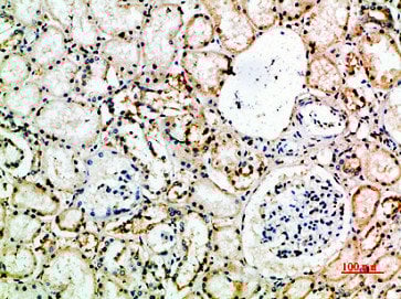
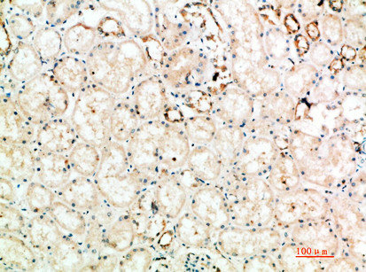
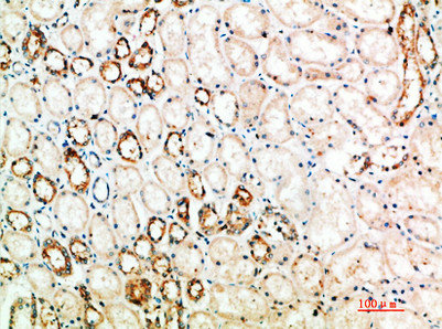
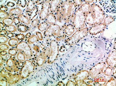
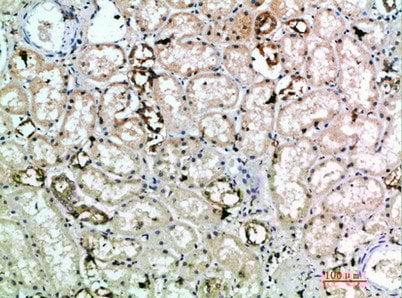
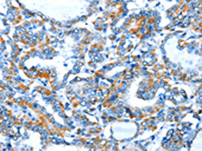
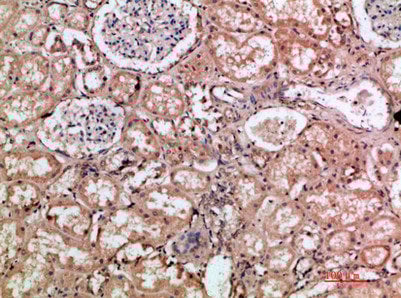 IHC (Immunohistochemisry)
(Immunohistochemical analysis of paraffin-embedded human-kidney, antibody was diluted at 1:200)
IHC (Immunohistochemisry)
(Immunohistochemical analysis of paraffin-embedded human-kidney, antibody was diluted at 1:200)
HMMR, Polyclonal Antibody (Cat# AAA236895)
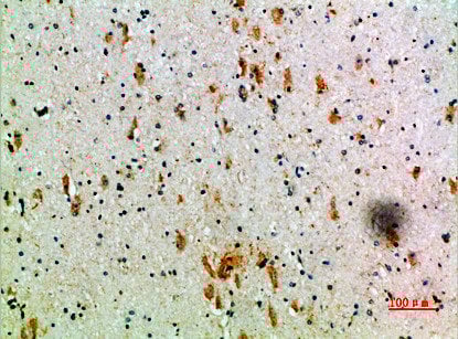
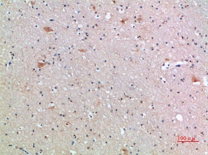
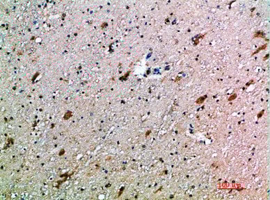
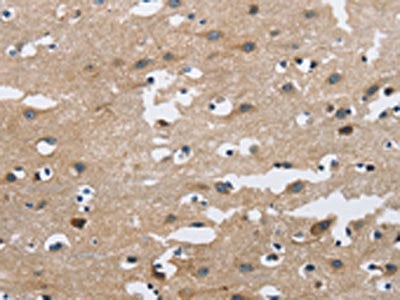
 IHC (Immunohistochemisry)
(Immunohistochemical analysis of paraffin-embedded human-brain, antibody was diluted at 1:200)
IHC (Immunohistochemisry)
(Immunohistochemical analysis of paraffin-embedded human-brain, antibody was diluted at 1:200)
FCGR3A/FCGR3B, Polyclonal Antibody (Cat# AAA236898)
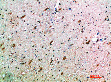
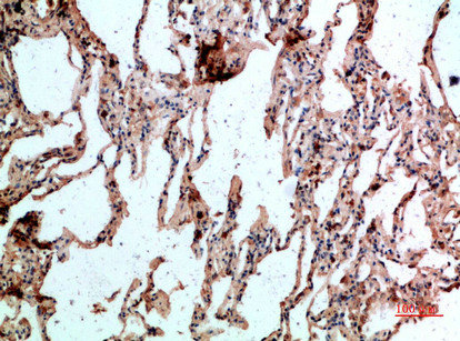
 IHC (Immunohistochemisry)
(Immunohistochemical analysis of paraffin-embedded human-brain, antibody was diluted at 1:200)
IHC (Immunohistochemisry)
(Immunohistochemical analysis of paraffin-embedded human-brain, antibody was diluted at 1:200)
FCGR1A, Polyclonal Antibody (Cat# AAA236899)
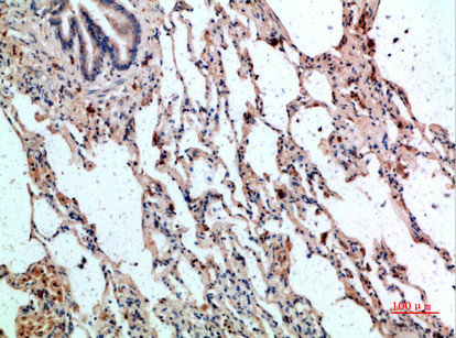
 IHC (Immunohiostchemistry)
(Immunohistochemical analysis of paraffin-embedded human-brain, antibody was diluted at 1:200)
IHC (Immunohiostchemistry)
(Immunohistochemical analysis of paraffin-embedded human-brain, antibody was diluted at 1:200)
TNFRSF9, Polyclonal Antibody (Cat# AAA236900)
 IHC (Immunohiostchemistry)
(Immunohistochemical analysis of paraffin-embedded human-small-intestine, antibody was diluted at 1:200)
IHC (Immunohiostchemistry)
(Immunohistochemical analysis of paraffin-embedded human-small-intestine, antibody was diluted at 1:200)
BTLA, Polyclonal Antibody (Cat# AAA236902)
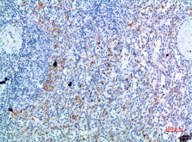
 IHC (Immunohistochemisry)
(Immunohistochemical analysis of paraffin-embedded human-brain, antibody was diluted at 1:200)
IHC (Immunohistochemisry)
(Immunohistochemical analysis of paraffin-embedded human-brain, antibody was diluted at 1:200)
CTLA4, Polyclonal Antibody (Cat# AAA236903)
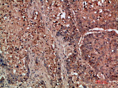
 IHC (Immunohistochemisry)
(Immunohistochemical analysis of paraffin-embedded human-brain, antibody was diluted at 1:200)
IHC (Immunohistochemisry)
(Immunohistochemical analysis of paraffin-embedded human-brain, antibody was diluted at 1:200)
KIR3DL2, Polyclonal Antibody (Cat# AAA236905)
 IHC (Immunohiostchemistry)
(Immunohistochemical analysis of paraffin-embedded human-lung-cancer, antibody was diluted at 1:200)
IHC (Immunohiostchemistry)
(Immunohistochemical analysis of paraffin-embedded human-lung-cancer, antibody was diluted at 1:200)
DDR2, Polyclonal Antibody (Cat# AAA236907)
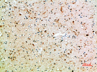
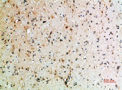
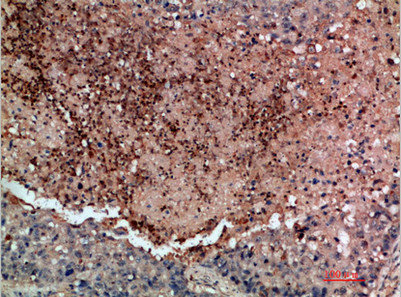
 IHC (Immunohistochemistry)
(Immunohistochemical analysis of paraffin-embedded human-brain, antibody was diluted at 1:200)
IHC (Immunohistochemistry)
(Immunohistochemical analysis of paraffin-embedded human-brain, antibody was diluted at 1:200)
FZD9, Polyclonal Antibody (Cat# AAA236908)
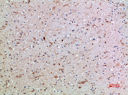
 IHC (Immunohistochemisry)
(Immunohistochemical analysis of paraffin-embedded human-brain, antibody was diluted at 1:200)
IHC (Immunohistochemisry)
(Immunohistochemical analysis of paraffin-embedded human-brain, antibody was diluted at 1:200)
KLRK1, Polyclonal Antibody (Cat# AAA236911)
 IHC (Immunohistochemisry)
(Immunohistochemical analysis of paraffin-embedded human-spleen, antibody was diluted at 1:200)
IHC (Immunohistochemisry)
(Immunohistochemical analysis of paraffin-embedded human-spleen, antibody was diluted at 1:200)
GYPC, Polyclonal Antibody (Cat# AAA236914)
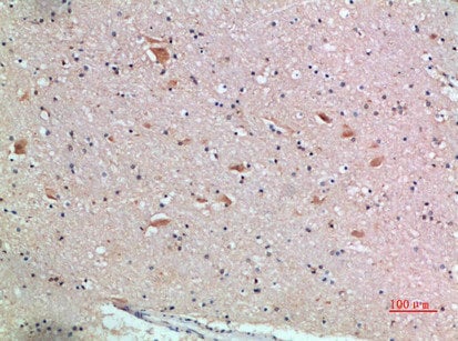
 IHC (Immunohistochemistry)
(Immunohistochemical analysis of paraffin-embedded human-brain, antibody was diluted at 1:200)
IHC (Immunohistochemistry)
(Immunohistochemical analysis of paraffin-embedded human-brain, antibody was diluted at 1:200)
PDGFC, Polyclonal Antibody (Cat# AAA236915)
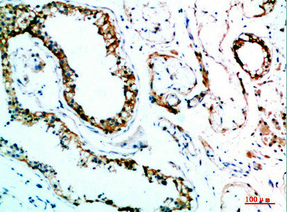
 IHC (Immunohiostchemistry)
(The image on the left is immunohistochemistry of paraffin-embedded Human tonsil tissue using AAA237204(TTR Antibody) at dilution 1/10, on the right is treated with fusion protein. (Original magnification: ×200))
IHC (Immunohiostchemistry)
(The image on the left is immunohistochemistry of paraffin-embedded Human tonsil tissue using AAA237204(TTR Antibody) at dilution 1/10, on the right is treated with fusion protein. (Original magnification: ×200))
TTR, Polyclonal Antibody (Cat# AAA237204)

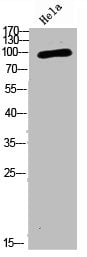
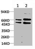
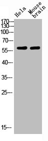
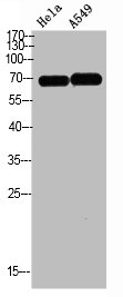
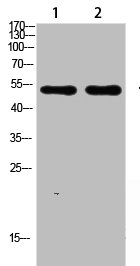
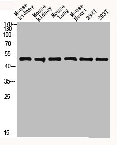
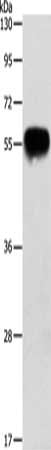
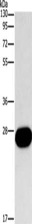
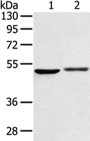
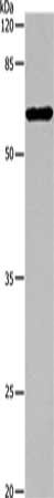
 SDS-PAGE
(Gel: 8%SDS-PAGE, Lysate: 30 ug, Lane 1-2: Hela cells, 293T cells, Primary antibody: AAA237206(VIM Antibody) at dilution 1/250, Secondary antibody: Goat anti rabbit IgG at 1/8000 dilution, Exposure time: 30 seconds)
SDS-PAGE
(Gel: 8%SDS-PAGE, Lysate: 30 ug, Lane 1-2: Hela cells, 293T cells, Primary antibody: AAA237206(VIM Antibody) at dilution 1/250, Secondary antibody: Goat anti rabbit IgG at 1/8000 dilution, Exposure time: 30 seconds)
VIM, Polyclonal Antibody (Cat# AAA237206)
 SDS-PAGE
(Gel: 8%SDS-PAGE, Lysate: 40 ug, Lane: HT29 cells, Primary antibody: AAA237207(TRIM29 Antibody) at dilution 1/400, Secondary antibody: Goat anti rabbit IgG at 1/8000 dilution, Exposure time: 10 seconds)
SDS-PAGE
(Gel: 8%SDS-PAGE, Lysate: 40 ug, Lane: HT29 cells, Primary antibody: AAA237207(TRIM29 Antibody) at dilution 1/400, Secondary antibody: Goat anti rabbit IgG at 1/8000 dilution, Exposure time: 10 seconds)
TRIM29, Polyclonal Antibody (Cat# AAA237207)
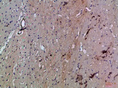
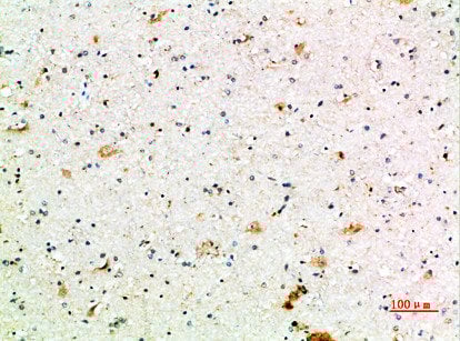
 IHC (Immunohiostchemistry)
(The image on the left is immunohistochemistry of paraffin-embedded Human brain tissue using AAA237208(SEPT9 Antibody) at dilution 1/15, on the right is treated with fusion protein. (Original magnification: ×200))
IHC (Immunohiostchemistry)
(The image on the left is immunohistochemistry of paraffin-embedded Human brain tissue using AAA237208(SEPT9 Antibody) at dilution 1/15, on the right is treated with fusion protein. (Original magnification: ×200))
SEPT9, Polyclonal Antibody (Cat# AAA237208)
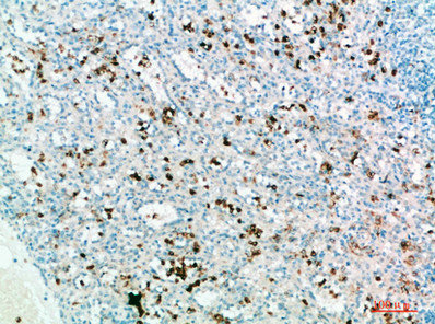
 IHC (Immunohiostchemistry)
(The image on the left is immunohistochemistry of paraffin-embedded Human cervical cancer tissue using AAA237211(ADAMTS15 Antibody) at dilution 1/60, on the right is treated with fusion protein. (Original magnification: ×200))
IHC (Immunohiostchemistry)
(The image on the left is immunohistochemistry of paraffin-embedded Human cervical cancer tissue using AAA237211(ADAMTS15 Antibody) at dilution 1/60, on the right is treated with fusion protein. (Original magnification: ×200))
ADAMTS15, Polyclonal Antibody (Cat# AAA237211)
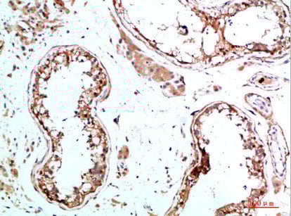
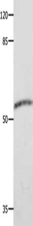
 SDS-PAGE
(Gel: 10%SDS-PAGE, Lysate: 40 ug, Lane: Human testis tissue, Primary antibody: AAA237212(APOH Antibody) at dilution 1/500, Secondary antibody: Goat anti rabbit IgG at 1/8000 dilution, Exposure time: 20 seconds)
SDS-PAGE
(Gel: 10%SDS-PAGE, Lysate: 40 ug, Lane: Human testis tissue, Primary antibody: AAA237212(APOH Antibody) at dilution 1/500, Secondary antibody: Goat anti rabbit IgG at 1/8000 dilution, Exposure time: 20 seconds)
APOH, Polyclonal Antibody (Cat# AAA237212)
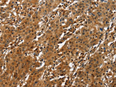
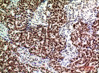
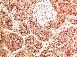
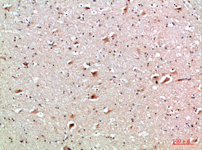
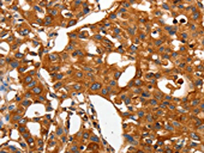
 IHC (Immunohiostchemistry)
(The image on the left is immunohistochemistry of paraffin-embedded Human liver cancer tissue using AAA237223(ATF4 Antibody) at dilution 1/50, on the right is treated with fusion protein. (Original magnification: ×200))
IHC (Immunohiostchemistry)
(The image on the left is immunohistochemistry of paraffin-embedded Human liver cancer tissue using AAA237223(ATF4 Antibody) at dilution 1/50, on the right is treated with fusion protein. (Original magnification: ×200))
ATF4, Polyclonal Antibody (Cat# AAA237223)
 SDS-PAGE
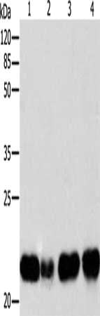
(Gel: 12%SDS-PAGE, Lysate: 40 ug, Lane 1-4: Human fetal brain tissue, 293T cells, mouse intestinum tenue tissue, lovo cells, Primary antibody: AAA237225(ATP5J Antibody) at dilution 1/300, Secondary antibody: Goat anti rabbit IgG at 1/8000 dilution, Exposure time: 1 minute)
SDS-PAGE
(Gel: 12%SDS-PAGE, Lysate: 40 ug, Lane 1-4: Human fetal brain tissue, 293T cells, mouse intestinum tenue tissue, lovo cells, Primary antibody: AAA237225(ATP5J Antibody) at dilution 1/300, Secondary antibody: Goat anti rabbit IgG at 1/8000 dilution, Exposure time: 1 minute)
ATP5J, Polyclonal Antibody (Cat# AAA237225)
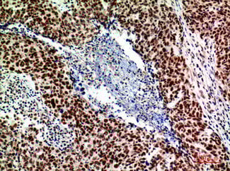
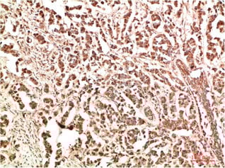
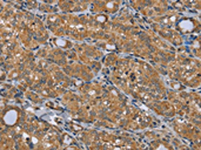
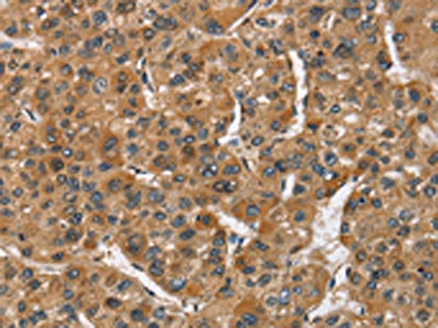
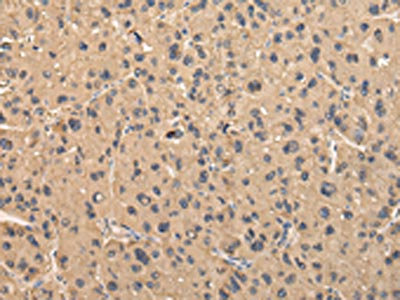
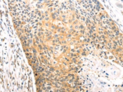
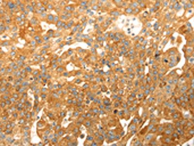
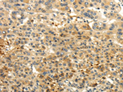
 IHC (Immunohiostchemistry)
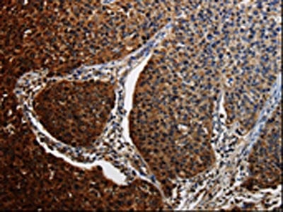
(The image on the left is immunohistochemistry of paraffin-embedded Human liver cancer tissue using AAA237227(BACE1 Antibody) at dilution 1/15, on the right is treated with fusion protein. (Original magnification: ×200))
IHC (Immunohiostchemistry)
(The image on the left is immunohistochemistry of paraffin-embedded Human liver cancer tissue using AAA237227(BACE1 Antibody) at dilution 1/15, on the right is treated with fusion protein. (Original magnification: ×200))
BACE1, Polyclonal Antibody (Cat# AAA237227)
 SDS-PAGE
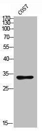
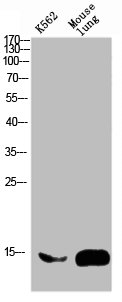
(Gel: 10%SDS-PAGE, Lysate: 40 ug, Lane: Mouse kidney tissue, Primary antibody: AAA237230(FGF9 Antibody) at dilution 1/800, Secondary antibody: Goat anti rabbit IgG at 1/8000 dilution, Exposure time: 20 seconds)
SDS-PAGE
(Gel: 10%SDS-PAGE, Lysate: 40 ug, Lane: Mouse kidney tissue, Primary antibody: AAA237230(FGF9 Antibody) at dilution 1/800, Secondary antibody: Goat anti rabbit IgG at 1/8000 dilution, Exposure time: 20 seconds)
FGF9, Polyclonal Antibody (Cat# AAA237230)
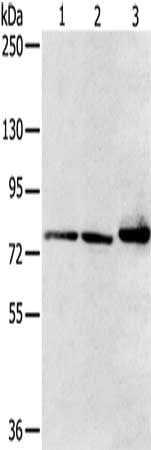
 SDS-PAGE
(Gel: 12%SDS-PAGE, Lysate: 30 ug, Lane 1-4: Human fetal brain tissue, Human liver tissue, HT29 cells, HepG2 cells, Primary antibody: AAA237231(BID Antibody) at dilution 1/260, Secondary antibody: Goat anti rabbit IgG at 1/8000 dilution, Exposure time: 10 seconds)
SDS-PAGE
(Gel: 12%SDS-PAGE, Lysate: 30 ug, Lane 1-4: Human fetal brain tissue, Human liver tissue, HT29 cells, HepG2 cells, Primary antibody: AAA237231(BID Antibody) at dilution 1/260, Secondary antibody: Goat anti rabbit IgG at 1/8000 dilution, Exposure time: 10 seconds)
BID, Polyclonal Antibody (Cat# AAA237231)
 SDS-PAGE
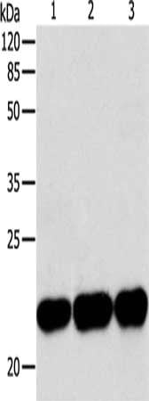
(Gel: 12%SDS-PAGE, Lysate: 30 ug, Lane 1-3: Human fetal brain tissue, HT29 cells, HepG2 cells, Primary antibody: AAA237232(BID Antibody) at dilution 1/220, Secondary antibody: Goat anti rabbit IgG at 1/8000 dilution, Exposure time: 10 seconds)
SDS-PAGE
(Gel: 12%SDS-PAGE, Lysate: 30 ug, Lane 1-3: Human fetal brain tissue, HT29 cells, HepG2 cells, Primary antibody: AAA237232(BID Antibody) at dilution 1/220, Secondary antibody: Goat anti rabbit IgG at 1/8000 dilution, Exposure time: 10 seconds)
BID, Polyclonal Antibody (Cat# AAA237232)
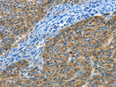
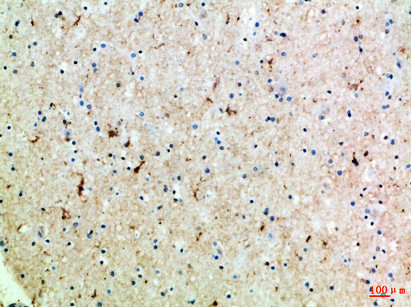
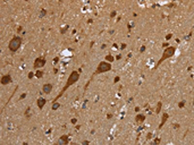
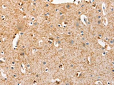
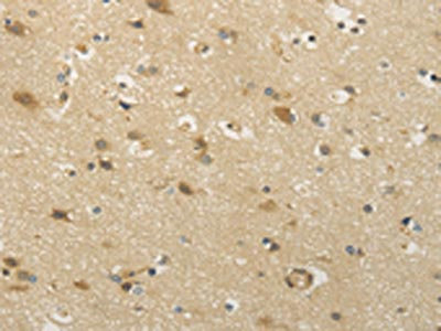
 IHC (Immunohiostchemistry)
(The image on the left is immunohistochemistry of paraffin-embedded Human brain tissue using AAA237233(BIN1 Antibody) at dilution 1/30, on the right is treated with fusion protein. (Original magnification: ×200))
IHC (Immunohiostchemistry)
(The image on the left is immunohistochemistry of paraffin-embedded Human brain tissue using AAA237233(BIN1 Antibody) at dilution 1/30, on the right is treated with fusion protein. (Original magnification: ×200))
BIN1, Polyclonal Antibody (Cat# AAA237233)
 SDS-PAGE
(Gel: 8%SDS-PAGE, Lysate: 40 ug, Lane 1-2: Human normal lung and fetal liver tissue, Primary antibody: AAA237235(BMP4 Antibody) at dilution 1/500 dilution, Secondary antibody: Goat anti rabbit IgG at 1/8000 dilution, Exposure time: 1 minute)
SDS-PAGE
(Gel: 8%SDS-PAGE, Lysate: 40 ug, Lane 1-2: Human normal lung and fetal liver tissue, Primary antibody: AAA237235(BMP4 Antibody) at dilution 1/500 dilution, Secondary antibody: Goat anti rabbit IgG at 1/8000 dilution, Exposure time: 1 minute)
BMP4, Polyclonal Antibody (Cat# AAA237235)
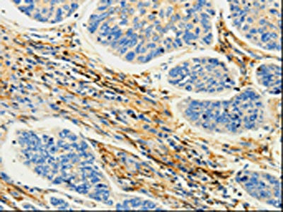
 IHC (Immunohiostchemistry)
(The image on the left is immunohistochemistry of paraffin-embedded Human cervical cancer tissue using AAA237239(BRCA1 Antibody) at dilution 1/25, on the right is treated with fusion protein. (Original magnification: ×200))
IHC (Immunohiostchemistry)
(The image on the left is immunohistochemistry of paraffin-embedded Human cervical cancer tissue using AAA237239(BRCA1 Antibody) at dilution 1/25, on the right is treated with fusion protein. (Original magnification: ×200))
BRCA1, Polyclonal Antibody (Cat# AAA237239)
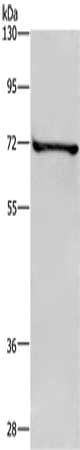
 SDS-PAGE
(Gel: 10%SDS-PAGE, Lysate: 30 ug, Lane: 293T cells, Primary antibody: AAA237242(CAPN1 Antibody) at dilution 1/450, Secondary antibody: Goat anti rabbit IgG at 1/8000 dilution, Exposure time: 1 minute)
SDS-PAGE
(Gel: 10%SDS-PAGE, Lysate: 30 ug, Lane: 293T cells, Primary antibody: AAA237242(CAPN1 Antibody) at dilution 1/450, Secondary antibody: Goat anti rabbit IgG at 1/8000 dilution, Exposure time: 1 minute)
CAPN1, Polyclonal Antibody (Cat# AAA237242)
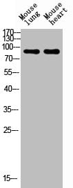
 SDS-PAGE
(Gel: 6%SDS-PAGE, Lysate: 40 ug, Lane 1-3: Mouse heart tissue, HT29 cells, mouse liver tissue, Primary antibody: AAA237245(CAPN7 Antibody) at dilution 1/300, Secondary antibody: Goat anti rabbit IgG at 1/8000 dilution, Exposure time: 1 second)
SDS-PAGE
(Gel: 6%SDS-PAGE, Lysate: 40 ug, Lane 1-3: Mouse heart tissue, HT29 cells, mouse liver tissue, Primary antibody: AAA237245(CAPN7 Antibody) at dilution 1/300, Secondary antibody: Goat anti rabbit IgG at 1/8000 dilution, Exposure time: 1 second)
CAPN7, Polyclonal Antibody (Cat# AAA237245)
 IHC (Immunohiostchemistry)
(The image on the left is immunohistochemistry of paraffin-embedded Human brain tissue using AAA237247(CAMK1G Antibody) at dilution 1/30, on the right is treated with fusion protein. (Original magnification: ×200))
IHC (Immunohiostchemistry)
(The image on the left is immunohistochemistry of paraffin-embedded Human brain tissue using AAA237247(CAMK1G Antibody) at dilution 1/30, on the right is treated with fusion protein. (Original magnification: ×200))
CAMK1G, Polyclonal Antibody (Cat# AAA237247)
 SDS-PAGE
(Gel: 10%SDS-PAGE, Lysate: 30 ug, Lane: NIH/3T3 cells, Primary antibody: AAA237250(CAP1 Antibody) at dilution 1/150, Secondary antibody: Goat anti rabbit IgG at 1/8000 dilution, Exposure time: 30 seconds)
SDS-PAGE
(Gel: 10%SDS-PAGE, Lysate: 30 ug, Lane: NIH/3T3 cells, Primary antibody: AAA237250(CAP1 Antibody) at dilution 1/150, Secondary antibody: Goat anti rabbit IgG at 1/8000 dilution, Exposure time: 30 seconds)
CAP1, Polyclonal Antibody (Cat# AAA237250)
 SDS-PAGE
(Gel: 8%SDS-PAGE, Lysate: 40 ug, Lane: Human fetal muscle tissue, Primary antibody: AAA237251(CAP2 Antibody) at dilution 1/600, Secondary antibody: Goat anti rabbit IgG at 1/8000 dilution, Exposure time: 1 minute)
SDS-PAGE
(Gel: 8%SDS-PAGE, Lysate: 40 ug, Lane: Human fetal muscle tissue, Primary antibody: AAA237251(CAP2 Antibody) at dilution 1/600, Secondary antibody: Goat anti rabbit IgG at 1/8000 dilution, Exposure time: 1 minute)
CAP2, Polyclonal Antibody (Cat# AAA237251)
 SDS-PAGE
(Gel: 8%SDS-PAGE, Lysate: 40 ug, Lane: Human fetal muscle tissue, Primary antibody: AAA237252(CAP2 Antibody) at dilution 1/700, Secondary antibody: Goat anti rabbit IgG at 1/8000 dilution, Exposure time: 1 minute)
SDS-PAGE
(Gel: 8%SDS-PAGE, Lysate: 40 ug, Lane: Human fetal muscle tissue, Primary antibody: AAA237252(CAP2 Antibody) at dilution 1/700, Secondary antibody: Goat anti rabbit IgG at 1/8000 dilution, Exposure time: 1 minute)
CAP2, Polyclonal Antibody (Cat# AAA237252)
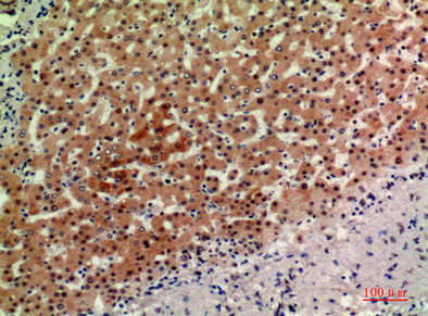
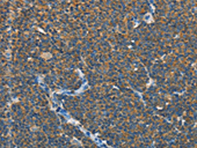
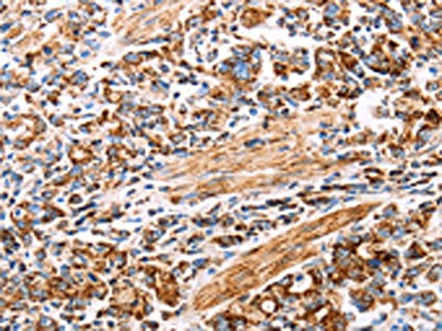
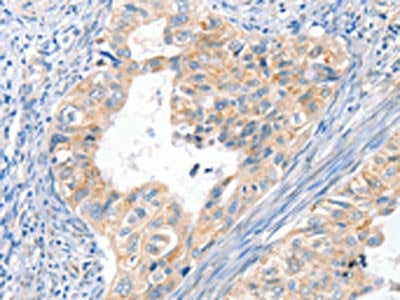
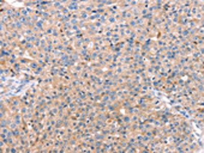
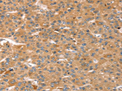
 IHC (Immunohiostchemistry)
(The image on the left is immunohistochemistry of paraffin-embedded Human liver cancer tissue using AAA237256(CA9 Antibody) at dilution 1/50, on the right is treated with fusion protein. (Original magnification: ×200))
IHC (Immunohiostchemistry)
(The image on the left is immunohistochemistry of paraffin-embedded Human liver cancer tissue using AAA237256(CA9 Antibody) at dilution 1/50, on the right is treated with fusion protein. (Original magnification: ×200))
CA9, Polyclonal Antibody (Cat# AAA237256)
What are Polyclonal Antibodies?
Polyclonal antibodies are antibodies that come from multiple B cell clones of a host animal. The typical hosts used for the majority of polyclonal antibody production are rabbits, goats, sheep, and donkeys. These polyclonal antibodies, once having identified their target, will bind to different epitopes located at different regions or sequences on the same protein/antigen. As a result, they are ideal at locating and binding to the target, even if the target is in very low concentrations (due to many different antibodies being able to bind to the same target molecule, which allows for significant amplification of a downstream signal).
Polyclonal antibodies are typically produced by injecting an antigen into a host animal, which causes the animal’s immune system to attack the foreign antigen by mass generating antibodies against it. After a period of time, serum is collected from the animal and purified using physicochemical fractionation, class-specific affinity purification, and/or antigen-affinity purification.
Key Uses of Polyclonal Antibodies
- Western Blotting: This method is used to find specific proteins in biological samples after separating them by size.
- Immunohistochemistry: IHC helps visualize the location of proteins in tissue sections using various staining techniques.
- ELISA: (Enzyme-Linked Immunosorbent Assay) is typically used to identify specific protein quantities in a sample. ELISAs can be either “Quantitative” or “Qualitative”.
- Flow Cytometry: technique that identifies and measures the specific protein on the surface or inside the cells in a fluid suspension.
- Immunoprecipitation: IP isolates and studies a specific protein from a complex mixture using antibodies.
Why Buy Polyclonal Antibodies from AAA Biotech?
1. Ideal for Various Applications
Our antibodies are generally going to be validated for use in multiple types of assays, including ELISA, Western Blotting, Immunohistochemistry, Immunoprecipitation, amongst others. They are ideal for a wide range of research applications.
2. Rigorous Quality Control
All of the antibodies in our catalog undergo strict quality testing to ensure specificity, sensitivity, and consistent performance. We are confident in the ability of our antibodies to provide you with accurate results.
3. Wide Assortment of Antibodies
Antibodies in are catalog can be found for both common and exotic species, and these antibodies are also available in both conjugated and recombinant forms to suit many diverse experimental needs.
4. Highly Purified
Our antibodies are available in purified forms with over 85% purity, as confirmed by SDS-PAGE. They are also available with tags such as His, Flag, GST, or MBP. We cater to customers worldwide.
FAQ
1. How are polyclonal antibodies produced?
Traditionally, polyclonal antibodies are produced by injecting an antigen into a host animal (such as a rabbit or goat), which then triggers an immune response from the host animal. The animal’s B cells produce antibodies that will recognize different parts of the injected antigen. These antibodies are then collected from the animal’s blood and purified for use.
2. How do polyclonal antibodies differ from monoclonal antibodies?
Polyclonal antibodies are a mix of antibodies that bind to different locations (epitopes) of the same antigen, while monoclonal antibodies are identical and bind to just one specific epitope. This makes polyclonal antibodies more versatile and better at detecting proteins that may be present in low quantities or in altered/modified forms.
3. How should I store polyclonal antibodies?
Polyclonal antibodies should be stored at 4°C for short-term use (up to a few weeks) and at -20°C or -80°C for long-term storage. Avoid repeated freeze-thaw cycles by dividing them into small aliquots. Always check the datasheet for specific storage instructions.