Filters
▼Clonality
▼Type
▼Reactivity
▼Gene Name
▼Isotype
▼Host
▼Application
▼Clone
▼Phospho Antibodies
Phospho-specific antibodies’ typical purpose is to enable researchers to detect changes in proteins. They will exclusively bind to the amino acid sequence on a protein that has been phosphorylated (which is both a physical & chemical change) and do not bind to the same amino acid sequence on said protein if it lacks said phosphorylation. This aids in being able to clearly see and understand the data produced from this particular protein modification.
Viewing 1800-1850 of 5298 product results
 IHC (Immunohistochemisry)
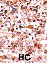
(Formalin-fixed and paraffin-embedded human cancer tissue reacted with the primary antibody, which was peroxidase-conjugated to the secondary antibody, followed by AEC staining. This data demonstrates the use of this antibody for immunohistochemistry; clinical relevance has not been evaluated. BC = breast carcinoma; HC = hepatocarcinoma.)
IHC (Immunohistochemisry)
(Formalin-fixed and paraffin-embedded human cancer tissue reacted with the primary antibody, which was peroxidase-conjugated to the secondary antibody, followed by AEC staining. This data demonstrates the use of this antibody for immunohistochemistry; clinical relevance has not been evaluated. BC = breast carcinoma; HC = hepatocarcinoma.)
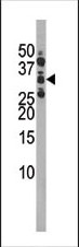
Phospho-CDK1 (S39), Polyclonal Antibody (Cat# AAA290375)
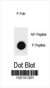 DB (Dot Blot)
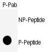
(Dot blot analysis of CCNB1 Antibody (Phospho S35) Phospho-specific Pab on nitrocellulose membrane. 50ng of Phospho-peptide or Non Phospho-peptide per dot were adsorbed. Antibody working concentrations are 0.6ug per ml.)
DB (Dot Blot)
(Dot blot analysis of CCNB1 Antibody (Phospho S35) Phospho-specific Pab on nitrocellulose membrane. 50ng of Phospho-peptide or Non Phospho-peptide per dot were adsorbed. Antibody working concentrations are 0.6ug per ml.)
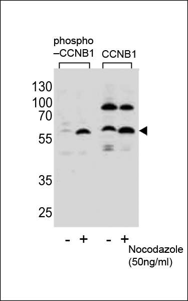
Phospho-CCNB1 (S35), Polyclonal Antibody (Cat# AAA290418)
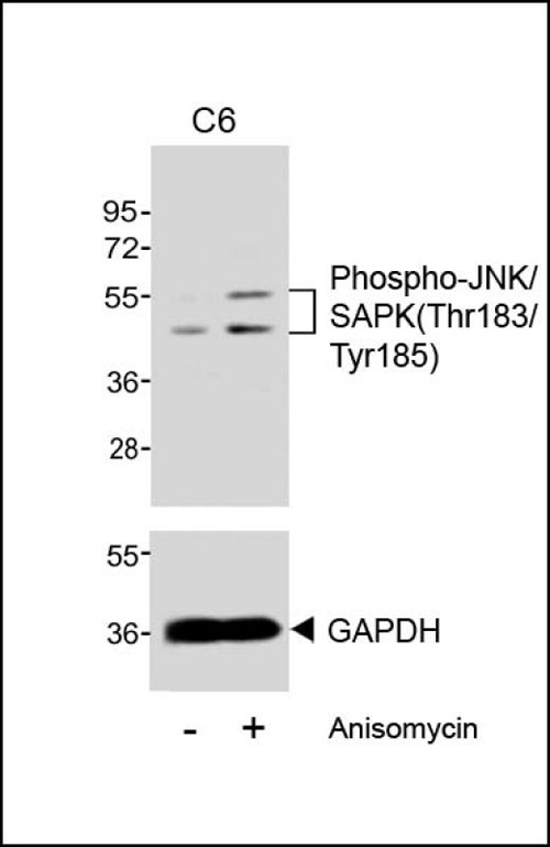
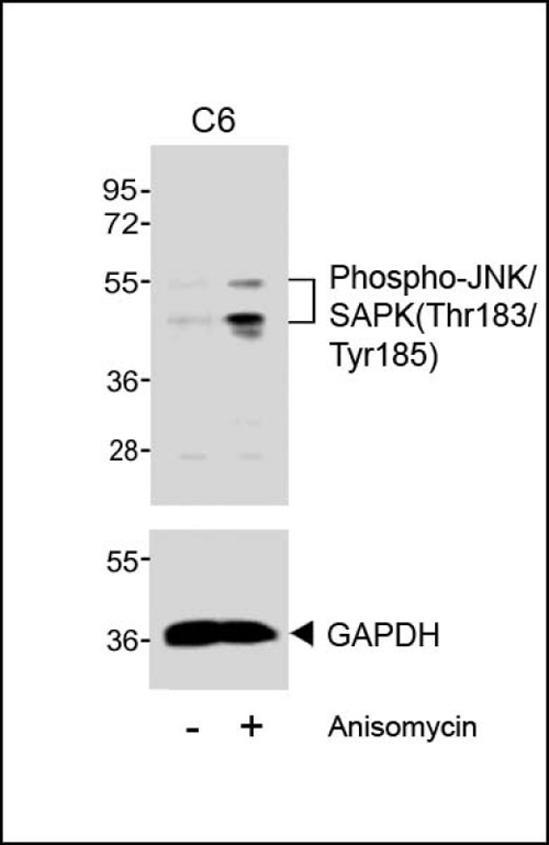 WB (Western Blot)
(Western blot analysis of extracts from C6 cells, untreated or treated with anisomycin (25 ug/ml), using Phospho-JNK/SAPK(Thr183/Tyr185) (upper) or GAPDH (lower).)
WB (Western Blot)
(Western blot analysis of extracts from C6 cells, untreated or treated with anisomycin (25 ug/ml), using Phospho-JNK/SAPK(Thr183/Tyr185) (upper) or GAPDH (lower).)
JNK/SAPK, Polyclonal Antibody (Cat# AAA290748)
Predicted: Chicken, Mouse, Rat
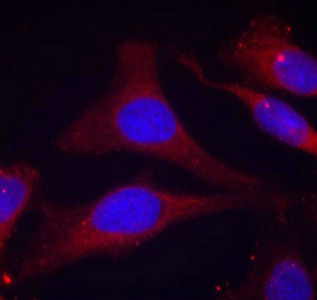
 IF (Immunofluorescence)
(Confocal immunofluorescent analysis of Phospho-P53-S9 Antibody with A2058 cell followed by Alexa Fluor 488-conjugated goat anti-rabbit lgG (green). Actin filaments have been labeled with Alexa Fluor 555 phalloidin (red).DAPI was used to stain the cell nuclear (blue).)
IF (Immunofluorescence)
(Confocal immunofluorescent analysis of Phospho-P53-S9 Antibody with A2058 cell followed by Alexa Fluor 488-conjugated goat anti-rabbit lgG (green). Actin filaments have been labeled with Alexa Fluor 555 phalloidin (red).DAPI was used to stain the cell nuclear (blue).)
Phospho-P53 (S9), Polyclonal Antibody (Cat# AAA290334)
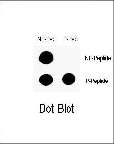
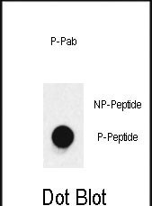
 DB (Dot Blot)
(Dot blot analysis of anti-RASGRF1-pS929 Pab (RB14017) on nitrocellulose membrane. 50ng of Phospho-peptide or Non Phospho-peptide per dot were adsorbed. Antibody working concentrations are 0.5ug per ml.)
DB (Dot Blot)
(Dot blot analysis of anti-RASGRF1-pS929 Pab (RB14017) on nitrocellulose membrane. 50ng of Phospho-peptide or Non Phospho-peptide per dot were adsorbed. Antibody working concentrations are 0.5ug per ml.)
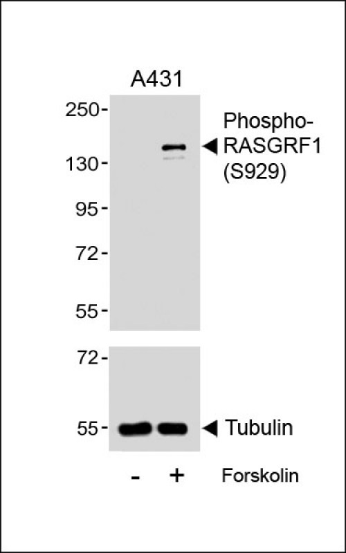
Phospho-RASGRF1, Polyclonal Antibody (Cat# AAA290977)
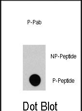
 DB (Dot Blot)
(Dot blot analysis of anti-Phospho-p38 MAPK-T180 Antibody (Cat.# AP3642a) on nitrocellulose membrane. 50ng of Phospho-peptide or Non Phospho-peptide per dot were adsorbed. Antibody working concentrations are 0.5ug per ml.)
DB (Dot Blot)
(Dot blot analysis of anti-Phospho-p38 MAPK-T180 Antibody (Cat.# AP3642a) on nitrocellulose membrane. 50ng of Phospho-peptide or Non Phospho-peptide per dot were adsorbed. Antibody working concentrations are 0.5ug per ml.)
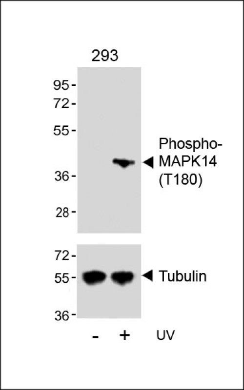
Phospho-MAPK14, Polyclonal Antibody (Cat# AAA290978)
Predicted Reactivity: Zebrafish, Mouse, Rat
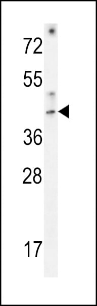
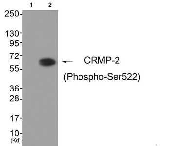 WB (Western Blot)
(Western blot analysis of extracts from HuvEc cells (Lane 2), using CRMP-2 (Phospho-Ser522) Antibody. The lane on the left is treated with antigen-specific peptide.)
WB (Western Blot)
(Western blot analysis of extracts from HuvEc cells (Lane 2), using CRMP-2 (Phospho-Ser522) Antibody. The lane on the left is treated with antigen-specific peptide.)
DPYSL2, Polyclonal Antibody (Cat# AAA243037)
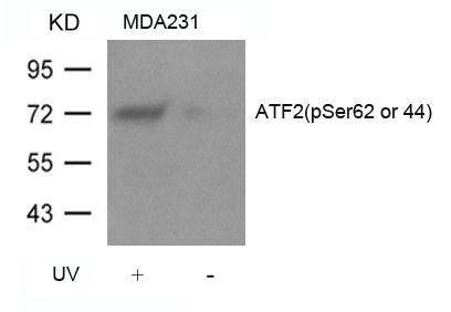
 IHC (Immunohiostchemistry)
(Immunohistochemical analysis of paraffin-embedded human breast carcinoma tissue using ATF2(Phospho-Ser62 or 44) Antibody.)
IHC (Immunohiostchemistry)
(Immunohistochemical analysis of paraffin-embedded human breast carcinoma tissue using ATF2(Phospho-Ser62 or 44) Antibody.)
ATF2, Polyclonal Antibody (Cat# AAA243048)

 IHC (Immunohiostchemistry)
(Immunohistochemical analysis of paraffin-embedded human tonsil carcinoma tissue using VASP(Phospho-Ser239) Antibody(left) or the same antibody preincubated with blocking peptide(right).)
IHC (Immunohiostchemistry)
(Immunohistochemical analysis of paraffin-embedded human tonsil carcinoma tissue using VASP(Phospho-Ser239) Antibody(left) or the same antibody preincubated with blocking peptide(right).)
VASP, Polyclonal Antibody (Cat# AAA243098)
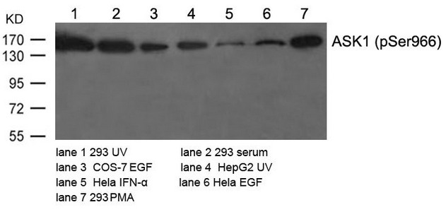
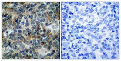
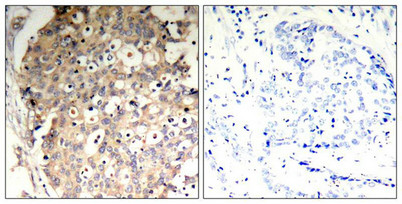
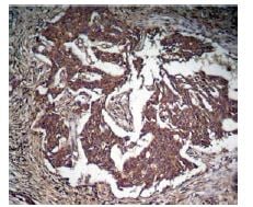
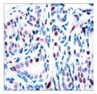
 IHC (Immunohiostchemistry)
(Immunohistochemical analysis of paraffin-embedded human breast carcinoma tissue using ASK1(Phospho-Ser966) Antibody(left) or the same antibody preincubated with blocking peptide(right).)
IHC (Immunohiostchemistry)
(Immunohistochemical analysis of paraffin-embedded human breast carcinoma tissue using ASK1(Phospho-Ser966) Antibody(left) or the same antibody preincubated with blocking peptide(right).)
MAP3K5, Polyclonal Antibody (Cat# AAA243106)
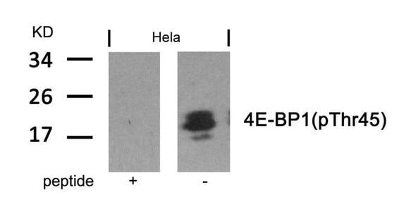
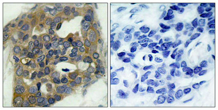
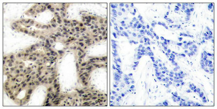 IHC (Immunohiostchemistry)
(Immunohistochemical analysis of paraffin-embedded human breast carcinoma tissue using 4E-BP1(Phospho-Thr45) Antibody(left) or the same antibody preincubated with blocking peptide(right).)
IHC (Immunohiostchemistry)
(Immunohistochemical analysis of paraffin-embedded human breast carcinoma tissue using 4E-BP1(Phospho-Thr45) Antibody(left) or the same antibody preincubated with blocking peptide(right).)
EIF4EBP1, Polyclonal Antibody (Cat# AAA243121)

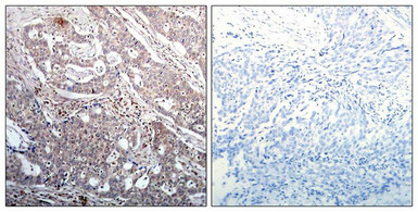
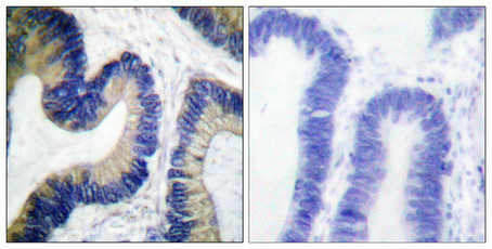
 IHC (Immunohiostchemistry)
(Immunohistochemical analysis of paraffin-embedded human breast carcinoma tissue using IRS-1(Phospho-Ser636) Antibody(left) or the same antibody preincubated with blocking peptide(right).)
IHC (Immunohiostchemistry)
(Immunohistochemical analysis of paraffin-embedded human breast carcinoma tissue using IRS-1(Phospho-Ser636) Antibody(left) or the same antibody preincubated with blocking peptide(right).)
IRS1, Polyclonal Antibody (Cat# AAA243125)
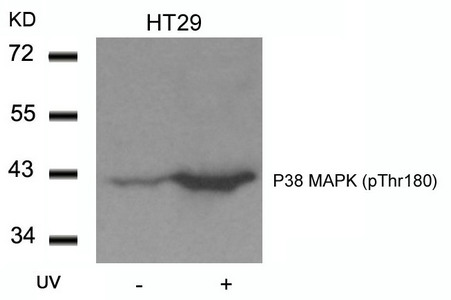
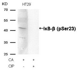 WB (Western Blot)
(Western blot analysis of extracts from HT29 cells, treated with CA or calf intestinal phosphatase (CIP), using IkappaB-beta (Phospho-Ser23) Antibody.)
WB (Western Blot)
(Western blot analysis of extracts from HT29 cells, treated with CA or calf intestinal phosphatase (CIP), using IkappaB-beta (Phospho-Ser23) Antibody.)
NFKBIB, Polyclonal Antibody (Cat# AAA243144)
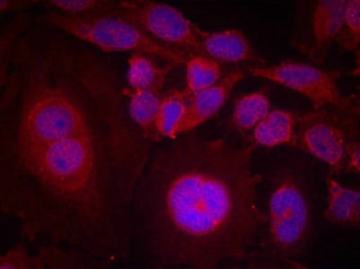
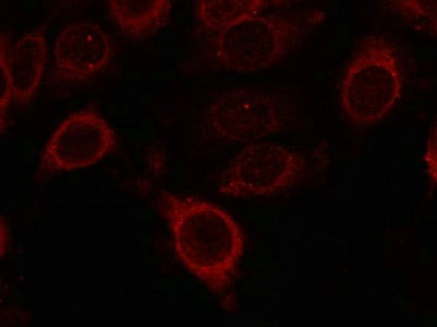
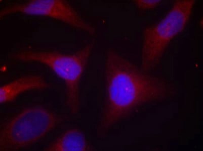
 IF (Immunofluorescence)
(Immunofluorescence staining of methanol-fixed Hela cells using SHP-1(Phospho-Tyr536) Antibody.)
IF (Immunofluorescence)
(Immunofluorescence staining of methanol-fixed Hela cells using SHP-1(Phospho-Tyr536) Antibody.)
PTPN6, Polyclonal Antibody (Cat# AAA243148)
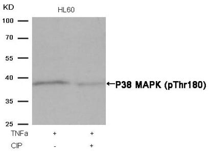 WB (Western Blot)
(Western blot analysis of extracts from HL60 cells, treated with TNFa or calf intestinal phosphatase (CIP), using P38 MAPK (Phospho-Thr180) Antibody.)
WB (Western Blot)
(Western blot analysis of extracts from HL60 cells, treated with TNFa or calf intestinal phosphatase (CIP), using P38 MAPK (Phospho-Thr180) Antibody.)
MAPK14, Polyclonal Antibody (Cat# AAA243307)
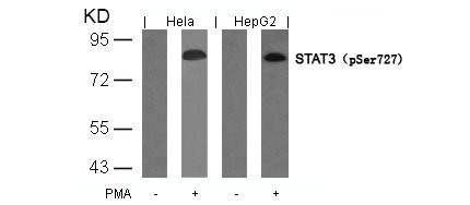
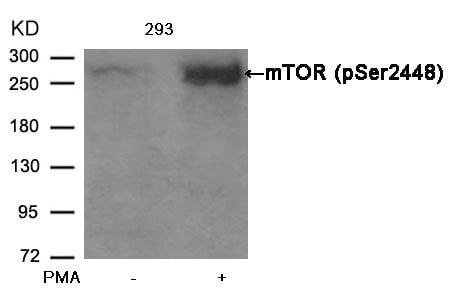
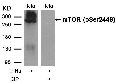
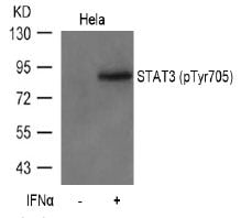
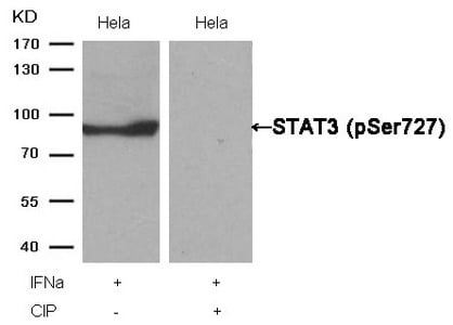 WB (Western Blot)
(Western blot analysis of extracts from Hela cells, treated with IFNa or calf intestinal phosphatase (CIP), using STAT3 (Phospho-Ser727) Antibody.)
WB (Western Blot)
(Western blot analysis of extracts from Hela cells, treated with IFNa or calf intestinal phosphatase (CIP), using STAT3 (Phospho-Ser727) Antibody.)
STAT3, Polyclonal Antibody (Cat# AAA243354)
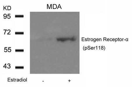
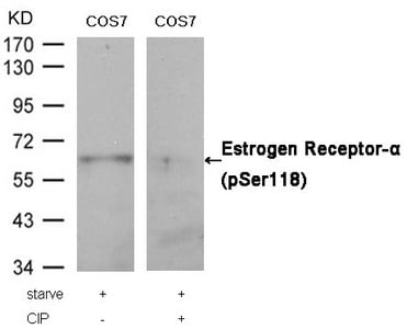 WB (Western Blot)
(Western blot analysis of extracts from COS7 cells, treated with starve or calf intestinal phosphatase (CIP), using Estrogen Receptor-alpha (Phospho-Ser118) Antibody.)
WB (Western Blot)
(Western blot analysis of extracts from COS7 cells, treated with starve or calf intestinal phosphatase (CIP), using Estrogen Receptor-alpha (Phospho-Ser118) Antibody.)
ESR1, Polyclonal Antibody (Cat# AAA243356)
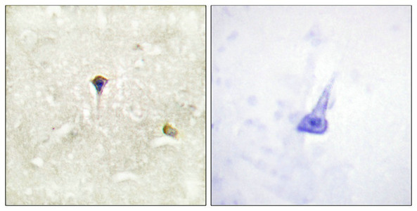
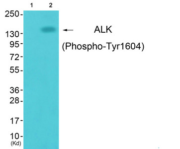
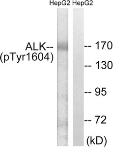
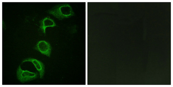 IF (Immunofluorescence)
(Immunofluorescence analysis of HeLa cells, using ALK (Phospho-Tyr1604) antibody. The picture on the right is treated with the synthesized peptide.)
IF (Immunofluorescence)
(Immunofluorescence analysis of HeLa cells, using ALK (Phospho-Tyr1604) antibody. The picture on the right is treated with the synthesized peptide.)
ALK, Polyclonal Antibody (Cat# AAA243378)
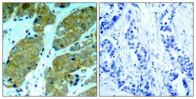
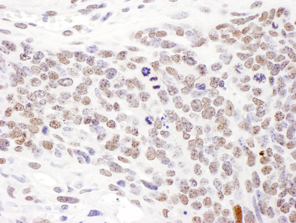
 IHC (Immunohistochemistry)
(Immunohistochemical analysis ofparaffin-embedded human breast carcinomatissue using mTOR (Phospho-Ser2448) Antibody)
IHC (Immunohistochemistry)
(Immunohistochemical analysis ofparaffin-embedded human breast carcinomatissue using mTOR (Phospho-Ser2448) Antibody)
MTOR, Polyclonal Antibody (Cat# AAA243381)
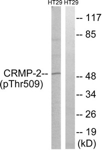
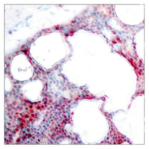
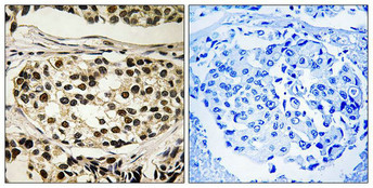
 IHC (Immunohiostchemistry)
(Immunohistochemical analysis of paraffin-embedded human brain tissue using CRMP-2 (Phospho-Thr509) antibody (left)or the same antibody preincubated with blocking peptide (right).)
IHC (Immunohiostchemistry)
(Immunohistochemical analysis of paraffin-embedded human brain tissue using CRMP-2 (Phospho-Thr509) antibody (left)or the same antibody preincubated with blocking peptide (right).)
DPYSL2, Polyclonal Antibody (Cat# AAA243219)
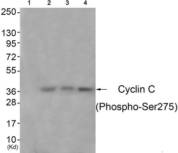
 IHC (Immunohiostchemistry)
(Immunohistochemical analysis of paraffin-embedded human breast carcinoma tissue using Cyclin C (Phospho-Ser275) antibody (left)or the same antibody preincubated with blocking peptide (right).)
IHC (Immunohiostchemistry)
(Immunohistochemical analysis of paraffin-embedded human breast carcinoma tissue using Cyclin C (Phospho-Ser275) antibody (left)or the same antibody preincubated with blocking peptide (right).)
CCNC, Polyclonal Antibody (Cat# AAA243221)
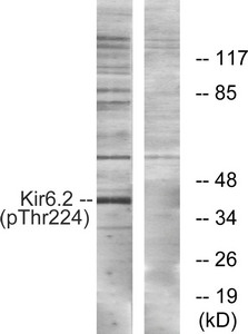
 IF (Immunofluorescence)
(Immunofluorescence analysis of HuvEc cells, using Kir6.2 (Phospho-Thr224) antibody. The picture on the right is treated with the synthesized peptide.)
IF (Immunofluorescence)
(Immunofluorescence analysis of HuvEc cells, using Kir6.2 (Phospho-Thr224) antibody. The picture on the right is treated with the synthesized peptide.)
KCNJ11, Polyclonal Antibody (Cat# AAA243233)
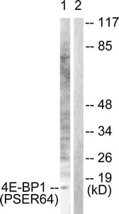
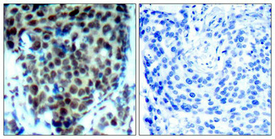
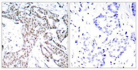
 IHC (Immunohiostchemistry)
(mmunohistochemical analysis of paraffin-embedded human colon carcinoma tissue using 4E-BP1 (Phospho-Ser64) antibody. The picture on the right is treated with the synthesized peptide.)
IHC (Immunohiostchemistry)
(mmunohistochemical analysis of paraffin-embedded human colon carcinoma tissue using 4E-BP1 (Phospho-Ser64) antibody. The picture on the right is treated with the synthesized peptide.)
EIF4EBP1, Polyclonal Antibody (Cat# AAA243239)
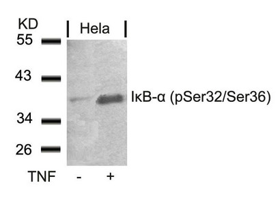
 IF (Immunofluorescence)
(Immunofluorescence staining of methanol-fixed Hela cells using IkB-a(Phospho-Ser32/Ser36) Antibody.)
IF (Immunofluorescence)
(Immunofluorescence staining of methanol-fixed Hela cells using IkB-a(Phospho-Ser32/Ser36) Antibody.)
NFKBIA, Polyclonal Antibody (Cat# AAA243289)
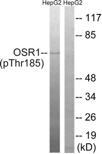
 IHC (Immunohiostchemistry)
(Immunohistochemical analysis of paraffin-embedded human breast carcinoma tissue using OSR1 (Phospho-Thr185) antibody (left)or the same antibody preincubated with blocking peptide (right).)
IHC (Immunohiostchemistry)
(Immunohistochemical analysis of paraffin-embedded human breast carcinoma tissue using OSR1 (Phospho-Thr185) antibody (left)or the same antibody preincubated with blocking peptide (right).)
OXSR1, Polyclonal Antibody (Cat# AAA243211)
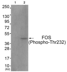
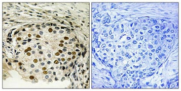
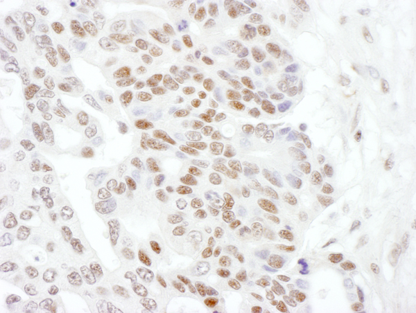
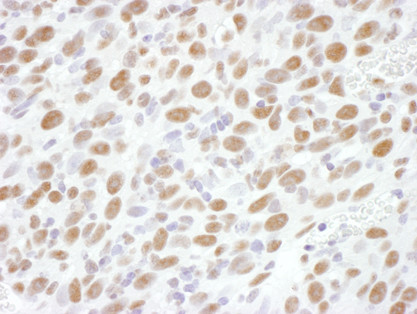
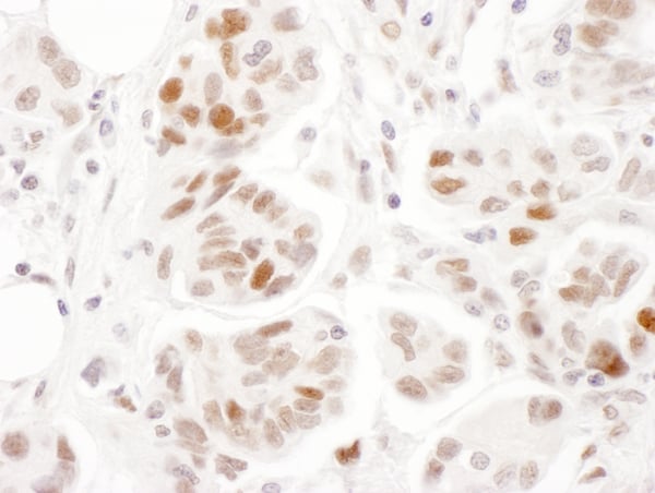
 IHC (Immunohiostchemistry)
(Immunohistochemical analysis of paraffin-embedded human breast carcinoma tissue using FOS (Phospho-Thr232) antibody (left)or the same antibody preincubated with blocking peptide (right).)
IHC (Immunohiostchemistry)
(Immunohistochemical analysis of paraffin-embedded human breast carcinoma tissue using FOS (Phospho-Thr232) antibody (left)or the same antibody preincubated with blocking peptide (right).)
FOS, Polyclonal Antibody (Cat# AAA243214)
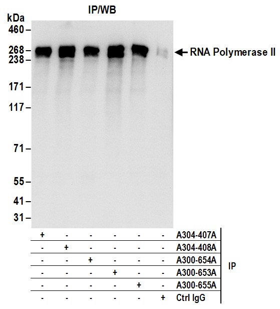
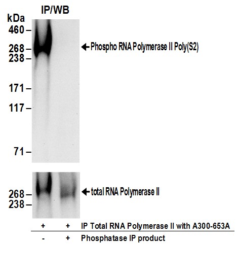 IP (Immunoprecipitation)
(Detection of human Phospho RNA Polymerase II (S2) by western blot of immunoprecipitates. Samples: Whole cell lysate (1 mg for IP; 20% of IP loaded) prepared using NETN buffer from 293T cells. Antibodies: RNA Polymerase II was immunoprecipitated using rabbit anti-RNA Polymerase II antibody Following immunoprecipitation the product of the IP reaction was mock treated (-) or treated with phosphatases (+). Affinity purified rabbit anti-Phospho RNA Polymerase II (S2) antibody AAA212674 was used for WB at 1 ug/ml (upper panel). To examine total RNA Polymerase II, the blot was stripped and then blotted with rabbit anti-RNA Polymerase II antibody (lower panel). Detection: Chemiluminescence with exposure times of 30 seconds (upper panel) and 15 seconds (lower panel).)
IP (Immunoprecipitation)
(Detection of human Phospho RNA Polymerase II (S2) by western blot of immunoprecipitates. Samples: Whole cell lysate (1 mg for IP; 20% of IP loaded) prepared using NETN buffer from 293T cells. Antibodies: RNA Polymerase II was immunoprecipitated using rabbit anti-RNA Polymerase II antibody Following immunoprecipitation the product of the IP reaction was mock treated (-) or treated with phosphatases (+). Affinity purified rabbit anti-Phospho RNA Polymerase II (S2) antibody AAA212674 was used for WB at 1 ug/ml (upper panel). To examine total RNA Polymerase II, the blot was stripped and then blotted with rabbit anti-RNA Polymerase II antibody (lower panel). Detection: Chemiluminescence with exposure times of 30 seconds (upper panel) and 15 seconds (lower panel).)
RNA Polymerase II, Polyclonal Antibody (Cat# AAA212674)
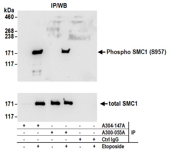
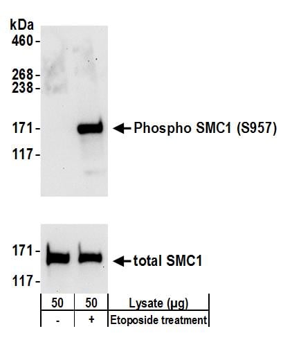 WB (Western Blot)
(Detection of human Phospho SMC1 (S957) by western blot. Samples: Whole cell lysate (50 ug) from 293T cells treated with 100 uM etoposide for 2 hours (+) or mock treated (-) cells. Antibodies: Affinity purified rabbit anti-Phospho SMC1 (S957) antibody AAA212520 (lot AAA212520-1) used for WB at 0.1 ug/ml. Detection: Chemiluminescence with an exposure time of 30 seconds. Lower panel shows western blot for total SMC1 using affinity purified rabbit anti-SMC1 antibody at 0.1 ug/ml with an exposure time of 30 seconds.)
WB (Western Blot)
(Detection of human Phospho SMC1 (S957) by western blot. Samples: Whole cell lysate (50 ug) from 293T cells treated with 100 uM etoposide for 2 hours (+) or mock treated (-) cells. Antibodies: Affinity purified rabbit anti-Phospho SMC1 (S957) antibody AAA212520 (lot AAA212520-1) used for WB at 0.1 ug/ml. Detection: Chemiluminescence with an exposure time of 30 seconds. Lower panel shows western blot for total SMC1 using affinity purified rabbit anti-SMC1 antibody at 0.1 ug/ml with an exposure time of 30 seconds.)
SMC1, Polyclonal Antibody (Cat# AAA212520)
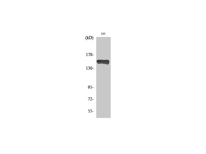 WB (Western Blot)
(Western Blot analysis of 293 cells withPhospho-PLCG1 (Tyr783) Polyclonal Antibody.)
WB (Western Blot)
(Western Blot analysis of 293 cells withPhospho-PLCG1 (Tyr783) Polyclonal Antibody.)
PLC gamma1, Polyclonal Antibody (Cat# AAA172029)
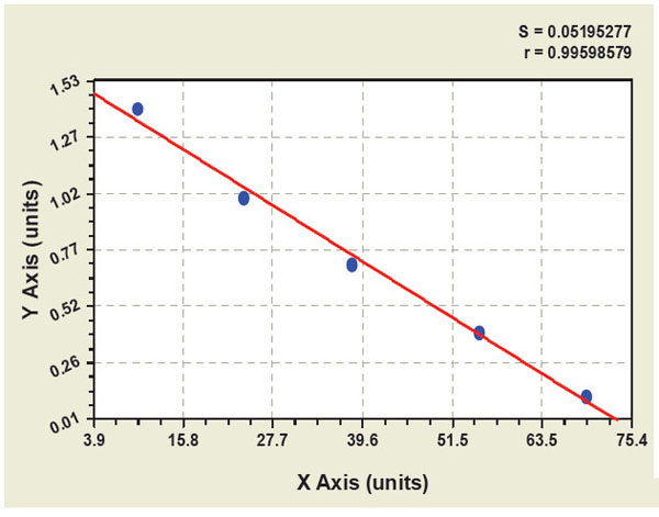 Standard Curve (Sample)
Phospho-Nuclear Factor kappa B, ELISA Kit (Cat# AAA91992)
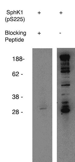 WB (Western Blot)
(Western blot using anti-mouse SphK1 (pS225) on mouse B-cell lysate. Antibody used at 1 ug/ml with phosphorylated blocking peptide (lane A) and without (laneB).)
WB (Western Blot)
(Western blot using anti-mouse SphK1 (pS225) on mouse B-cell lysate. Antibody used at 1 ug/ml with phosphorylated blocking peptide (lane A) and without (laneB).)
phospho-Sphingosine Kinase 1,(pS225), Mouse Reactive, Polyclonal Antibody (Cat# AAA60497)
Purification: Antigen Immunoaffiinity Purification
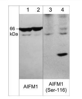 WB (Western Blot)
(Western blot image of human jurkat cells untreated (lanes 1 & 3) or treated with calyculin A (100nM, 30 min.) (lanes 2 and 4). The blot was probed with rabbit polyclonals anti-AIFM1 (C-terminal region) at 1:500 (lanes 1 & 2) and anti-AIFM1 (Ser-116) phospho-specific antibody at 1:1000 (lanes 3 & 4).)
WB (Western Blot)
(Western blot image of human jurkat cells untreated (lanes 1 & 3) or treated with calyculin A (100nM, 30 min.) (lanes 2 and 4). The blot was probed with rabbit polyclonals anti-AIFM1 (C-terminal region) at 1:500 (lanes 1 & 2) and anti-AIFM1 (Ser-116) phospho-specific antibody at 1:1000 (lanes 3 & 4).)
AIFM1, Polyclonal Antibody (Cat# AAA71569)
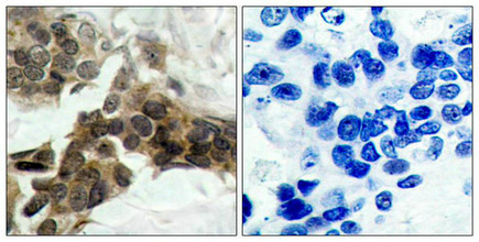
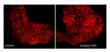 ICC (Immunocytochemistry)
(Immunocytochemical labeling of beta-Catenin in pervanadate-treated A431 cells. The cells were labeled with mouse monoclonal beta-Catenin (CM1181) or rabbit polyclonal beta-Catenin (Tyr-489) antibodies, then the antibodies were detected using appropriate secondary antibodies conjugated to Cy3.)
ICC (Immunocytochemistry)
(Immunocytochemical labeling of beta-Catenin in pervanadate-treated A431 cells. The cells were labeled with mouse monoclonal beta-Catenin (CM1181) or rabbit polyclonal beta-Catenin (Tyr-489) antibodies, then the antibodies were detected using appropriate secondary antibodies conjugated to Cy3.)
beta-Catenin, Polyclonal Antibody (Cat# AAA71609)
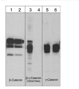
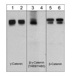
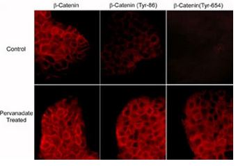 ICC (Immunocytochemistry)
(Immunocytochemical labeling of phosphorylated beta-Catenin in control and pervanadate-treated A431 cells. The cells were labeled with mouse monoclonal beta-Catenin (CM1181) or rabbit polyclonal beta-Catenin (Tyr-86) or beta-Catenin (Y654) antibodies, then the antibodies were detected using appropriate secondary antibodies conjugated to Cy3.)
ICC (Immunocytochemistry)
(Immunocytochemical labeling of phosphorylated beta-Catenin in control and pervanadate-treated A431 cells. The cells were labeled with mouse monoclonal beta-Catenin (CM1181) or rabbit polyclonal beta-Catenin (Tyr-86) or beta-Catenin (Y654) antibodies, then the antibodies were detected using appropriate secondary antibodies conjugated to Cy3.)
beta-Catenin, Polyclonal Antibody (Cat# AAA71610)
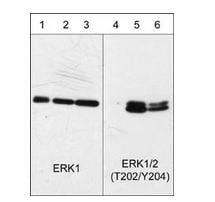
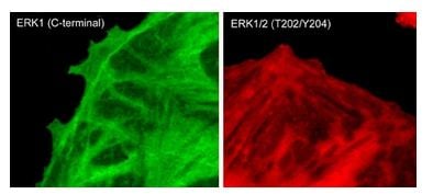 ICC (Immunocytochemistry)
(Immunocytochemical labeling of phosphorylated ERK1 in paraformaldehyde-fixed and NP-40-permeabilized rat A7r5 cells treated with calyculin A. The fixed cells were labeled with mouse monoclonal antibodies to anti-ERK1 (EM2331) and anti-ERK1/2 (Thr-202/Tyr-204) (EM2061). The antibodies were detected using Goat anti-Mouse secondary antibodies conjugated to DyLight 488 (left) and DyLight 594 (right).)
ICC (Immunocytochemistry)
(Immunocytochemical labeling of phosphorylated ERK1 in paraformaldehyde-fixed and NP-40-permeabilized rat A7r5 cells treated with calyculin A. The fixed cells were labeled with mouse monoclonal antibodies to anti-ERK1 (EM2331) and anti-ERK1/2 (Thr-202/Tyr-204) (EM2061). The antibodies were detected using Goat anti-Mouse secondary antibodies conjugated to DyLight 488 (left) and DyLight 594 (right).)
ERK1, Monoclonal Antibody (Cat# AAA71624)
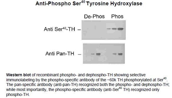 Application Data
Tyrosine Hydroxylase (Ser40), Polyclonal Antibody (Cat# AAA72806)
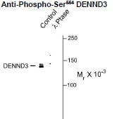 WB (Western Blot)
(Western blot of HeLa lysate showing specific immunolabeling of the ~142kD DENND3 protein phosphorylated at Ser554 (control). Phosphospecificity is shown in the second lane (lambda-phosphatase: lambda-Ptase). The blot is identical to the control except that the lysate was incubated in lambda-Ptase (800 units/1mg protein for 30 min). The immunolabeling is completely eliminated by treatment with lambda-Ptase.)
WB (Western Blot)
(Western blot of HeLa lysate showing specific immunolabeling of the ~142kD DENND3 protein phosphorylated at Ser554 (control). Phosphospecificity is shown in the second lane (lambda-phosphatase: lambda-Ptase). The blot is identical to the control except that the lysate was incubated in lambda-Ptase (800 units/1mg protein for 30 min). The immunolabeling is completely eliminated by treatment with lambda-Ptase.)
Ser554 DENND3, Polyclonal Antibody (Cat# AAA72827)
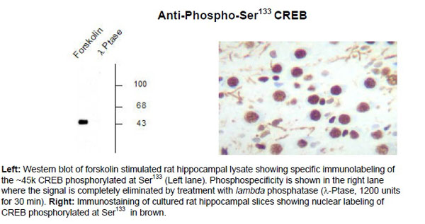 Application Data
CREB (Ser133), Polyclonal Antibody (Cat# AAA72783)
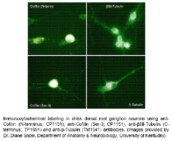
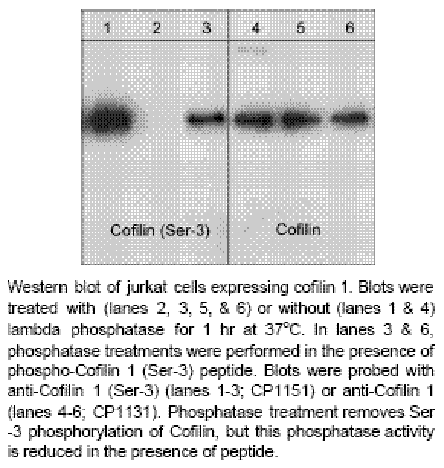 Application Data
Cofilin 1 (Ser-3), Polyclonal Antibody (Cat# AAA71517)
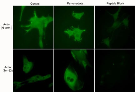 ICC (Immunocytochemistry)
(Immunocytochemical labeling using anti-Actin (N-terminal) and anti-Actin (Tyr-53) polyclonal antibodies in C2C12 cells control (left) or treated with pervanadate (1 mM) for 30 min (middle). The cells were fixed in paraformaldehyde and permeabilized in acetone. Both antibodies were used in the presence of blocking peptide: Actin (N-terminal) peptide or phospho-Actin (Tyr-53) peptide, respectively (right).)
ICC (Immunocytochemistry)
(Immunocytochemical labeling using anti-Actin (N-terminal) and anti-Actin (Tyr-53) polyclonal antibodies in C2C12 cells control (left) or treated with pervanadate (1 mM) for 30 min (middle). The cells were fixed in paraformaldehyde and permeabilized in acetone. Both antibodies were used in the presence of blocking peptide: Actin (N-terminal) peptide or phospho-Actin (Tyr-53) peptide, respectively (right).)
Actin, Polyclonal Antibody (Cat# AAA71548)
 Application Data
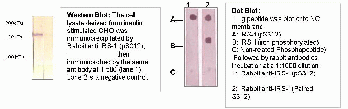
IRS-1 (pS312),, Antibody (Cat# AAA71386)
 WB (Western Blot)
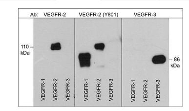
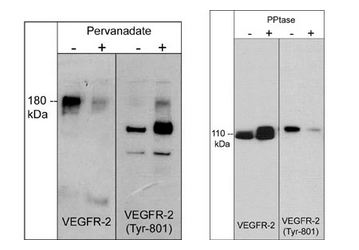
(Left: Western blot image of HUVEC cells untreated (-) or treated with pervanadate (1mM) for 30 min. (+). Right: Western blot image of GST-recombinant VEGFR-2 kinase without (-) or with (+) akaline phosphatase treatment. Both sets of blots were probed with rabbit polyclonal anti-VEGFR-2 (a.a. 1304-1317) or anti-VEGFR-2 (Tyr-801).)
WB (Western Blot)
(Left: Western blot image of HUVEC cells untreated (-) or treated with pervanadate (1mM) for 30 min. (+). Right: Western blot image of GST-recombinant VEGFR-2 kinase without (-) or with (+) akaline phosphatase treatment. Both sets of blots were probed with rabbit polyclonal anti-VEGFR-2 (a.a. 1304-1317) or anti-VEGFR-2 (Tyr-801).)
VEGFR-2, Polyclonal Antibody (Cat# AAA71731)
 ICC (Immunocytochemistry)
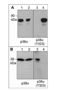
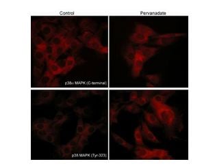
(Immunocytochemical labeling of p38 MAPK in pervanadate-treated mouse C2C12. The cells were labeled with mouse monoclonal p38alpha MAPK and rabbit polyclonal p38 MAPK (Tyr-323) antibodies, then the antibodies were detected using appropriate secondary antibodies conjugated to Cy3.)
ICC (Immunocytochemistry)
(Immunocytochemical labeling of p38 MAPK in pervanadate-treated mouse C2C12. The cells were labeled with mouse monoclonal p38alpha MAPK and rabbit polyclonal p38 MAPK (Tyr-323) antibodies, then the antibodies were detected using appropriate secondary antibodies conjugated to Cy3.)
p38alpha MAP Kinase, Polyclonal Antibody (Cat# AAA71695)
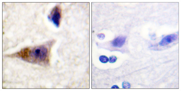
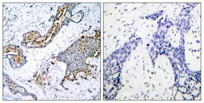
 IHC (Immunohiostchemistry)
(Immunohistochemistry analysis of paraffin-embedded human brain tissue using Caspase 1 (Phospho-Ser376) antibody. Western blot analysis of extracts from 293 cells, using Caspase 1 (Phospho-Ser376) antibody.)
IHC (Immunohiostchemistry)
(Immunohistochemistry analysis of paraffin-embedded human brain tissue using Caspase 1 (Phospho-Ser376) antibody. Western blot analysis of extracts from 293 cells, using Caspase 1 (Phospho-Ser376) antibody.)
Caspase 1, Antibody (Cat# AAA112121)
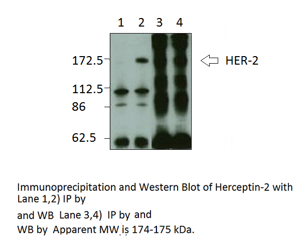
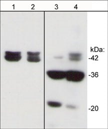
 WB (Western Blot)
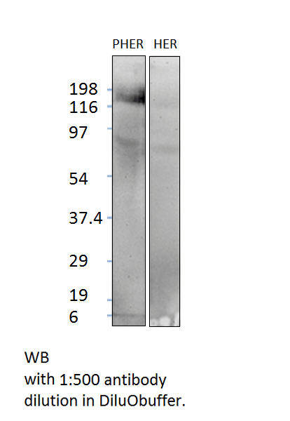
Phospho-ErbB 2, Polyclonal Antibody (Cat# AAA76011)
Predicted: Human
Phospho-MERTK, Polyclonal Antibody (Cat# AAA76018)
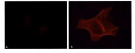
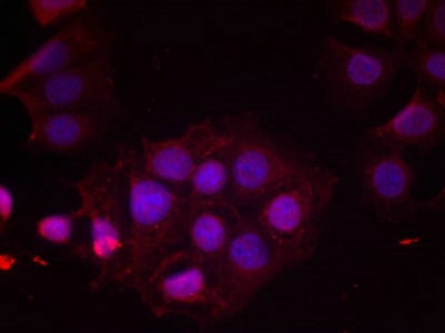
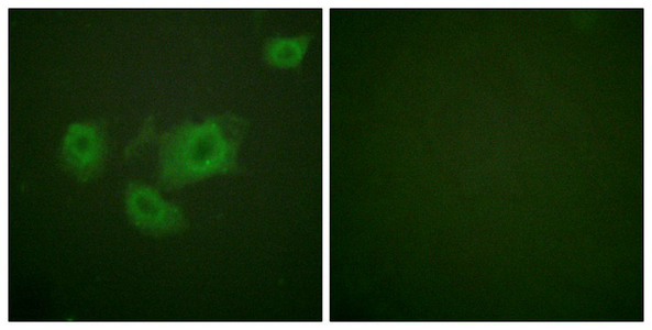
 IF (Immunofluorescence)
(Immunofluorescence analysis of transfected HeLa cells stained with the antiphospho- Tau (Tyr 18) antibody, dilution 1:200: A: Tau; B: Tau + Fyn)
IF (Immunofluorescence)
(Immunofluorescence analysis of transfected HeLa cells stained with the antiphospho- Tau (Tyr 18) antibody, dilution 1:200: A: Tau; B: Tau + Fyn)
Tau, Monoclonal Antibody (Cat# AAA77834)
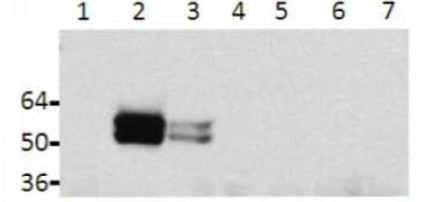
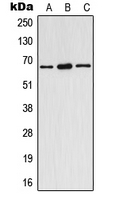 WB (Western Blot)
(Western blot analysis of Estrogen Receptor alpha (pY537) expression in HeLa (A), SP2/0 (B), rat spleen (C) whole cell lysates.)
WB (Western Blot)
(Western blot analysis of Estrogen Receptor alpha (pY537) expression in HeLa (A), SP2/0 (B), rat spleen (C) whole cell lysates.)
Estrogen Receptor alpha (pY537), Polyclonal Antibody (Cat# AAA105233)
 Application Data
STAT3, Polyclonal Antibody (Cat# AAA47824)
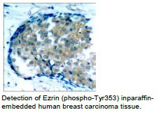
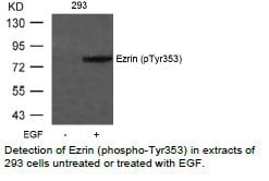 Application Data
Ezrin, Polyclonal Antibody (Cat# AAA47838)
What Are Phospho Antibodies?
Protein phosphorylation is a process where a phosphate group is added to certain amino acid residues of a protein – usually serine (S), threonine (T), or tyrosine (Y) - by enzymes called kinases. This process is integral in controlling cellular signaling, cellular growth, and other biological functions.
Our catalog includes a wide range of phospho-specific antibodies that can accurately detect this important marker. They perform strongly in widely-used laboratory applications such as Western blot, flow cytometry, immunohistochemistry, and immunofluorescence microscopy. We value your trust in us and are committed to providing top-quality products and services. All of our antibodies are guaranteed to work for the applications and species indicated on our website & associated product pages.
What Are The Key Applications of Phospho Antibodies?
1. Western Blotting
One of the first steps a researcher can take in utilizing these phospho-specific antibodies, is to check if the antibody works using a technique referred to as “Western blot”. For those unfamiliar, Western Blot aids in showing whether the protein that the antibody recognizes is appearing at the correct/expected size. These phospho-specific antibodies should also be able to detect changes in the target protein’s phosphorylation (on/off state) when cells are stimulated in certain ways.
2. Staining of Fixed Cells (Immunocytochemistry)
Another routine use of these phospho-specific antibodies, is to test if the antibody is able to demonstrate similar performance when used on fixed cells (intact cells that have been preserved) as it did in the Western blot tests. It is an important aspect in many cases to confirm that the antibody works in actual intact cell samples. Ideally, the method used for cellular fixation should be the same as what is used in pathology labs (like using 10% formalin). To check if the antibody works well in tissue sections (FFPE), researchers will often test it on fixed cells that are processed similar to tissue samples.
3. Specificity Tests Using Peptides
In order to make sure that the antibody is only binding to the right target:
- Laboratory technicians will mix the antibody with phospho-peptides (short segments of the protein containing the phosphate group modification).
- If the antibody signal disappears, it is confirmation that it is binding to the correct phosphorylated location.
- A more robust test is to use both the phosphorylated and non-phosphorylated (dephosphorylated) versions of the protein. The antibody should react only with the phosphorylated one.
- Another method sometimes utilized is to treat the sample with an enzyme, such as alkaline phosphatase, that specifically removes phosphate groups. If the antibody signal disappears after this, it also confirms specificity.
4. Genetic Confirmation
As a final step, scientists can genetically manipulate the nucleotide sequence and alter the target protein by removing the exact site where phosphorylation happens. If the antibody no longer appears to detect the modified protein, it is strong evidence supporting the antibody being specific for that phosphorylated site.
Why Buy Phospho Antibodies Through Us?
- The production laboratory adheres to strict and consistent protocols prior to releasing any of these phospho-specific antibodies:
- Standard methods and proper controls in all tests to ensure high quality.
- These antibodies are tested and validated in different cell types and species.
- High quality control criterion to ensure each batch is consistent, so you will obtain reliable results every time.
FAQ
1. What Are Phospho-Specific Antibodies?
Phospho-specific antibodies are made to detect proteins only when they have a phosphate group linked to a specific amino acid residue. This empowers scientists understand if a protein is "turned on" or active, based on its phosphorylation state.
2. How to Detect Phosphorylated Proteins in a Western Blot?
To find out if a protein is phosphorylated using Western blot:
- Use a phospho-specific antibody that binds only to the phosphorylated form of the protein.
- You can also use a “regular” antibody for the same amino acid sequence of the protein that the phospho-specific antibody is binding to (but in this case, this antibody will not bind if there is a phosphate group present) in order to compare how much of it is phosphorylated versus how much is non-phosphorylated (or “total” protein, if the “normal” antibody’s epitopes are non-phospho-site-specific).
3. How to Choose the Best Antibody?
Here are some simple tips to help you pick the right antibody:
- Know your target
- Match your sample characteristics
- Confirm the intended use is appropriate
- Check “host” and “type”
- Check the “quality” of the presented data/images
- Appraise whether the available validation meets your needs