Filters
▼Clonality
▼Type
▼Reactivity
▼Gene Name
▼Isotype
▼Host
▼Application
▼Clone
▼Phospho Antibodies
Phospho-specific antibodies’ typical purpose is to enable researchers to detect changes in proteins. They will exclusively bind to the amino acid sequence on a protein that has been phosphorylated (which is both a physical & chemical change) and do not bind to the same amino acid sequence on said protein if it lacks said phosphorylation. This aids in being able to clearly see and understand the data produced from this particular protein modification.
Viewing 1950-2000 of 5298 product results
 WB (Western Blot)
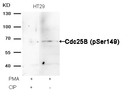
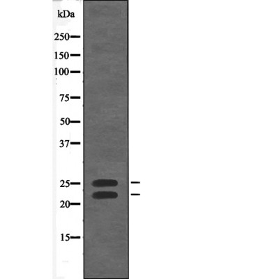
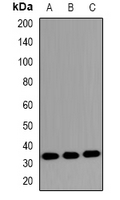
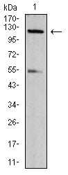
(Western blot analysis of extracts from HT29 cells, treated with PMA or calf intestinal phosphatase (CIP), using Cdc25B (Phospho-Ser149) Antibody.)
WB (Western Blot)
(Western blot analysis of extracts from HT29 cells, treated with PMA or calf intestinal phosphatase (CIP), using Cdc25B (Phospho-Ser149) Antibody.)
Cdc25B, Polyclonal Antibody (Cat# AAA307919)
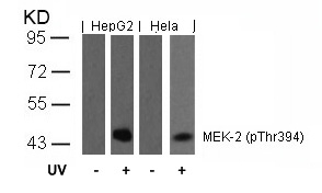
 IF (Immunofluorescence)
(Immunofluorescence staining of methanol-fixed Hela cells using MEK-2(Phospho-Thr394) Antibody.)
IF (Immunofluorescence)
(Immunofluorescence staining of methanol-fixed Hela cells using MEK-2(Phospho-Thr394) Antibody.)
MEK-2, Polyclonal Antibody (Cat# AAA308227)
 WB (Western Blot)
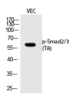
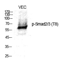
(Western Blot analysis of VEC cells using Phospho-Smad2/3 (T8) Polyclonal Antibody)
WB (Western Blot)
(Western Blot analysis of VEC cells using Phospho-Smad2/3 (T8) Polyclonal Antibody)
Smad2/3, Polyclonal Antibody (Cat# AAA308249)
 WB (Western Blot)
(Western blot analysis DYRK1A/B (Phospho-Tyr321/273) using EGF treated K562 whole cell lysates)
WB (Western Blot)
(Western blot analysis DYRK1A/B (Phospho-Tyr321/273) using EGF treated K562 whole cell lysates)
DYRK1A/B, Polyclonal Antibody (Cat# AAA309129)
 WB (Western Blot)
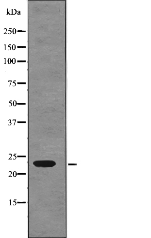
(Western blot analysis TIE1 (Phospho-Tyr1117) using Serum treated LOVO whole cell lysates)
WB (Western Blot)
(Western blot analysis TIE1 (Phospho-Tyr1117) using Serum treated LOVO whole cell lysates)
TIE1, Polyclonal Antibody (Cat# AAA309131)
 WB (Western Blot)
(Western blot analysis Bim (Phospho-Thr56/116) using EGF treated 293 whole cell lysates)
WB (Western Blot)
(Western blot analysis Bim (Phospho-Thr56/116) using EGF treated 293 whole cell lysates)
Bim, Polyclonal Antibody (Cat# AAA309133)
 WB (Western Blot)
(Western blot analysis R-Ras (Phospho-Tyr66) using PMA treated NIH-3T3 whole cell lysates)
WB (Western Blot)
(Western blot analysis R-Ras (Phospho-Tyr66) using PMA treated NIH-3T3 whole cell lysates)
R-Ras, Polyclonal Antibody (Cat# AAA309144)
 WB (Western Blot)
(Western blot analysis PKM2(phospho-Ser37))
WB (Western Blot)
(Western blot analysis PKM2(phospho-Ser37))
PKM2, Polyclonal Antibody (Cat# AAA309147)
 IF (Immunofluorescence)
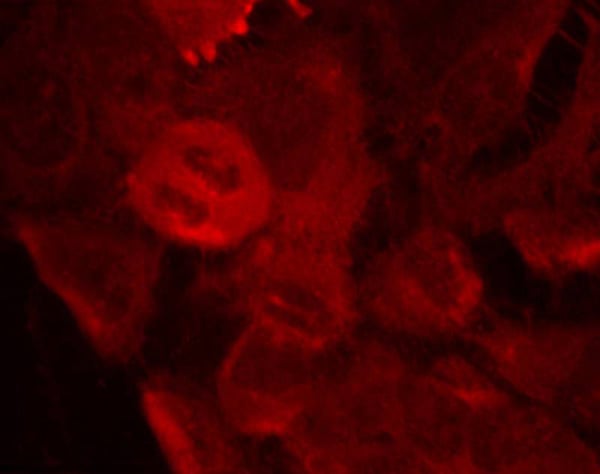
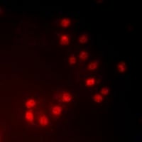
(Immunofluorescent analysis of MKI67IP (phospho-Thr234) staining in HeLa cells. Formalin-fixed cells were permeabilized with 0.1% Triton X-100 in TBS for 5-10 minutes and blocked with 3% BSA-PBS for 30 minutes at room temperature. Cells were probed with the primary antibody in 3% BSA-PBS and incubated overnight at 4 C in a hidified chamber. Cells were washed with PBST and incubated with a DyLight 594-conjugated secondary antibody (red) in PBS at room temperature in the dark.)
IF (Immunofluorescence)
(Immunofluorescent analysis of MKI67IP (phospho-Thr234) staining in HeLa cells. Formalin-fixed cells were permeabilized with 0.1% Triton X-100 in TBS for 5-10 minutes and blocked with 3% BSA-PBS for 30 minutes at room temperature. Cells were probed with the primary antibody in 3% BSA-PBS and incubated overnight at 4 C in a hidified chamber. Cells were washed with PBST and incubated with a DyLight 594-conjugated secondary antibody (red) in PBS at room temperature in the dark.)
MKI67IP (phospho-Thr234), Polyclonal Antibody (Cat# AAA310520)
 IHC (Immunohiostchemistry)
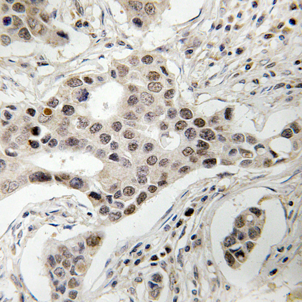
(Immunohistochemical analysis of NCOA2 (phospho-Ser736) staining in human breast carcinoma formalin fixed paraffin embedded tissue section. The section was then incubated with the antibody at room temperature and detected using an HRP conjugated compact polymer system. DAB was used as the chromogen. The section was then counterstained with haematoxylin and mounted with DPX.)
IHC (Immunohiostchemistry)
(Immunohistochemical analysis of NCOA2 (phospho-Ser736) staining in human breast carcinoma formalin fixed paraffin embedded tissue section. The section was then incubated with the antibody at room temperature and detected using an HRP conjugated compact polymer system. DAB was used as the chromogen. The section was then counterstained with haematoxylin and mounted with DPX.)
NCOA2 (phospho-Ser736), Polyclonal Antibody (Cat# AAA310502)
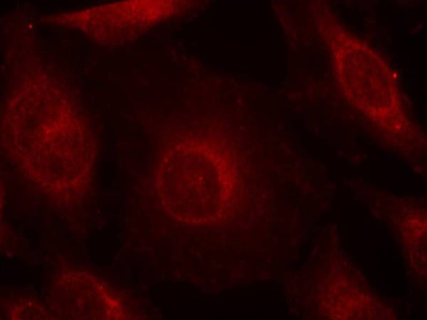
 IF (Immunofluorescence)
(Immunofluorescent analysis of Beta-NaCH (phospho-Thr615) staining in COS7 cells. Formalin-fixed cells were permeabilized with 0.1% Triton X-100 in TBS for 5-10 minutes and blocked with 3% BSA-PBS for 30 minutes at room temperature. Cells were probed with the primary antibody in 3% BSA-PBS and incubated overnight at 4 C in a hidified chamber. Cells were washed with PBST and incubated with Alexa Fluor 647-conjugated secondary antibody (red) in PBS at room temperature in the dark.)
IF (Immunofluorescence)
(Immunofluorescent analysis of Beta-NaCH (phospho-Thr615) staining in COS7 cells. Formalin-fixed cells were permeabilized with 0.1% Triton X-100 in TBS for 5-10 minutes and blocked with 3% BSA-PBS for 30 minutes at room temperature. Cells were probed with the primary antibody in 3% BSA-PBS and incubated overnight at 4 C in a hidified chamber. Cells were washed with PBST and incubated with Alexa Fluor 647-conjugated secondary antibody (red) in PBS at room temperature in the dark.)
Beta-NaCH (phospho-Thr615), Polyclonal Antibody (Cat# AAA310504)
 IHC (Immunohiostchemistry)
(Immunohistochemical analysis of c-SRC/FYN/c-YES (phospho-Tyr419/420/426) staining in human colon carcinoma formalin fixed paraffin embedded tissue section. The section was then incubated with the antibody at room temperature and detected using an HRP conjugated compact polymer system. DAB was used as the chromogen. The section was then counterstained with haematoxylin and mounted with DPX.)
IHC (Immunohiostchemistry)
(Immunohistochemical analysis of c-SRC/FYN/c-YES (phospho-Tyr419/420/426) staining in human colon carcinoma formalin fixed paraffin embedded tissue section. The section was then incubated with the antibody at room temperature and detected using an HRP conjugated compact polymer system. DAB was used as the chromogen. The section was then counterstained with haematoxylin and mounted with DPX.)
c-SRC/FYN/c-YES (phospho-Tyr419/420/426), Polyclonal Antibody (Cat# AAA310505)
 IF (Immunofluorescence)
(Immunofluorescent analysis of Insulin Receptor (phospho-Tyr1361) staining in HEK293T cells. Formalin-fixed cells were permeabilized with 0.1% Triton X-100 in TBS for 5-10 minutes and blocked with 3% BSA-PBS for 30 minutes at room temperature. Cells were probed with the primary antibody in 3% BSA-PBS and incubated overnight at 4 C in a hidified chamber. Cells were washed with PBST and incubated with a DyLight 594-conjugated secondary antibody (red) in PBS at room temperature in the dark. DAPI was used to stain the cell nuclei (blue).)
IF (Immunofluorescence)
(Immunofluorescent analysis of Insulin Receptor (phospho-Tyr1361) staining in HEK293T cells. Formalin-fixed cells were permeabilized with 0.1% Triton X-100 in TBS for 5-10 minutes and blocked with 3% BSA-PBS for 30 minutes at room temperature. Cells were probed with the primary antibody in 3% BSA-PBS and incubated overnight at 4 C in a hidified chamber. Cells were washed with PBST and incubated with a DyLight 594-conjugated secondary antibody (red) in PBS at room temperature in the dark. DAPI was used to stain the cell nuclei (blue).)
Insulin Receptor (phospho-Tyr1361), Polyclonal Antibody (Cat# AAA310510)
 IHC (Immunohistochemisry)
(Immunohistochemical analysis of paraffin-embedded mouse colon tissue using anti-Phospho-Cdk2 (Y15) antibody. Counter stained with hematoxylin.)
IHC (Immunohistochemisry)
(Immunohistochemical analysis of paraffin-embedded mouse colon tissue using anti-Phospho-Cdk2 (Y15) antibody. Counter stained with hematoxylin.)
CDK2, Monoclonal Antibody (Cat# AAA311024)
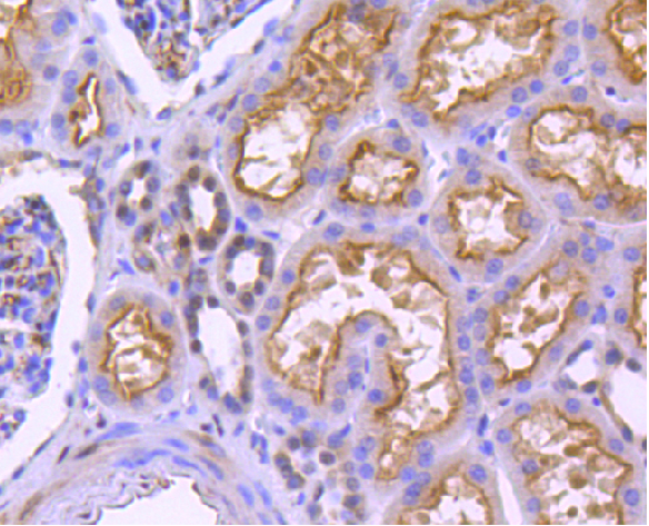
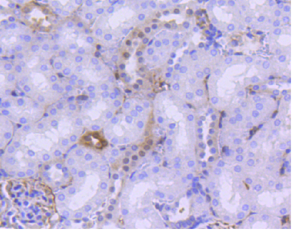 IHC (Immunohistochemisry)
(Immunohistochemical analysis of paraffin-embedded mouse kidney tissue using anti- phospho-YAP1 (S127) antibody. Counter stained with hematoxylin.)
IHC (Immunohistochemisry)
(Immunohistochemical analysis of paraffin-embedded mouse kidney tissue using anti- phospho-YAP1 (S127) antibody. Counter stained with hematoxylin.)
YAP1, Monoclonal Antibody (Cat# AAA311026)
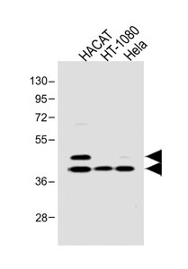
 WB (Western Blot)
(Western Blot analysis of 1 Hela, 2 treated with LPS 100ng/mL 20mim,using primary antibody at 1:1000 dilution.)
WB (Western Blot)
(Western Blot analysis of 1 Hela, 2 treated with LPS 100ng/mL 20mim,using primary antibody at 1:1000 dilution.)
TRIM28, Polyclonal Antibody (Cat# AAA311048)
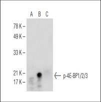
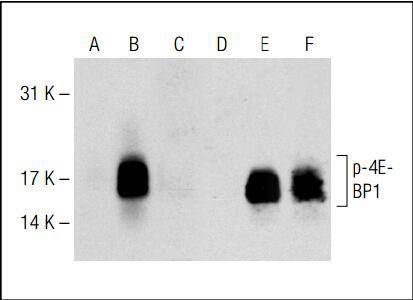 WB (Western Blot)
(Western blot analysis of 4E-BP1 phosphorylation in nontransfected (A, D), untreated human 4E-BP1 transfected (B, E) and lambda protein phosphatase treated human 4E-BP1 transfected (C, F) 293T whole cell lysates. Antibodies tested include p-4E-BP1/2/3 (A, B, C) and 4E-BP1 (D, E, F).)
WB (Western Blot)
(Western blot analysis of 4E-BP1 phosphorylation in nontransfected (A, D), untreated human 4E-BP1 transfected (B, E) and lambda protein phosphatase treated human 4E-BP1 transfected (C, F) 293T whole cell lysates. Antibodies tested include p-4E-BP1/2/3 (A, B, C) and 4E-BP1 (D, E, F).)
4E-BP1/2/3, Monoclonal Antibody (Cat# AAA310988)
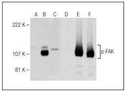
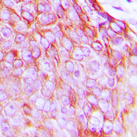
 Application Data
(B.Immunoperoxidase staining of formalin fixed, paraffin-embedded human adrenal gland tissue showing cytoplasmic staining of glandular cells.)
Application Data
(B.Immunoperoxidase staining of formalin fixed, paraffin-embedded human adrenal gland tissue showing cytoplasmic staining of glandular cells.)
FAK, Monoclonal Antibody (Cat# AAA310990)
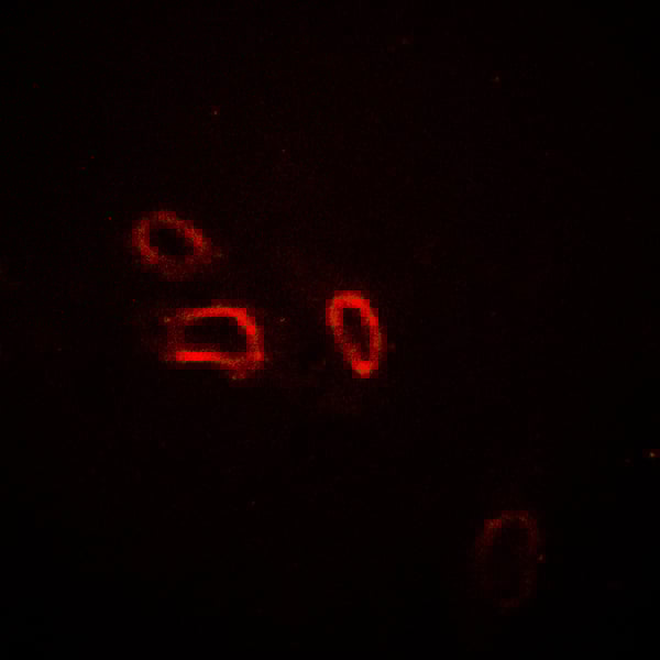
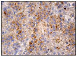
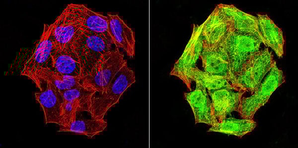 ICC (Immunocytochemistry)
(ICC staining phospho-NLRC4 (Ser-533) (green) and Actin filaments (red) in Hela cells. The nuclear counter stain is DAPI (blue). Cells were fixed in paraformaldehyde, permeabilised with 0.25% Triton X100/PBS.)
ICC (Immunocytochemistry)
(ICC staining phospho-NLRC4 (Ser-533) (green) and Actin filaments (red) in Hela cells. The nuclear counter stain is DAPI (blue). Cells were fixed in paraformaldehyde, permeabilised with 0.25% Triton X100/PBS.)
NLRC4, Monoclonal Antibody (Cat# AAA310993)
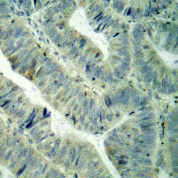
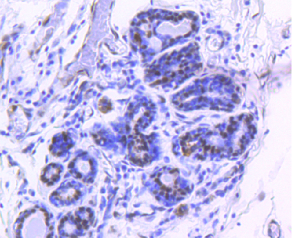
 IHC (Immunohiostchemistry)
(Immunohistochemical analysis of paraffin-embedded human breast carcinoma tissue using anti-Phospho-GATA3 (S308) antibody. Counter stained with hematoxylin.)
IHC (Immunohiostchemistry)
(Immunohistochemical analysis of paraffin-embedded human breast carcinoma tissue using anti-Phospho-GATA3 (S308) antibody. Counter stained with hematoxylin.)
GATA3, Monoclonal Antibody (Cat# AAA311004)
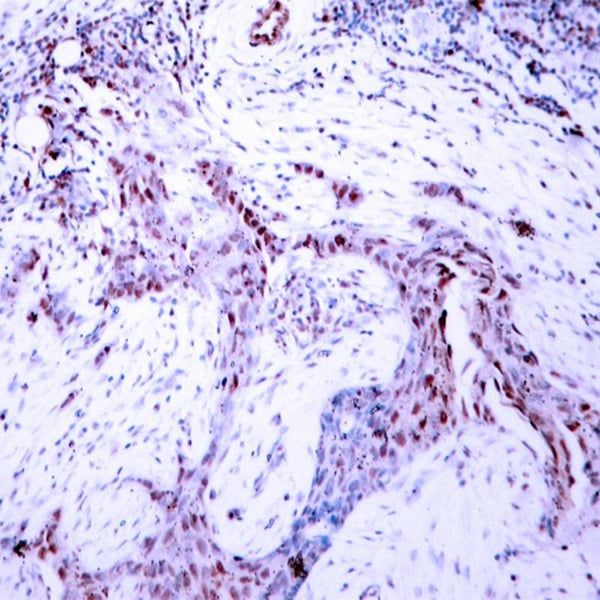
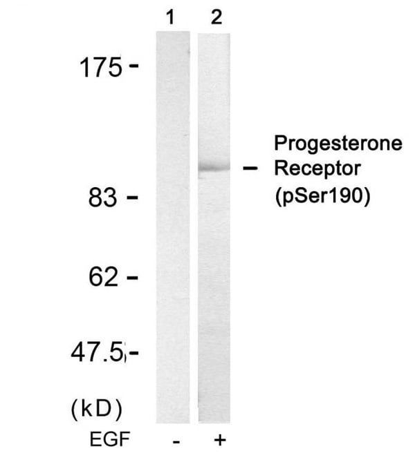
 IF (Immunofluorescence)
(Immunofluorescence staining of methanol-fixed MCF cells using Progesterone Receptor(Phospho-Ser190) Antibody.)
IF (Immunofluorescence)
(Immunofluorescence staining of methanol-fixed MCF cells using Progesterone Receptor(Phospho-Ser190) Antibody.)
Progesterone Receptor, Polyclonal Antibody (Cat# AAA307083)
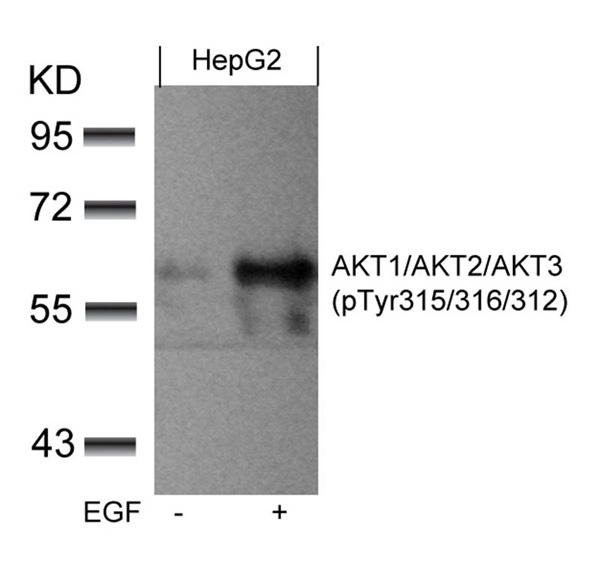
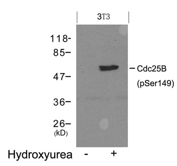
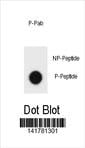
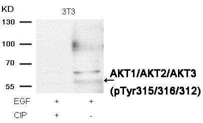 WB (Western Blot)
(Western blot analysis of extracts from 3T3 cells, treated with EGF or calf intestinal phosphatase (CIP), using AKT1/AKT2/AKT3 (phospho-Tyr315/316/312) Antibody.)
WB (Western Blot)
(Western blot analysis of extracts from 3T3 cells, treated with EGF or calf intestinal phosphatase (CIP), using AKT1/AKT2/AKT3 (phospho-Tyr315/316/312) Antibody.)
AKT1/AKT2/AKT3, Polyclonal Antibody (Cat# AAA307089)

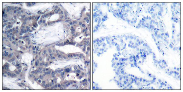
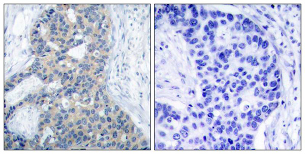 IHC (Immunohiostchemistry)
(Immunohistochemical analysis of paraffin-embedded human breast carcinoma tissue using Stathmin 1(Phospho-Ser38) Antibody (left) or the same antibody preincubated with blocking peptide(right).)
IHC (Immunohiostchemistry)
(Immunohistochemical analysis of paraffin-embedded human breast carcinoma tissue using Stathmin 1(Phospho-Ser38) Antibody (left) or the same antibody preincubated with blocking peptide(right).)
Stathmin 1, Polyclonal Antibody (Cat# AAA307105)
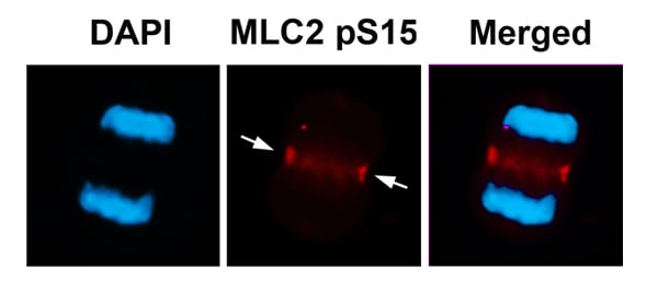
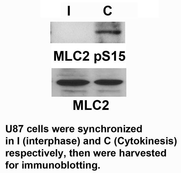 Application Data
MLC2, Polyclonal Antibody (Cat# AAA307119)
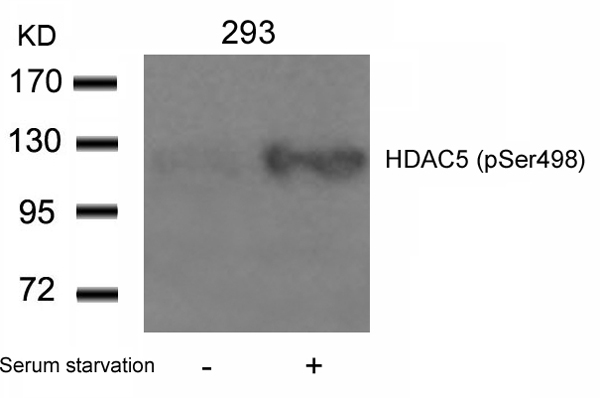
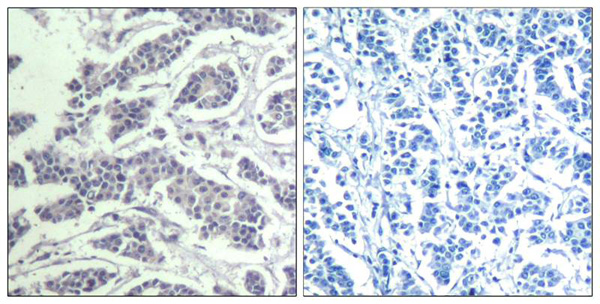
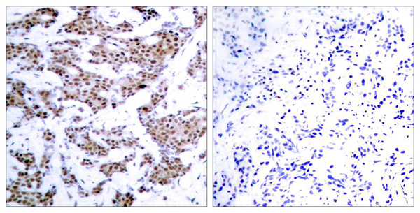
 IHC (Immunohiostchemistry)
(Immunohistochemical analysis of paraffin-embedded human breast carcinoma tissue using HDAC5(Phospho-Ser498) Antibody (left) or the same antibody preincubated with blocking peptide(right).)
IHC (Immunohiostchemistry)
(Immunohistochemical analysis of paraffin-embedded human breast carcinoma tissue using HDAC5(Phospho-Ser498) Antibody (left) or the same antibody preincubated with blocking peptide(right).)
HDAC5, Polyclonal Antibody (Cat# AAA307132)
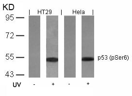
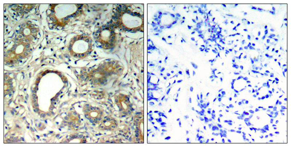
 IHC (Immunohiostchemistry)
(Immunohistochemical analysis of paraffin-embedded human breast carcinoma tissue using p53(Phospho-Ser6) Antibody (left) or the same antibody preincubated with blocking peptide(right).)
IHC (Immunohiostchemistry)
(Immunohistochemical analysis of paraffin-embedded human breast carcinoma tissue using p53(Phospho-Ser6) Antibody (left) or the same antibody preincubated with blocking peptide(right).)
p53, Polyclonal Antibody (Cat# AAA307535)

 IHC (Immunohiostchemistry)
(Immunohistochemical analysis of paraffin-embedded human breast carcinoma tissue using Smad3(Phospho-Ser425) Antibody (left) or the same antibody preincubated with blocking peptide(right).)
IHC (Immunohiostchemistry)
(Immunohistochemical analysis of paraffin-embedded human breast carcinoma tissue using Smad3(Phospho-Ser425) Antibody (left) or the same antibody preincubated with blocking peptide(right).)
Smad3, Polyclonal Antibody (Cat# AAA307666)
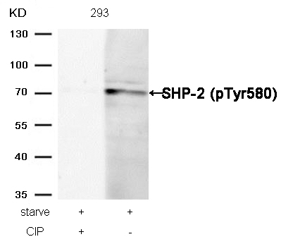 WB (Western Blot)
(Western blot analysis of extracts from 293 cells, treated with starve or calf intestinal phosphatase (CIP), using SHP-2 (Phospho-Tyr580) Antibody.)
WB (Western Blot)
(Western blot analysis of extracts from 293 cells, treated with starve or calf intestinal phosphatase (CIP), using SHP-2 (Phospho-Tyr580) Antibody.)
SHP-2, Polyclonal Antibody (Cat# AAA307487)

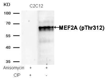 WB (Western Blot)
(Western blot analysis of extracts from C2C12 cells, treated with Anisomycin or calf intestinal phosphatase (CIP), using MEF2A (Phospho-Thr312) Antibody.)
WB (Western Blot)
(Western blot analysis of extracts from C2C12 cells, treated with Anisomycin or calf intestinal phosphatase (CIP), using MEF2A (Phospho-Thr312) Antibody.)
MEF2A, Polyclonal Antibody (Cat# AAA307490)
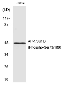
 WB (Western Blot)
(Western Blot analysis of VEC A549 cells using Phospho-AP-1/Jun D (S73/100) Polyclonal Antibody)
WB (Western Blot)
(Western Blot analysis of VEC A549 cells using Phospho-AP-1/Jun D (S73/100) Polyclonal Antibody)
AP-1/Jun D, Polyclonal Antibody (Cat# AAA307706)
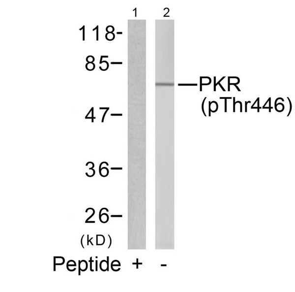
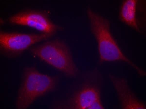 IF (Immunofluorescence)
(Immunofluorescence staining of methanol-fixed Hela cells using PKR(Phospho-Thr446) Antibody.)
IF (Immunofluorescence)
(Immunofluorescence staining of methanol-fixed Hela cells using PKR(Phospho-Thr446) Antibody.)
PKR, Polyclonal Antibody (Cat# AAA307253)
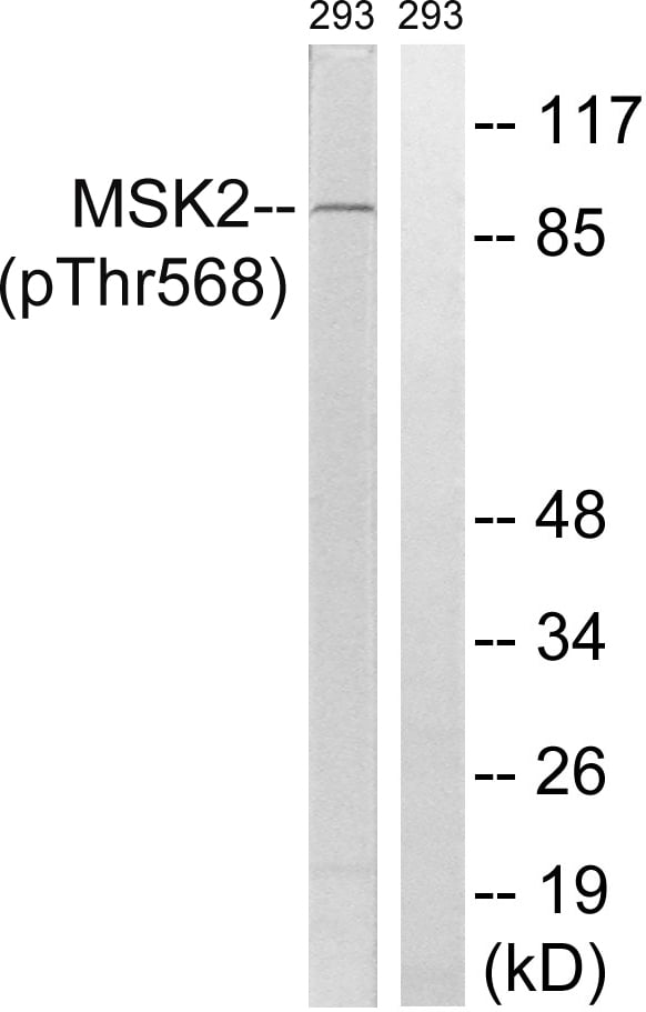
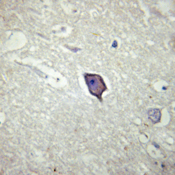
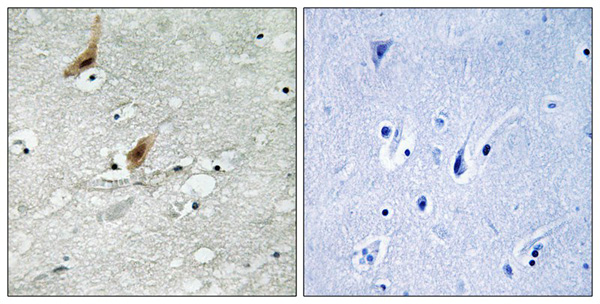 IHC (Immunohiostchemistry)
(Immunohistochemistry analysis of paraffin-embedded human brain tissue using MSK2 (Phospho-Thr568) antibody. The picture on the right is treated with the synthesized peptide.)
IHC (Immunohiostchemistry)
(Immunohistochemistry analysis of paraffin-embedded human brain tissue using MSK2 (Phospho-Thr568) antibody. The picture on the right is treated with the synthesized peptide.)
MSK2, Polyclonal Antibody (Cat# AAA307406)
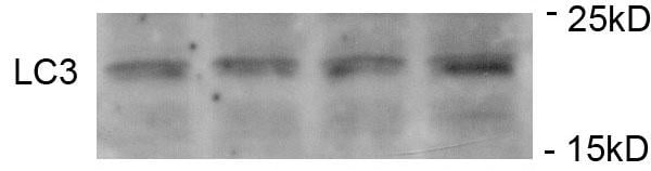
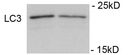
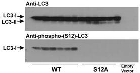
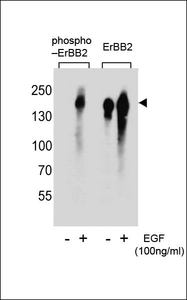
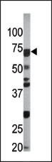
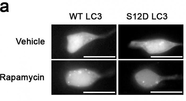 WB (Western Blot)
(Something like SH-SY5Y cells expressing GFP-LC3-WT or-S12D treated with rapamycin or vehicle for 1h.)
WB (Western Blot)
(Something like SH-SY5Y cells expressing GFP-LC3-WT or-S12D treated with rapamycin or vehicle for 1h.)
Phospho-LC3C (S12), Polyclonal Antibody (Cat# AAA285518)
Predicted Reactivity: Zebrafish, Bovine, Mouse, Rat
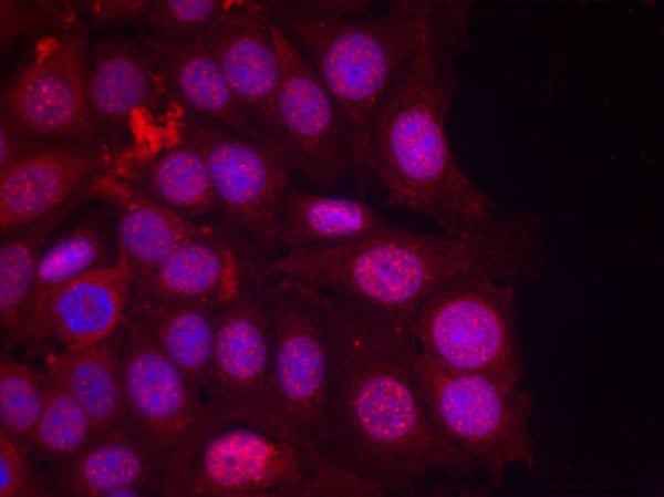
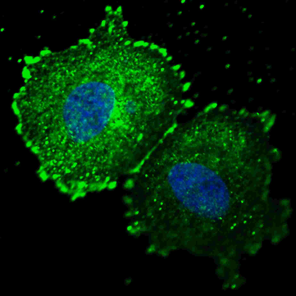
 IF (Immunofluorescence)
(Confocal immunofluorescent analysis of Phospho-HER4-Y1162 Antibody with MCF-7 cell followed by Alexa Fluor 488-conjugated goat anti-rabbit lgG (green). Actin filaments have been labeled with Alexa Fluor 555 phalloidin (red).DAPI was used to stain the cell nuclear (blue).)
IF (Immunofluorescence)
(Confocal immunofluorescent analysis of Phospho-HER4-Y1162 Antibody with MCF-7 cell followed by Alexa Fluor 488-conjugated goat anti-rabbit lgG (green). Actin filaments have been labeled with Alexa Fluor 555 phalloidin (red).DAPI was used to stain the cell nuclear (blue).)
Phospho-HER4 (Y1162), Polyclonal Antibody (Cat# AAA284789)
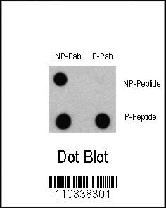
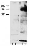
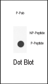
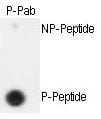
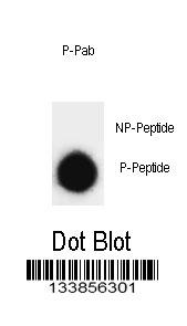
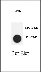
 DB (Dot Blot)
(Dot blot analysis of ERBB2 Antibody (Phospho Y1005) Phospho-specific Pab on nitrocellulose membrane. 50ng of Phospho-peptide or Non Phospho-peptide per dot were adsorbed. Antibody working concentrations are 0.6ug per ml.)
DB (Dot Blot)
(Dot blot analysis of ERBB2 Antibody (Phospho Y1005) Phospho-specific Pab on nitrocellulose membrane. 50ng of Phospho-peptide or Non Phospho-peptide per dot were adsorbed. Antibody working concentrations are 0.6ug per ml.)
Phospho-ERBB2 (Y1005), Polyclonal Antibody (Cat# AAA284919)
 DB (Dot Blot)
(Dot blot analysis of anti-Phospho-IL6ST-Y905 Phospho-specific Pab (AAA283923) on nitrocellulose membrane. 50ng of Phospho-peptide or Non Phospho-peptide per dot were adsorbed. Antibody working concentrations are 0.5ug per ml.)
DB (Dot Blot)
(Dot blot analysis of anti-Phospho-IL6ST-Y905 Phospho-specific Pab (AAA283923) on nitrocellulose membrane. 50ng of Phospho-peptide or Non Phospho-peptide per dot were adsorbed. Antibody working concentrations are 0.5ug per ml.)
Phospho-IL6ST (Y905), Polyclonal Antibody (Cat# AAA283923)
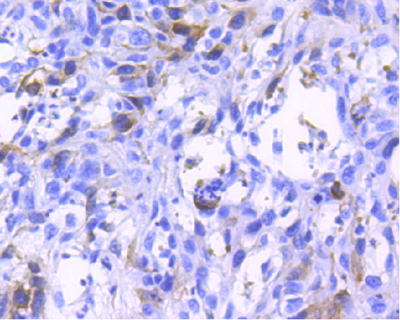
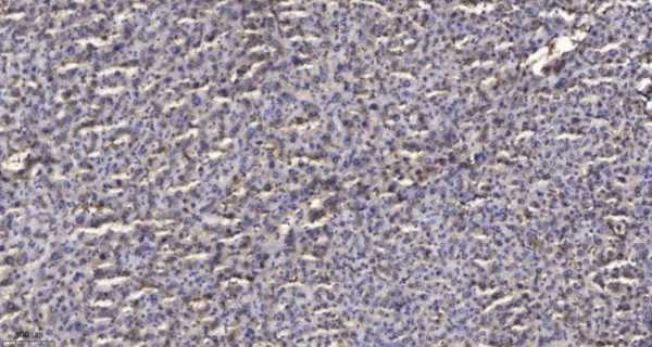
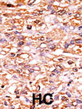
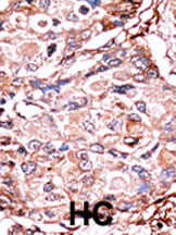
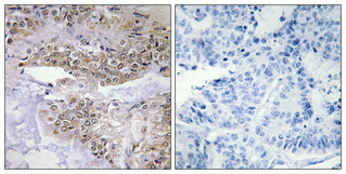
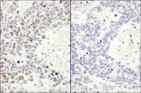
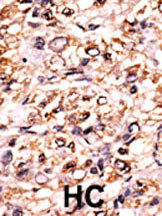 IHC (Immunohiostchemistry)
(Formalin-fixed and paraffin-embedded human cancer tissue reacted with the primary antibody, which was peroxidase-conjugated to the secondary antibody, followed by DAB staining. This data demonstrates the use of this antibody for immunohistochemistry; clinical relevance has not been evaluated. BC = breast carcinoma; HC = hepatocarcinoma.)
IHC (Immunohiostchemistry)
(Formalin-fixed and paraffin-embedded human cancer tissue reacted with the primary antibody, which was peroxidase-conjugated to the secondary antibody, followed by DAB staining. This data demonstrates the use of this antibody for immunohistochemistry; clinical relevance has not been evaluated. BC = breast carcinoma; HC = hepatocarcinoma.)
Phospho-SEPARIN (S1126), Polyclonal Antibody (Cat# AAA283977)
 DB (Dot Blot)
(Dot blot analysis of anti-Phospho-ErbB2-pY1248(M) Phospho-specific Pab on nitrocellulose membrane. 50ng of Phospho-peptide or Non Phospho-peptide per dot were adsorbed. Antibody working concentrations are 0.5ug per ml.)
DB (Dot Blot)
(Dot blot analysis of anti-Phospho-ErbB2-pY1248(M) Phospho-specific Pab on nitrocellulose membrane. 50ng of Phospho-peptide or Non Phospho-peptide per dot were adsorbed. Antibody working concentrations are 0.5ug per ml.)
Phospho-ErbB2 (Y1248), Polyclonal Antibody (Cat# AAA288096)
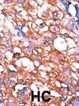
 IHC (Immunohiostchemistry)
(Formalin-fixed and paraffin-embedded human cancer tissue reacted with the primary antibody, which was peroxidase-conjugated to the secondary antibody, followed by AEC staining. This data demonstrates the use of this antibody for immunohistochemistry; clinical relevance has not been evaluated. BC = breast carcinoma; HC = hepatocarcinoma.)
IHC (Immunohiostchemistry)
(Formalin-fixed and paraffin-embedded human cancer tissue reacted with the primary antibody, which was peroxidase-conjugated to the secondary antibody, followed by AEC staining. This data demonstrates the use of this antibody for immunohistochemistry; clinical relevance has not been evaluated. BC = breast carcinoma; HC = hepatocarcinoma.)
Phospho-CDC6 (S54), Polyclonal Antibody (Cat# AAA287691)
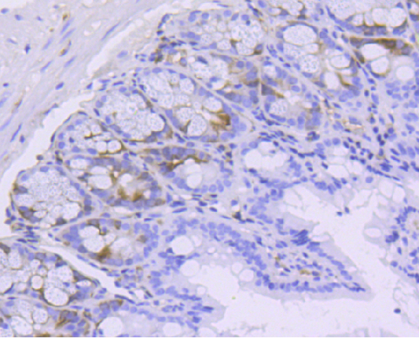
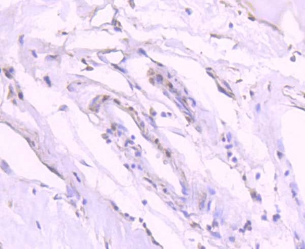
 IHC (Immunohiostchemistry)
(Formalin-fixed and paraffin-embedded human cancer tissue reacted with the primary antibody, which was peroxidase-conjugated to the secondary antibody, followed by AEC staining. This data demonstrates the use of this antibody for immunohistochemistry; clinical relevance has not been evaluated. BC = breast carcinoma; HC = hepatocarcinoma.)
IHC (Immunohiostchemistry)
(Formalin-fixed and paraffin-embedded human cancer tissue reacted with the primary antibody, which was peroxidase-conjugated to the secondary antibody, followed by AEC staining. This data demonstrates the use of this antibody for immunohistochemistry; clinical relevance has not been evaluated. BC = breast carcinoma; HC = hepatocarcinoma.)
Phospho-p27Kip1 (S178), Polyclonal Antibody (Cat# AAA289387)
 DB (Dot Blot)
(Dot blot analysis of Phospho-SNAP25-pT138 Antibody Phospho-specific Pab on nitrocellulose membrane. 50ng of Phospho-peptide or Non Phospho-peptide per dot were adsorbed. Antibody working concentrations are 0.6ug per ml.)
DB (Dot Blot)
(Dot blot analysis of Phospho-SNAP25-pT138 Antibody Phospho-specific Pab on nitrocellulose membrane. 50ng of Phospho-peptide or Non Phospho-peptide per dot were adsorbed. Antibody working concentrations are 0.6ug per ml.)
Phospho-SNAP25 (T138), Polyclonal Antibody (Cat# AAA289167)
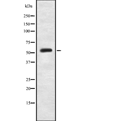
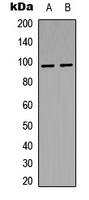
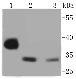
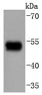
 WB (Western Blot)
(The anti-Phospho-SMAD3-S213 Pab is used in Western blot to detect Phospho-SMAD3-S213 in Ramos tissue lysate)
WB (Western Blot)
(The anti-Phospho-SMAD3-S213 Pab is used in Western blot to detect Phospho-SMAD3-S213 in Ramos tissue lysate)
Phospho-SMAD3 (S213), Polyclonal Antibody (Cat# AAA285661)
 DB (Dot Blot)
(Dot blot analysis of anti-Phospho-SMAD2-S118 Antibody on nitrocellulose membrane. 50ng of Phospho-peptide or Non Phospho-peptide per dot were adsorbed. Antibody working concentrations are 0.5ug per ml.)
DB (Dot Blot)
(Dot blot analysis of anti-Phospho-SMAD2-S118 Antibody on nitrocellulose membrane. 50ng of Phospho-peptide or Non Phospho-peptide per dot were adsorbed. Antibody working concentrations are 0.5ug per ml.)
Phospho-SMAD2 (S118), Polyclonal Antibody (Cat# AAA288611)
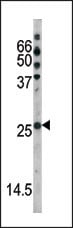
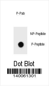 DB (Dot Blot)
(Dot blot analysis of Phospho-ERBB2-T1172 Antibody Phospho-specific Pab on nitrocellulose membrane. 50ng of Phospho-peptide or Non Phospho-peptide per dot were adsorbed. Antibody working concentrations are 0.6ug per ml.)
DB (Dot Blot)
(Dot blot analysis of Phospho-ERBB2-T1172 Antibody Phospho-specific Pab on nitrocellulose membrane. 50ng of Phospho-peptide or Non Phospho-peptide per dot were adsorbed. Antibody working concentrations are 0.6ug per ml.)
Phospho-ERBB2 (T1172), Polyclonal Antibody (Cat# AAA289958)
 DB (Dot Blot)
(Dot blot analysis of anti-raptor-pS863 Pab (RB13350) on nitrocellulose membrane. 50ng of Phospho-peptide or Non Phospho-peptide per dot were adsorbed. Antibody working concentrations are 0.5ug per ml.)
DB (Dot Blot)
(Dot blot analysis of anti-raptor-pS863 Pab (RB13350) on nitrocellulose membrane. 50ng of Phospho-peptide or Non Phospho-peptide per dot were adsorbed. Antibody working concentrations are 0.5ug per ml.)
Phospho-Raptor (S863), Polyclonal Antibody (Cat# AAA289679)

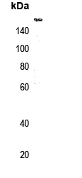
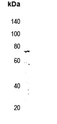
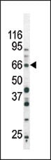
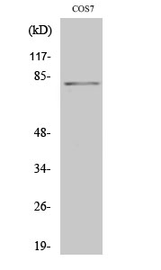 WB (Western Blot)
(Dilution: Western Blot: 1/500 - 1/2000. Immunohistochemistry: 1/100 - 1/300. Immunoprecipitation: 2-5 ug/mg lysate. ELISA: 1/5000. Not yet tested in other applications.)
WB (Western Blot)
(Dilution: Western Blot: 1/500 - 1/2000. Immunohistochemistry: 1/100 - 1/300. Immunoprecipitation: 2-5 ug/mg lysate. ELISA: 1/5000. Not yet tested in other applications.)
Stat4, Polyclonal Antibody (Cat# AAA293935)

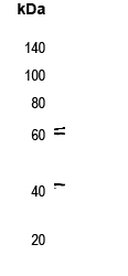
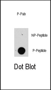
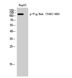 WB (Western Blot)
(Dilution: Western Blot: 1/500 - 1/2000. ELISA: 1/10000. Not yet tested in other applications.)
WB (Western Blot)
(Dilution: Western Blot: 1/500 - 1/2000. ELISA: 1/10000. Not yet tested in other applications.)
Flg/Bek, Polyclonal Antibody (Cat# AAA293984)
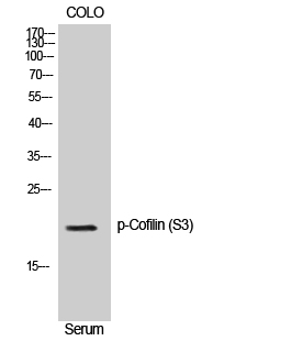
 IHC (Immunohiostchemistry)
(Dilution: Western Blot: 1/500 - 1/2000. Immunohistochemistry: 1/100 - 1/300. ELISA: 1/10000. Not yet tested in other applications.)
IHC (Immunohiostchemistry)
(Dilution: Western Blot: 1/500 - 1/2000. Immunohistochemistry: 1/100 - 1/300. ELISA: 1/10000. Not yet tested in other applications.)
Cofilin, Polyclonal Antibody (Cat# AAA293878)
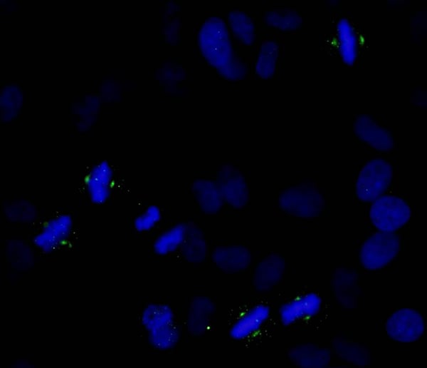
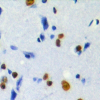
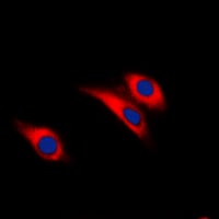
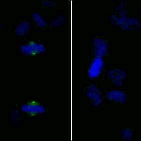 ICC (Immunocytochemistry)
(Detection of human Phospho-Aurora A (T288) by immunocytochemistry. Samples: NBF-fixed asynchronous HeLa cells. Mock phosphatase treated section (left) or calf intestinal phosphatase-treated section (right) immunostained for Phospho-Aurora A. Antibody: Affinity purified rabbit anti-Phospho-Aurora A (T288) (Cat. No. AAA213768) used at a dilution of 1:250. Detection: Anti-rabbit IgG-FITC conjugated (Cat. No. used at a dilution of 1:100.)
ICC (Immunocytochemistry)
(Detection of human Phospho-Aurora A (T288) by immunocytochemistry. Samples: NBF-fixed asynchronous HeLa cells. Mock phosphatase treated section (left) or calf intestinal phosphatase-treated section (right) immunostained for Phospho-Aurora A. Antibody: Affinity purified rabbit anti-Phospho-Aurora A (T288) (Cat. No. AAA213768) used at a dilution of 1:250. Detection: Anti-rabbit IgG-FITC conjugated (Cat. No. used at a dilution of 1:100.)
Aurora A, Polyclonal Antibody (Cat# AAA213768)
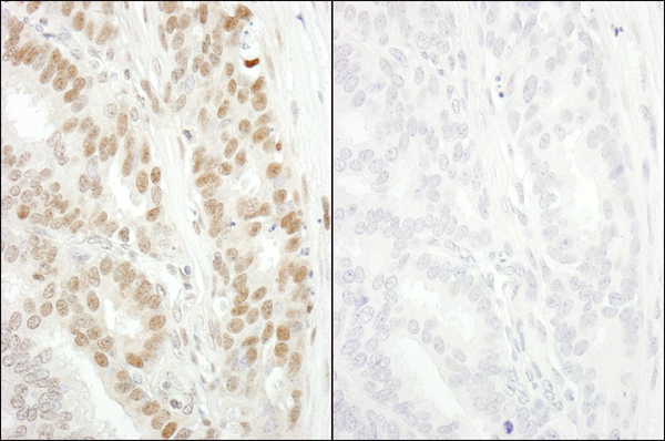
 IHC (Immunohiostchemistry)
(Detection of mouse Phospho-MCM2 (S53) by immunohistochemistry. Samples: FFPE serial sections of mouse teratoma. Mock phosphatase treated section (left) or calf intestinal phosphatase treated section (right) immunostained for Phospho-MCM2 (S53). Antibody: Affinity purified rabbit anti-Phospho-MCM2 (S53) (Cat. No. AAA213769 Lot3) used at a dilution of 1:250. Detection: DAB)
IHC (Immunohiostchemistry)
(Detection of mouse Phospho-MCM2 (S53) by immunohistochemistry. Samples: FFPE serial sections of mouse teratoma. Mock phosphatase treated section (left) or calf intestinal phosphatase treated section (right) immunostained for Phospho-MCM2 (S53). Antibody: Affinity purified rabbit anti-Phospho-MCM2 (S53) (Cat. No. AAA213769 Lot3) used at a dilution of 1:250. Detection: DAB)
MCM2, Polyclonal Antibody (Cat# AAA213769)
What Are Phospho Antibodies?
Protein phosphorylation is a process where a phosphate group is added to certain amino acid residues of a protein – usually serine (S), threonine (T), or tyrosine (Y) - by enzymes called kinases. This process is integral in controlling cellular signaling, cellular growth, and other biological functions.
Our catalog includes a wide range of phospho-specific antibodies that can accurately detect this important marker. They perform strongly in widely-used laboratory applications such as Western blot, flow cytometry, immunohistochemistry, and immunofluorescence microscopy. We value your trust in us and are committed to providing top-quality products and services. All of our antibodies are guaranteed to work for the applications and species indicated on our website & associated product pages.
What Are The Key Applications of Phospho Antibodies?
1. Western Blotting
One of the first steps a researcher can take in utilizing these phospho-specific antibodies, is to check if the antibody works using a technique referred to as “Western blot”. For those unfamiliar, Western Blot aids in showing whether the protein that the antibody recognizes is appearing at the correct/expected size. These phospho-specific antibodies should also be able to detect changes in the target protein’s phosphorylation (on/off state) when cells are stimulated in certain ways.
2. Staining of Fixed Cells (Immunocytochemistry)
Another routine use of these phospho-specific antibodies, is to test if the antibody is able to demonstrate similar performance when used on fixed cells (intact cells that have been preserved) as it did in the Western blot tests. It is an important aspect in many cases to confirm that the antibody works in actual intact cell samples. Ideally, the method used for cellular fixation should be the same as what is used in pathology labs (like using 10% formalin). To check if the antibody works well in tissue sections (FFPE), researchers will often test it on fixed cells that are processed similar to tissue samples.
3. Specificity Tests Using Peptides
In order to make sure that the antibody is only binding to the right target:
- Laboratory technicians will mix the antibody with phospho-peptides (short segments of the protein containing the phosphate group modification).
- If the antibody signal disappears, it is confirmation that it is binding to the correct phosphorylated location.
- A more robust test is to use both the phosphorylated and non-phosphorylated (dephosphorylated) versions of the protein. The antibody should react only with the phosphorylated one.
- Another method sometimes utilized is to treat the sample with an enzyme, such as alkaline phosphatase, that specifically removes phosphate groups. If the antibody signal disappears after this, it also confirms specificity.
4. Genetic Confirmation
As a final step, scientists can genetically manipulate the nucleotide sequence and alter the target protein by removing the exact site where phosphorylation happens. If the antibody no longer appears to detect the modified protein, it is strong evidence supporting the antibody being specific for that phosphorylated site.
Why Buy Phospho Antibodies Through Us?
- The production laboratory adheres to strict and consistent protocols prior to releasing any of these phospho-specific antibodies:
- Standard methods and proper controls in all tests to ensure high quality.
- These antibodies are tested and validated in different cell types and species.
- High quality control criterion to ensure each batch is consistent, so you will obtain reliable results every time.
FAQ
1. What Are Phospho-Specific Antibodies?
Phospho-specific antibodies are made to detect proteins only when they have a phosphate group linked to a specific amino acid residue. This empowers scientists understand if a protein is "turned on" or active, based on its phosphorylation state.
2. How to Detect Phosphorylated Proteins in a Western Blot?
To find out if a protein is phosphorylated using Western blot:
- Use a phospho-specific antibody that binds only to the phosphorylated form of the protein.
- You can also use a “regular” antibody for the same amino acid sequence of the protein that the phospho-specific antibody is binding to (but in this case, this antibody will not bind if there is a phosphate group present) in order to compare how much of it is phosphorylated versus how much is non-phosphorylated (or “total” protein, if the “normal” antibody’s epitopes are non-phospho-site-specific).
3. How to Choose the Best Antibody?
Here are some simple tips to help you pick the right antibody:
- Know your target
- Match your sample characteristics
- Confirm the intended use is appropriate
- Check “host” and “type”
- Check the “quality” of the presented data/images
- Appraise whether the available validation meets your needs