Filters
▼Clonality
▼Type
▼Reactivity
▼Gene Name
▼Isotype
▼Host
▼Application
▼Clone
▼Phospho Antibodies
Phospho-specific antibodies’ typical purpose is to enable researchers to detect changes in proteins. They will exclusively bind to the amino acid sequence on a protein that has been phosphorylated (which is both a physical & chemical change) and do not bind to the same amino acid sequence on said protein if it lacks said phosphorylation. This aids in being able to clearly see and understand the data produced from this particular protein modification.
Viewing 2150-2200 of 5298 product results
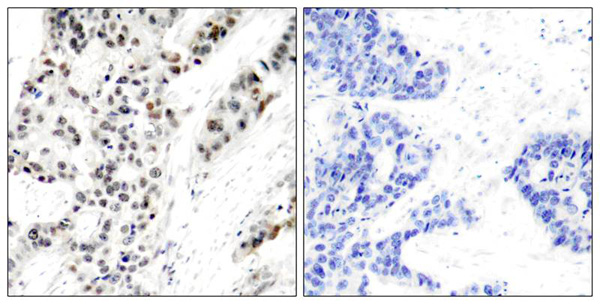
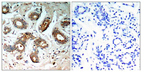
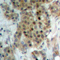
 Application Data
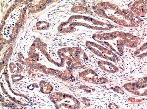
(Immunoperoxidase staining of formalin-fixed, paraffin-embedded human breast carcinoma tissue showing cytoplasmic staining.)
Application Data
(Immunoperoxidase staining of formalin-fixed, paraffin-embedded human breast carcinoma tissue showing cytoplasmic staining.)
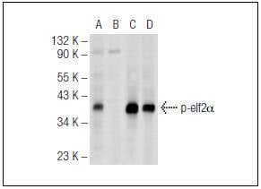
eIF-2a, Polyclonal Antibody (Cat# AAA310989)
 Application Data
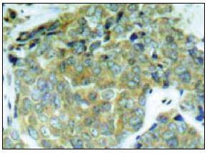
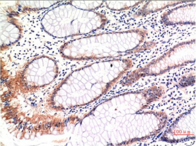
(Immunoperoxidase staining of formalin-fixed, paraffin-embedded human colon carcinoma tissue showing cytoplasmic staining.)
Application Data
(Immunoperoxidase staining of formalin-fixed, paraffin-embedded human colon carcinoma tissue showing cytoplasmic staining.)
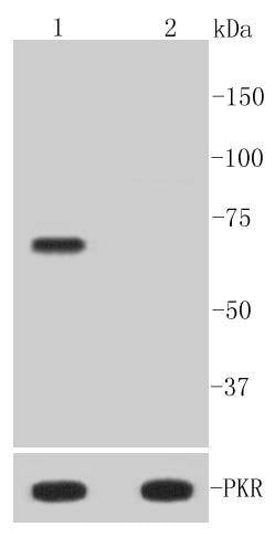
PKR, Polyclonal Antibody (Cat# AAA310992)
 IHC (Immunohiostchemistry)
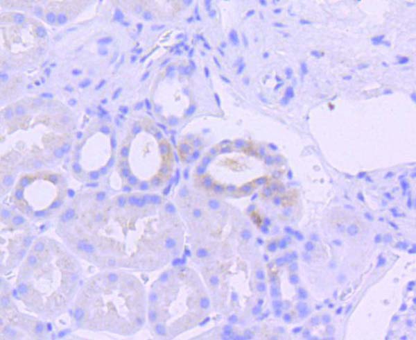
(Immunohistochemical analysis of paraffin-embedded human kidney tissue using anti-Phospho-PKR (T446) antibody. Counter stained with hematoxylin.)
IHC (Immunohiostchemistry)
(Immunohistochemical analysis of paraffin-embedded human kidney tissue using anti-Phospho-PKR (T446) antibody. Counter stained with hematoxylin.)
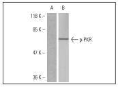
PKR, Monoclonal Antibody (Cat# AAA311000)
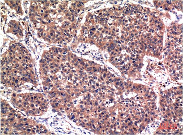
 IHC (Immunohiostchemistry)
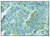
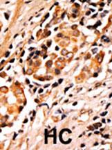
(Formalin-fixed and paraffin-embedded human cancer tissue reacted with the primary antibody, which was peroxidase-conjugated to the secondary antibody, followed by AEC staining. This data demonstrates the use of this antibody for immunohistochemistry; clinical relevance has not been evaluated. BC = breast carcinoma; HC = hepatocarcinoma.)
IHC (Immunohiostchemistry)
(Formalin-fixed and paraffin-embedded human cancer tissue reacted with the primary antibody, which was peroxidase-conjugated to the secondary antibody, followed by AEC staining. This data demonstrates the use of this antibody for immunohistochemistry; clinical relevance has not been evaluated. BC = breast carcinoma; HC = hepatocarcinoma.)
Phospho-Bad (S75), Polyclonal Antibody (Cat# AAA288792)
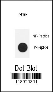 DB (Dot Blot)
(Dot blot analysis of anti-Phospho-Nanog-S71 Antibody on nitrocellulose membrane. 50ng of Phospho-peptide or Non Phospho-peptide per dot were adsorbed. Antibody working concentrations are 0.5ug per ml.)
DB (Dot Blot)
(Dot blot analysis of anti-Phospho-Nanog-S71 Antibody on nitrocellulose membrane. 50ng of Phospho-peptide or Non Phospho-peptide per dot were adsorbed. Antibody working concentrations are 0.5ug per ml.)
Phospho-Nanog (S71), Polyclonal Antibody (Cat# AAA288713)
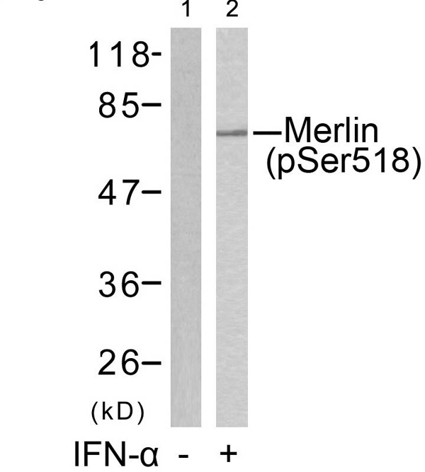
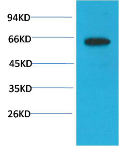
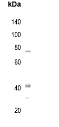
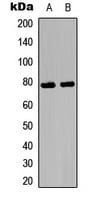
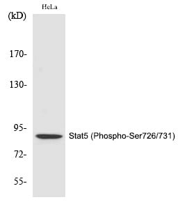 WB (Western Blot)
(Western Blot analysis of HeLa cells using Phospho-Stat5 (S726/731) Polyclonal Antibody)
WB (Western Blot)
(Western Blot analysis of HeLa cells using Phospho-Stat5 (S726/731) Polyclonal Antibody)
Stat5, Polyclonal Antibody (Cat# AAA306249)
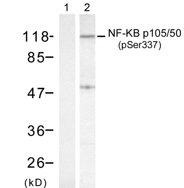


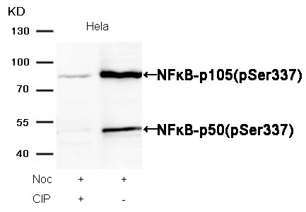 WB (Western Blot)
(Western blot analysis of extracts from Hela cells, treated with Noc or calf intestinal phosphatase (CIP), using NFkappaB-p105/p50(Phospho-Ser337) Antibody.)
WB (Western Blot)
(Western blot analysis of extracts from Hela cells, treated with Noc or calf intestinal phosphatase (CIP), using NFkappaB-p105/p50(Phospho-Ser337) Antibody.)
NFkappaB-p105/p50, Polyclonal Antibody (Cat# AAA306251)
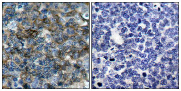 IHC (Immunohiostchemistry)
(Immunohistochemical analysis of paraffin-embedded human tonsil carcinoma tissue using VASP(Phospho-Ser157) Antibody (left) or the same antibody preincubated with blocking peptide(right).)
IHC (Immunohiostchemistry)
(Immunohistochemical analysis of paraffin-embedded human tonsil carcinoma tissue using VASP(Phospho-Ser157) Antibody (left) or the same antibody preincubated with blocking peptide(right).)
VASP, Polyclonal Antibody (Cat# AAA306280)

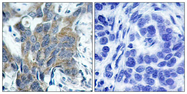
 IHC (Immunohiostchemistry)
(Immunohistochemical analysis of paraffin-embedded human breast carcinoma tissue using IkB-e(Phospho-Ser22) Antibody (left) or the same antibody preincubated with blocking peptide(right).)
IHC (Immunohiostchemistry)
(Immunohistochemical analysis of paraffin-embedded human breast carcinoma tissue using IkB-e(Phospho-Ser22) Antibody (left) or the same antibody preincubated with blocking peptide(right).)
IkB-epsilon, Polyclonal Antibody (Cat# AAA304773)
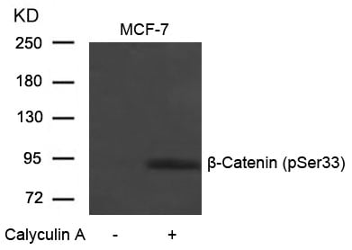
 IHC (Immunohiostchemistry)
(Immunohistochemical analysis of paraffin-embedded human breast carcinoma tissue using b-Catenin(Phospho-Ser33) Antibody (left) or the same antibody preincubated with blocking peptide(right).)
IHC (Immunohiostchemistry)
(Immunohistochemical analysis of paraffin-embedded human breast carcinoma tissue using b-Catenin(Phospho-Ser33) Antibody (left) or the same antibody preincubated with blocking peptide(right).)
beta-Catenin, Polyclonal Antibody (Cat# AAA305918)
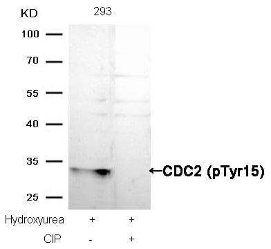
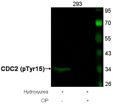 WB (Western Blot)
(Western blot analysis of extracts from 293 cells, treated with Hydroxyurea or calf intestinal phosphatase (CIP), using CDC2 (Phospho-Tyr15) Antibody.)
WB (Western Blot)
(Western blot analysis of extracts from 293 cells, treated with Hydroxyurea or calf intestinal phosphatase (CIP), using CDC2 (Phospho-Tyr15) Antibody.)
CDC2, Polyclonal Antibody (Cat# AAA305946)
 IF (Immunofluorescence)
(Immunofluorescence staining of methanol-fixed Hela cells using PAK1/PAK2/PAK3(Phospho-Thr423/Thr402/Thr421) Antibody.)
IF (Immunofluorescence)
(Immunofluorescence staining of methanol-fixed Hela cells using PAK1/PAK2/PAK3(Phospho-Thr423/Thr402/Thr421) Antibody.)
PAK1/PAK2/PAK3, Polyclonal Antibody (Cat# AAA306153)
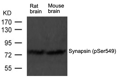
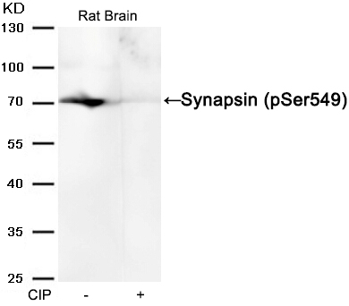 WB (Western Blot)
(Western blot analysis of extracts from Rat brain tissue or calf intestinal phosphatase (CIP), using Synapsin (phospho-Ser549) Antibody.)
WB (Western Blot)
(Western blot analysis of extracts from Rat brain tissue or calf intestinal phosphatase (CIP), using Synapsin (phospho-Ser549) Antibody.)
Synapsin, Polyclonal Antibody (Cat# AAA306032)
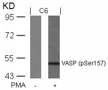
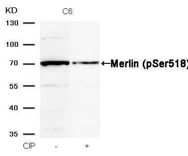 WB (Western Blot)
(Western blot analysis of extracts from C6 cells, treated with calf intestinal phosphatase (CIP), using Merlin (Phospho-Ser518) Antibody.)
WB (Western Blot)
(Western blot analysis of extracts from C6 cells, treated with calf intestinal phosphatase (CIP), using Merlin (Phospho-Ser518) Antibody.)
Merlin, Polyclonal Antibody (Cat# AAA306383)
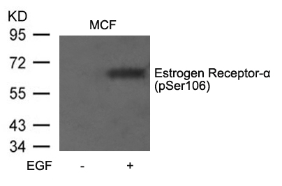
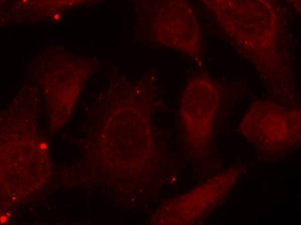
 IF (Immunofluorescence)
(Immunofluorescence staining of methanol-fixed MCF cells using Estrogen Receptor-a(Phospho-Ser106) Antibody.)
IF (Immunofluorescence)
(Immunofluorescence staining of methanol-fixed MCF cells using Estrogen Receptor-a(Phospho-Ser106) Antibody.)
Estrogen Receptor-alpha, Polyclonal Antibody (Cat# AAA305846)
 IHC (Immunohiostchemistry)
(Immunohistochemical analysis of paraffin-embedded Mouse Brain Tissue using CaMKIIb/ g /&d (Phospho Thr287) Mouse mAb diluted at 1:200.)
IHC (Immunohiostchemistry)
(Immunohistochemical analysis of paraffin-embedded Mouse Brain Tissue using CaMKIIb/ g /&d (Phospho Thr287) Mouse mAb diluted at 1:200.)
CaMKIIbeta/gamma/delta, Monoclonal Antibody (Cat# AAA309599)
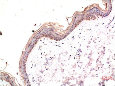 IHC (Immunohistochemistry)
(Immunohistochemical analysis of paraffin-embedded Human Skin Tissue using Phospho-MLKL S358 Mouse mAb diluted at 1:200.)
IHC (Immunohistochemistry)
(Immunohistochemical analysis of paraffin-embedded Human Skin Tissue using Phospho-MLKL S358 Mouse mAb diluted at 1:200.)
Akt, Monoclonal Antibody (Cat# AAA309601)
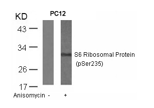
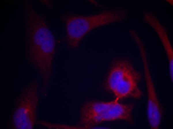 IF (Immunofluorescence)
(Immunofluorescence staining of methanol-fixed Hela cells using S6 Ribosomal Protein(Phospho-Ser235) Antibody.)
IF (Immunofluorescence)
(Immunofluorescence staining of methanol-fixed Hela cells using S6 Ribosomal Protein(Phospho-Ser235) Antibody.)
S6 Ribosomal Protein, Polyclonal Antibody (Cat# AAA307250)
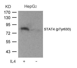
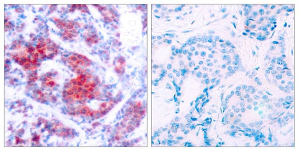 IHC (Immunohiostchemistry)
(Immunohistochemical analysis of paraffin-embedded human breast carcinoma tissue using STAT4(Phospho-Tyr693) Antibody (left) or the same antibody preincubated with blocking peptide(right).)
IHC (Immunohiostchemistry)
(Immunohistochemical analysis of paraffin-embedded human breast carcinoma tissue using STAT4(Phospho-Tyr693) Antibody (left) or the same antibody preincubated with blocking peptide(right).)
STAT4, Polyclonal Antibody (Cat# AAA307272)
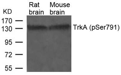
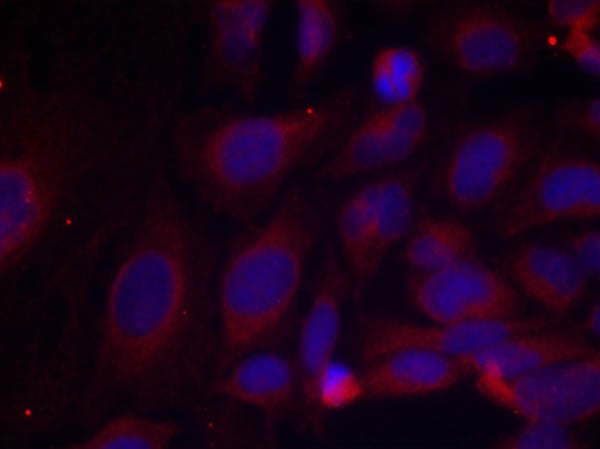
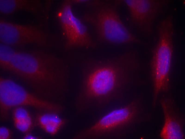 IF (Immunofluorescence)
(Immunofluorescence staining of methanol-fixed Hela cells using TrkA(Phospho-Ser791) Antibody.)
IF (Immunofluorescence)
(Immunofluorescence staining of methanol-fixed Hela cells using TrkA(Phospho-Ser791) Antibody.)
TrkA, Polyclonal Antibody (Cat# AAA307279)
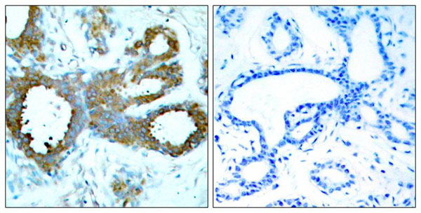
 IHC (Immunohiostchemistry)
(Immunohistochemical analysis of paraffin-embedded human breast carcinoma tissue using NF-kappaB p105 (phospho-Ser893) antibody ().)
IHC (Immunohiostchemistry)
(Immunohistochemical analysis of paraffin-embedded human breast carcinoma tissue using NF-kappaB p105 (phospho-Ser893) antibody ().)
NFkappaB-p105/p50, Polyclonal Antibody (Cat# AAA307302)
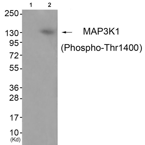
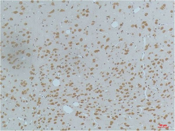
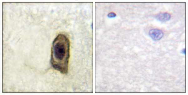
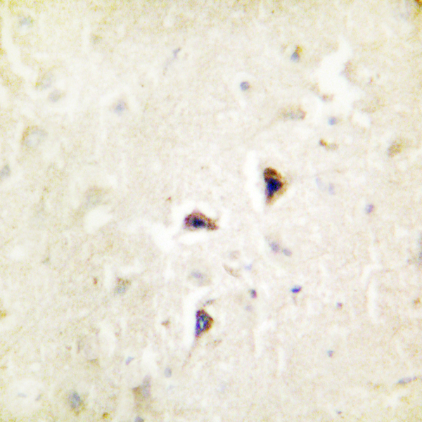
 IHC (Immunohiostchemistry)
(Immunohistochemical analysis of paraffin-embedded human brain tissue using MAP3K1 (Phospho-Thr1400) antibody (left)or the same antibody preincubated with blocking peptide (right).)
IHC (Immunohiostchemistry)
(Immunohistochemical analysis of paraffin-embedded human brain tissue using MAP3K1 (Phospho-Thr1400) antibody (left)or the same antibody preincubated with blocking peptide (right).)
MAP3K1, Polyclonal Antibody (Cat# AAA307845)
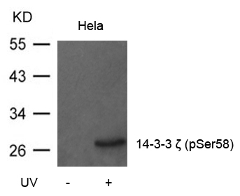
 IF (Immunofluorescence)
(Immunofluorescence staining of methanol-fixed Hela cells using 14-3-3z(Phospho-Ser58) Antibody.)
IF (Immunofluorescence)
(Immunofluorescence staining of methanol-fixed Hela cells using 14-3-3z(Phospho-Ser58) Antibody.)
14-3-3 zeta, Polyclonal Antibody (Cat# AAA307925)
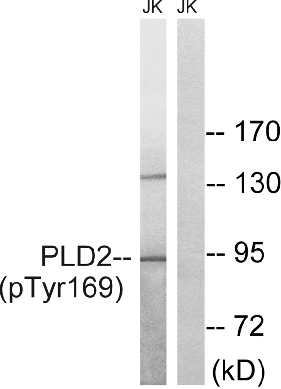
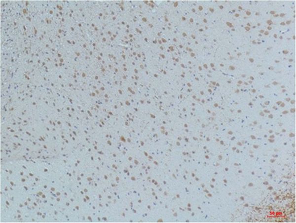
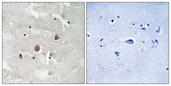
 IHC (Immunohiostchemistry)
(Immunohistochemical analysis of paraffin-embedded human brain tissue using PLD2 (Phospho-Tyr169) antibody (left)or the same antibody preincubated with blocking peptide (right).)
IHC (Immunohiostchemistry)
(Immunohistochemical analysis of paraffin-embedded human brain tissue using PLD2 (Phospho-Tyr169) antibody (left)or the same antibody preincubated with blocking peptide (right).)
PLD2, Polyclonal Antibody (Cat# AAA307786)
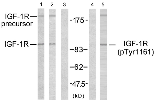
 IF (Immunofluorescence)
(Immunofluorescence staining of methanol-fixed MCF7 cells using IGF-1R (phospho-Tyr1161) antibody (, Red).)
IF (Immunofluorescence)
(Immunofluorescence staining of methanol-fixed MCF7 cells using IGF-1R (phospho-Tyr1161) antibody (, Red).)
IGF-1R, Polyclonal Antibody (Cat# AAA307594)
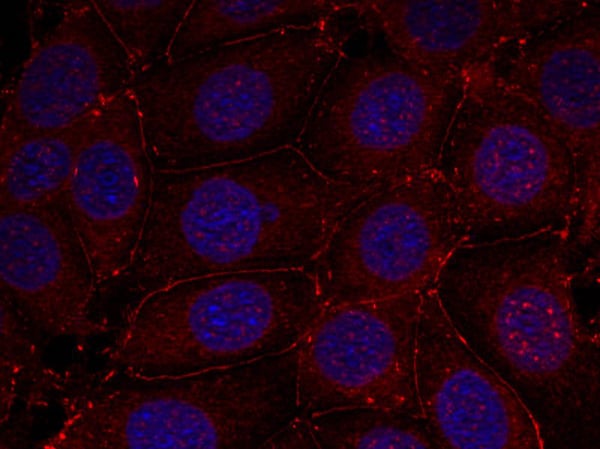
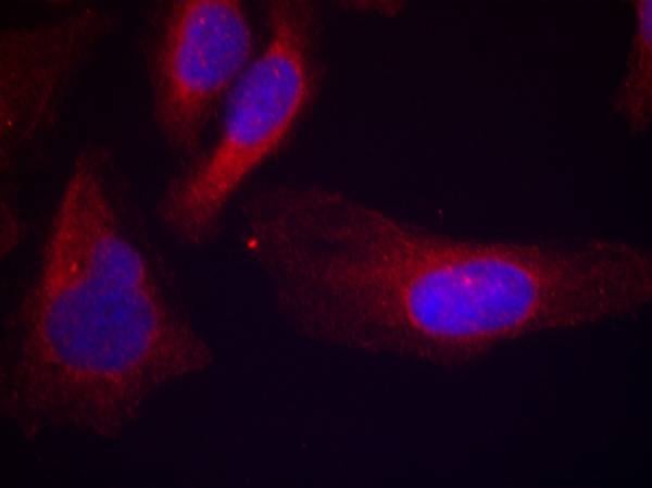 IF (Immunofluorescence)
(Immunofluorescence staining of methanol-fixed Hela cells using c-Cbl(phospho-Tyr700) Antibody.)
IF (Immunofluorescence)
(Immunofluorescence staining of methanol-fixed Hela cells using c-Cbl(phospho-Tyr700) Antibody.)
c-Cbl, Polyclonal Antibody (Cat# AAA307623)

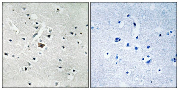
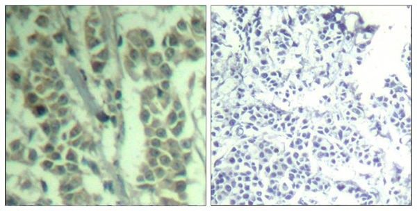
 IHC (Immunohiostchemistry)
(Immunohistochemical analysis of paraffin-embedded human brain, using CSFR (Phospho-Tyr561) antibody (left)or the same antibody preincubated with blocking peptide (right).)
IHC (Immunohiostchemistry)
(Immunohistochemical analysis of paraffin-embedded human brain, using CSFR (Phospho-Tyr561) antibody (left)or the same antibody preincubated with blocking peptide (right).)
CSFR, Polyclonal Antibody (Cat# AAA307652)
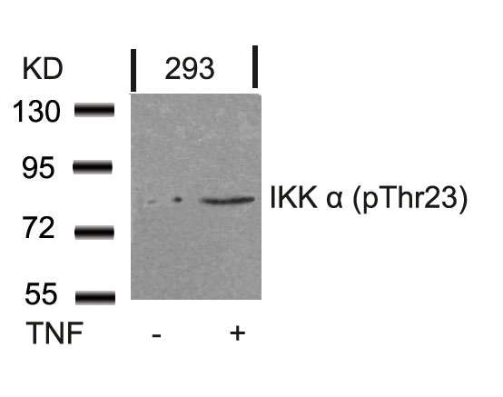
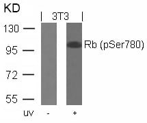
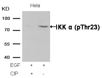 WB (Western Blot)
(Western blot analysis of extracts from Hela cells, treated with EGF or calf intestinal phosphatase (CIP), using IKK alpha (Phospho-Thr23) Antibody.)
WB (Western Blot)
(Western blot analysis of extracts from Hela cells, treated with EGF or calf intestinal phosphatase (CIP), using IKK alpha (Phospho-Thr23) Antibody.)
IKK alpha, Polyclonal Antibody (Cat# AAA307661)
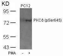
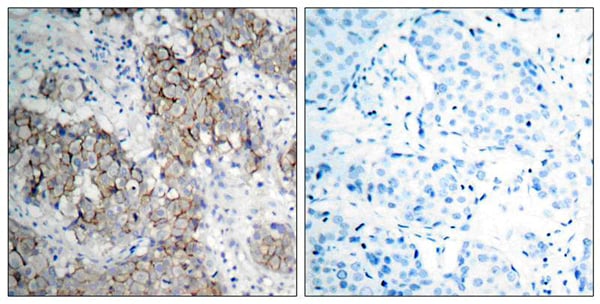
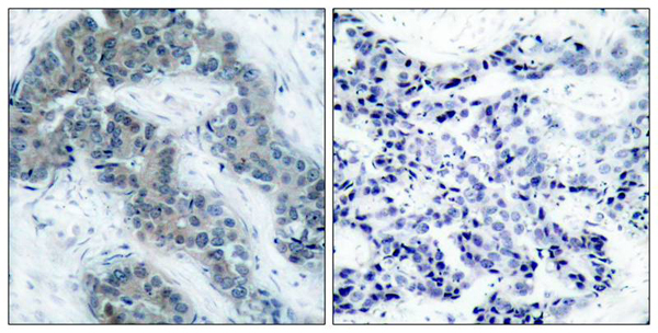
 IHC (Immunohiostchemistry)
(Immunohistochemical analysis of paraffin-embedded human breast carcinoma tissue using PKCd(Phospho-Ser645) Antibody (left) or the same antibody preincubated with blocking peptide(right).)
IHC (Immunohiostchemistry)
(Immunohistochemical analysis of paraffin-embedded human breast carcinoma tissue using PKCd(Phospho-Ser645) Antibody (left) or the same antibody preincubated with blocking peptide(right).)
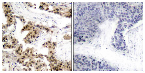
PKCdelta, Polyclonal Antibody (Cat# AAA307444)

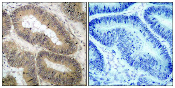
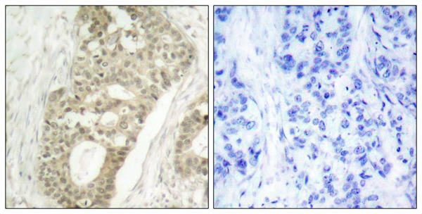
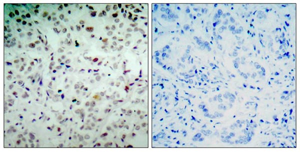
 IHC (Immunohiostchemistry)
(Immunohistochemical analysis of paraffin-embedded human breast carcinoma tissue using JAK2(Phospho-Tyr1007) Antibody (left) or the same antibody preincubated with blocking peptide(right).)
IHC (Immunohiostchemistry)
(Immunohistochemical analysis of paraffin-embedded human breast carcinoma tissue using JAK2(Phospho-Tyr1007) Antibody (left) or the same antibody preincubated with blocking peptide(right).)
JAK2, Polyclonal Antibody (Cat# AAA307448)
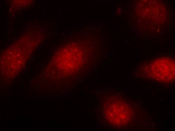 IF (Immunofluorescence)
(Immunofluorescence staining of methanol-fixed Hela cells using Rb(Phospho-Ser780) Antibody.)
IF (Immunofluorescence)
(Immunofluorescence staining of methanol-fixed Hela cells using Rb(Phospho-Ser780) Antibody.)
Rb, Polyclonal Antibody (Cat# AAA307496)
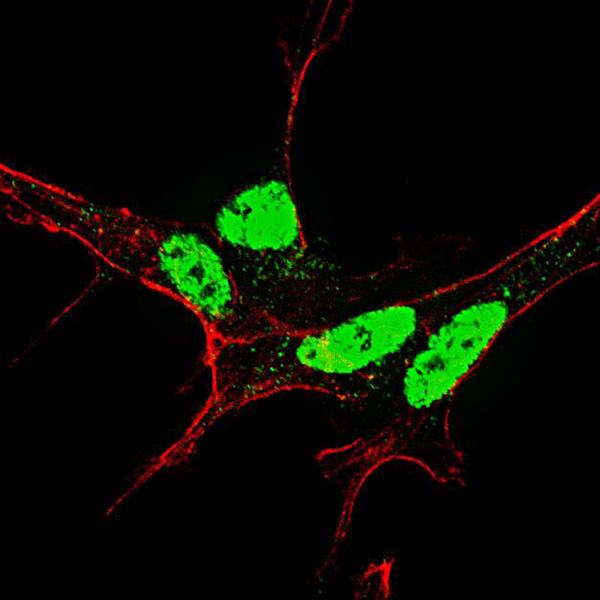
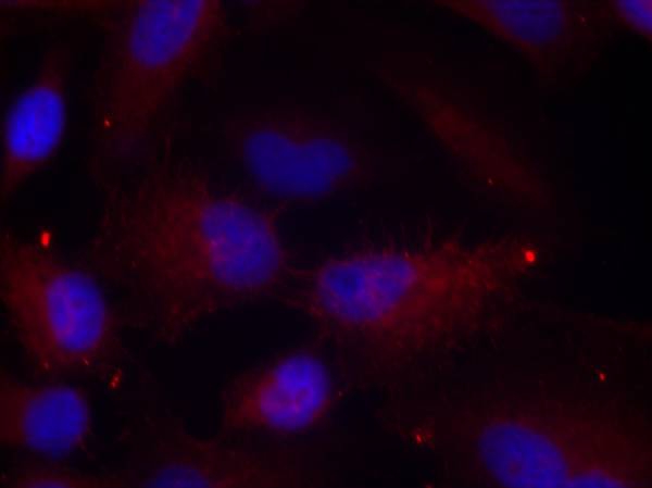
 IF (Immunofluorescence)
(Immunofluorescent analysis of Rabphilin 3A (phospho-hospho-Ser237) staining in HeLa cells. Formalin-fixed cells were permeabilized with 0.1% Triton X-100 in TBS for 5-10 minutes and blocked with 3% BSA-PBS for 30 minutes at room temperature. Cells were probed with the primary antibody in 3% BSA-PBS and incubated overnight at 4 C in a hidified chamber. Cells were washed with PBST and incubated with Alexa Fluor 647-conjugated secondary antibody (red) in PBS at room temperature in the dark.)
IF (Immunofluorescence)
(Immunofluorescent analysis of Rabphilin 3A (phospho-hospho-Ser237) staining in HeLa cells. Formalin-fixed cells were permeabilized with 0.1% Triton X-100 in TBS for 5-10 minutes and blocked with 3% BSA-PBS for 30 minutes at room temperature. Cells were probed with the primary antibody in 3% BSA-PBS and incubated overnight at 4 C in a hidified chamber. Cells were washed with PBST and incubated with Alexa Fluor 647-conjugated secondary antibody (red) in PBS at room temperature in the dark.)
Rabphilin 3A (phospho-Ser237), Polyclonal Antibody (Cat# AAA310503)
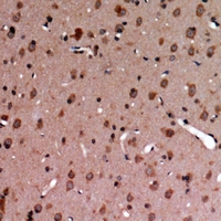 IHC (Immunohiostchemistry)
(Immunohistochemical analysis of TYRO3/MERTK (phospho-Tyr749/681) staining in human brain formalin fixed paraffin embedded tissue section. The section was then incubated with the antibody at room temperature and detected using an HRP conjugated compact polymer system. DAB was used as the chromogen. The section was then counterstained with haematoxylin and mounted with DPX.)
IHC (Immunohiostchemistry)
(Immunohistochemical analysis of TYRO3/MERTK (phospho-Tyr749/681) staining in human brain formalin fixed paraffin embedded tissue section. The section was then incubated with the antibody at room temperature and detected using an HRP conjugated compact polymer system. DAB was used as the chromogen. The section was then counterstained with haematoxylin and mounted with DPX.)
TYRO3/MERTK (phospho-Tyr749/681), Polyclonal Antibody (Cat# AAA310506)

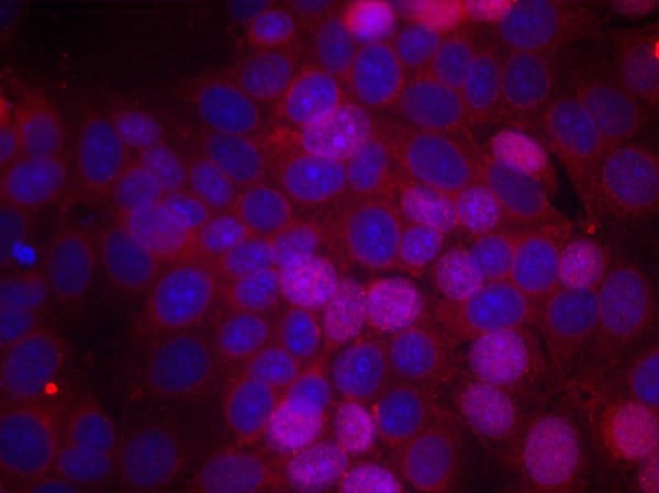
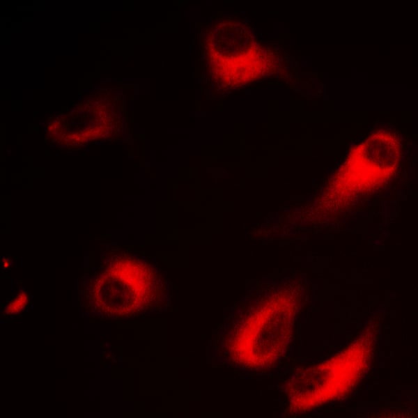
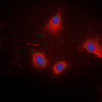 IF (Immunofluorescence)
(Immunofluorescent analysis of Ezrin/Radixin/Moesin (phospho-Thr567/564/558) staining in SKOV3 cells. Formalin-fixed cells were permeabilized with 0.1% Triton X-100 in TBS for 5-10 minutes and blocked with 3% BSA-PBS for 30 minutes at room temperature. Cells were probed with the primary antibody in 3% BSA-PBS and incubated overnight at 4 C in a hidified chamber. Cells were washed with PBST and incubated with a DyLight 594-conjugated secondary antibody (red) in PBS at room temperature in the dark. DAPI was used to stain the cell nuclei (blue).)
IF (Immunofluorescence)
(Immunofluorescent analysis of Ezrin/Radixin/Moesin (phospho-Thr567/564/558) staining in SKOV3 cells. Formalin-fixed cells were permeabilized with 0.1% Triton X-100 in TBS for 5-10 minutes and blocked with 3% BSA-PBS for 30 minutes at room temperature. Cells were probed with the primary antibody in 3% BSA-PBS and incubated overnight at 4 C in a hidified chamber. Cells were washed with PBST and incubated with a DyLight 594-conjugated secondary antibody (red) in PBS at room temperature in the dark. DAPI was used to stain the cell nuclei (blue).)
Ezrin/Radixin/Moesin (phospho-Thr567/564/558), Polyclonal Antibody (Cat# AAA310514)
 IF (Immunofluorescence)
(Immunofluorescent analysis of NEK9 (phospho-Thr210) staining in THP1 cells. Formalin-fixed cells were permeabilized with 0.1% Triton X-100 in TBS for 5-10 minutes and blocked with 3% BSA-PBS for 30 minutes at room temperature. Cells were probed with the primary antibody in 3% BSA-PBS and incubated overnight at 4 C in a hidified chamber. Cells were washed with PBST and incubated with a DyLight 594-conjugated secondary antibody (red) in PBS at room temperature in the dark.)
IF (Immunofluorescence)
(Immunofluorescent analysis of NEK9 (phospho-Thr210) staining in THP1 cells. Formalin-fixed cells were permeabilized with 0.1% Triton X-100 in TBS for 5-10 minutes and blocked with 3% BSA-PBS for 30 minutes at room temperature. Cells were probed with the primary antibody in 3% BSA-PBS and incubated overnight at 4 C in a hidified chamber. Cells were washed with PBST and incubated with a DyLight 594-conjugated secondary antibody (red) in PBS at room temperature in the dark.)
NEK9 (phospho-Thr210), Polyclonal Antibody (Cat# AAA310515)
KDM4C, Polyclonal Antibody (Cat# AAA312929)
Synapsin I, Polyclonal Antibody (Cat# AAA312932)
CD4, Polyclonal Antibody (Cat# AAA312939)
Nemo-like kinase, Polyclonal Antibody (Cat# AAA313002)
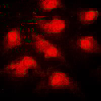
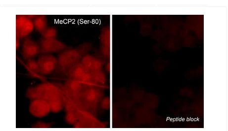 ICC (Immunocytochemistry)
(Immunocytochemical labeling of MeCP2 phosphorylation in rat PC12 cells differentiated with NGF. The cells were probed with MeCP2 (Ser-80) rabbit polyclonal antibody (MP4601) in the absence (left) or presence (right) of blocking peptide (MX4605). The antibody was detected using appropriate secondary antibody conjugated to DyLight 594.)
ICC (Immunocytochemistry)
(Immunocytochemical labeling of MeCP2 phosphorylation in rat PC12 cells differentiated with NGF. The cells were probed with MeCP2 (Ser-80) rabbit polyclonal antibody (MP4601) in the absence (left) or presence (right) of blocking peptide (MX4605). The antibody was detected using appropriate secondary antibody conjugated to DyLight 594.)
MeCP2, Polyclonal Antibody (Cat# AAA71669)
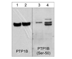
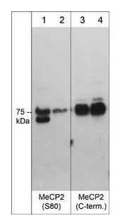
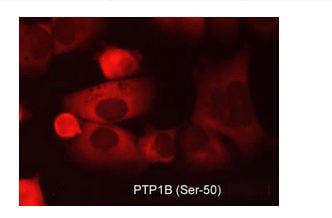 ICC (Immunocytochemistry)
(Immunocytochemical labeling of PTP1B in aldehyde-fixed and NP-40 permeabilized human NCI-H1915 lung carcinoma cells. The cells were labeled with rabbit polyclonal anti-PTP1B (Ser-50) (PP2411) phosphospecific antibody. The antibody was detected using appropriate secondary antibody conjugated to DyLight 594.)
ICC (Immunocytochemistry)
(Immunocytochemical labeling of PTP1B in aldehyde-fixed and NP-40 permeabilized human NCI-H1915 lung carcinoma cells. The cells were labeled with rabbit polyclonal anti-PTP1B (Ser-50) (PP2411) phosphospecific antibody. The antibody was detected using appropriate secondary antibody conjugated to DyLight 594.)
PTP1B, Polyclonal Antibody (Cat# AAA71693)
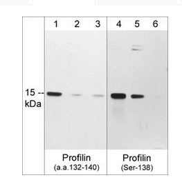
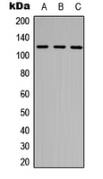
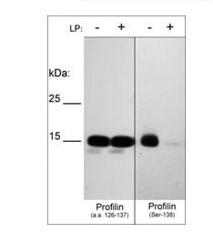 WB (Western Blot)
(Western blot of human recombinant Profilin-1 phosphorylated in vitro with PKCalpha kinase then untreated (-) or treated with lambda phosphatase (+). The blots were probed with anti-Profilin (a.a. 126-137) (left panel) or anti-Profilin (Ser-138) phospho-specific (right panel) antibodies at 1:1000.)
WB (Western Blot)
(Western blot of human recombinant Profilin-1 phosphorylated in vitro with PKCalpha kinase then untreated (-) or treated with lambda phosphatase (+). The blots were probed with anti-Profilin (a.a. 126-137) (left panel) or anti-Profilin (Ser-138) phospho-specific (right panel) antibodies at 1:1000.)
Profilin, Polyclonal Antibody (Cat# AAA71697)
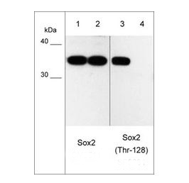
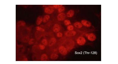 ICC (Immunocytochemistry)
(Immunocytochemical labeling of phosphorylated Sox2 in aldehyde fixed and NP-40 permeabilized human NCI-H446 lung carcinoma cells. The cells were labeled with rabbit polyclonal anti-Sox2 (Thr-128) phospho-specific (SP0381). The antibody was detected using goat anti-rabbit Ig:DyLight 594.)
ICC (Immunocytochemistry)
(Immunocytochemical labeling of phosphorylated Sox2 in aldehyde fixed and NP-40 permeabilized human NCI-H446 lung carcinoma cells. The cells were labeled with rabbit polyclonal anti-Sox2 (Thr-128) phospho-specific (SP0381). The antibody was detected using goat anti-rabbit Ig:DyLight 594.)
Sox2, Polyclonal Antibody (Cat# AAA71709)
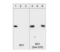
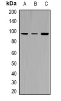
 WB (Western Blot)
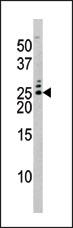
(Western blot of HeLa stimulated with calyculin A (lanes 1-4). The blots were untreated (lane 1 & 3) or treated with lambda phosphatase (lane 2 & 4), then probed with anti-SK1 (Central region) SP1621 (lanes 1 & 2) or anti-SK1 (Ser-225) SP1641 (lanes 3 & 4).)
WB (Western Blot)
(Western blot of HeLa stimulated with calyculin A (lanes 1-4). The blots were untreated (lane 1 & 3) or treated with lambda phosphatase (lane 2 & 4), then probed with anti-SK1 (Central region) SP1621 (lanes 1 & 2) or anti-SK1 (Ser-225) SP1641 (lanes 3 & 4).)
Sphingosine Kinase 1, Polyclonal Antibody (Cat# AAA71711)
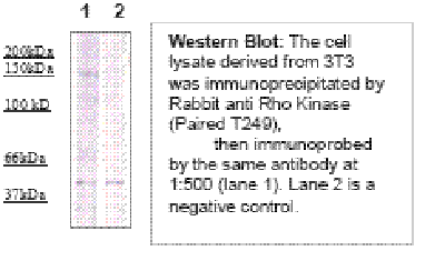 Application Data
Rho Kinase/ROCKII (Paired T249), Antibody (Cat# AAA71407)
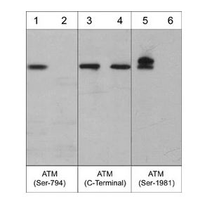
 ICC (Immunocytochemistry)
(Immunocytochemical labeling of ATM phosphorylation in control (Top row) or calyculin A-treated A431 cells (Bottom row). The cells were labeled with mouse monoclonal ATM (C-terminal region) (AM3611) and ATM (Ser-1981) (AM3661). The antibodies were detected using goat anti-mouse-DyLight 594.)
ICC (Immunocytochemistry)
(Immunocytochemical labeling of ATM phosphorylation in control (Top row) or calyculin A-treated A431 cells (Bottom row). The cells were labeled with mouse monoclonal ATM (C-terminal region) (AM3611) and ATM (Ser-1981) (AM3661). The antibodies were detected using goat anti-mouse-DyLight 594.)
ATM, Monoclonal Antibody (Cat# AAA71562)
CRMP2 Phospho-Regulation, Antibody Sampler Kit (Cat# AAA71573)
 ICC (Immunocytochemistry)
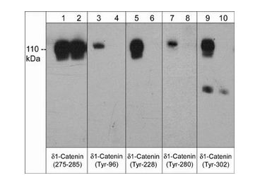
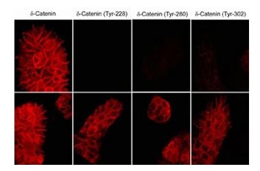
(Immunocytochemical labeling of delta1-Catenin in untreated (Top) or pervanadate-treated (bottom) A431 cells. The cells were labeled with mouse monoclonal delta1-Catenin (a.a. 275-285), delta1-Catenin (Tyr-228), delta1-Catenin (Tyr-280), or delta1-Catenin (Tyr-302) antibodies. The antibodies were detected using donkey anti-mouse secondary antibodies conjugated to Cy3.)
ICC (Immunocytochemistry)
(Immunocytochemical labeling of delta1-Catenin in untreated (Top) or pervanadate-treated (bottom) A431 cells. The cells were labeled with mouse monoclonal delta1-Catenin (a.a. 275-285), delta1-Catenin (Tyr-228), delta1-Catenin (Tyr-280), or delta1-Catenin (Tyr-302) antibodies. The antibodies were detected using donkey anti-mouse secondary antibodies conjugated to Cy3.)
delta1-Catenin, Monoclonal Antibody (Cat# AAA71595)
 ICC (Immunocytochemistry)
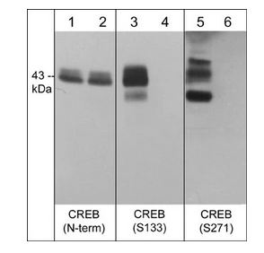
(Immunocytochemical labeling of phosphorylated CREB in calyculin A-treated A431 cells. The cells were fixed in paraformaldehyde and permeabilized using NP-40 before labeling with mouse monoclonal CREB (Ser-133). The antibody was detected using goat anti-mouse DyLight 594.)
ICC (Immunocytochemistry)
(Immunocytochemical labeling of phosphorylated CREB in calyculin A-treated A431 cells. The cells were fixed in paraformaldehyde and permeabilized using NP-40 before labeling with mouse monoclonal CREB (Ser-133). The antibody was detected using goat anti-mouse DyLight 594.)
CREB, Monoclonal Antibody (Cat# AAA71596)
 ICC (Immunocytochemistry)
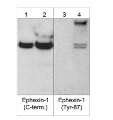
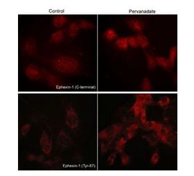
(Immunocytochemical labeling of phosphorylated Exphexin-1 in pervanadate-treated mouse C2C12. The cells were labeled with rabbit polyclonal Ephexin-1 (C-terminal region) and Ephexin-1 (Tyr-87) antibodies, then the antibodies were detected using appropriate secondary antibodies conjugated to Cy3.)
ICC (Immunocytochemistry)
(Immunocytochemical labeling of phosphorylated Exphexin-1 in pervanadate-treated mouse C2C12. The cells were labeled with rabbit polyclonal Ephexin-1 (C-terminal region) and Ephexin-1 (Tyr-87) antibodies, then the antibodies were detected using appropriate secondary antibodies conjugated to Cy3.)
Ephexin-1, Polyclonal Antibody (Cat# AAA71632)
What Are Phospho Antibodies?
Protein phosphorylation is a process where a phosphate group is added to certain amino acid residues of a protein – usually serine (S), threonine (T), or tyrosine (Y) - by enzymes called kinases. This process is integral in controlling cellular signaling, cellular growth, and other biological functions.
Our catalog includes a wide range of phospho-specific antibodies that can accurately detect this important marker. They perform strongly in widely-used laboratory applications such as Western blot, flow cytometry, immunohistochemistry, and immunofluorescence microscopy. We value your trust in us and are committed to providing top-quality products and services. All of our antibodies are guaranteed to work for the applications and species indicated on our website & associated product pages.
What Are The Key Applications of Phospho Antibodies?
1. Western Blotting
One of the first steps a researcher can take in utilizing these phospho-specific antibodies, is to check if the antibody works using a technique referred to as “Western blot”. For those unfamiliar, Western Blot aids in showing whether the protein that the antibody recognizes is appearing at the correct/expected size. These phospho-specific antibodies should also be able to detect changes in the target protein’s phosphorylation (on/off state) when cells are stimulated in certain ways.
2. Staining of Fixed Cells (Immunocytochemistry)
Another routine use of these phospho-specific antibodies, is to test if the antibody is able to demonstrate similar performance when used on fixed cells (intact cells that have been preserved) as it did in the Western blot tests. It is an important aspect in many cases to confirm that the antibody works in actual intact cell samples. Ideally, the method used for cellular fixation should be the same as what is used in pathology labs (like using 10% formalin). To check if the antibody works well in tissue sections (FFPE), researchers will often test it on fixed cells that are processed similar to tissue samples.
3. Specificity Tests Using Peptides
In order to make sure that the antibody is only binding to the right target:
- Laboratory technicians will mix the antibody with phospho-peptides (short segments of the protein containing the phosphate group modification).
- If the antibody signal disappears, it is confirmation that it is binding to the correct phosphorylated location.
- A more robust test is to use both the phosphorylated and non-phosphorylated (dephosphorylated) versions of the protein. The antibody should react only with the phosphorylated one.
- Another method sometimes utilized is to treat the sample with an enzyme, such as alkaline phosphatase, that specifically removes phosphate groups. If the antibody signal disappears after this, it also confirms specificity.
4. Genetic Confirmation
As a final step, scientists can genetically manipulate the nucleotide sequence and alter the target protein by removing the exact site where phosphorylation happens. If the antibody no longer appears to detect the modified protein, it is strong evidence supporting the antibody being specific for that phosphorylated site.
Why Buy Phospho Antibodies Through Us?
- The production laboratory adheres to strict and consistent protocols prior to releasing any of these phospho-specific antibodies:
- Standard methods and proper controls in all tests to ensure high quality.
- These antibodies are tested and validated in different cell types and species.
- High quality control criterion to ensure each batch is consistent, so you will obtain reliable results every time.
FAQ
1. What Are Phospho-Specific Antibodies?
Phospho-specific antibodies are made to detect proteins only when they have a phosphate group linked to a specific amino acid residue. This empowers scientists understand if a protein is "turned on" or active, based on its phosphorylation state.
2. How to Detect Phosphorylated Proteins in a Western Blot?
To find out if a protein is phosphorylated using Western blot:
- Use a phospho-specific antibody that binds only to the phosphorylated form of the protein.
- You can also use a “regular” antibody for the same amino acid sequence of the protein that the phospho-specific antibody is binding to (but in this case, this antibody will not bind if there is a phosphate group present) in order to compare how much of it is phosphorylated versus how much is non-phosphorylated (or “total” protein, if the “normal” antibody’s epitopes are non-phospho-site-specific).
3. How to Choose the Best Antibody?
Here are some simple tips to help you pick the right antibody:
- Know your target
- Match your sample characteristics
- Confirm the intended use is appropriate
- Check “host” and “type”
- Check the “quality” of the presented data/images
- Appraise whether the available validation meets your needs