Filters
▼Clonality
▼Type
▼Reactivity
▼Gene Name
▼Isotype
▼Host
▼Application
▼Clone
▼Monoclonal Antibodies
Get accurate results in your research with our Monoclonal Antibodies, which are specially made to target exactly what you require for your research, and will produce consistent, reliable performance in lab tests.
Viewing 4300-4350 of 27597 product results
 IP (Immunoprecipitation)
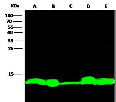
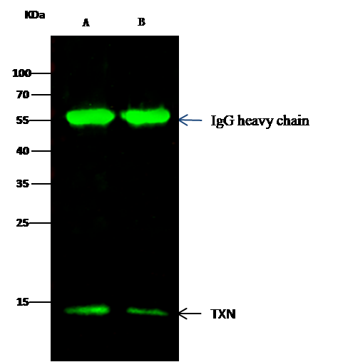
(TXN was immunoprecipitated using:Lane A:0.5 mg Hela Whole Cell LysateLane B:0.5 mg 293T Whole Cell Lysate2 uL anti-TXN rabbit monoclonal antibody and 15 ul of 50 % Protein G agarose.Primary antibody:Anti-TXN rabbit monoclonal antibody,at 1:100 dilutionSecondary antibody:Dylight 800-labeled antibody to rabbit IgG (H+L), at 1:5000 dilutionDeveloped using the odssey technique.Performed under reducing conditions.Predicted band size: 13 kDaObserved band size: 13 kDa)
IP (Immunoprecipitation)
(TXN was immunoprecipitated using:Lane A:0.5 mg Hela Whole Cell LysateLane B:0.5 mg 293T Whole Cell Lysate2 uL anti-TXN rabbit monoclonal antibody and 15 ul of 50 % Protein G agarose.Primary antibody:Anti-TXN rabbit monoclonal antibody,at 1:100 dilutionSecondary antibody:Dylight 800-labeled antibody to rabbit IgG (H+L), at 1:5000 dilutionDeveloped using the odssey technique.Performed under reducing conditions.Predicted band size: 13 kDaObserved band size: 13 kDa)
Thioredoxin/TRX, Monoclonal Antibody (Cat# AAA254886)
 IP (Immunoprecipitation)
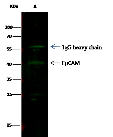
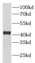
(EPCAM was immunoprecipitated using:Lane A:0.5 mg MCF-7 Whole Cell Lysate0.5 uL anti-EPCAM mouse monoclonal antibody and 15 ul of 50 % Protein G agarose.Primary antibody:Anti-EPCAM mouse monoclonal antibody,at 1:500 dilutionSecondary antibody:Dylight 800-labeled antibody to Mouse IgG (H+L), at 1:7500 dilutionDeveloped using the odssey technique.Performed under reducing conditions.Predicted band size: 40 kDaObserved band size: 40 kDa)
IP (Immunoprecipitation)
(EPCAM was immunoprecipitated using:Lane A:0.5 mg MCF-7 Whole Cell Lysate0.5 uL anti-EPCAM mouse monoclonal antibody and 15 ul of 50 % Protein G agarose.Primary antibody:Anti-EPCAM mouse monoclonal antibody,at 1:500 dilutionSecondary antibody:Dylight 800-labeled antibody to Mouse IgG (H+L), at 1:7500 dilutionDeveloped using the odssey technique.Performed under reducing conditions.Predicted band size: 40 kDaObserved band size: 40 kDa)
EpCAM, Monoclonal Antibody (Cat# AAA255084)
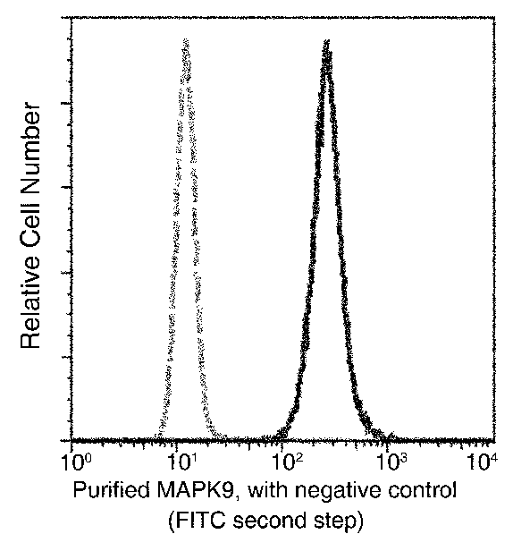 FCM/FACS (Flow Cytometry)
(Flow cytometric analysis of Human MAPK9 expression on HeLa cells. The cells were treated according to manufacturer's manual (BD Pharmingen'), stained with purified anti-Human MAPK9, then a FITC-conjugated second step antibody. The fluorescence histograms were derived from gated events with the forward and side light-scatter characteristics of intact cells.)
FCM/FACS (Flow Cytometry)
(Flow cytometric analysis of Human MAPK9 expression on HeLa cells. The cells were treated according to manufacturer's manual (BD Pharmingen'), stained with purified anti-Human MAPK9, then a FITC-conjugated second step antibody. The fluorescence histograms were derived from gated events with the forward and side light-scatter characteristics of intact cells.)
JNK2, Monoclonal Antibody (Cat# AAA255131)
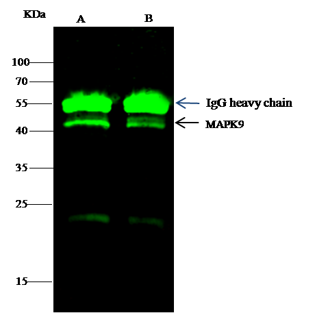
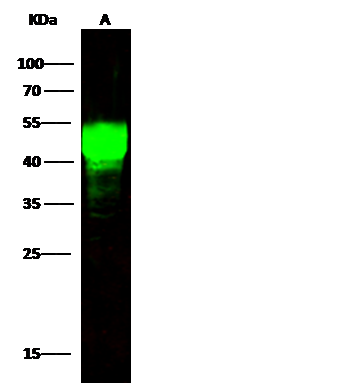
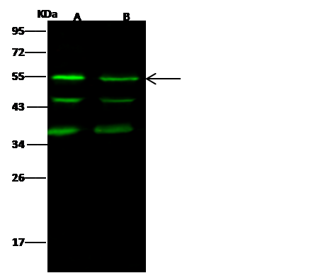 WB (Western Blot)
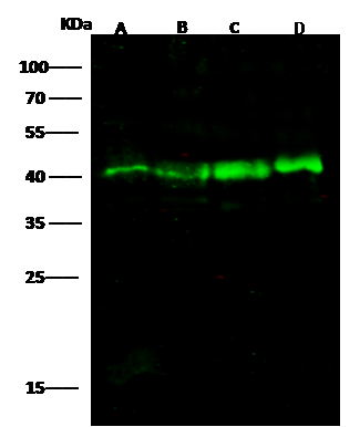
(Anti-MAPK9 rabbit monoclonal antibody at 1:500 dilutionLane A: HepG2 Whole Cell LysateLane B: A549 Whole Cell LysateLysates/proteins at 30 ug per lane.SecondaryGoat Anti-Rabbit IgG H&L (Dylight800) at 1/10000 dilution.Developed using the Odyssey technique.Performed under reducing conditions.Predicted band size:48 kDaObserved band size:54 kDa(We are unsure as to the identity of these extra bands.))
WB (Western Blot)
(Anti-MAPK9 rabbit monoclonal antibody at 1:500 dilutionLane A: HepG2 Whole Cell LysateLane B: A549 Whole Cell LysateLysates/proteins at 30 ug per lane.SecondaryGoat Anti-Rabbit IgG H&L (Dylight800) at 1/10000 dilution.Developed using the Odyssey technique.Performed under reducing conditions.Predicted band size:48 kDaObserved band size:54 kDa(We are unsure as to the identity of these extra bands.))
JNK2, Monoclonal Antibody (Cat# AAA255132)
 FCM/FACS (Flow Cytometry)
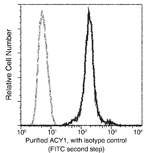
(Flow cytometric analysis of Human ACY1 expression on HepG2 cells. The cells were treated according to manufacturer's manual (BD Pharmingen'), stained with purified anti-Human ACY1, then a FITC-conjugated second step antibody. The fluorescence histograms were derived from gated events with the forward and side light-scatter characteristics of intact cells.)
FCM/FACS (Flow Cytometry)
(Flow cytometric analysis of Human ACY1 expression on HepG2 cells. The cells were treated according to manufacturer's manual (BD Pharmingen'), stained with purified anti-Human ACY1, then a FITC-conjugated second step antibody. The fluorescence histograms were derived from gated events with the forward and side light-scatter characteristics of intact cells.)
Aminoacylase 1, Monoclonal Antibody (Cat# AAA255000)
 IP (Immunoprecipitation)
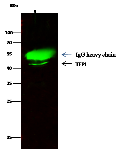
(TFPI was immunoprecipitated using:Lane A:0.5 mg MCF-7 Whole Cell Lysate0.5 uL anti-TFPI rabbit monoclonal antibody and 15 ul of 50 % Protein G agarose.Primary antibody:Anti-TFPI rabbit monoclonal antibody,at 1:1000 dilutionSecondary antibody:Dylight 800-labeled antibody to rabbit IgG (H+L), at 1:5000 dilutionDeveloped using the odssey technique.Performed under reducing conditions.Predicted band size: 35 kDaObserved band size: 45 kDa)
IP (Immunoprecipitation)
(TFPI was immunoprecipitated using:Lane A:0.5 mg MCF-7 Whole Cell Lysate0.5 uL anti-TFPI rabbit monoclonal antibody and 15 ul of 50 % Protein G agarose.Primary antibody:Anti-TFPI rabbit monoclonal antibody,at 1:1000 dilutionSecondary antibody:Dylight 800-labeled antibody to rabbit IgG (H+L), at 1:5000 dilutionDeveloped using the odssey technique.Performed under reducing conditions.Predicted band size: 35 kDaObserved band size: 45 kDa)
TFPI, Monoclonal Antibody (Cat# AAA255006)
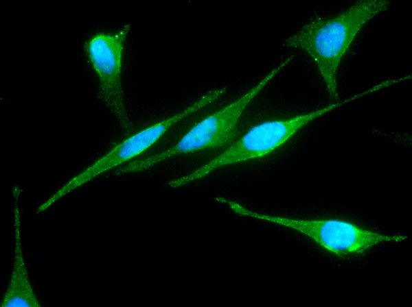
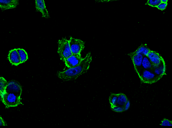
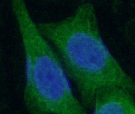
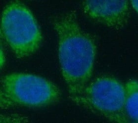
 IF (Immunofluorescence)
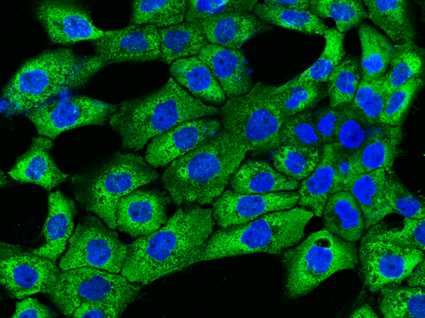
(Immunofluorescence staining of TFPI in A431 cells. Cells were fixed with 4% PFA, permeabilzed with 0.3% Triton X-100 in PBS, blocked with 10% serum, and incubated with rabbit anti-human TFPI monoclonal antibody (1:60) at 4 degree C overnight. Then cells were stained with the Alexa Fluor 488-conjugated Goat Anti-rabbit IgG secondary antibody (green) and counterstained with DAPI (blue).)
IF (Immunofluorescence)
(Immunofluorescence staining of TFPI in A431 cells. Cells were fixed with 4% PFA, permeabilzed with 0.3% Triton X-100 in PBS, blocked with 10% serum, and incubated with rabbit anti-human TFPI monoclonal antibody (1:60) at 4 degree C overnight. Then cells were stained with the Alexa Fluor 488-conjugated Goat Anti-rabbit IgG secondary antibody (green) and counterstained with DAPI (blue).)
TFPI, Monoclonal Antibody (Cat# AAA255007)
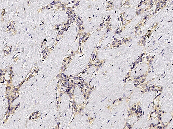
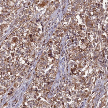
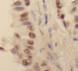
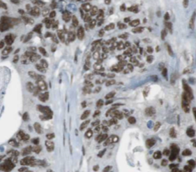
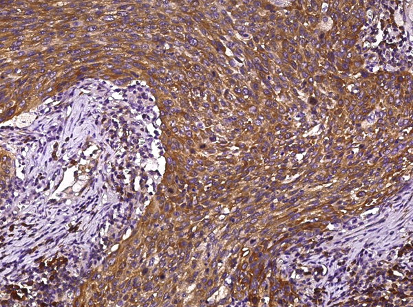 IHC (Immunohiostchemistry)
(Immunochemical staining of FGFR1 in lung carcinoma with mouse monoclonal antibody (1:60, formalin-fixed paraffin embedded sections).)
IHC (Immunohiostchemistry)
(Immunochemical staining of FGFR1 in lung carcinoma with mouse monoclonal antibody (1:60, formalin-fixed paraffin embedded sections).)
FGFR1, Monoclonal Antibody (Cat# AAA255031)
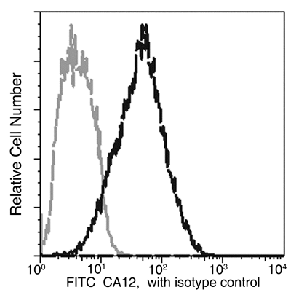 FCM/FACS (Flow Cytometry)
(Flow cytometric analysis of Human CA12 expression on MCF-7 cells. Cells were stained with FITC-conjugated anti-Human CA12. The fluorescence histograms were derived from gated events with the forward and side light-scatter characteristics of intact cells.Flow cytometry was performed on a BD FC Calibur flow cytometry system.)
FCM/FACS (Flow Cytometry)
(Flow cytometric analysis of Human CA12 expression on MCF-7 cells. Cells were stained with FITC-conjugated anti-Human CA12. The fluorescence histograms were derived from gated events with the forward and side light-scatter characteristics of intact cells.Flow cytometry was performed on a BD FC Calibur flow cytometry system.)
carbonic anhydrase XII, Monoclonal Antibody (Cat# AAA255032)
Azurocidin/CAP37, Monoclonal Antibody (Cat# AAA255054)
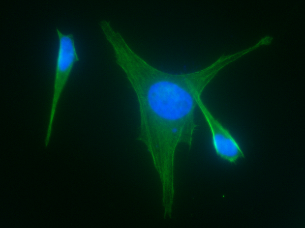
 IF (Immunofluorescence)
(Immunofluorescence staining of CD324 in MCF7 cells. Cells were fixed with 4% PFA, permeabilzed with 0.3% Triton X-100 in PBS, blocked with 10% serum, and incubated with rabbit anti-human CD324 monoclonal antibody (1:60) at 4 degree C overnight. Then cells were stained with the Alexa Fluor 488-conjugated Goat Anti-rabbit IgG secondary antibody (green) and counterstained with DAPI (blue).)
IF (Immunofluorescence)
(Immunofluorescence staining of CD324 in MCF7 cells. Cells were fixed with 4% PFA, permeabilzed with 0.3% Triton X-100 in PBS, blocked with 10% serum, and incubated with rabbit anti-human CD324 monoclonal antibody (1:60) at 4 degree C overnight. Then cells were stained with the Alexa Fluor 488-conjugated Goat Anti-rabbit IgG secondary antibody (green) and counterstained with DAPI (blue).)
E-Cadherin, Monoclonal Antibody (Cat# AAA254688)

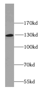
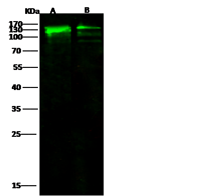 WB (Western Blot)
(Anti-E-cad rabbit monoclonal antibody at 1:500 dilutionLane A: MCF7 Whole Cell LysateLane B: A431 Whole Cell LysateLysates/proteins at 30 ug per lane.SecondaryGoat Anti-Rabbit IgG H&L (Dylight800) at 1/10000 dilution.Developed using the Odyssey technique.Performed under reducing conditions.Predicted band size:97 kDaObserved band size:130 kDa)
WB (Western Blot)
(Anti-E-cad rabbit monoclonal antibody at 1:500 dilutionLane A: MCF7 Whole Cell LysateLane B: A431 Whole Cell LysateLysates/proteins at 30 ug per lane.SecondaryGoat Anti-Rabbit IgG H&L (Dylight800) at 1/10000 dilution.Developed using the Odyssey technique.Performed under reducing conditions.Predicted band size:97 kDaObserved band size:130 kDa)
E-Cadherin, Monoclonal Antibody (Cat# AAA254689)
LAYN, Monoclonal Antibody (Cat# AAA254700)

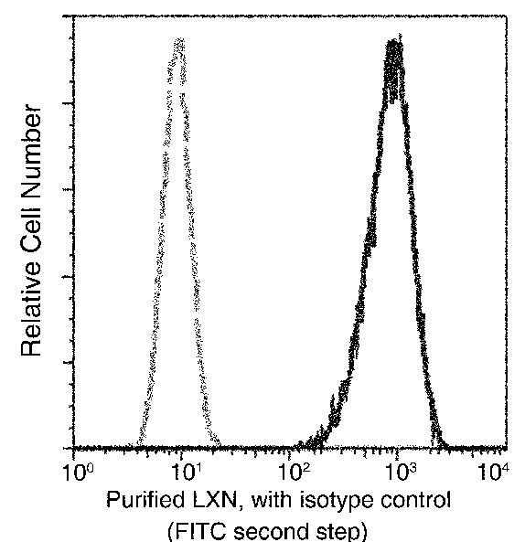
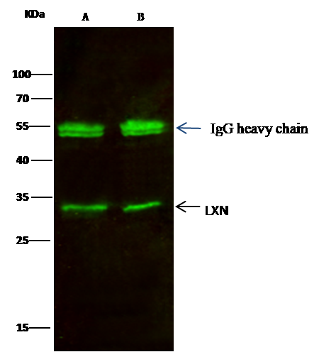 IP (Immunoprecipitation)
(LXN was immunoprecipitated using:Lane A:0.5 mg A549 Whole Cell LysateLane B:0.5 mg Hela Whole Cell Lysate2 uL anti-LXN rabbit monoclonal antibody and 15 ul of 50 % Protein G agarose.Primary antibody:Anti-LXN rabbit monoclonal antibody,at 1:200 dilutionSecondary antibody:Dylight 800-labeled antibody to rabbit IgG (H+L), at 1:5000 dilutionDeveloped using the odssey technique.Performed under reducing conditions.Predicted band size: 25.7 kDaObserved band size: 25.7 kDa)
IP (Immunoprecipitation)
(LXN was immunoprecipitated using:Lane A:0.5 mg A549 Whole Cell LysateLane B:0.5 mg Hela Whole Cell Lysate2 uL anti-LXN rabbit monoclonal antibody and 15 ul of 50 % Protein G agarose.Primary antibody:Anti-LXN rabbit monoclonal antibody,at 1:200 dilutionSecondary antibody:Dylight 800-labeled antibody to rabbit IgG (H+L), at 1:5000 dilutionDeveloped using the odssey technique.Performed under reducing conditions.Predicted band size: 25.7 kDaObserved band size: 25.7 kDa)
Latexin, Monoclonal Antibody (Cat# AAA254707)
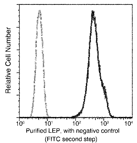
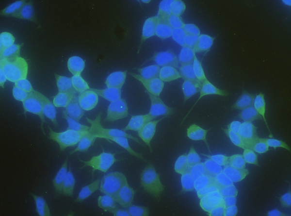
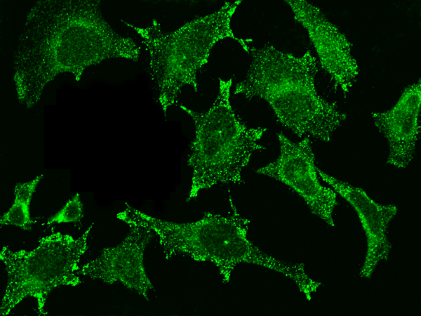
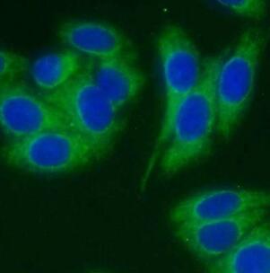
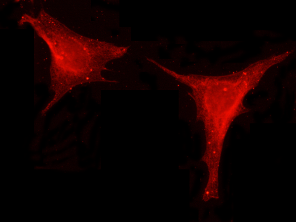 IF (Immunofluorescence)
(Immunofluorescence staining of LEP in Hela cells. Cells were fixed with 4% PFA, permeabilzed with 0.1% Triton X-100 in PBS,blocked with 10% serum, and incubated with rabbit anti-human LEP monoclonal antibody (dilution ratio 1:60) at 4 degree C overnight. Then cells were stained with the Alexa Fluor594-conjugated Goat Anti-rabbit IgG secondary antibody (red). Positive staining was localized to Cytoplasm.)
IF (Immunofluorescence)
(Immunofluorescence staining of LEP in Hela cells. Cells were fixed with 4% PFA, permeabilzed with 0.1% Triton X-100 in PBS,blocked with 10% serum, and incubated with rabbit anti-human LEP monoclonal antibody (dilution ratio 1:60) at 4 degree C overnight. Then cells were stained with the Alexa Fluor594-conjugated Goat Anti-rabbit IgG secondary antibody (red). Positive staining was localized to Cytoplasm.)
Leptin, Monoclonal Antibody (Cat# AAA254724)

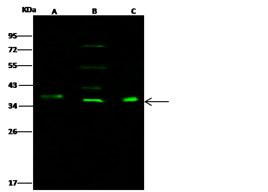 WB (Western Blot)
(Anti-BMP2 mouse monoclonal antibody at 1:500 dilutionLane A: Caco2 Whole Cell LysateLane B: THP1 Whole Cell LysateLane C: HeLa Whole Cell LysateLysates/proteins at 30 ug per lane.SecondaryGoat Anti-Mouse IgG H&L (Dylight800) at 1/15000 dilution.Developed using the Odyssey technique.Performed under reducing conditions.Predicted band size:45 kDaObserved band size:39 kDa(We are unsure as to the identity of these extra bands.))
WB (Western Blot)
(Anti-BMP2 mouse monoclonal antibody at 1:500 dilutionLane A: Caco2 Whole Cell LysateLane B: THP1 Whole Cell LysateLane C: HeLa Whole Cell LysateLysates/proteins at 30 ug per lane.SecondaryGoat Anti-Mouse IgG H&L (Dylight800) at 1/15000 dilution.Developed using the Odyssey technique.Performed under reducing conditions.Predicted band size:45 kDaObserved band size:39 kDa(We are unsure as to the identity of these extra bands.))
BMP-2, Monoclonal Antibody (Cat# AAA254915)
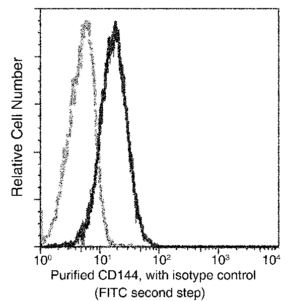
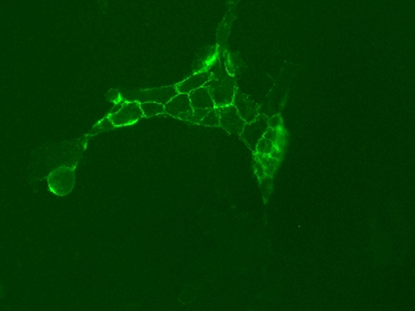 IF (Immunofluorescence)
(Immunofluorescence staining of CDH5 in HUVEC cells. Cells were fixed with 4% PFA,blocked with 10% serum, and incubated with mouse anti-human CDH5 monoclonal antibody (dilution ratio 1:30) at 4 degree C overnight. Then cells were stained with the Alexa Fluor488-conjugated Goat Anti-mouse IgG secondary antibody (green). Positive staining was localized to Cell membrane.)
IF (Immunofluorescence)
(Immunofluorescence staining of CDH5 in HUVEC cells. Cells were fixed with 4% PFA,blocked with 10% serum, and incubated with mouse anti-human CDH5 monoclonal antibody (dilution ratio 1:30) at 4 degree C overnight. Then cells were stained with the Alexa Fluor488-conjugated Goat Anti-mouse IgG secondary antibody (green). Positive staining was localized to Cell membrane.)
VE-Cadherin, Monoclonal Antibody (Cat# AAA254924)
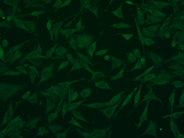
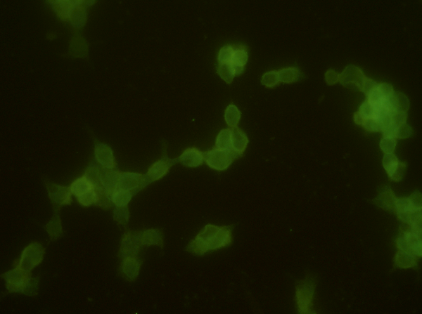
 IF (Immunofluorescence)
(Immunofluorescence staining of CST3 in Hela cells. Cells were fixed with 4% PFA, permeabilzed with 0.1% Triton X-100 in PBS,blocked with 10% serum, and incubated with rabbit anti-Y CST3 monoclonal antibody (dilution ratio 1:60) at 4 degree C overnight. Then cells were stained with the Alexa Fluor488-conjugated Goat Anti-rabbit IgG secondary antibody (green). Positive staining was localized to Cytoplasm.)
IF (Immunofluorescence)
(Immunofluorescence staining of CST3 in Hela cells. Cells were fixed with 4% PFA, permeabilzed with 0.1% Triton X-100 in PBS,blocked with 10% serum, and incubated with rabbit anti-Y CST3 monoclonal antibody (dilution ratio 1:60) at 4 degree C overnight. Then cells were stained with the Alexa Fluor488-conjugated Goat Anti-rabbit IgG secondary antibody (green). Positive staining was localized to Cytoplasm.)
Cystatin C, Monoclonal Antibody (Cat# AAA254931)
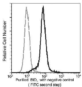
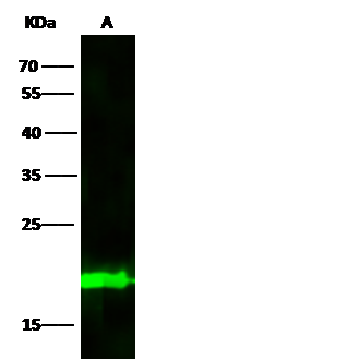 WB (Western Blot)
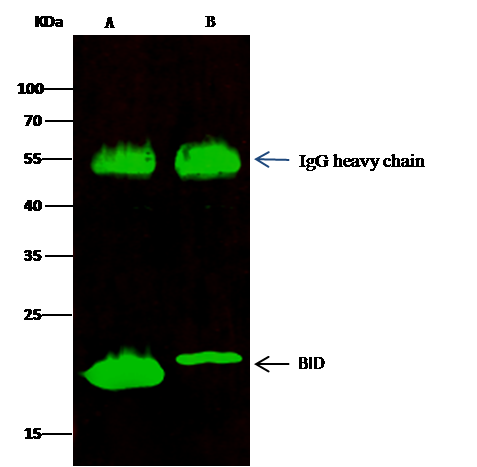
(Anti-BID mouse monoclonal antibody at 1:500 dilutionLane A: Jurkat Whole Cell LysateLysates/proteins at 30 ug per lane.SecondaryGoat Anti-Mouse IgG H&L (Dylight800) at 1/15000 dilution.Developed using the Odyssey technique.Performed under reducing conditions.Predicted band size:22 kDaObserved band size:22 kDa)
WB (Western Blot)
(Anti-BID mouse monoclonal antibody at 1:500 dilutionLane A: Jurkat Whole Cell LysateLysates/proteins at 30 ug per lane.SecondaryGoat Anti-Mouse IgG H&L (Dylight800) at 1/15000 dilution.Developed using the Odyssey technique.Performed under reducing conditions.Predicted band size:22 kDaObserved band size:22 kDa)
BID, Monoclonal Antibody (Cat# AAA254943)
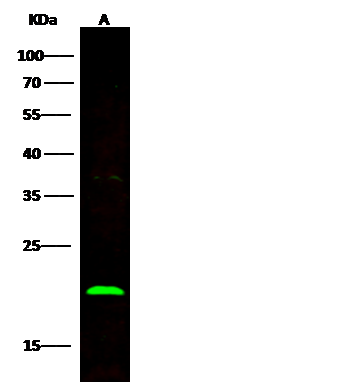 WB (Western Blot)
(Anti-BID rabbit monoclonal antibody at 1:500 dilutionLane A: Jurkat Whole Cell LysateLysates/proteins at 30 ug per lane.SecondaryGoat Anti-Rabbit IgG H&L (Dylight800) at 1/10000 dilution.Developed using the Odyssey technique.Performed under reducing conditions.Predicted band size:22 kDaObserved band size:22 kDa)
WB (Western Blot)
(Anti-BID rabbit monoclonal antibody at 1:500 dilutionLane A: Jurkat Whole Cell LysateLysates/proteins at 30 ug per lane.SecondaryGoat Anti-Rabbit IgG H&L (Dylight800) at 1/10000 dilution.Developed using the Odyssey technique.Performed under reducing conditions.Predicted band size:22 kDaObserved band size:22 kDa)
BID, Monoclonal Antibody (Cat# AAA254944)
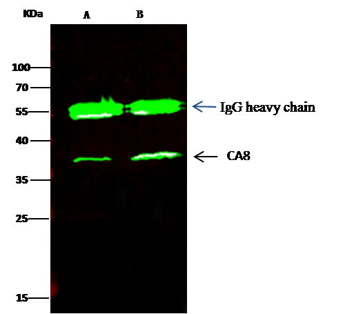 IP (Immunoprecipitation)
(CA8 was immunoprecipitated using:Lane A:0.5 mg Hela Whole Cell LysateLane B:0.5 mg A549 Whole Cell Lysate0.5 uL anti-CA8 rabbit monoclonal antibody and 15 ul of 50 % Protein G agarose.Primary antibody:Anti-CA8 rabbit monoclonal antibody,at 1:150 dilutionSecondary antibody:Dylight 800-labeled antibody to rabbit IgG (H+L), at 1:5000 dilutionDeveloped using the odssey technique.Performed under reducing conditions.Predicted band size: 37 kDaObserved band size: 37 kDa)
IP (Immunoprecipitation)
(CA8 was immunoprecipitated using:Lane A:0.5 mg Hela Whole Cell LysateLane B:0.5 mg A549 Whole Cell Lysate0.5 uL anti-CA8 rabbit monoclonal antibody and 15 ul of 50 % Protein G agarose.Primary antibody:Anti-CA8 rabbit monoclonal antibody,at 1:150 dilutionSecondary antibody:Dylight 800-labeled antibody to rabbit IgG (H+L), at 1:5000 dilutionDeveloped using the odssey technique.Performed under reducing conditions.Predicted band size: 37 kDaObserved band size: 37 kDa)
Carbonic Anhydrase 8/CA8, Monoclonal Antibody (Cat# AAA254947)
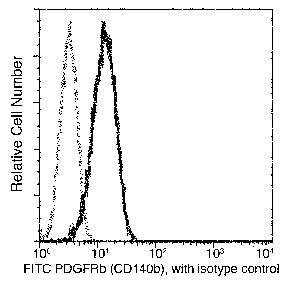 FCM/FACS (Flow Cytometry)
(Flow cytometric analysis of Human PDGFRbeta (CD140b) expression on MG63 cells. Cells were stained with FITC-conjugated anti-Human PDGFRbeta (CD140b). The fluorescence histograms were derived from gated events with the forward and side light-scatter characteristics of intact cells.)
FCM/FACS (Flow Cytometry)
(Flow cytometric analysis of Human PDGFRbeta (CD140b) expression on MG63 cells. Cells were stained with FITC-conjugated anti-Human PDGFRbeta (CD140b). The fluorescence histograms were derived from gated events with the forward and side light-scatter characteristics of intact cells.)
PDGFRB, Monoclonal Antibody (Cat# AAA254981)
CD36, Monoclonal Antibody (Cat# AAA255139)
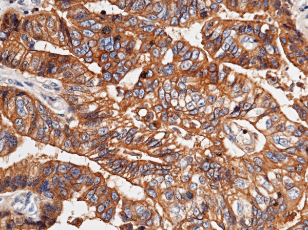
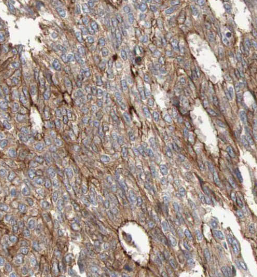
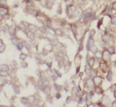
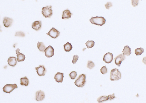
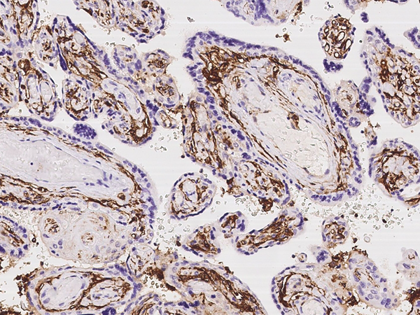 IHC (Immunohistochemisry)
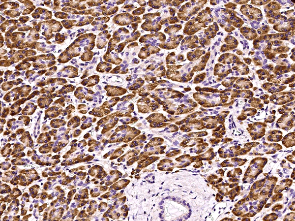
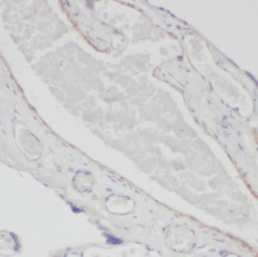
(Immunochemical staining of human CD36 in human placenta with rabbit monoclonal antibody at 1:200 dilution, formalin-fixed paraffin embedded sections.)
IHC (Immunohistochemisry)
(Immunochemical staining of human CD36 in human placenta with rabbit monoclonal antibody at 1:200 dilution, formalin-fixed paraffin embedded sections.)
CD36, Monoclonal Antibody (Cat# AAA255141)
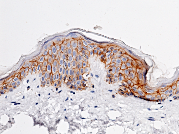
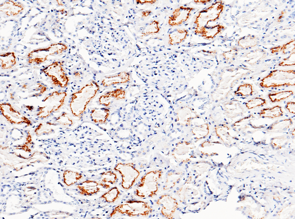
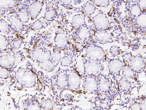
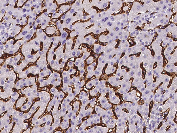
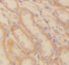
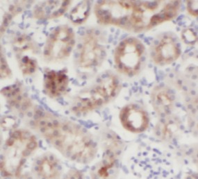
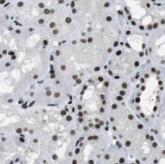
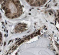
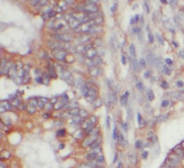
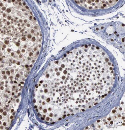
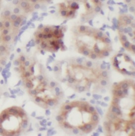
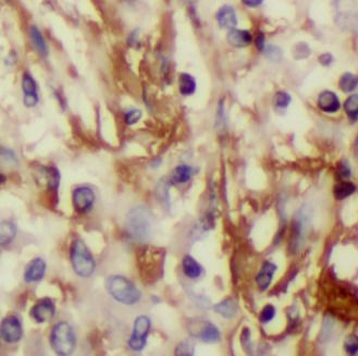
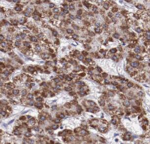
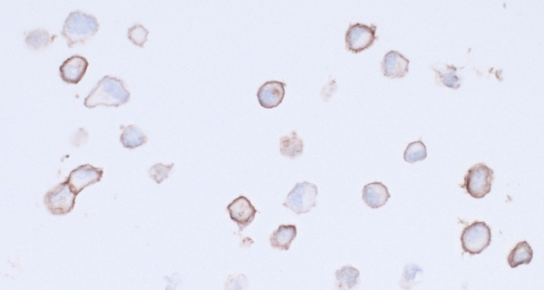
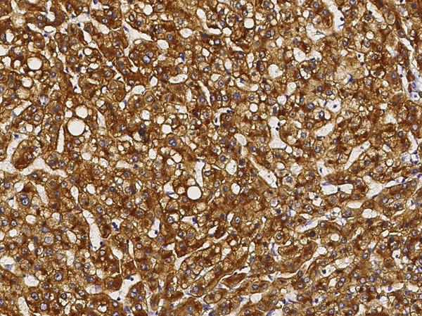 IHC (Immunohiostchemistry)
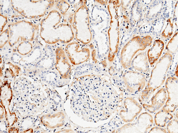
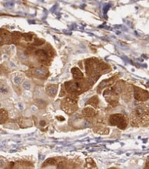
(Immunochemical staining of human ASGR1 in human cirrhosis with rabbit monoclonal antibody (1:200, formalin-fixed paraffin embedded sections).)
IHC (Immunohiostchemistry)
(Immunochemical staining of human ASGR1 in human cirrhosis with rabbit monoclonal antibody (1:200, formalin-fixed paraffin embedded sections).)
Asialoglycoprotein Receptor, Monoclonal Antibody (Cat# AAA255154)
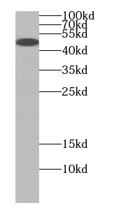
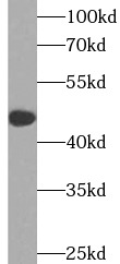
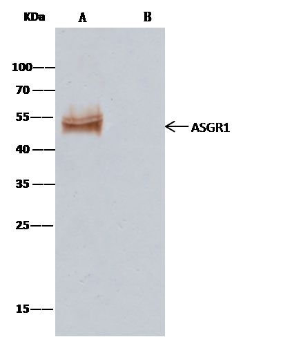 IP (Immunoprecipitation)
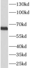
(ASGR1 was immunoprecipitated using:Lane A:0.5 mg HepG2 Whole Cell Lysate2 uL anti-ASGR1 rabbit monoclonal antibody and 15 ul of 50 % Protein G agarose.Primary antibody:Anti-ASGR1 rabbit monoclonal antibody,at 1:100 dilutionSecondary antibody:Clean-Blot IP Detection Reagent (HRP) at 1:1000 dilutionDeveloped using the DAB staining technique.Performed under reducing conditions.Predicted band size: 50 kDaObserved band size: 50 kDa)
IP (Immunoprecipitation)
(ASGR1 was immunoprecipitated using:Lane A:0.5 mg HepG2 Whole Cell Lysate2 uL anti-ASGR1 rabbit monoclonal antibody and 15 ul of 50 % Protein G agarose.Primary antibody:Anti-ASGR1 rabbit monoclonal antibody,at 1:100 dilutionSecondary antibody:Clean-Blot IP Detection Reagent (HRP) at 1:1000 dilutionDeveloped using the DAB staining technique.Performed under reducing conditions.Predicted band size: 50 kDaObserved band size: 50 kDa)
Asialoglycoprotein Receptor, Monoclonal Antibody (Cat# AAA255155)
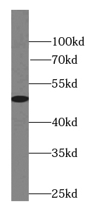
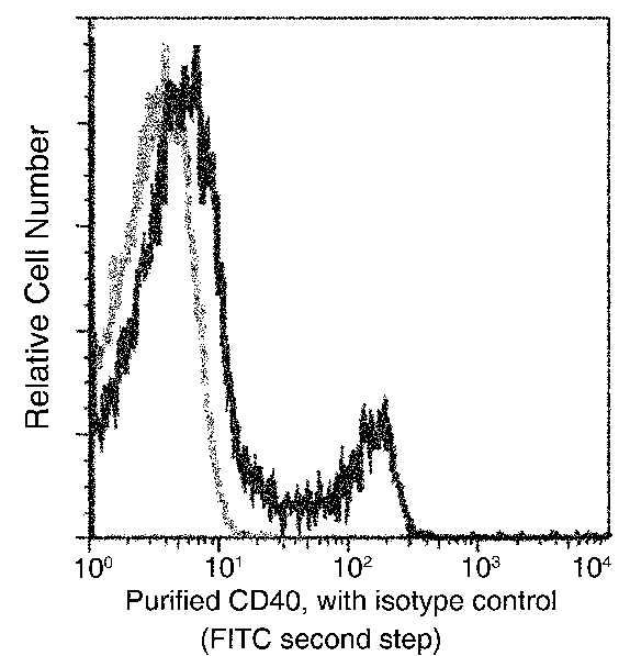 FCM/FACS (Flow Cytometry)
(Flow cytometric analysis of human CD40 expression on human whole blood lymphocytes. Human whole blood lymphocytes were stained with purified anti-Human CD40, then a FITC-conjugated second step antibody. The histogram were derived from gated events with the forward and side light-scatter characteristics of viable lymphocytes.)
FCM/FACS (Flow Cytometry)
(Flow cytometric analysis of human CD40 expression on human whole blood lymphocytes. Human whole blood lymphocytes were stained with purified anti-Human CD40, then a FITC-conjugated second step antibody. The histogram were derived from gated events with the forward and side light-scatter characteristics of viable lymphocytes.)
CD40, Monoclonal Antibody (Cat# AAA255156)
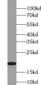
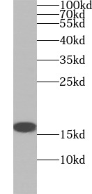
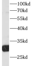
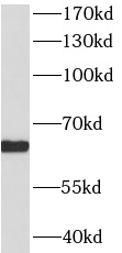
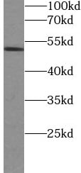
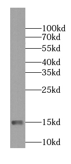 WB (Western Blot)
(human kidney tissue were subjected to SDS PAGE followed by western blot with AAA248009 (NDUFS5 antibody) at dilution of 1:1000)
WB (Western Blot)
(human kidney tissue were subjected to SDS PAGE followed by western blot with AAA248009 (NDUFS5 antibody) at dilution of 1:1000)
NDUFS5, Monoclonal Antibody (Cat# AAA248009)
Protein A+G purification
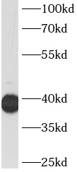
 WB (Western Blot)
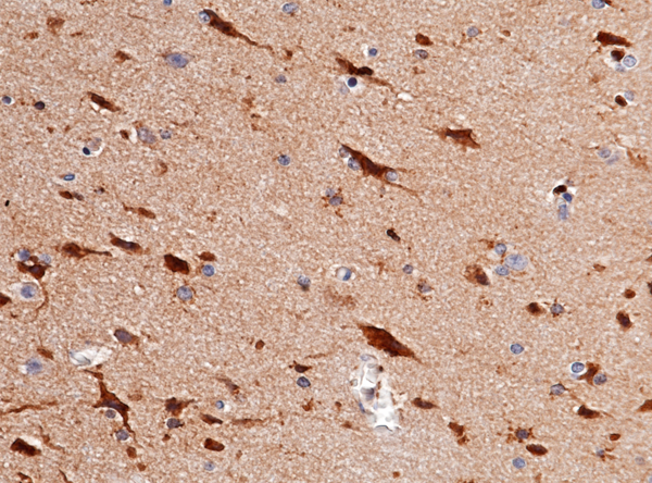
(human brain tissue were subjected to SDS PAGE followed by western blot with AAA248010 (NENF antibody) at dilution of 1:1000)
WB (Western Blot)
(human brain tissue were subjected to SDS PAGE followed by western blot with AAA248010 (NENF antibody) at dilution of 1:1000)
NENF, Monoclonal Antibody (Cat# AAA248010)
Protein A+G purification
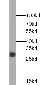
 WB (Western Blot)
(Hela cells were subjected to SDS PAGE followed by western blot with AAA248018 (NR2F6 Antibody) at dilution of 1:2000)
WB (Western Blot)
(Hela cells were subjected to SDS PAGE followed by western blot with AAA248018 (NR2F6 Antibody) at dilution of 1:2000)
NR2F6, Monoclonal Antibody (Cat# AAA248018)
Purification: Protein A+G purification
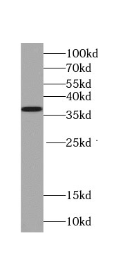
 WB (Western Blot)
(HepG2 cells were subjected to SDS PAGE followed by western blot with AAA248048 (ALPP Antibody) at dilution of 1:1000)
WB (Western Blot)
(HepG2 cells were subjected to SDS PAGE followed by western blot with AAA248048 (ALPP Antibody) at dilution of 1:1000)
PLAP, Monoclonal Antibody (Cat# AAA248048)
Protein A+G purification
 WB (Western Blot)
(human brain tissue were subjected to SDS PAGE followed by western blot with AAA248052 (TTR antibody) at dilution of 1:1000)
WB (Western Blot)
(human brain tissue were subjected to SDS PAGE followed by western blot with AAA248052 (TTR antibody) at dilution of 1:1000)
Prealbumin/transthyretin, Monoclonal Antibody (Cat# AAA248052)
Protein A+G purification
 WB (Western Blot)
(HeLa cells were subjected to SDS PAGE followed by western blot with AAA248054 (PSMD4 Antibody) at dilution of 1:1000)
WB (Western Blot)
(HeLa cells were subjected to SDS PAGE followed by western blot with AAA248054 (PSMD4 Antibody) at dilution of 1:1000)
PSMD4, Monoclonal Antibody (Cat# AAA248054)
Protein A+G purification
 WB (Western Blot)
(HSCT6 cells were subjected to SDS PAGE followed by western blot with AAA248059 (Rac1 antibody) at dilution of 1:2000)
WB (Western Blot)
(HSCT6 cells were subjected to SDS PAGE followed by western blot with AAA248059 (Rac1 antibody) at dilution of 1:2000)
Rac1, Monoclonal Antibody (Cat# AAA248059)
Purification: Protein A+G purification
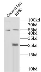
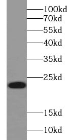
 WB (Western Blot)
(RAW 264.7 cells were subjected to SDS PAGE followed by western blot with AAA248073 (RPS3 Antibody) at dilution of 1:8000)
WB (Western Blot)
(RAW 264.7 cells were subjected to SDS PAGE followed by western blot with AAA248073 (RPS3 Antibody) at dilution of 1:8000)
RPS3, Monoclonal Antibody (Cat# AAA248073)
Purity: > = 95% as determined by SDS-PAGE
 WB (Western Blot)
(HeLa cells were subjected to SDS PAGE followed by western blot with AAA248086 (SIRT1 Antibody) at dilution of 1:3000)
WB (Western Blot)
(HeLa cells were subjected to SDS PAGE followed by western blot with AAA248086 (SIRT1 Antibody) at dilution of 1:3000)
SIRT1, Monoclonal Antibody (Cat# AAA248086)
Protein A+G purification
 WB (Western Blot)
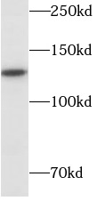
(HepG2 cells were subjected to SDS PAGE followed by western blot with AAA247934 (HDAC1 antibody) at dilution of 1:30000)
WB (Western Blot)
(HepG2 cells were subjected to SDS PAGE followed by western blot with AAA247934 (HDAC1 antibody) at dilution of 1:30000)
HDAC1, Monoclonal Antibody (Cat# AAA247934)
Protein A+G purification
 WB (Western Blot)
(0.5 ul human plasma was subjected to SDS PAGE followed by western blot with AAA247936 (HP Antibody) at dilution of 1:20000)
WB (Western Blot)
(0.5 ul human plasma was subjected to SDS PAGE followed by western blot with AAA247936 (HP Antibody) at dilution of 1:20000)
HP, Monoclonal Antibody (Cat# AAA247936)
Protein A+G purification
 WB (Western Blot)
(Recombinant protein were subjected to SDS PAGE followed by western blot with AAA247951 (IL12A Antibody) at dilution of 1:2000)
WB (Western Blot)
(Recombinant protein were subjected to SDS PAGE followed by western blot with AAA247951 (IL12A Antibody) at dilution of 1:2000)
IL12A, Monoclonal Antibody (Cat# AAA247951)
Protein A+G purification
 WB (Western Blot)
(HepG2 cells were subjected to SDS PAGE followed by western blot with AAA247966 (INHBA antibody) at dilution of 1:1000)
WB (Western Blot)
(HepG2 cells were subjected to SDS PAGE followed by western blot with AAA247966 (INHBA antibody) at dilution of 1:1000)
Inhibin beta A, Monoclonal Antibody (Cat# AAA247966)
Purity: > = 95% as determined by SDS-PAGE
 WB (Western Blot)
(HEK-293 cells were subjected to SDS PAGE followed by western blot with AAA247970 (JNK Antibody) at dilution of 1:3000)
WB (Western Blot)
(HEK-293 cells were subjected to SDS PAGE followed by western blot with AAA247970 (JNK Antibody) at dilution of 1:3000)
JNK, Monoclonal Antibody (Cat# AAA247970)
Protein A+G purification
 WB (Western Blot)
(Human brain lysate were subjected to SDS PAGE followed by western blot with AAA247991 (MGEA5 antibody) at dilution of 1:600)
WB (Western Blot)
(Human brain lysate were subjected to SDS PAGE followed by western blot with AAA247991 (MGEA5 antibody) at dilution of 1:600)
MGEA5, Monoclonal Antibody (Cat# AAA247991)
Protein A+G purification
 WB (Western Blot)
(HeLa cells were subjected to SDS PAGE followed by western blot with AAA248114 (TNFAIP1 antibody) at dilution of 1:3000)
WB (Western Blot)
(HeLa cells were subjected to SDS PAGE followed by western blot with AAA248114 (TNFAIP1 antibody) at dilution of 1:3000)
TNFAIP1, Monoclonal Antibody (Cat# AAA248114)
Protein A+G purification
 WB (Western Blot)
(HepG2 cells were subjected to SDS PAGE followed by western blot with AAA249539(Phospho-GSK3B (Ser9) antibody) at dilution of 1:1000)
WB (Western Blot)
(HepG2 cells were subjected to SDS PAGE followed by western blot with AAA249539(Phospho-GSK3B (Ser9) antibody) at dilution of 1:1000)
Phospho-GSK3B, Monoclonal Antibody (Cat# AAA249539)
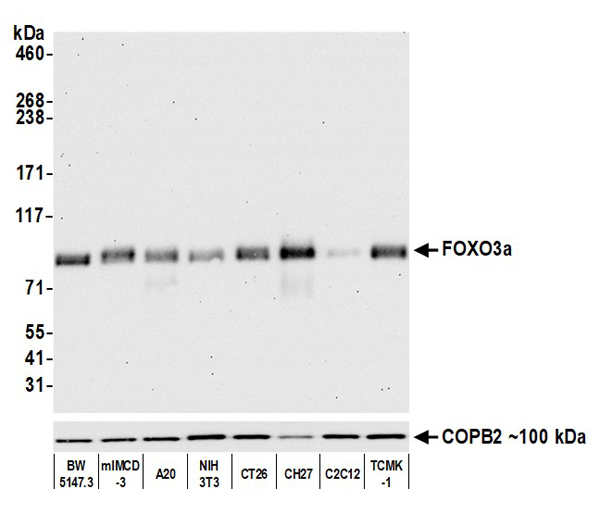
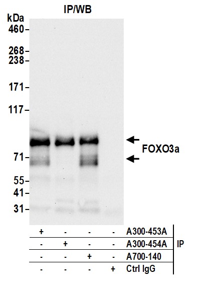
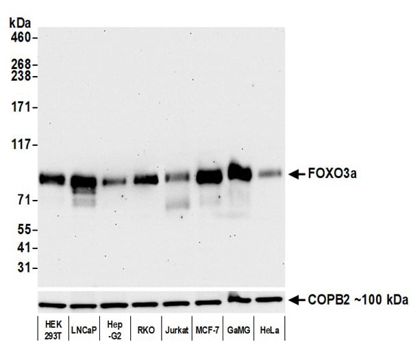 WB (Western Blot)
(Detection of human FOXO3a by western blot. Samples: Whole cell lysate (10 ug) from HEK293T, LNCaP, Hep-G2, RKO, Jurkat, MCF-7, GaMG, and HeLa cells prepared using NETN lysis buffer. Antibody: Rabbit anti-FOXO3a recombinant monoclonal antibody (AAA213601 lot 1) used at 1:1000. Secondary: HRP-conjugated goat anti-rabbit IgG . Detection: Chemiluminescence with an exposure time of 30 seconds.)
WB (Western Blot)
(Detection of human FOXO3a by western blot. Samples: Whole cell lysate (10 ug) from HEK293T, LNCaP, Hep-G2, RKO, Jurkat, MCF-7, GaMG, and HeLa cells prepared using NETN lysis buffer. Antibody: Rabbit anti-FOXO3a recombinant monoclonal antibody (AAA213601 lot 1) used at 1:1000. Secondary: HRP-conjugated goat anti-rabbit IgG . Detection: Chemiluminescence with an exposure time of 30 seconds.)
FOXO3a, Monoclonal Recombinant Antibody (Cat# AAA213601)
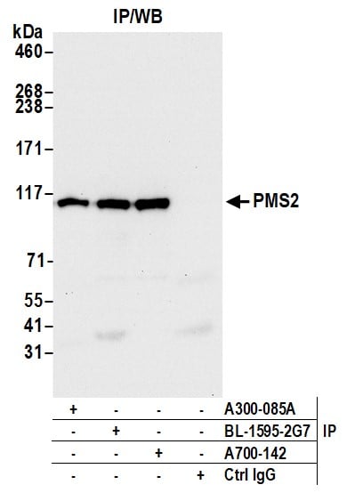
 WB (Western Blot)
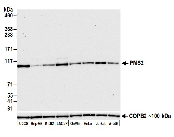
(Detection of human PMS2 by western blot. Samples: Whole cell lysate (25 ug) from U2OS, Hep-G2, K-562, LNCaP, GaMG, HeLa, Jurkat, and A-549 cells prepared using NETN lysis buffer. Antibody: Rabbit anti-PMS2 recombinant monoclonal antibody (AAA213603 lot 1) used at 1:1000. Secondary: HRP-conjugated goat anti-rabbit IgG . Detection: Chemiluminescence with an exposure time of 30 seconds. Lower Panel: Rabbit anti-COPB2 antibody .)
WB (Western Blot)
(Detection of human PMS2 by western blot. Samples: Whole cell lysate (25 ug) from U2OS, Hep-G2, K-562, LNCaP, GaMG, HeLa, Jurkat, and A-549 cells prepared using NETN lysis buffer. Antibody: Rabbit anti-PMS2 recombinant monoclonal antibody (AAA213603 lot 1) used at 1:1000. Secondary: HRP-conjugated goat anti-rabbit IgG . Detection: Chemiluminescence with an exposure time of 30 seconds. Lower Panel: Rabbit anti-COPB2 antibody .)
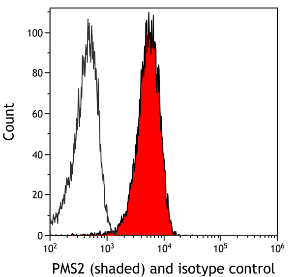
PMS2, Monoclonal Recombinant Antibody (Cat# AAA213603)
 WB (Western Blot)
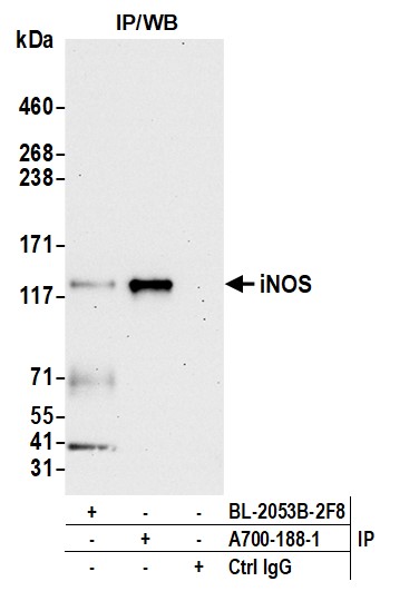
(Detection of human iNOS by western blot. Samples: Whole cell lysate (10 ug) from DLD-1 and DLD-1 induced (IFN-gamma (56 ng/mL), IL-1beta (145 pg/mL), and TNFa (10 ng/mL)) prepared using NETN lysis buffer. Antibody: Rabbit anti-iNOS recombinant monoclonal antibody (AAA213633 lot 1) used at 1:1000. Secondary: HRP-conjugated goat anti-rabbit IgG . Detection: Chemiluminescence with an exposure time of 30 seconds. Lower Panel: Rabbit anti-COPB2 antibody .)
WB (Western Blot)
(Detection of human iNOS by western blot. Samples: Whole cell lysate (10 ug) from DLD-1 and DLD-1 induced (IFN-gamma (56 ng/mL), IL-1beta (145 pg/mL), and TNFa (10 ng/mL)) prepared using NETN lysis buffer. Antibody: Rabbit anti-iNOS recombinant monoclonal antibody (AAA213633 lot 1) used at 1:1000. Secondary: HRP-conjugated goat anti-rabbit IgG . Detection: Chemiluminescence with an exposure time of 30 seconds. Lower Panel: Rabbit anti-COPB2 antibody .)
iNOS, Monoclonal Recombinant Antibody (Cat# AAA213633)
 WB (Western Blot)
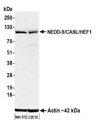
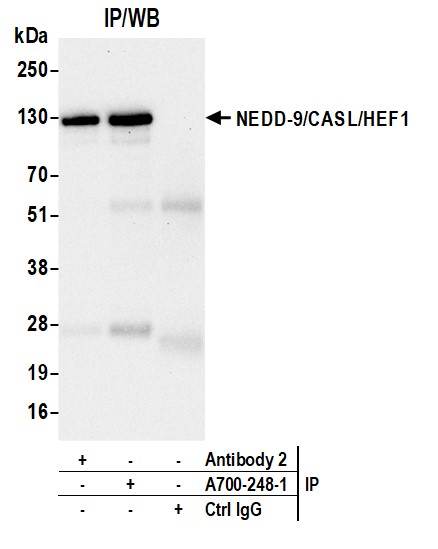
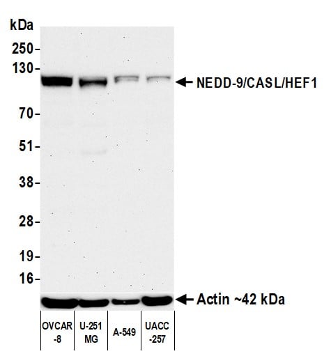
(Detection of human NEDD-9/CASL/HEF1 by western blot. Samples: Whole cell lysate (50 ug) from OVCAR-8, U-251MG, A-549, and UACC-257 cells prepared using NETN lysis buffer. Antibody: Rabbit anti-NEDD-9/CASL/HEF1 recombinant monoclonal antibody (AAA213662 lot 1) used at 1:1000. Secondary: HRP-conjugated goat anti-rabbit IgG . Detection: Chemiluminescence with an exposure time of 3 minutes. Lower Panel: Rabbit anti-Actin recombinant monoclonal antibody .)
WB (Western Blot)
(Detection of human NEDD-9/CASL/HEF1 by western blot. Samples: Whole cell lysate (50 ug) from OVCAR-8, U-251MG, A-549, and UACC-257 cells prepared using NETN lysis buffer. Antibody: Rabbit anti-NEDD-9/CASL/HEF1 recombinant monoclonal antibody (AAA213662 lot 1) used at 1:1000. Secondary: HRP-conjugated goat anti-rabbit IgG . Detection: Chemiluminescence with an exposure time of 3 minutes. Lower Panel: Rabbit anti-Actin recombinant monoclonal antibody .)
NEDD-9/CASL/HEF1, Monoclonal Recombinant Antibody (Cat# AAA213662)
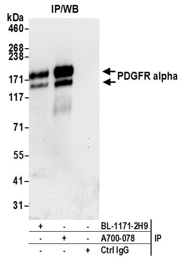
 WB (Western Blot)
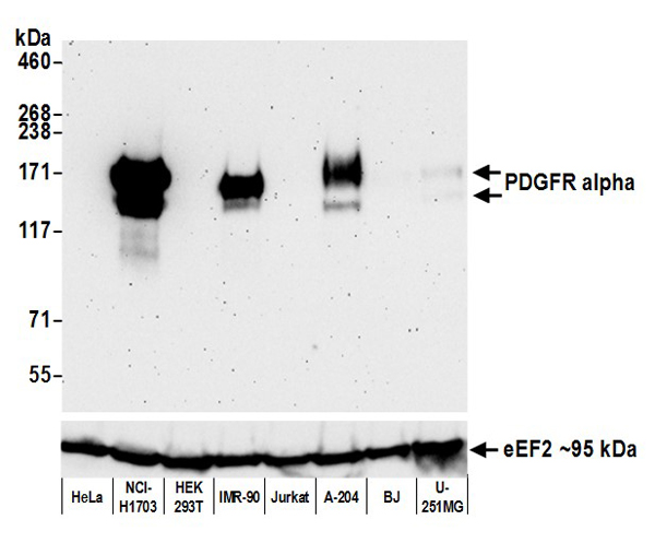
(Detection of human PDGFR alpha by western blot. Samples: Whole cell lysate (50 ug) from HeLa, NCI-H1703, HEK293T, IMR-90, Jurkat, A-204, BJ, and U-251MG cells prepared using NETN lysis buffer. Antibody: Rabbit anti-PDGFR alpha recombinant monoclonal antibody (AAA213564 lot 1) used at 1:1000. Secondary: HRP-conjugated goat anti-rabbit IgG . Detection: Chemiluminescence with an exposure time of 3 minutes. Lower Panel: Rabbit anti-eEF2 .)
WB (Western Blot)
(Detection of human PDGFR alpha by western blot. Samples: Whole cell lysate (50 ug) from HeLa, NCI-H1703, HEK293T, IMR-90, Jurkat, A-204, BJ, and U-251MG cells prepared using NETN lysis buffer. Antibody: Rabbit anti-PDGFR alpha recombinant monoclonal antibody (AAA213564 lot 1) used at 1:1000. Secondary: HRP-conjugated goat anti-rabbit IgG . Detection: Chemiluminescence with an exposure time of 3 minutes. Lower Panel: Rabbit anti-eEF2 .)
PDGFR alpha, Monoclonal Recombinant Antibody (Cat# AAA213564)
CMTM6, Monoclonal Antibody (Cat# AAA214360)
What are Monoclonal Antibodies?
Monoclonal antibodies are specialized laboratory-produced proteins developed for binding to specific biological antigens or other molecular targets. Since they come from a single cell (or clone), they are especially consistent and accurate in the data they are involved in producing.
This type of antibody material has been shown to be a powerful tool in finding and subsequently destroying harmful cells in an organism, such as those found in cancers or various autoimmune diseases. This makes them excellent aids in medical testing and research, which is why they are so widely used.
AAA Biotech offers a comprehensive range of high-quality monoclonal antibodies that perform effectively in various laboratory tests, including (amongst others) ELISA, western blotting, immunohistochemistry, and flow cytometry. All of the products in our catalog are thoroughly quality tested to make sure that they are reliable and will consistently perform well in your research.
What Are The Uses of Monoclonal Antibodies
Monoclonal antibodies are used in many lab tests, including (amongst others) ELISA, western blotting, immunohistochemistry, and flow cytometry.
ELISA is a test that helps detect a specific substance/analyte in a sample. It uses antibodies (often monoclonal) bound to a solid surface (such as the well of a microplate) to “capture” the substance/analyte in the sample and immobilize it so that the detection antibody component can then bind to it and produce a signal, which can then be measured.
Western blotting identifies specific proteins in a sample. The sample is first separated on a gel, and then antibodies are applied that will typically bind to the target, which will all be localized to a single band in a lane.
Immunohistochemistry helps locate specific proteins in cells or tissue samples using antibodies.
Flow cytometry looks at and sorts cells. It uses antibodies that are conjugated to reporter molecules called “fluorophores”, which, under special lights, emit light themselves, which can then be measured by a detector instrument.
How Monoclonal Antibodies Are Used as Medicine?
Please note that all of the products listed in AAA Biotech’s also known as AAA Bio or AAABio catalog are strictly for research-use only (RUO).
Monoclonal antibodies can also be used as therapeutic/medical treatments, particularly in the context of cancers. They are designed to find and bind to specific cells or proteins, helping the immune system recognize and attack the cancer. These treatments work in different ways, such as:
- Radioimmunotherapy attaches a small amount of radioactive molecule to the antibody, so it delivers the radiation directly to the cancer cells that the antibody is specifically binding to.
- Antibody-directed enzyme prodrug therapy uses antibodies that are specifically bound to special enzymes. These enzymes activate a harmless drug in the body and turn it into a cancer-killing drug only near the cancer cells—this helps avoid harming healthy cells.
- Immunoliposomes are tiny “bubbles” filled with medicine/drug and coated with antibodies. They carry the drug straight to the cancer cells.
Why Buy Monoclonal Antibodies From Us?
At AAA Biotech, we provide high-performance monoclonal antibodies designed to support a wide range of research needs.
1. Validated for Versatile Applications
The antibodies in our catalog are extensively validated and compatible with multiple techniques, including (but not limited to) ELISA, flow cytometry (FC), immunocytochemistry (ICC), immunofluorescence (IF), immunohistochemistry (IHC), immunoprecipitation (IP), and western blotting (WB).
2. Wide Selection & Specialized Options
We offer antibodies for common and rare species, that are available in various conjugated forms, and also in recombinant formats. Essentially, there is almost anything one might need to meet their experimental model’s requirements.
3. High-Quality Proteins
Our proteins meet high purity standards—90% or more as confirmed by SDS-PAGE. Many are available with tags like His, Flag, GST, or MBP, and we also supply native and biologically active proteins for functional studies.
Frequently Asked Questions
1. Are your monoclonal antibodies validated for specific applications?
Yes, our antibodies are tested and validated for use in methods such as ELISA, western blot, IHC, flow cytometry, and more. Refer to specific product pages or datasheets for individual product information.
2. How do I choose the right monoclonal antibody for my application?
Review the product details directly for application validation, species reactivity, and target information. You may also contact our support team at any time for help.
3. How quickly can I receive my order?
Most orders are processed and shipped within 1–3 business days, depending on product availability and your shipping location.