Filters
▼Clonality
▼Type
▼Reactivity
▼Gene Name
▼Isotype
▼Host
▼Application
▼Clone
▼Monoclonal Antibodies
Get accurate results in your research with our Monoclonal Antibodies, which are specially made to target exactly what you require for your research, and will produce consistent, reliable performance in lab tests.
Viewing 4200-4250 of 27597 product results
 IHC (Immunohistochemisry)
(DAB staining on IHC-P; Samples: Rat Spinal Cord Tissue.)
IHC (Immunohistochemisry)
(DAB staining on IHC-P; Samples: Rat Spinal Cord Tissue.)
Receptor Activator Of Nuclear Factor Kappa B Ligand (RANkL), Monoclonal Antibody (Cat# AAA146519)
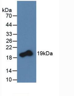
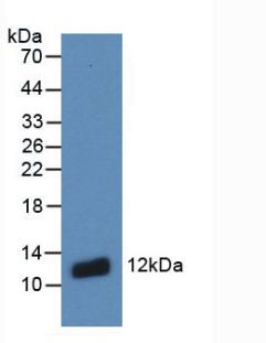
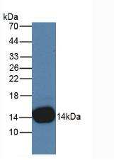
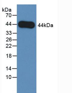
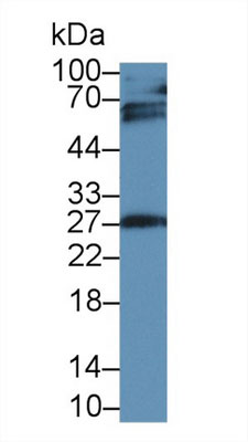
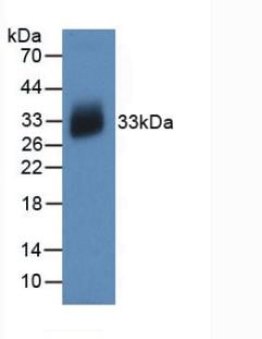
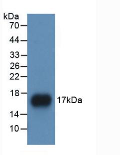
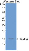
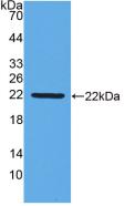
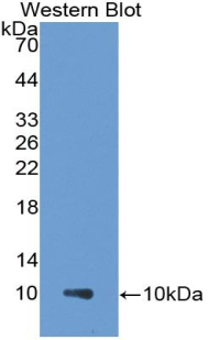
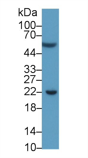
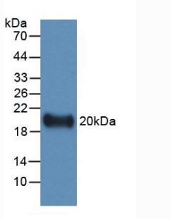 WB (Western Blot)
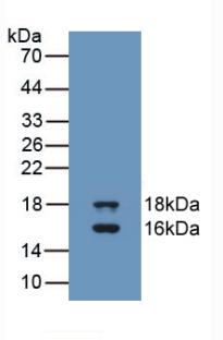
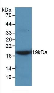
(Western Blot: Sample: Recombinant IL19, Human.)
WB (Western Blot)
(Western Blot: Sample: Recombinant IL19, Human.)
Interleukin 19 (IL19), Monoclonal Antibody (Cat# AAA146530)


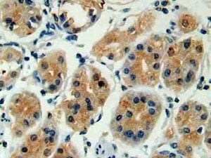
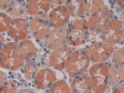
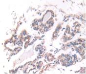
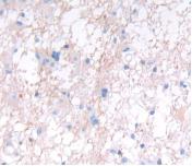
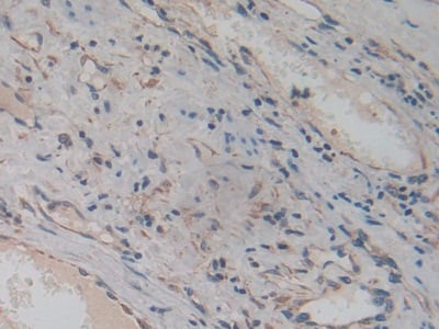
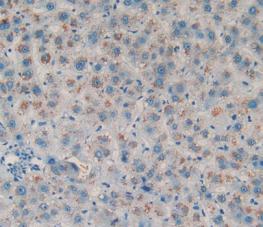
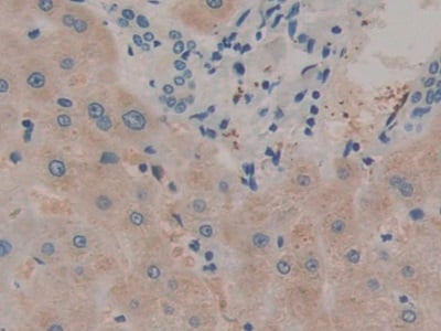
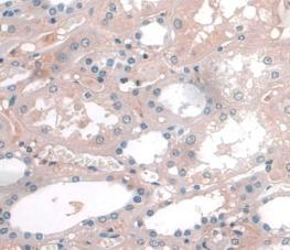
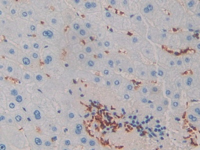
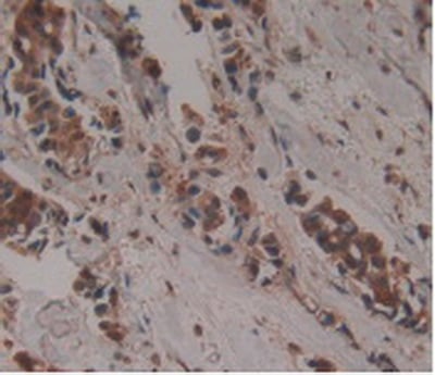
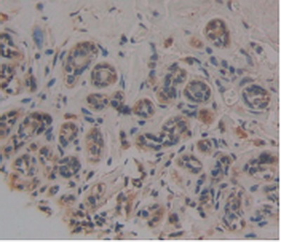
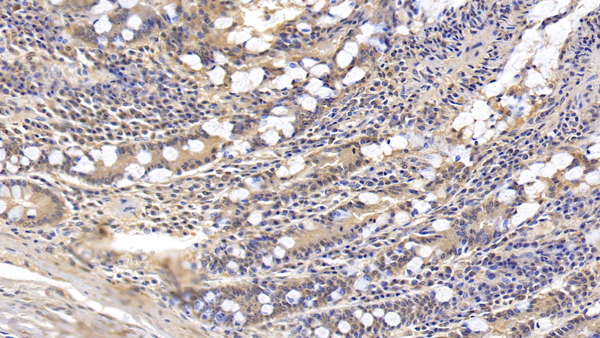
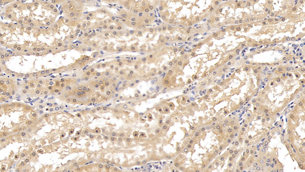
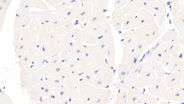
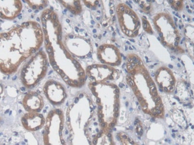 IHC (Immunohistochemisry)
(DAB staining on IHC-P; Samples: Human Kidney Tissue))
IHC (Immunohistochemisry)
(DAB staining on IHC-P; Samples: Human Kidney Tissue))
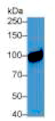
Aspartate Aminotransferase (AST), Monoclonal Antibody (Cat# AAA146531)
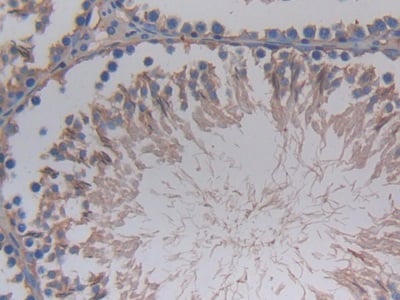
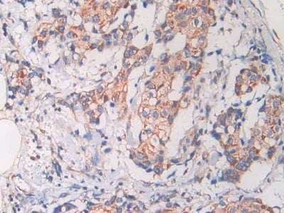
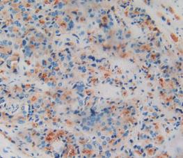
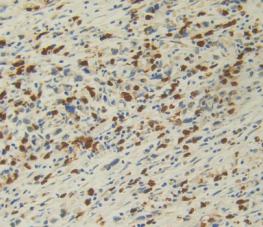
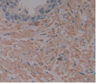
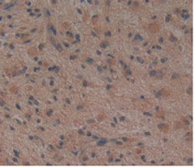
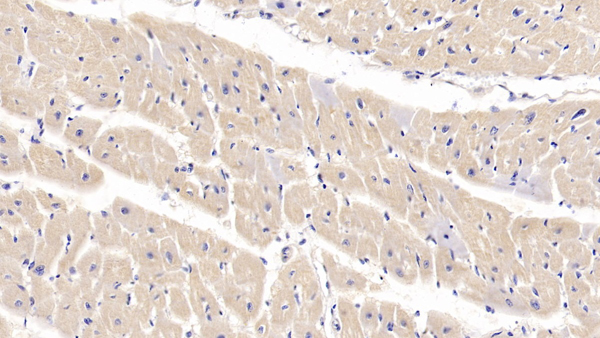
 IHC (Immunohistochemisry)
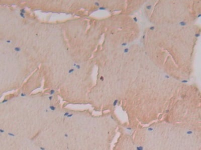
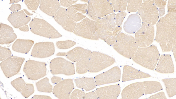
(DAB staining on IHC-P; Samples: Rat Skeletal muscle Tissue))
IHC (Immunohistochemisry)
(DAB staining on IHC-P; Samples: Rat Skeletal muscle Tissue))
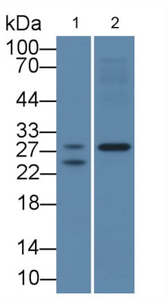
Corticosteroid Binding Globulin (CBG), Monoclonal Antibody (Cat# AAA146533)
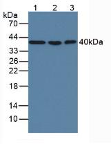
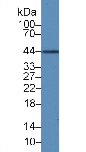
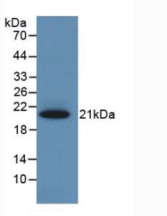
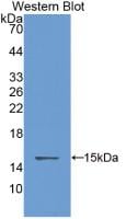
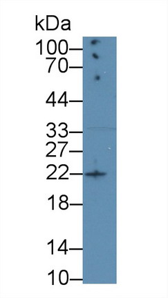
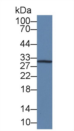
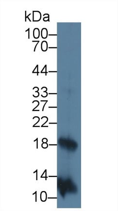
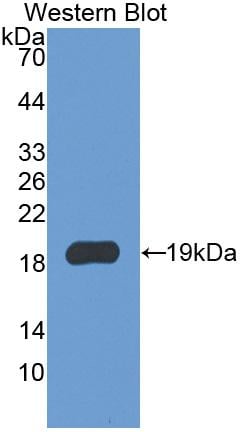
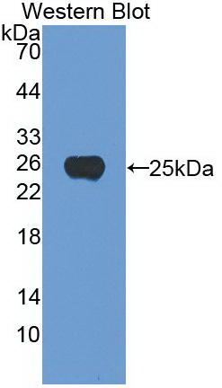
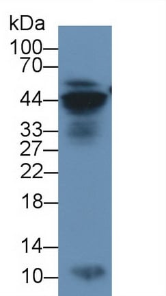
 WB (Western Blot)
(Western Blot: Sample: Recombinant MBP, Rat.)
WB (Western Blot)
(Western Blot: Sample: Recombinant MBP, Rat.)
Major Basic Protein (MBP), Monoclonal Antibody (Cat# AAA146538)
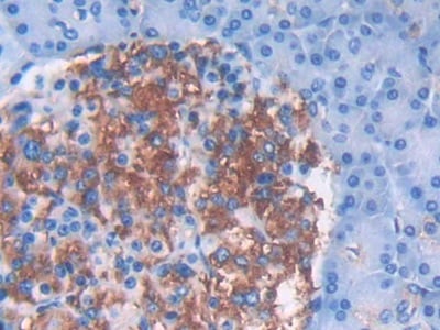
 IHC (Immunohistochemisry)
(DAB staining on IHC-P; Samples: Human Stomach TissuePrimary Ab: 30ug/ml Mouse Anti-Human IL28A AntibodySecond Ab: 2ug/ml HRP-Linked Caprine Anti-Mouse IgG Polyclonal Antibody)
IHC (Immunohistochemisry)
(DAB staining on IHC-P; Samples: Human Stomach TissuePrimary Ab: 30ug/ml Mouse Anti-Human IL28A AntibodySecond Ab: 2ug/ml HRP-Linked Caprine Anti-Mouse IgG Polyclonal Antibody)
Interleukin 28A (IL28A), Monoclonal Antibody (Cat# AAA146539)
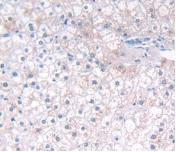
 IHC (Immunohistochemisry)
(DAB staining on IHC-P; Samples: Human Stomach cancer Tissue;Primary Ab: 20ug/ml Mouse Anti-Human IL28A AntibodySecondary Ab: 2ug/ml HRP-Linked Caprine Anti-Mouse IgG Polyclonal Antibody)
IHC (Immunohistochemisry)
(DAB staining on IHC-P; Samples: Human Stomach cancer Tissue;Primary Ab: 20ug/ml Mouse Anti-Human IL28A AntibodySecondary Ab: 2ug/ml HRP-Linked Caprine Anti-Mouse IgG Polyclonal Antibody)
Interleukin 28A (IL28A), Monoclonal Antibody (Cat# AAA146540)

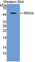
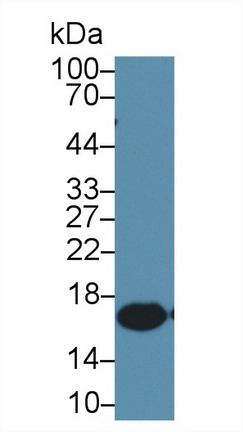
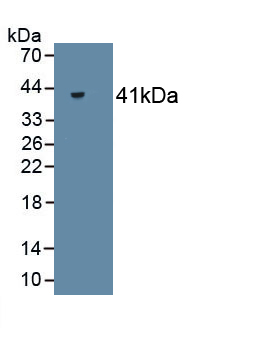
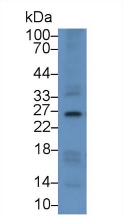
 WB (Western Blot)
(Western Blot: Sample: Recombinant S100A8, Human.)
WB (Western Blot)
(Western Blot: Sample: Recombinant S100A8, Human.)
S100 Calcium Binding Protein A8 (S100A8), Monoclonal Antibody (Cat# AAA146545)
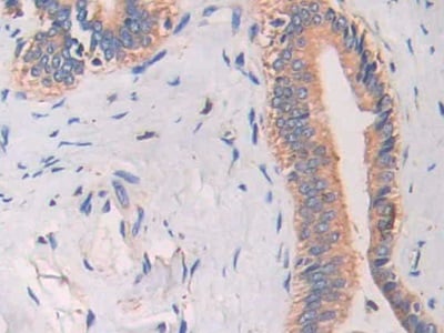 IHC (Immunohiostchemistry)
(DAB staining on IHC-P; Samples: Human Bile duct cancer Tissue)
IHC (Immunohiostchemistry)
(DAB staining on IHC-P; Samples: Human Bile duct cancer Tissue)
Cathelicidin Antimicrobial Peptide (CAMP), Monoclonal Antibody (Cat# AAA146565)
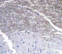
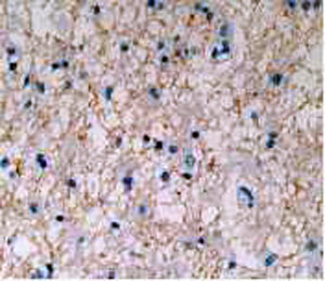
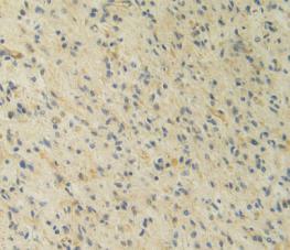
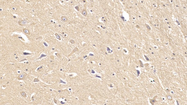
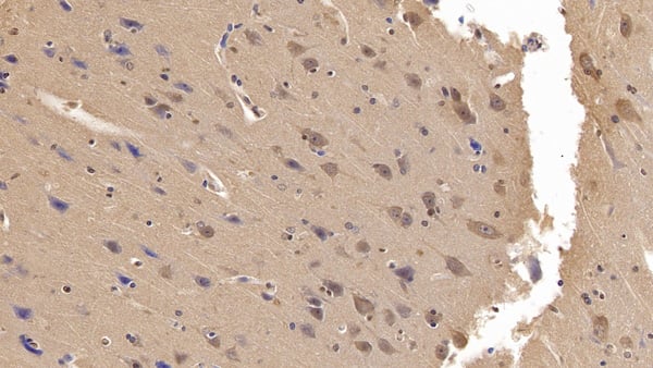
 IHC (Immunohistochemistry)
(DAB staining on IHC-P; Samples: Human Glioma Tissue.)
IHC (Immunohistochemistry)
(DAB staining on IHC-P; Samples: Human Glioma Tissue.)
Interferon Alpha 5 (IFNa5), Monoclonal Antibody (Cat# AAA146573)
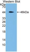
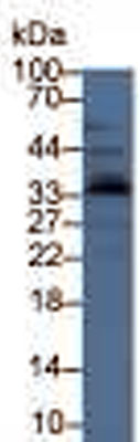
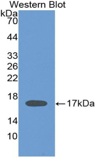
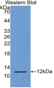
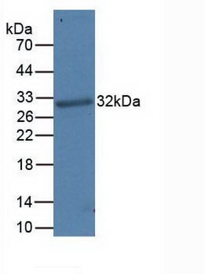
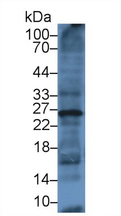
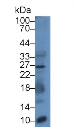
 WB (Western Blot)
(Western Blot: Sample: Recombinant CSTB, Rat.)
WB (Western Blot)
(Western Blot: Sample: Recombinant CSTB, Rat.)
Cystatin B (CSTB), Monoclonal Antibody (Cat# AAA146576)

 IHC (Immunohiostchemistry)
(DAB staining on IHC-P; Sample: Human Liver cancer Tissue; Primary Ab: 40ug/ml Mouse Anti-Multi-species Ang1-7 Antibody Second Ab: 2ug/mL HRP-Linked Caprine Anti-Mouse IgG Polyclonal Antibody)
IHC (Immunohiostchemistry)
(DAB staining on IHC-P; Sample: Human Liver cancer Tissue; Primary Ab: 40ug/ml Mouse Anti-Multi-species Ang1-7 Antibody Second Ab: 2ug/mL HRP-Linked Caprine Anti-Mouse IgG Polyclonal Antibody)
Angiotensin 1-7 (Ang1-7), Monoclonal Antibody (Cat# AAA146580)
Immunoglobulin G1 (IgG1), Monoclonal Antibody (Cat# AAA146009)
Complement Component 5a (C5a), Monoclonal Antibody (Cat# AAA146068)
Immunoglobulin G (IgG), Monoclonal Antibody (Cat# AAA146095)
Interleukin 1 Beta (IL1b), Monoclonal Antibody (Cat# AAA146099)
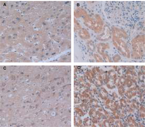 IHC (Immunohistochemistry)
(A. Human Liver TissueB. Human Kidney TissueC. Human Glioma TissueD. Human Stomach Tissue)
IHC (Immunohistochemistry)
(A. Human Liver TissueB. Human Kidney TissueC. Human Glioma TissueD. Human Stomach Tissue)
Procollagen III N-Terminal Propeptide (PIIINP), Monoclonal Antibody (Cat# AAA146104)
 IHC (Immunohistochemistry)
(DAB staining on IHC-P; Samples: Human Liver Tissue)
IHC (Immunohistochemistry)
(DAB staining on IHC-P; Samples: Human Liver Tissue)
Amphiregulin (AREG), Monoclonal Antibody (Cat# AAA146471)
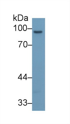
 WB (Western Blot)
(Western Blot; Sample: Rat Uterus lysate; Primary Ab: 2ug/ml Mouse Anti-Human IGFBP4 Antibody Second Ab: 0.2ug/mL HaRP-Linked Caprine Anti-Mouse IgG Polyclonal Antibody)
WB (Western Blot)
(Western Blot; Sample: Rat Uterus lysate; Primary Ab: 2ug/ml Mouse Anti-Human IGFBP4 Antibody Second Ab: 0.2ug/mL HaRP-Linked Caprine Anti-Mouse IgG Polyclonal Antibody)
Insulin Like Growth Factor Binding Protein 4 (IGFBP4), Monoclonal Antibody (Cat# AAA146477)
 IHC (Immunohistochemistry)
(DAB staining on IHC-P; Samples: Human Glioma Tissue)
IHC (Immunohistochemistry)
(DAB staining on IHC-P; Samples: Human Glioma Tissue)
Interleukin 12A (IL12A), Monoclonal Antibody (Cat# AAA146480)
 IHC (Immunohistochemistry)
(DAB staining on IHC-P;Samples: Human Kidney Tissue.)
IHC (Immunohistochemistry)
(DAB staining on IHC-P;Samples: Human Kidney Tissue.)
Apolipoprotein A1 (APOA1), Monoclonal Antibody (Cat# AAA146500)
Histamine (HA), Monoclonal Antibody (Cat# AAA146182)
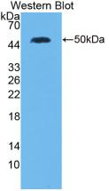
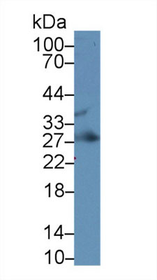
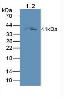
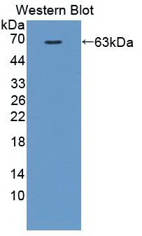
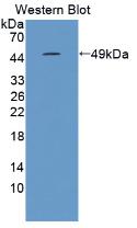
 WB (Western Blot)
(Western Blot: Sample: Recombinant FGF23, Human.)
WB (Western Blot)
(Western Blot: Sample: Recombinant FGF23, Human.)
Fibroblast Growth Factor 23, Monoclonal Antibody (Cat# AAA141270)
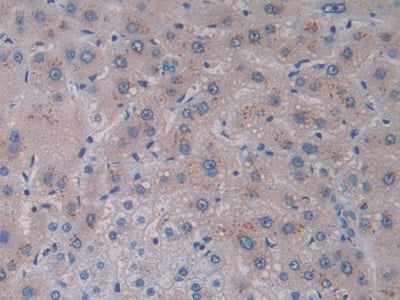
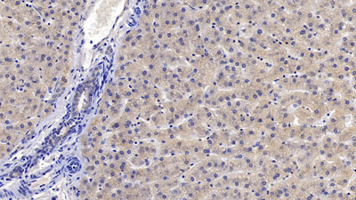
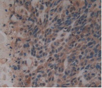
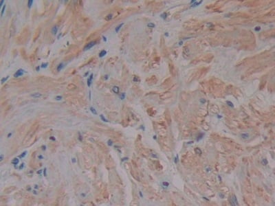
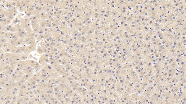
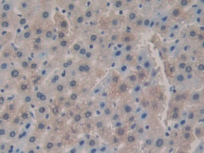
 IHC (Immunohistochemisry)
(DAB staining on IHC-P; Samples: Human Liver Tissue))
IHC (Immunohistochemisry)
(DAB staining on IHC-P; Samples: Human Liver Tissue))
Interleukin 2, Monoclonal Antibody (Cat# AAA141271)
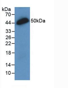
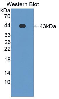
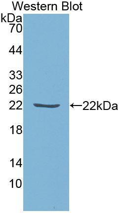
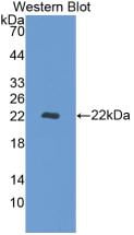
 WB (Western Blot)
(Western Blot: Sample: Recombinant IFNb, Gallus.)
WB (Western Blot)
(Western Blot: Sample: Recombinant IFNb, Gallus.)
Interferon Beta, Monoclonal Antibody (Cat# AAA141273)
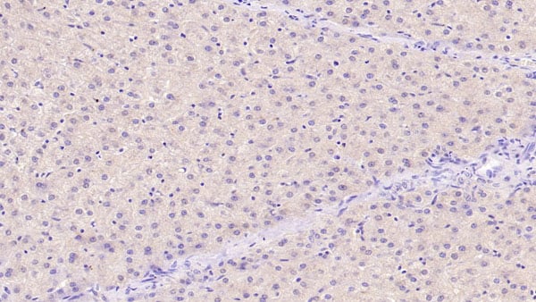
 IHC (Immunohistochemisry)
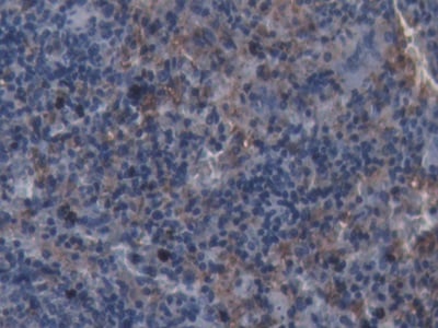
(DAB staining on IHC-P; Samples: Rat Spleen Tissue))
IHC (Immunohistochemisry)
(DAB staining on IHC-P; Samples: Rat Spleen Tissue))
Allograft Inflammatory Factor 1, Monoclonal Antibody (Cat# AAA141279)
 WB (Western Blot)
(Western Blot; Sample: Recombinant SPD, Human.)
WB (Western Blot)
(Western Blot; Sample: Recombinant SPD, Human.)
Surfactant Associated Protein D, Monoclonal Antibody (Cat# AAA141286)
 IHC (Immunohistochemistry)
(DAB staining on IHC-P;Samples: Human Breast Cancer Tissue)
IHC (Immunohistochemistry)
(DAB staining on IHC-P;Samples: Human Breast Cancer Tissue)
Growth Differentiation Factor 3, Monoclonal Antibody (Cat# AAA141289)
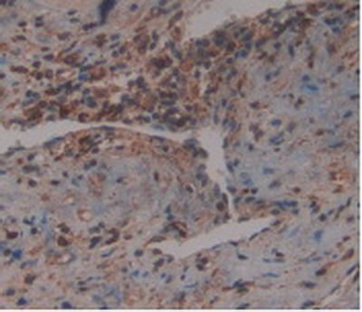
 IHC (Immunohistochemisry)
(DAB staining on IHC-P; Samples: Human Prostate Tissue))
IHC (Immunohistochemisry)
(DAB staining on IHC-P; Samples: Human Prostate Tissue))
Matrix Metalloproteinase 2, Monoclonal Antibody (Cat# AAA141294)
 IHC (Immunohiostchemistry)
(DAB staining on IHC-P; Samples: Human Liver Tissue)
IHC (Immunohiostchemistry)
(DAB staining on IHC-P; Samples: Human Liver Tissue)
Tissue Inhibitors Of Metalloproteinase 1, Monoclonal Antibody (Cat# AAA141300)
 WB (Western Blot)
(Western Blot: Sample: Recombinant S100P, Human)
WB (Western Blot)
(Western Blot: Sample: Recombinant S100P, Human)
S100 Calcium Binding Protein P, Monoclonal Antibody (Cat# AAA141302)
 WB (Western Blot)
(Western Blot: Sample: Recombinant MMP9, Rat.)
WB (Western Blot)
(Western Blot: Sample: Recombinant MMP9, Rat.)
Matrix Metalloproteinase 9, Monoclonal Antibody (Cat# AAA141363)
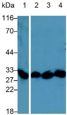 WB (Western Blot)
(Western Blot;Sample:Lane 1: Porcine Thymus lysate;Lane 2: A431 cell lysate;Lane 3: Jurkat cell lysate;Lane 4: THP1 cell lysatePrimary Ab: 1ug/ml Mouse Anti-Porcine IL1b AntibodySecond Ab: 0.2ug/mL HRP-Linked Caprine Anti-Mouse IgG Polyclonal Antibody)
WB (Western Blot)
(Western Blot;Sample:Lane 1: Porcine Thymus lysate;Lane 2: A431 cell lysate;Lane 3: Jurkat cell lysate;Lane 4: THP1 cell lysatePrimary Ab: 1ug/ml Mouse Anti-Porcine IL1b AntibodySecond Ab: 0.2ug/mL HRP-Linked Caprine Anti-Mouse IgG Polyclonal Antibody)
Interleukin 1 Beta, Monoclonal Antibody (Cat# AAA141364)
 WB (Western Blot)
(Western Blot: Sample: Recombinant PARC, Human.)
WB (Western Blot)
(Western Blot: Sample: Recombinant PARC, Human.)
Pulmonary Activation Regulated Chemokine, Monoclonal Antibody (Cat# AAA141335)


 IHC (Immunohistochemistry)
(DAB staining on IHC-P;Samples:Human Glioma Tissue.)
IHC (Immunohistochemistry)
(DAB staining on IHC-P;Samples:Human Glioma Tissue.)
Noggin, Monoclonal Antibody (Cat# AAA141336)
 WB (Western Blot)
(Western Blot:Sample: Recombinant IL10, Equine.)
WB (Western Blot)
(Western Blot:Sample: Recombinant IL10, Equine.)
Interleukin 10, Monoclonal Antibody (Cat# AAA141340)
 WB (Western Blot)
(Western Blot: Sample: RecombinantREV1,Human))
WB (Western Blot)
(Western Blot: Sample: RecombinantREV1,Human))
REV1 Homolog, Monoclonal Antibody (Cat# AAA141341)
 IHC (Immunohistochemistry)
(Formalin-fixed, paraffin-embedded human squamous cell carcinoma stained with Cytokeratin 5/6 Mouse Monoclonal Antibody (KRT5. 6/4866).)
IHC (Immunohistochemistry)
(Formalin-fixed, paraffin-embedded human squamous cell carcinoma stained with Cytokeratin 5/6 Mouse Monoclonal Antibody (KRT5. 6/4866).)
Cytokeratin 5/6, Monoclonal Antibody (Cat# AAA216091)
 WB (Western Blot)
(Western Blot: Sample: Recombinant PDCD1LG1, Human.)
WB (Western Blot)
(Western Blot: Sample: Recombinant PDCD1LG1, Human.)
Programmed Cell Death Protein 1 Ligand 1 (PDL1), Monoclonal Antibody (Cat# AAA147910)
Bone Morphogenetic Protein 10 (BMP10), Monoclonal Antibody (Cat# AAA148009)
 WB (Western Blot)
(Western Blot:Sample: Recombinant CKM, Rabbit.)
WB (Western Blot)
(Western Blot:Sample: Recombinant CKM, Rabbit.)
Creatine Kinase, Muscle (CKM), Monoclonal Antibody (Cat# AAA149423)
 IHC (Immunohistochemistry)
(DAB staining on IHC-P; Samples: Human Kidney Tissue; Primary Ab: 30ug/ml Mouse Anti-Human FAPa AntibodySecond Ab: 2ug/mL HRP-Linked Caprine Anti-Mouse IgG Polyclonal Antibody)
IHC (Immunohistochemistry)
(DAB staining on IHC-P; Samples: Human Kidney Tissue; Primary Ab: 30ug/ml Mouse Anti-Human FAPa AntibodySecond Ab: 2ug/mL HRP-Linked Caprine Anti-Mouse IgG Polyclonal Antibody)
Fibroblast Activation Protein Alpha (FAPa), Monoclonal Antibody (Cat# AAA149569)
 IHC (Immunohistochemisry)
(DAB staining on IHC-P; Samples: Human Skeletal muscle Tissue; Primary Ab: 40ug/ml Mouse Anti-Human TNNT2 AntibodySecond Ab: 2ug/mL HRP-Linked Caprine Anti-Mouse IgG Polyclonal Antibody)
IHC (Immunohistochemisry)
(DAB staining on IHC-P; Samples: Human Skeletal muscle Tissue; Primary Ab: 40ug/ml Mouse Anti-Human TNNT2 AntibodySecond Ab: 2ug/mL HRP-Linked Caprine Anti-Mouse IgG Polyclonal Antibody)
Troponin T Type 2 (TNNT2), Monoclonal Antibody (Cat# AAA149576)
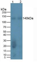
 WB (Western Blot)
(Western Blot; Sample: Porcine Kidney lysatePrimary Ab: 2ug/ml Mouse Anti-Human NEP AntibodySecond Ab: 0.2ug/mL HRP-Linked Caprine Anti-Mouse IgG Polyclonal Antibody)
WB (Western Blot)
(Western Blot; Sample: Porcine Kidney lysatePrimary Ab: 2ug/ml Mouse Anti-Human NEP AntibodySecond Ab: 0.2ug/mL HRP-Linked Caprine Anti-Mouse IgG Polyclonal Antibody)
Neprilysin (CD10), Monoclonal Antibody (Cat# AAA151125)
 WB (Western Blot)
(Western Blot; Sample: Human HT1080 cell lysate; Primary Ab: 2ug/mL Mouse Anti-Human S100 Antibody Second Ab: 0.2ug/mL HRP-Linked Caprine Anti-Mouse IgG Polyclonal Antibody)
WB (Western Blot)
(Western Blot; Sample: Human HT1080 cell lysate; Primary Ab: 2ug/mL Mouse Anti-Human S100 Antibody Second Ab: 0.2ug/mL HRP-Linked Caprine Anti-Mouse IgG Polyclonal Antibody)
Calcium Binding Protein (S100), Monoclonal Antibody (Cat# AAA150784)
 WB (Western Blot)
(Western Blot; Sample: Rat Liver lysate; Primary Ab: 3ug/mL Mouse Anti-Multi-species Ub AntibodySecond Ab: 0.2ug/mL HRP-Linked Caprine Anti-Mouse IgG Polyclonal Antibody)
WB (Western Blot)
(Western Blot; Sample: Rat Liver lysate; Primary Ab: 3ug/mL Mouse Anti-Multi-species Ub AntibodySecond Ab: 0.2ug/mL HRP-Linked Caprine Anti-Mouse IgG Polyclonal Antibody)
Ubiquitin (Ub), Monoclonal Antibody (Cat# AAA150799)
Fibroblast Activation Protein Alpha (FAPa), Monoclonal Antibody (Cat# AAA151417)
 WB (Western Blot)
(Western Blot; Sample: Lane1: Rat Cerebrum lysate; Lane2: Mouse Cerebrum lysate; Lane3: Mouse Cerebellum lysate Primary Ab: 2ug/ml Mouse AntiMouse GFAP Antibody Second Ab: 0.2ug/mL HRPLinked Caprine AntiMouse IgG Polyclonal Antibody (Catalog: SAA544Mu19))
WB (Western Blot)
(Western Blot; Sample: Lane1: Rat Cerebrum lysate; Lane2: Mouse Cerebrum lysate; Lane3: Mouse Cerebellum lysate Primary Ab: 2ug/ml Mouse AntiMouse GFAP Antibody Second Ab: 0.2ug/mL HRPLinked Caprine AntiMouse IgG Polyclonal Antibody (Catalog: SAA544Mu19))
Glial Fibrillary Acidic Protein (GFAP), Monoclonal Antibody (Cat# AAA151453)
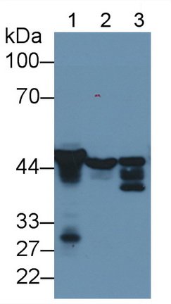
 WB (Western Blot)
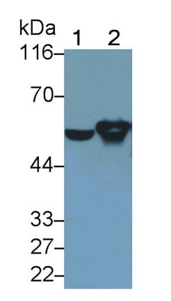
(Western Blot; Sample: Lane1: Rat Cerebrum lysate; Lane2: Rat Cerebellum lysate Primary Ab: 0.1ug/ml Mouse AntiRat GFAP Antibody Second Ab: 0.2ug/mL HRPLinked Caprine AntiMouse IgG Polyclonal Antibody (Catalog: SAA544Mu19))
WB (Western Blot)
(Western Blot; Sample: Lane1: Rat Cerebrum lysate; Lane2: Rat Cerebellum lysate Primary Ab: 0.1ug/ml Mouse AntiRat GFAP Antibody Second Ab: 0.2ug/mL HRPLinked Caprine AntiMouse IgG Polyclonal Antibody (Catalog: SAA544Mu19))
Glial Fibrillary Acidic Protein (GFAP), Monoclonal Antibody (Cat# AAA151455)
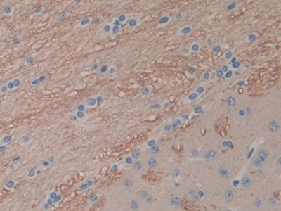
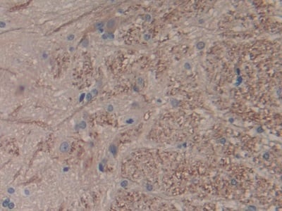
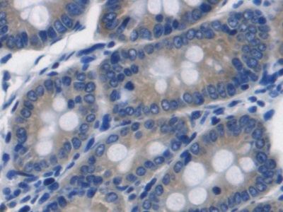
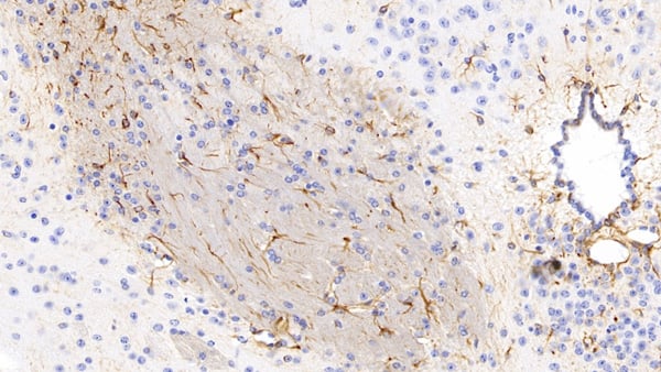
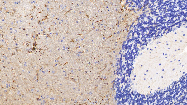
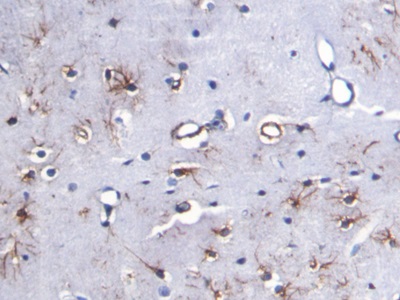
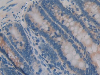 IHC (Immunohistochemistry)
(DAB staining on IHCP;Sample: Rat Intestine Tissue; Primary Ab: 30ug/ml Mouse AntiRat IL1a AntibodySecond Ab: 2ug/mL HRPLinked Caprine AntiMouse IgG Polyclonal Antibody(Catalog: SAA544Mu19))
IHC (Immunohistochemistry)
(DAB staining on IHCP;Sample: Rat Intestine Tissue; Primary Ab: 30ug/ml Mouse AntiRat IL1a AntibodySecond Ab: 2ug/mL HRPLinked Caprine AntiMouse IgG Polyclonal Antibody(Catalog: SAA544Mu19))
Interleukin 1 Alpha (IL1a), Monoclonal Antibody (Cat# AAA151458)
What are Monoclonal Antibodies?
Monoclonal antibodies are specialized laboratory-produced proteins developed for binding to specific biological antigens or other molecular targets. Since they come from a single cell (or clone), they are especially consistent and accurate in the data they are involved in producing.
This type of antibody material has been shown to be a powerful tool in finding and subsequently destroying harmful cells in an organism, such as those found in cancers or various autoimmune diseases. This makes them excellent aids in medical testing and research, which is why they are so widely used.
AAA Biotech offers a comprehensive range of high-quality monoclonal antibodies that perform effectively in various laboratory tests, including (amongst others) ELISA, western blotting, immunohistochemistry, and flow cytometry. All of the products in our catalog are thoroughly quality tested to make sure that they are reliable and will consistently perform well in your research.
What Are The Uses of Monoclonal Antibodies
Monoclonal antibodies are used in many lab tests, including (amongst others) ELISA, western blotting, immunohistochemistry, and flow cytometry.
ELISA is a test that helps detect a specific substance/analyte in a sample. It uses antibodies (often monoclonal) bound to a solid surface (such as the well of a microplate) to “capture” the substance/analyte in the sample and immobilize it so that the detection antibody component can then bind to it and produce a signal, which can then be measured.
Western blotting identifies specific proteins in a sample. The sample is first separated on a gel, and then antibodies are applied that will typically bind to the target, which will all be localized to a single band in a lane.
Immunohistochemistry helps locate specific proteins in cells or tissue samples using antibodies.
Flow cytometry looks at and sorts cells. It uses antibodies that are conjugated to reporter molecules called “fluorophores”, which, under special lights, emit light themselves, which can then be measured by a detector instrument.
How Monoclonal Antibodies Are Used as Medicine?
Please note that all of the products listed in AAA Biotech’s also known as AAA Bio or AAABio catalog are strictly for research-use only (RUO).
Monoclonal antibodies can also be used as therapeutic/medical treatments, particularly in the context of cancers. They are designed to find and bind to specific cells or proteins, helping the immune system recognize and attack the cancer. These treatments work in different ways, such as:
- Radioimmunotherapy attaches a small amount of radioactive molecule to the antibody, so it delivers the radiation directly to the cancer cells that the antibody is specifically binding to.
- Antibody-directed enzyme prodrug therapy uses antibodies that are specifically bound to special enzymes. These enzymes activate a harmless drug in the body and turn it into a cancer-killing drug only near the cancer cells—this helps avoid harming healthy cells.
- Immunoliposomes are tiny “bubbles” filled with medicine/drug and coated with antibodies. They carry the drug straight to the cancer cells.
Why Buy Monoclonal Antibodies From Us?
At AAA Biotech, we provide high-performance monoclonal antibodies designed to support a wide range of research needs.
1. Validated for Versatile Applications
The antibodies in our catalog are extensively validated and compatible with multiple techniques, including (but not limited to) ELISA, flow cytometry (FC), immunocytochemistry (ICC), immunofluorescence (IF), immunohistochemistry (IHC), immunoprecipitation (IP), and western blotting (WB).
2. Wide Selection & Specialized Options
We offer antibodies for common and rare species, that are available in various conjugated forms, and also in recombinant formats. Essentially, there is almost anything one might need to meet their experimental model’s requirements.
3. High-Quality Proteins
Our proteins meet high purity standards—90% or more as confirmed by SDS-PAGE. Many are available with tags like His, Flag, GST, or MBP, and we also supply native and biologically active proteins for functional studies.
Frequently Asked Questions
1. Are your monoclonal antibodies validated for specific applications?
Yes, our antibodies are tested and validated for use in methods such as ELISA, western blot, IHC, flow cytometry, and more. Refer to specific product pages or datasheets for individual product information.
2. How do I choose the right monoclonal antibody for my application?
Review the product details directly for application validation, species reactivity, and target information. You may also contact our support team at any time for help.
3. How quickly can I receive my order?
Most orders are processed and shipped within 1–3 business days, depending on product availability and your shipping location.