Filters
▼Clonality
▼Type
▼Reactivity
▼Gene Name
▼Isotype
▼Host
▼Application
▼Clone
▼Monoclonal Antibodies
Get accurate results in your research with our Monoclonal Antibodies, which are specially made to target exactly what you require for your research, and will produce consistent, reliable performance in lab tests.
Viewing 250-300 of 27560 product results
 WB (Western Blot)
(Western blot analysis of TWEAKR expression in HUVEC cell lysate)
WB (Western Blot)
(Western blot analysis of TWEAKR expression in HUVEC cell lysate)
TWEAKR, Monoclonal Antibody (Cat# AAA124487)
 WB (Western Blot)
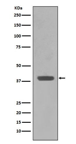
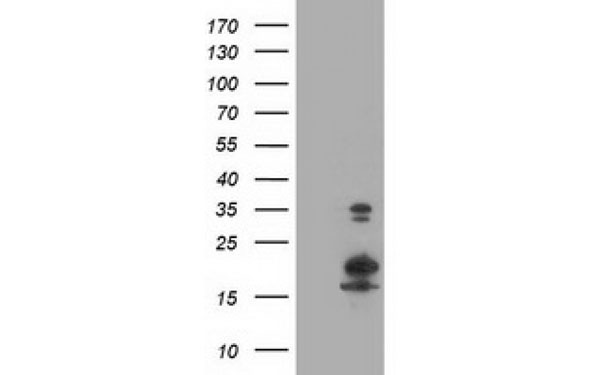
(Western blot analysis of MAD2L1BP expression in A431 cell lysate (AAA124492).Electrophoresis was performed on a 5-20% SDS-PAGE gel at 70V (Stacking gel) / 90V (Resolving gel) for 2-3 hours. The sample well of each lane was loaded with 50ug of sample under reducing conditions.After Electrophoresis, proteins were transferred to a Nitrocellulose membrane at 150mA for 50-90 minutes. Blocked the membrane with 5% Non-fat Milk/ TBS for 1.5 hour at RT. The membrane was incubated with rabbit anti-MAD2L1BP monoclonal antibody overnight at 4 degree C, then washed with TBS-0.1%Tween 3 times with 5 minutes each and probed with a goat anti-rabbit IgG-HRP secondary antibody at a dilution of 1:10000 for 1.5 hour at RT. The signal is developed using an Enhanced Chemiluminescent detection (ECL) kit with Tanon 5200 system. A specific band was detected for MAD2L1BP)
WB (Western Blot)
(Western blot analysis of MAD2L1BP expression in A431 cell lysate (AAA124492).Electrophoresis was performed on a 5-20% SDS-PAGE gel at 70V (Stacking gel) / 90V (Resolving gel) for 2-3 hours. The sample well of each lane was loaded with 50ug of sample under reducing conditions.After Electrophoresis, proteins were transferred to a Nitrocellulose membrane at 150mA for 50-90 minutes. Blocked the membrane with 5% Non-fat Milk/ TBS for 1.5 hour at RT. The membrane was incubated with rabbit anti-MAD2L1BP monoclonal antibody overnight at 4 degree C, then washed with TBS-0.1%Tween 3 times with 5 minutes each and probed with a goat anti-rabbit IgG-HRP secondary antibody at a dilution of 1:10000 for 1.5 hour at RT. The signal is developed using an Enhanced Chemiluminescent detection (ECL) kit with Tanon 5200 system. A specific band was detected for MAD2L1BP)
MAD2L1BP/Mad2L1 Binding Protein, Monoclonal Antibody (Cat# AAA124492)
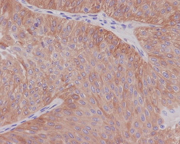
 IHC (Immunohiostchemistry)
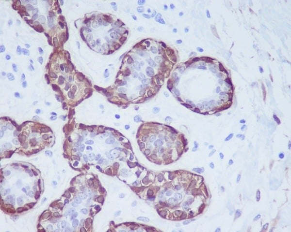
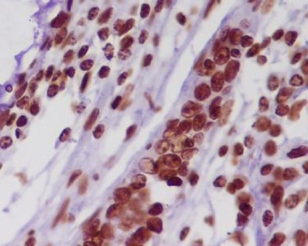
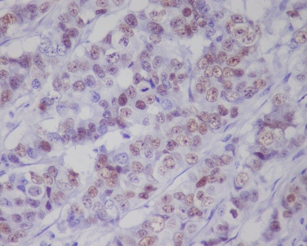
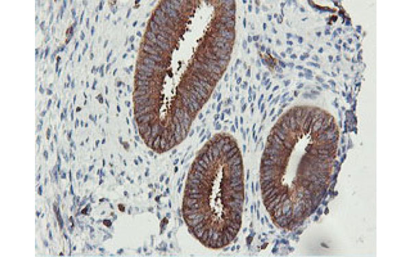
(Immunohistochemical analysis of paraffin-embedded human breast, using Calponin Antibody(AAA124495)CNN1 was detected in paraffin-embedded tissue section. Heat mediated antigen retrieval was performed in citrate buffer (pH6, epitope retrieval solution) for 20 mins. The tissue section was blocked with 10% goat serum. The tissue section was then incubated with 1ug/ml rabbit anti-CNN1 Antibody (AAA124495)overnight at 4 degree C. Biotinylated goat anti-rabbit IgG was used as secondary antibody and incubated for 30 minutes at 37 degree C. The tissue section was developed using Strepavidin-Biotin-Complex (SABC) with DAB as the chromogen.)
IHC (Immunohiostchemistry)
(Immunohistochemical analysis of paraffin-embedded human breast, using Calponin Antibody(AAA124495)CNN1 was detected in paraffin-embedded tissue section. Heat mediated antigen retrieval was performed in citrate buffer (pH6, epitope retrieval solution) for 20 mins. The tissue section was blocked with 10% goat serum. The tissue section was then incubated with 1ug/ml rabbit anti-CNN1 Antibody (AAA124495)overnight at 4 degree C. Biotinylated goat anti-rabbit IgG was used as secondary antibody and incubated for 30 minutes at 37 degree C. The tissue section was developed using Strepavidin-Biotin-Complex (SABC) with DAB as the chromogen.)
Calponin, Monoclonal Antibody (Cat# AAA124495)
 WB (Western Blot)
(Western blot analysis of alpha Tubulin 4A in NIH/3T3 cell lysate (AAA124498).Electrophoresis was performed on a 5-20% SDS-PAGE gel at 70V (Stacking gel) / 90V (Resolving gel) for 2-3 hours. The sample well of each lane was loaded with 50ug of sample under reducing conditions.After Electrophoresis, proteins were transferred to a Nitrocellulose membrane at 150mA for 50-90 minutes. Blocked the membrane with 5% Non-fat Milk/ TBS for 1.5 hour at RT. The membrane was incubated with rabbit anti-TUBA1B monoclonal antibody overnight at 4 degree C, then washed with TBS-0.1%Tween 3 times with 5 minutes each and probed with a goat anti-rabbit IgG-HRP secondary antibody at a dilution of 1:10000 for 1.5 hour at RT. The signal is developed using an Enhanced Chemiluminescent detection (ECL) kit with Tanon 5200 system. A specific band was detected for TUBA1B)
WB (Western Blot)
(Western blot analysis of alpha Tubulin 4A in NIH/3T3 cell lysate (AAA124498).Electrophoresis was performed on a 5-20% SDS-PAGE gel at 70V (Stacking gel) / 90V (Resolving gel) for 2-3 hours. The sample well of each lane was loaded with 50ug of sample under reducing conditions.After Electrophoresis, proteins were transferred to a Nitrocellulose membrane at 150mA for 50-90 minutes. Blocked the membrane with 5% Non-fat Milk/ TBS for 1.5 hour at RT. The membrane was incubated with rabbit anti-TUBA1B monoclonal antibody overnight at 4 degree C, then washed with TBS-0.1%Tween 3 times with 5 minutes each and probed with a goat anti-rabbit IgG-HRP secondary antibody at a dilution of 1:10000 for 1.5 hour at RT. The signal is developed using an Enhanced Chemiluminescent detection (ECL) kit with Tanon 5200 system. A specific band was detected for TUBA1B)
alpha Tubulin, Monoclonal Antibody (Cat# AAA124498)
 WB (Western Blot)
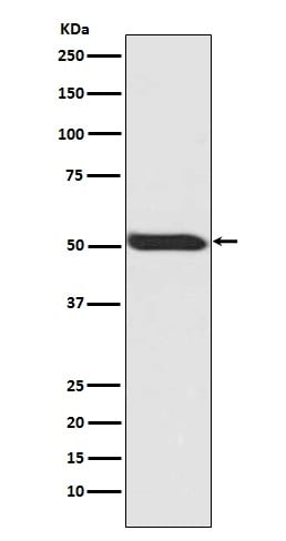
(Western blot analysis of HSPA12A expression in U-87 MG cell lysate (AAA124507).Electrophoresis was performed on a 5-20% SDS-PAGE gel at 70V (Stacking gel) / 90V (Resolving gel) for 2-3 hours. The sample well of each lane was loaded with 50ug of sample under reducing conditions.After Electrophoresis, proteins were transferred to a Nitrocellulose membrane at 150mA for 50-90 minutes. Blocked the membrane with 5% Non-fat Milk/ TBS for 1.5 hour at RT. The membrane was incubated with rabbit anti-HSPA12A monoclonal antibody overnight at 4 degree C, then washed with TBS-0.1%Tween 3 times with 5 minutes each and probed with a goat anti-rabbit IgG-HRP secondary antibody at a dilution of 1:10000 for 1.5 hour at RT. The signal is developed using an Enhanced Chemiluminescent detection (ECL) kit with Tanon 5200 system. A specific band was detected for HSPA12A)
WB (Western Blot)
(Western blot analysis of HSPA12A expression in U-87 MG cell lysate (AAA124507).Electrophoresis was performed on a 5-20% SDS-PAGE gel at 70V (Stacking gel) / 90V (Resolving gel) for 2-3 hours. The sample well of each lane was loaded with 50ug of sample under reducing conditions.After Electrophoresis, proteins were transferred to a Nitrocellulose membrane at 150mA for 50-90 minutes. Blocked the membrane with 5% Non-fat Milk/ TBS for 1.5 hour at RT. The membrane was incubated with rabbit anti-HSPA12A monoclonal antibody overnight at 4 degree C, then washed with TBS-0.1%Tween 3 times with 5 minutes each and probed with a goat anti-rabbit IgG-HRP secondary antibody at a dilution of 1:10000 for 1.5 hour at RT. The signal is developed using an Enhanced Chemiluminescent detection (ECL) kit with Tanon 5200 system. A specific band was detected for HSPA12A)
HSPA12A, Monoclonal Antibody (Cat# AAA124507)
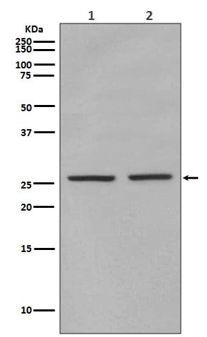
 WB (Western Blot)
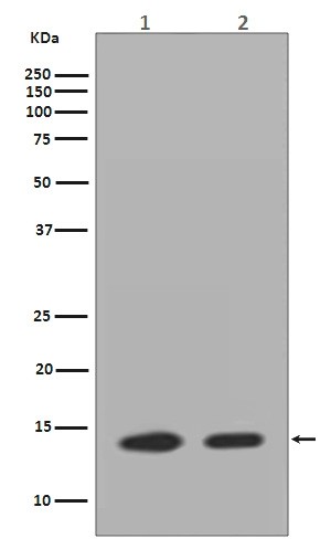
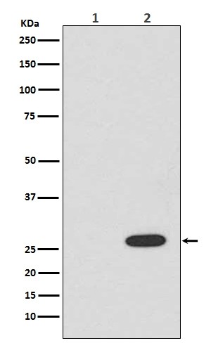
(Western blot analysis of Histone H2AZ expression in(1)Neuro-2a cell lysate;(2)HeLa cell lysate (AAA124509).Electrophoresis was performed on a 5-20% SDS-PAGE gel at 70V (Stacking gel) / 90V (Resolving gel) for 2-3 hours. The sample well of each lane was loaded with 50ug of sample under reducing conditions.After Electrophoresis, proteins were transferred to a Nitrocellulose membrane at 150mA for 50-90 minutes. Blocked the membrane with 5% Non-fat Milk/ TBS for 1.5 hour at RT. The membrane was incubated with rabbit anti-HIST1H2AB monoclonal antibody overnight at 4 degree C, then washed with TBS-0.1%Tween 3 times with 5 minutes each and probed with a goat anti-rabbit IgG-HRP secondary antibody at a dilution of 1:10000 for 1.5 hour at RT. The signal is developed using an Enhanced Chemiluminescent detection (ECL) kit with Tanon 5200 system. A specific band was detected for HIST1H2AB)
WB (Western Blot)
(Western blot analysis of Histone H2AZ expression in(1)Neuro-2a cell lysate;(2)HeLa cell lysate (AAA124509).Electrophoresis was performed on a 5-20% SDS-PAGE gel at 70V (Stacking gel) / 90V (Resolving gel) for 2-3 hours. The sample well of each lane was loaded with 50ug of sample under reducing conditions.After Electrophoresis, proteins were transferred to a Nitrocellulose membrane at 150mA for 50-90 minutes. Blocked the membrane with 5% Non-fat Milk/ TBS for 1.5 hour at RT. The membrane was incubated with rabbit anti-HIST1H2AB monoclonal antibody overnight at 4 degree C, then washed with TBS-0.1%Tween 3 times with 5 minutes each and probed with a goat anti-rabbit IgG-HRP secondary antibody at a dilution of 1:10000 for 1.5 hour at RT. The signal is developed using an Enhanced Chemiluminescent detection (ECL) kit with Tanon 5200 system. A specific band was detected for HIST1H2AB)
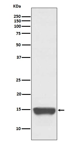
Histone H2A.Z, Monoclonal Antibody (Cat# AAA124509)
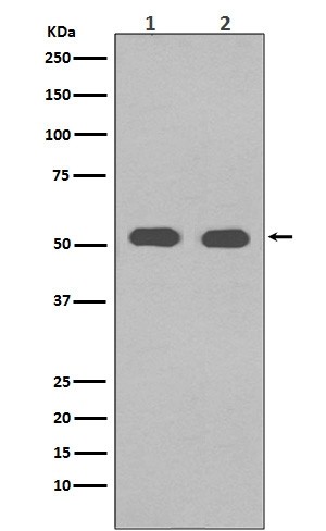
 WB (Western Blot)
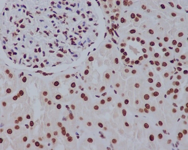
(Western blot analysis of Histone H2A expression in HeLa cell lysate (AAA124511).Electrophoresis was performed on a 5-20% SDS-PAGE gel at 70V (Stacking gel) / 90V (Resolving gel) for 2-3 hours. The sample well of each lane was loaded with 50ug of sample under reducing conditions.After Electrophoresis, proteins were transferred to a Nitrocellulose membrane at 150mA for 50-90 minutes. Blocked the membrane with 5% Non-fat Milk/ TBS for 1.5 hour at RT. The membrane was incubated with rabbit anti-HIST1H2AB monoclonal antibody overnight at 4 degree C, then washed with TBS-0.1%Tween 3 times with 5 minutes each and probed with a goat anti-rabbit IgG-HRP secondary antibody at a dilution of 1:10000 for 1.5 hour at RT. The signal is developed using an Enhanced Chemiluminescent detection (ECL) kit with Tanon 5200 system. A specific band was detected for HIST1H2AB)
WB (Western Blot)
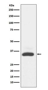
(Western blot analysis of Histone H2A expression in HeLa cell lysate (AAA124511).Electrophoresis was performed on a 5-20% SDS-PAGE gel at 70V (Stacking gel) / 90V (Resolving gel) for 2-3 hours. The sample well of each lane was loaded with 50ug of sample under reducing conditions.After Electrophoresis, proteins were transferred to a Nitrocellulose membrane at 150mA for 50-90 minutes. Blocked the membrane with 5% Non-fat Milk/ TBS for 1.5 hour at RT. The membrane was incubated with rabbit anti-HIST1H2AB monoclonal antibody overnight at 4 degree C, then washed with TBS-0.1%Tween 3 times with 5 minutes each and probed with a goat anti-rabbit IgG-HRP secondary antibody at a dilution of 1:10000 for 1.5 hour at RT. The signal is developed using an Enhanced Chemiluminescent detection (ECL) kit with Tanon 5200 system. A specific band was detected for HIST1H2AB)
Histone H2A, Monoclonal Antibody (Cat# AAA124511)
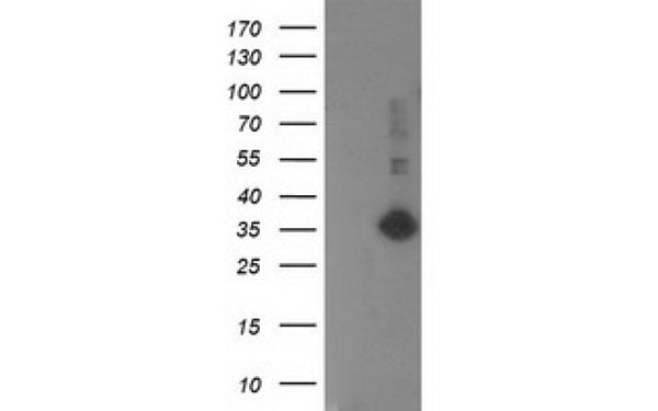
 WB (Western Blot)
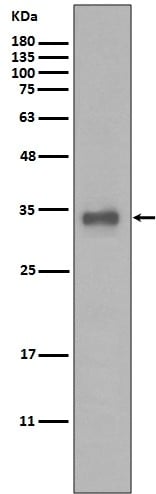
(Western blot analysis of Maltose Binding Protein expression in E.coli lysate.)
WB (Western Blot)
(Western blot analysis of Maltose Binding Protein expression in E.coli lysate.)
Maltose Binding Protein, Monoclonal Antibody (Cat# AAA124513)
 WB (Western Blot)
(Western blot analysis of RFP expression in (1) 293T cell lysate; (2) 293T cell lysate transfected with RFP (AAA124516).Electrophoresis was performed on a 5-20% SDS-PAGE gel at 70V (Stacking gel) / 90V (Resolving gel) for 2-3 hours. The sample well of each lane was loaded with 50ug of sample under reducing conditions.After Electrophoresis, proteins were transferred to a Nitrocellulose membrane at 150mA for 50-90 minutes. Blocked the membrane with 5% Non-fat Milk/ TBS for 1.5 hour at RT. The membrane was incubated with rabbit anti-RFP monoclonal antibody overnight at 4 degree C, then washed with TBS-0.1%Tween 3 times with 5 minutes each and probed with a goat anti-rabbit IgG-HRP secondary antibody at a dilution of 1:10000 for 1.5 hour at RT. The signal is developed using an Enhanced Chemiluminescent detection (ECL) kit with Tanon 5200 system. A specific band was detected for RFP)
WB (Western Blot)
(Western blot analysis of RFP expression in (1) 293T cell lysate; (2) 293T cell lysate transfected with RFP (AAA124516).Electrophoresis was performed on a 5-20% SDS-PAGE gel at 70V (Stacking gel) / 90V (Resolving gel) for 2-3 hours. The sample well of each lane was loaded with 50ug of sample under reducing conditions.After Electrophoresis, proteins were transferred to a Nitrocellulose membrane at 150mA for 50-90 minutes. Blocked the membrane with 5% Non-fat Milk/ TBS for 1.5 hour at RT. The membrane was incubated with rabbit anti-RFP monoclonal antibody overnight at 4 degree C, then washed with TBS-0.1%Tween 3 times with 5 minutes each and probed with a goat anti-rabbit IgG-HRP secondary antibody at a dilution of 1:10000 for 1.5 hour at RT. The signal is developed using an Enhanced Chemiluminescent detection (ECL) kit with Tanon 5200 system. A specific band was detected for RFP)
RFP, Monoclonal Antibody (Cat# AAA124516)
 WB (Western Blot)
(Western blot analysis of Phospho-p53 (Ser392) expression in HEK293 whole cell lysate (AAA124519).Electrophoresis was performed on a 5-20% SDS-PAGE gel at 70V (Stacking gel) / 90V (Resolving gel) for 2-3 hours. The sample well of each lane was loaded with 50ug of sample under reducing conditions.After Electrophoresis, proteins were transferred to a Nitrocellulose membrane at 150mA for 50-90 minutes. Blocked the membrane with 5% Non-fat Milk/ TBS for 1.5 hour at RT. The membrane was incubated with rabbit anti-TP53 monoclonal antibody overnight at 4 degree C, then washed with TBS-0.1%Tween 3 times with 5 minutes each and probed with a goat anti-rabbit IgG-HRP secondary antibody at a dilution of 1:10000 for 1.5 hour at RT. The signal is developed using an Enhanced Chemiluminescent detection (ECL) kit with Tanon 5200 system. A specific band was detected for TP53)
WB (Western Blot)
(Western blot analysis of Phospho-p53 (Ser392) expression in HEK293 whole cell lysate (AAA124519).Electrophoresis was performed on a 5-20% SDS-PAGE gel at 70V (Stacking gel) / 90V (Resolving gel) for 2-3 hours. The sample well of each lane was loaded with 50ug of sample under reducing conditions.After Electrophoresis, proteins were transferred to a Nitrocellulose membrane at 150mA for 50-90 minutes. Blocked the membrane with 5% Non-fat Milk/ TBS for 1.5 hour at RT. The membrane was incubated with rabbit anti-TP53 monoclonal antibody overnight at 4 degree C, then washed with TBS-0.1%Tween 3 times with 5 minutes each and probed with a goat anti-rabbit IgG-HRP secondary antibody at a dilution of 1:10000 for 1.5 hour at RT. The signal is developed using an Enhanced Chemiluminescent detection (ECL) kit with Tanon 5200 system. A specific band was detected for TP53)
p53, Monoclonal Antibody (Cat# AAA124519)
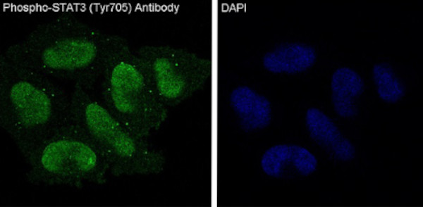 IF (Immunofluorescence)
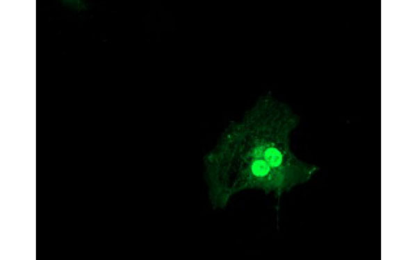
(Immunofluorescent analysis of HeLa cells treated with IFN-alpha, using Phospho-STAT3 (Y705) Antibody.)
IF (Immunofluorescence)
(Immunofluorescent analysis of HeLa cells treated with IFN-alpha, using Phospho-STAT3 (Y705) Antibody.)
STAT3, Monoclonal Antibody (Cat# AAA124520)
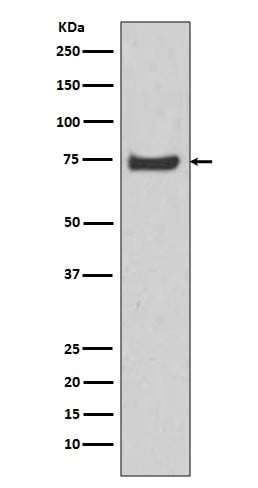
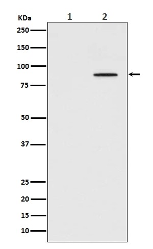
 WB (Western Blot)
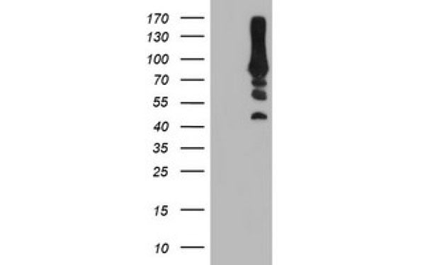
(Western blot analysis of Nrf2 phosphorylation expression in HepG2 cell lysate (AAA124527).Electrophoresis was performed on a 5-20% SDS-PAGE gel at 70V (Stacking gel) / 90V (Resolving gel) for 2-3 hours. The sample well of each lane was loaded with 50ug of sample under reducing conditions.After Electrophoresis, proteins were transferred to a Nitrocellulose membrane at 150mA for 50-90 minutes. Blocked the membrane with 5% Non-fat Milk/ TBS for 1.5 hour at RT. The membrane was incubated with rabbit anti-NFE2L2 monoclonal antibody overnight at 4 degree C, then washed with TBS-0.1%Tween 3 times with 5 minutes each and probed with a goat anti-rabbit IgG-HRP secondary antibody at a dilution of 1:10000 for 1.5 hour at RT. The signal is developed using an Enhanced Chemiluminescent detection (ECL) kit with Tanon 5200 system. A specific band was detected for NFE2L2)
WB (Western Blot)
(Western blot analysis of Nrf2 phosphorylation expression in HepG2 cell lysate (AAA124527).Electrophoresis was performed on a 5-20% SDS-PAGE gel at 70V (Stacking gel) / 90V (Resolving gel) for 2-3 hours. The sample well of each lane was loaded with 50ug of sample under reducing conditions.After Electrophoresis, proteins were transferred to a Nitrocellulose membrane at 150mA for 50-90 minutes. Blocked the membrane with 5% Non-fat Milk/ TBS for 1.5 hour at RT. The membrane was incubated with rabbit anti-NFE2L2 monoclonal antibody overnight at 4 degree C, then washed with TBS-0.1%Tween 3 times with 5 minutes each and probed with a goat anti-rabbit IgG-HRP secondary antibody at a dilution of 1:10000 for 1.5 hour at RT. The signal is developed using an Enhanced Chemiluminescent detection (ECL) kit with Tanon 5200 system. A specific band was detected for NFE2L2)
Nrf2, Monoclonal Antibody (Cat# AAA124527)
 WB (Western Blot)
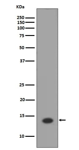
(Western blot analysis of eIF4E (phosphoS209) expression in (1) HEK293 cell lysate; (2) Mouse spleen lysate (AAA124528).Electrophoresis was performed on a 5-20% SDS-PAGE gel at 70V (Stacking gel) / 90V (Resolving gel) for 2-3 hours. The sample well of each lane was loaded with 50ug of sample under reducing conditions.After Electrophoresis, proteins were transferred to a Nitrocellulose membrane at 150mA for 50-90 minutes. Blocked the membrane with 5% Non-fat Milk/ TBS for 1.5 hour at RT. The membrane was incubated with rabbit anti-EIF4E monoclonal antibody overnight at 4 degree C, then washed with TBS-0.1%Tween 3 times with 5 minutes each and probed with a goat anti-rabbit IgG-HRP secondary antibody at a dilution of 1:10000 for 1.5 hour at RT. The signal is developed using an Enhanced Chemiluminescent detection (ECL) kit with Tanon 5200 system. A specific band was detected for EIF4E)
WB (Western Blot)
(Western blot analysis of eIF4E (phosphoS209) expression in (1) HEK293 cell lysate; (2) Mouse spleen lysate (AAA124528).Electrophoresis was performed on a 5-20% SDS-PAGE gel at 70V (Stacking gel) / 90V (Resolving gel) for 2-3 hours. The sample well of each lane was loaded with 50ug of sample under reducing conditions.After Electrophoresis, proteins were transferred to a Nitrocellulose membrane at 150mA for 50-90 minutes. Blocked the membrane with 5% Non-fat Milk/ TBS for 1.5 hour at RT. The membrane was incubated with rabbit anti-EIF4E monoclonal antibody overnight at 4 degree C, then washed with TBS-0.1%Tween 3 times with 5 minutes each and probed with a goat anti-rabbit IgG-HRP secondary antibody at a dilution of 1:10000 for 1.5 hour at RT. The signal is developed using an Enhanced Chemiluminescent detection (ECL) kit with Tanon 5200 system. A specific band was detected for EIF4E)
eIF4E, Monoclonal Antibody (Cat# AAA124528)
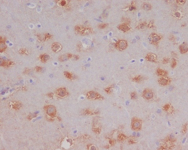
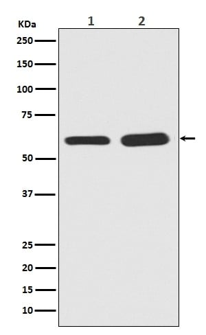 WB (Western Blot)
(Western blot analysis of Phospho-Smad5 in (1)Mouse brain tissue lysate; (2)Rat brain tissue lysate (AAA124535).Electrophoresis was performed on a 5-20% SDS-PAGE gel at 70V (Stacking gel) / 90V (Resolving gel) for 2-3 hours. The sample well of each lane was loaded with 50ug of sample under reducing conditions.After Electrophoresis, proteins were transferred to a Nitrocellulose membrane at 150mA for 50-90 minutes. Blocked the membrane with 5% Non-fat Milk/ TBS for 1.5 hour at RT. The membrane was incubated with rabbit anti-SMAD5 monoclonal antibody overnight at 4 degree C, then washed with TBS-0.1%Tween 3 times with 5 minutes each and probed with a goat anti-rabbit IgG-HRP secondary antibody at a dilution of 1:10000 for 1.5 hour at RT. The signal is developed using an Enhanced Chemiluminescent detection (ECL) kit with Tanon 5200 system. A specific band was detected for SMAD5)
WB (Western Blot)
(Western blot analysis of Phospho-Smad5 in (1)Mouse brain tissue lysate; (2)Rat brain tissue lysate (AAA124535).Electrophoresis was performed on a 5-20% SDS-PAGE gel at 70V (Stacking gel) / 90V (Resolving gel) for 2-3 hours. The sample well of each lane was loaded with 50ug of sample under reducing conditions.After Electrophoresis, proteins were transferred to a Nitrocellulose membrane at 150mA for 50-90 minutes. Blocked the membrane with 5% Non-fat Milk/ TBS for 1.5 hour at RT. The membrane was incubated with rabbit anti-SMAD5 monoclonal antibody overnight at 4 degree C, then washed with TBS-0.1%Tween 3 times with 5 minutes each and probed with a goat anti-rabbit IgG-HRP secondary antibody at a dilution of 1:10000 for 1.5 hour at RT. The signal is developed using an Enhanced Chemiluminescent detection (ECL) kit with Tanon 5200 system. A specific band was detected for SMAD5)
Smad5, Monoclonal Antibody (Cat# AAA124535)
 WB (Western Blot)
(Western blot analysis of Phospho-PAK1/2/3 expression in HeLa Cell lysate treated with lambda phosphatase (AAA124537).Electrophoresis was performed on a 5-20% SDS-PAGE gel at 70V (Stacking gel) / 90V (Resolving gel) for 2-3 hours. The sample well of each lane was loaded with 50ug of sample under reducing conditions.After Electrophoresis, proteins were transferred to a Nitrocellulose membrane at 150mA for 50-90 minutes. Blocked the membrane with 5% Non-fat Milk/ TBS for 1.5 hour at RT. The membrane was incubated with rabbit anti-PAK3 monoclonal antibody overnight at 4 degree C, then washed with TBS-0.1%Tween 3 times with 5 minutes each and probed with a goat anti-rabbit IgG-HRP secondary antibody at a dilution of 1:10000 for 1.5 hour at RT. The signal is developed using an Enhanced Chemiluminescent detection (ECL) kit with Tanon 5200 system. A specific band was detected for PAK3)
WB (Western Blot)
(Western blot analysis of Phospho-PAK1/2/3 expression in HeLa Cell lysate treated with lambda phosphatase (AAA124537).Electrophoresis was performed on a 5-20% SDS-PAGE gel at 70V (Stacking gel) / 90V (Resolving gel) for 2-3 hours. The sample well of each lane was loaded with 50ug of sample under reducing conditions.After Electrophoresis, proteins were transferred to a Nitrocellulose membrane at 150mA for 50-90 minutes. Blocked the membrane with 5% Non-fat Milk/ TBS for 1.5 hour at RT. The membrane was incubated with rabbit anti-PAK3 monoclonal antibody overnight at 4 degree C, then washed with TBS-0.1%Tween 3 times with 5 minutes each and probed with a goat anti-rabbit IgG-HRP secondary antibody at a dilution of 1:10000 for 1.5 hour at RT. The signal is developed using an Enhanced Chemiluminescent detection (ECL) kit with Tanon 5200 system. A specific band was detected for PAK3)
PAK1/2/3, Monoclonal Antibody (Cat# AAA124537)
 WB (Western Blot)
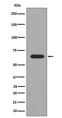
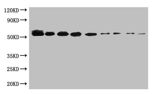
(WB: SUMO-tagged fusion protein(20ng/ml) was subjected to SDS-PAGE followed by Western Blot with AAA119665 at dilution ofLane 1: 1000 Lane 4: 8000 Lane 7: 64000Lane 2: 2000 Lane 5: 16000 Lane 8: 128000Lane 3: 4000 Lane 6: 32000 Lane 9: 256000SecondaryGoat polyclonal to Mouse IgG at 1/5000 dilutionPredicted band size: 55kdObserved band size: 55kd)
WB (Western Blot)
(WB: SUMO-tagged fusion protein(20ng/ml) was subjected to SDS-PAGE followed by Western Blot with AAA119665 at dilution ofLane 1: 1000 Lane 4: 8000 Lane 7: 64000Lane 2: 2000 Lane 5: 16000 Lane 8: 128000Lane 3: 4000 Lane 6: 32000 Lane 9: 256000SecondaryGoat polyclonal to Mouse IgG at 1/5000 dilutionPredicted band size: 55kdObserved band size: 55kd)
Sumo tag, Monoclonal Antibody (Cat# AAA119665)
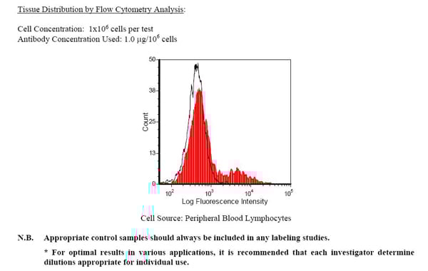 Application Data
HLA-DR, Monoclonal Antibody (Cat# AAA74184)
HBsAg surface antigen IgG1, Monoclonal Antibody (Cat# AAA71866)
 Application Data
Mouse Anti-Rat IgG2b (gamma 2b chain specific), Monoclonal Secondary Antibody (Cat# AAA78693)
Mouse Anti-Rat Kappa (kappa chain specific), Monoclonal Secondary Antibody (Cat# AAA78697)
 WB (Western Blot)
(All lanes: Mouse Anti-BSA monoclonal antibody at 1ug/mlLane 1: Bovine serum AlbuminPredicted band size: 67kdObserved band size: 67kd)
WB (Western Blot)
(All lanes: Mouse Anti-BSA monoclonal antibody at 1ug/mlLane 1: Bovine serum AlbuminPredicted band size: 67kdObserved band size: 67kd)
BSA, Monoclonal Antibody (Cat# AAA114275)
Mouse anti Human secretory component (free and bound), Monoclonal Secondary Antibody (Cat# AAA77530)
Mouse anti Human IgG2 (Fc subclass specific), Monoclonal Secondary Antibody (Cat# AAA77531)
Influenza A haemagglutinin H3, Monoclonal Antibody (Cat# AAA78059)
Protein A Chromatography
Parvovirus, Monoclonal Antibody (Cat# AAA78061)
Influenza Virus Type A, Monoclonal Antibody (Cat# AAA78062)
Chromatography on protein A Sepharose for MAbs InA108, InA245, InA180 and InA224
Listeria monocytogenes, Monoclonal Antibody (Cat# AAA78082)
dPAPP-A, Monoclonal Antibody (Cat# AAA78086)
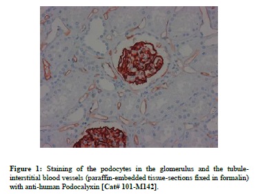
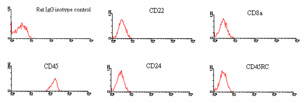
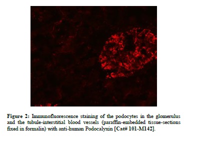 Application Data
Podocalyxin, Monoclonal Antibody (Cat# AAA79094)
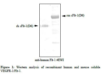
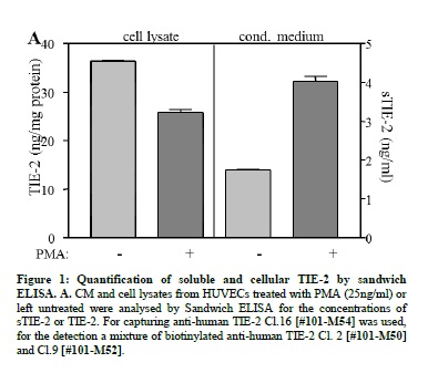
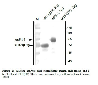 Application Data
VEGFR-1/Flt-1, Monoclonal Antibody (Cat# AAA79097)
 Application Data
VEGF-A, Monoclonal Antibody (Cat# AAA79113)
 Application Data
VEGFR-2/KDR, Monoclonal Antibody (Cat# AAA79115)

 IF (Immunofluorescence)
(Immunofluorescent staining of COS7 cells transiently transfected with recombinant BIN3 protein using BIN3 antibody)
IF (Immunofluorescence)
(Immunofluorescent staining of COS7 cells transiently transfected with recombinant BIN3 protein using BIN3 antibody)
BIN3, Monoclonal Antibody (Cat# AAA106528)
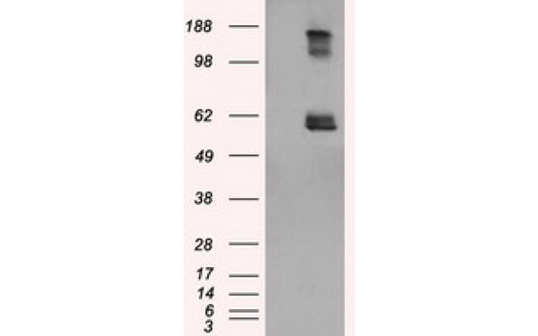
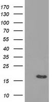 WB (Western Blot)
(Western Blot analysis using BIRC5 antibodyWestern Blot analysis of HEK293T cell lysates (5 ug) transfected with either recombinant BIRC5 protein (Right) or empty vector (Left) detected with BIRC5 antibody)
WB (Western Blot)
(Western Blot analysis using BIRC5 antibodyWestern Blot analysis of HEK293T cell lysates (5 ug) transfected with either recombinant BIRC5 protein (Right) or empty vector (Left) detected with BIRC5 antibody)
BIRC5, Monoclonal Antibody (Cat# AAA106558)
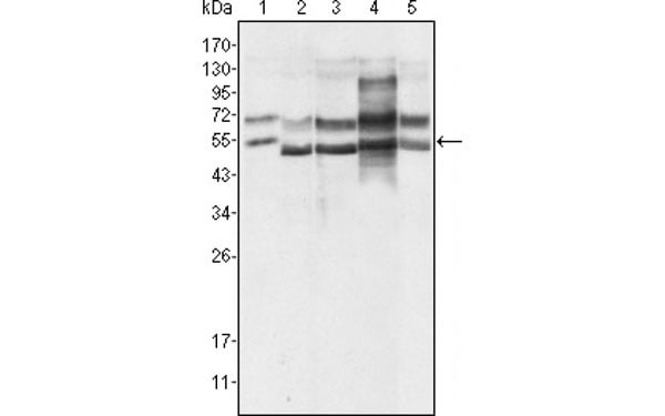 WB (Western Blot)
(Western Blot showing using GABPA antibody used against Hela (1), A549 (2), MCF-7 (3), NIH/3T3 (4) and SMMC-7721 (5) cell lysate.)
WB (Western Blot)
(Western Blot showing using GABPA antibody used against Hela (1), A549 (2), MCF-7 (3), NIH/3T3 (4) and SMMC-7721 (5) cell lysate.)
GABPA, Monoclonal Antibody (Cat# AAA106559)
Human IgA antibody, Monoclonal Antibody (Cat# AAA106571)
Human IgA antibody was purified by Ion exchange chromatography.
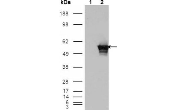
 WB (Western Blot)
(Western Blot analysis of HEK293T cell lysates (5 ug) transfected with either recombinant SAMHD1 protein (Right) or empty vector (Left) detected with SAMHD1 antibody)
WB (Western Blot)
(Western Blot analysis of HEK293T cell lysates (5 ug) transfected with either recombinant SAMHD1 protein (Right) or empty vector (Left) detected with SAMHD1 antibody)
SAMHD1, Monoclonal Antibody (Cat# AAA106579)
 WB (Western Blot)
(Western Blot analysis of HEK293T cell lysates (5 ug) transfected with either recombinant MAOA protein (Right) or empty vector (Left) detected with MAOA antibody)
WB (Western Blot)
(Western Blot analysis of HEK293T cell lysates (5 ug) transfected with either recombinant MAOA protein (Right) or empty vector (Left) detected with MAOA antibody)
MAOA, Monoclonal Antibody (Cat# AAA107288)
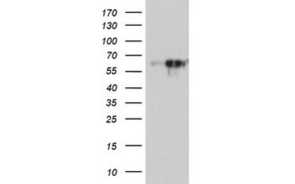
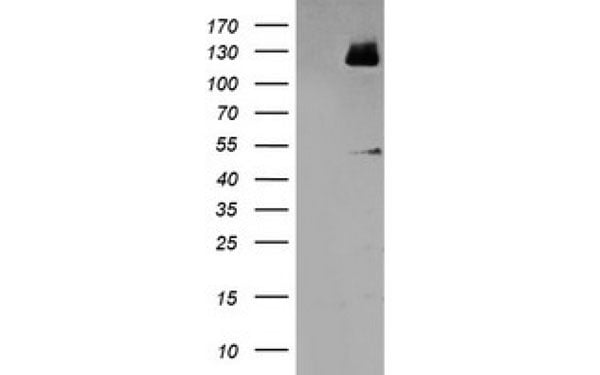 WB (Western Blot)
(Western Blot analysis of HEK293T cell lysates (5 ug) transfected with either recombinant MEF2C protein (Right) or empty vector (Left) detected with MEF2C antibody)
WB (Western Blot)
(Western Blot analysis of HEK293T cell lysates (5 ug) transfected with either recombinant MEF2C protein (Right) or empty vector (Left) detected with MEF2C antibody)
MEF2C, Monoclonal Antibody (Cat# AAA107318)
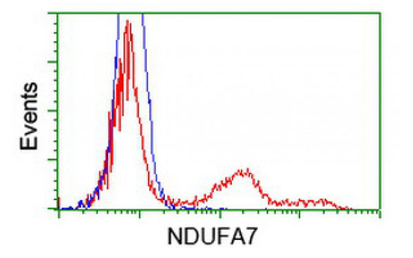
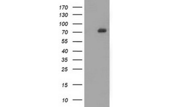
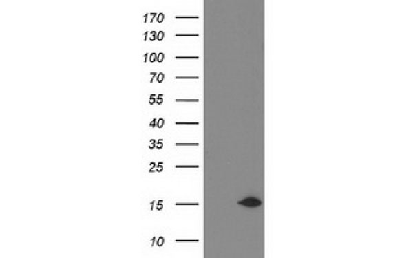 WB (Western Blot)
(Western Blot analysis of HEK293T cell lysates (5 ug) transfected with either recombinant NDUFA7 protein (Right) or empty vector (Left) detected with NDUFA7 antibody)
WB (Western Blot)
(Western Blot analysis of HEK293T cell lysates (5 ug) transfected with either recombinant NDUFA7 protein (Right) or empty vector (Left) detected with NDUFA7 antibody)
NDUFA7, Monoclonal Antibody (Cat# AAA107324)
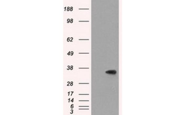
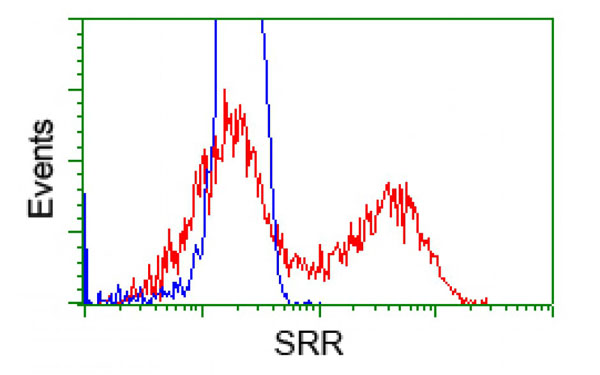
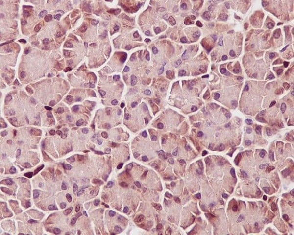
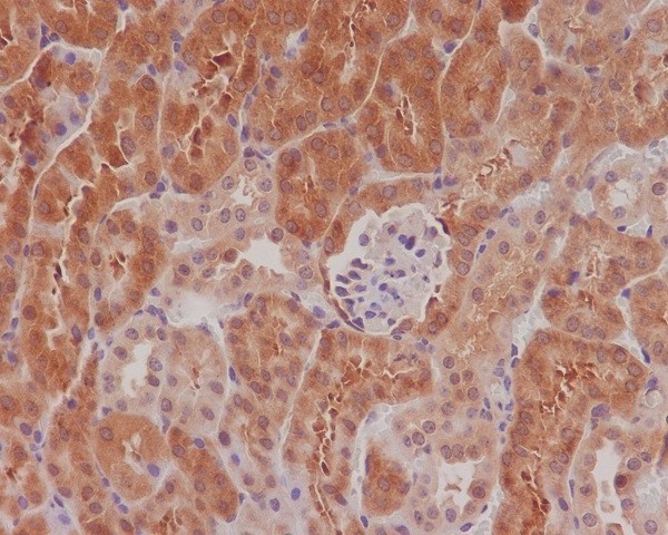
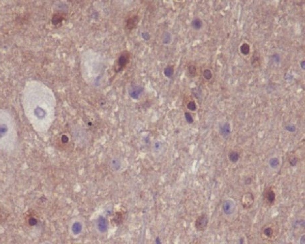
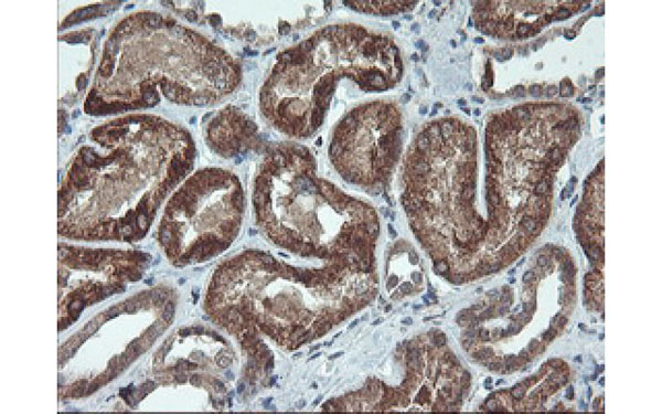
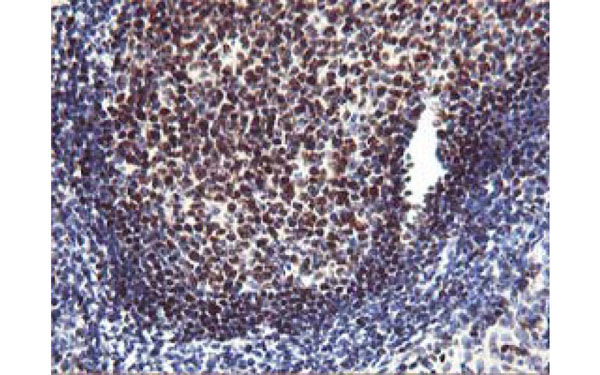
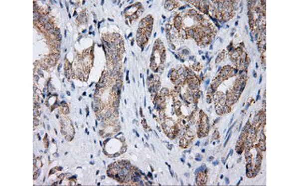
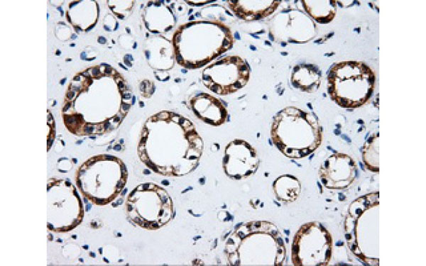 IHC (Immunohistochemisry)
(Immunohistochemical analysis of SRR protein in paraffin embedded Human kidney tissue using SRR antibody)
IHC (Immunohistochemisry)
(Immunohistochemical analysis of SRR protein in paraffin embedded Human kidney tissue using SRR antibody)
SRR, Monoclonal Antibody (Cat# AAA107354)
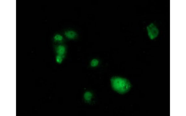
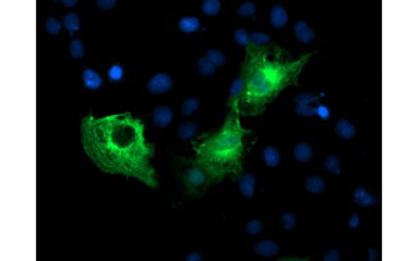
 IF (Immunofluorescence)
(Immunofluorescent staining of COS7 cells transiently transfected with recombinant LSM1 protein using LSM1 antibody)
IF (Immunofluorescence)
(Immunofluorescent staining of COS7 cells transiently transfected with recombinant LSM1 protein using LSM1 antibody)
LSM1, Monoclonal Antibody (Cat# AAA107357)
 IF (Immunofluorescence)
(Immunofluorescent staining of COS7 cells transiently transfected with recombinant PFKP protein using PFKP antibody)
IF (Immunofluorescence)
(Immunofluorescent staining of COS7 cells transiently transfected with recombinant PFKP protein using PFKP antibody)
PFKP, Monoclonal Antibody (Cat# AAA107030)
 FCM/FACS (Flow Cytometry)
(Staining of C57BL/6 splenocytes with CD45R antibody and 0.06 ug of Rat IgM kappa Isotype Control (FITC) (left) or 0.06 ug of CD43 antibody (FITC) (right). Cells in the lymphocyte gate were used for analysis.)
FCM/FACS (Flow Cytometry)
(Staining of C57BL/6 splenocytes with CD45R antibody and 0.06 ug of Rat IgM kappa Isotype Control (FITC) (left) or 0.06 ug of CD43 antibody (FITC) (right). Cells in the lymphocyte gate were used for analysis.)
CD43, Monoclonal Antibody (Cat# AAA107049)
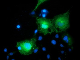
 IF (Immunofluorescence)
(Immunofluorescent staining of COS7 cells transiently transfected with recombinant LIPG protein using LIPG antibody)
IF (Immunofluorescence)
(Immunofluorescent staining of COS7 cells transiently transfected with recombinant LIPG protein using LIPG antibody)
LIPG, Monoclonal Antibody (Cat# AAA107056)
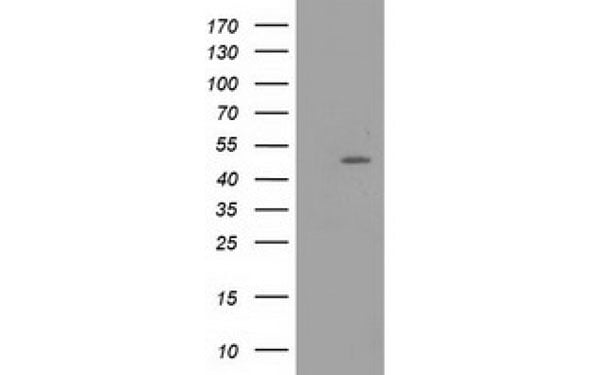 WB (Western Blot)
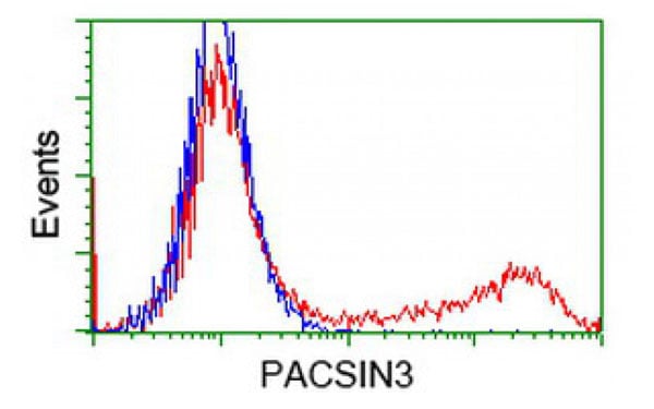
(Western Blot analysis of HEK293T cell lysates (5 ug) transfected with either recombinant PACSIN3 protein (Right) or empty vector (Left) detected with PACSIN3 antibody)
WB (Western Blot)
(Western Blot analysis of HEK293T cell lysates (5 ug) transfected with either recombinant PACSIN3 protein (Right) or empty vector (Left) detected with PACSIN3 antibody)
PACSIN3, Monoclonal Antibody (Cat# AAA106773)
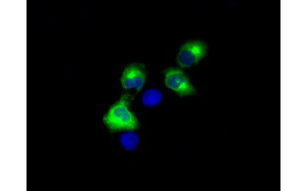
 IF (Immunofluorescence)
(Immunofluorescent staining of COS7 cells transiently transfected with recombinant IGF2BP2 protein using IGF2BP2 antibody)
IF (Immunofluorescence)
(Immunofluorescent staining of COS7 cells transiently transfected with recombinant IGF2BP2 protein using IGF2BP2 antibody)
IGF2BP2, Monoclonal Antibody (Cat# AAA106783)
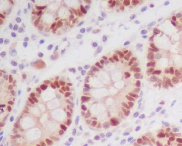
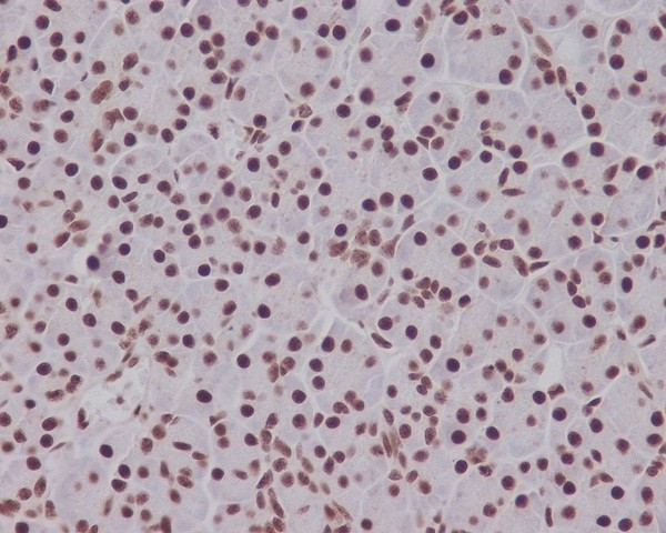
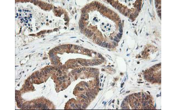
 IHC (Immunohistochemisry)
(Immunohistochemical analysis of NIF3L1 protein in paraffin embedded Human Kidney tissue using NIF3L1 antibody)
IHC (Immunohistochemisry)
(Immunohistochemical analysis of NIF3L1 protein in paraffin embedded Human Kidney tissue using NIF3L1 antibody)
NIF3L1, Monoclonal Antibody (Cat# AAA106789)
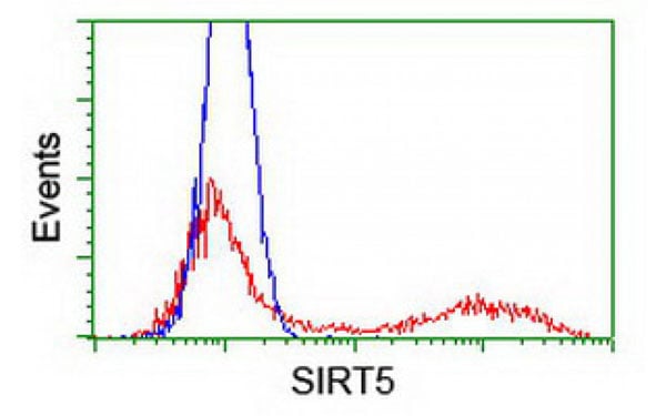
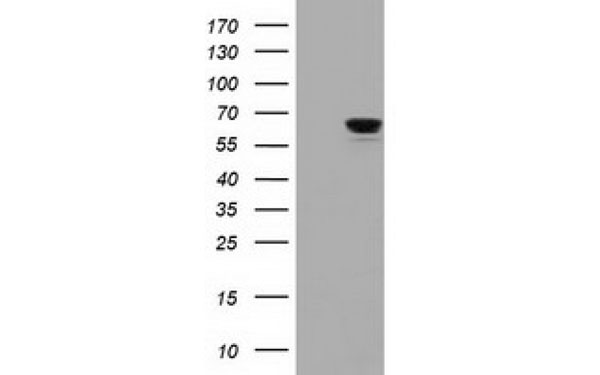
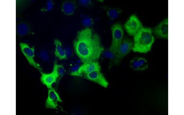
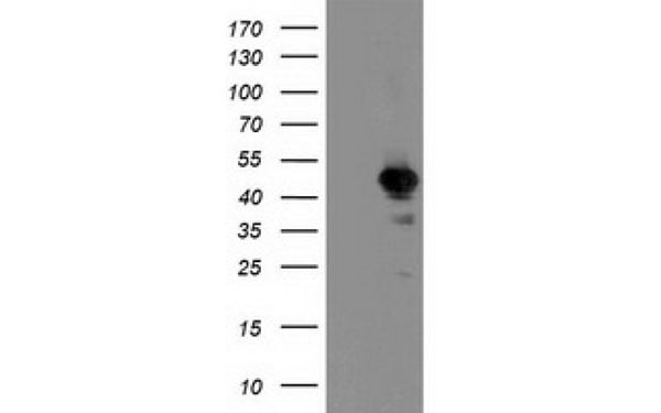
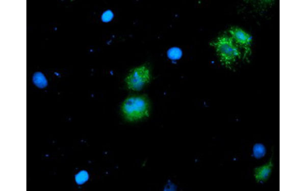
 WB (Western Blot)
(Western Blot analysis of HEK293T cell lysates (5 ug) transfected with either recombinant SIRT5 protein (Right) or empty vector (Left) detected with SIRT5 antibody)
WB (Western Blot)
(Western Blot analysis of HEK293T cell lysates (5 ug) transfected with either recombinant SIRT5 protein (Right) or empty vector (Left) detected with SIRT5 antibody)
SIRT5, Monoclonal Antibody (Cat# AAA106799)
What are Monoclonal Antibodies?
Monoclonal antibodies are specialized laboratory-produced proteins developed for binding to specific biological antigens or other molecular targets. Since they come from a single cell (or clone), they are especially consistent and accurate in the data they are involved in producing.
This type of antibody material has been shown to be a powerful tool in finding and subsequently destroying harmful cells in an organism, such as those found in cancers or various autoimmune diseases. This makes them excellent aids in medical testing and research, which is why they are so widely used.
AAA Biotech offers a comprehensive range of high-quality monoclonal antibodies that perform effectively in various laboratory tests, including (amongst others) ELISA, western blotting, immunohistochemistry, and flow cytometry. All of the products in our catalog are thoroughly quality tested to make sure that they are reliable and will consistently perform well in your research.
What Are The Uses of Monoclonal Antibodies
Monoclonal antibodies are used in many lab tests, including (amongst others) ELISA, western blotting, immunohistochemistry, and flow cytometry.
ELISA is a test that helps detect a specific substance/analyte in a sample. It uses antibodies (often monoclonal) bound to a solid surface (such as the well of a microplate) to “capture” the substance/analyte in the sample and immobilize it so that the detection antibody component can then bind to it and produce a signal, which can then be measured.
Western blotting identifies specific proteins in a sample. The sample is first separated on a gel, and then antibodies are applied that will typically bind to the target, which will all be localized to a single band in a lane.
Immunohistochemistry helps locate specific proteins in cells or tissue samples using antibodies.
Flow cytometry looks at and sorts cells. It uses antibodies that are conjugated to reporter molecules called “fluorophores”, which, under special lights, emit light themselves, which can then be measured by a detector instrument.
How Monoclonal Antibodies Are Used as Medicine?
Please note that all of the products listed in AAA Biotech’s also known as AAA Bio or AAABio catalog are strictly for research-use only (RUO).
Monoclonal antibodies can also be used as therapeutic/medical treatments, particularly in the context of cancers. They are designed to find and bind to specific cells or proteins, helping the immune system recognize and attack the cancer. These treatments work in different ways, such as:
- Radioimmunotherapy attaches a small amount of radioactive molecule to the antibody, so it delivers the radiation directly to the cancer cells that the antibody is specifically binding to.
- Antibody-directed enzyme prodrug therapy uses antibodies that are specifically bound to special enzymes. These enzymes activate a harmless drug in the body and turn it into a cancer-killing drug only near the cancer cells—this helps avoid harming healthy cells.
- Immunoliposomes are tiny “bubbles” filled with medicine/drug and coated with antibodies. They carry the drug straight to the cancer cells.
Why Buy Monoclonal Antibodies From Us?
At AAA Biotech, we provide high-performance monoclonal antibodies designed to support a wide range of research needs.
1. Validated for Versatile Applications
The antibodies in our catalog are extensively validated and compatible with multiple techniques, including (but not limited to) ELISA, flow cytometry (FC), immunocytochemistry (ICC), immunofluorescence (IF), immunohistochemistry (IHC), immunoprecipitation (IP), and western blotting (WB).
2. Wide Selection & Specialized Options
We offer antibodies for common and rare species, that are available in various conjugated forms, and also in recombinant formats. Essentially, there is almost anything one might need to meet their experimental model’s requirements.
3. High-Quality Proteins
Our proteins meet high purity standards—90% or more as confirmed by SDS-PAGE. Many are available with tags like His, Flag, GST, or MBP, and we also supply native and biologically active proteins for functional studies.
Frequently Asked Questions
1. Are your monoclonal antibodies validated for specific applications?
Yes, our antibodies are tested and validated for use in methods such as ELISA, western blot, IHC, flow cytometry, and more. Refer to specific product pages or datasheets for individual product information.
2. How do I choose the right monoclonal antibody for my application?
Review the product details directly for application validation, species reactivity, and target information. You may also contact our support team at any time for help.
3. How quickly can I receive my order?
Most orders are processed and shipped within 1–3 business days, depending on product availability and your shipping location.