Filters
▼Clonality
▼Type
▼Reactivity
▼Gene Name
▼Isotype
▼Host
▼Application
▼Clone
▼Monoclonal Antibodies
Get accurate results in your research with our Monoclonal Antibodies, which are specially made to target exactly what you require for your research, and will produce consistent, reliable performance in lab tests.
Viewing 2800-2850 of 27597 product results
 FCM/FACS (Flow Cytometry)
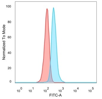
(Flow cytometric analysis of PFA-fixed HeLa cells. GTF2IRD2 Mouse Monoclonal Antibody (PCRP-GTF2IRD2-1B4) followed by goat anti-mouse IgG-CF488 (blue); unstained cells (red).)
FCM/FACS (Flow Cytometry)
(Flow cytometric analysis of PFA-fixed HeLa cells. GTF2IRD2 Mouse Monoclonal Antibody (PCRP-GTF2IRD2-1B4) followed by goat anti-mouse IgG-CF488 (blue); unstained cells (red).)
GTF2IRD2, Monoclonal Antibody (Cat# AAA216007)
 FCM/FACS (Flow Cytometry)
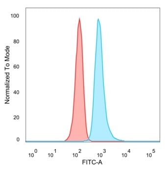
(Flow Cytometric Analysis of PFA-fixed HeLa cells. THG1/RHOXF2 Mouse Monoclonal Antibody (PCRP-RHOXF2-1D7) followed by goat anti-mouse IgG-CF488 (blue); unstained cells (red).)
FCM/FACS (Flow Cytometry)
(Flow Cytometric Analysis of PFA-fixed HeLa cells. THG1/RHOXF2 Mouse Monoclonal Antibody (PCRP-RHOXF2-1D7) followed by goat anti-mouse IgG-CF488 (blue); unstained cells (red).)
THG1/RHOXF2, Monoclonal Antibody (Cat# AAA216008)
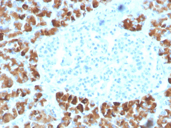 IHC (Immunohistochemistry)
(Formalin-fixed, paraffin-embedded human pancreas stained with INSM2 Mouse Monoclonal Antibody (INSM2/4291).)
IHC (Immunohistochemistry)
(Formalin-fixed, paraffin-embedded human pancreas stained with INSM2 Mouse Monoclonal Antibody (INSM2/4291).)
Insulinoma Associated 2/INSM2, Monoclonal Antibody (Cat# AAA216009)
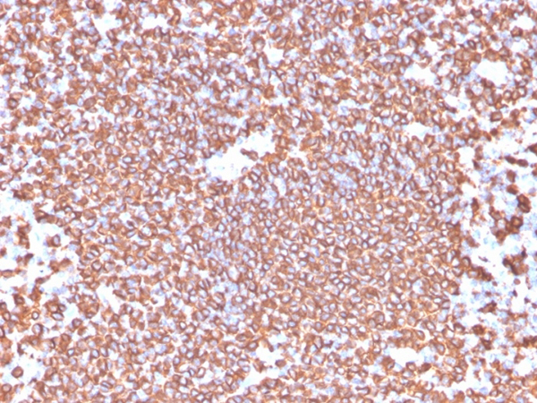
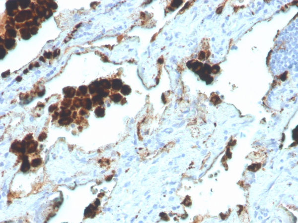
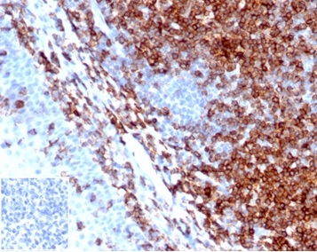
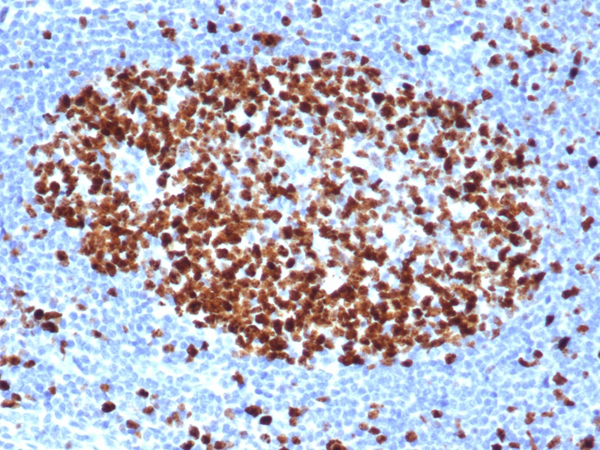
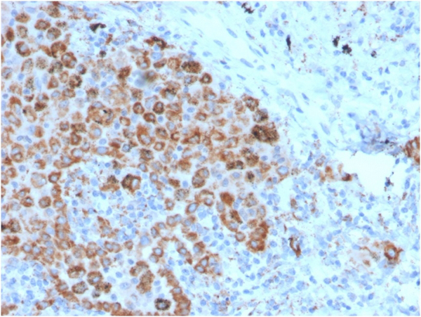 IHC (Immunohistochemistry)
(Formalin-fixed, paraffin-embeddedhuman lymph node stained with TIM3 Mouse Monoclonal Antibody (TIM3/4025).)
IHC (Immunohistochemistry)
(Formalin-fixed, paraffin-embeddedhuman lymph node stained with TIM3 Mouse Monoclonal Antibody (TIM3/4025).)
TIM3/HAVCR2/CD366, Monoclonal Antibody (Cat# AAA216013)
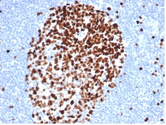
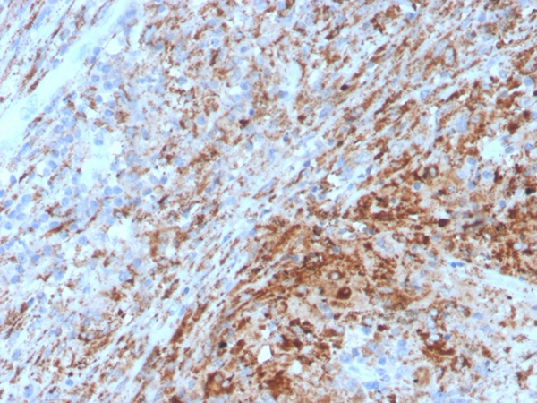 IHC (Immunohistochemistry)
(Formalin-fixed, paraffin-embedded human lymph node stained with TIM3 Mouse Monoclonal Antibody (TIM3/4029).)
IHC (Immunohistochemistry)
(Formalin-fixed, paraffin-embedded human lymph node stained with TIM3 Mouse Monoclonal Antibody (TIM3/4029).)
TIM3/HAVCR2/CD366, Monoclonal Antibody (Cat# AAA216016)
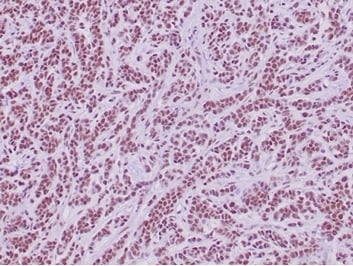
 IHC (Immunohistochemistry)
(Formalin-fixed, paraffin-embedded human prostatestained with p63 Recombinant Rabbit Monoclonal Antibody (TP63/4396R).)
IHC (Immunohistochemistry)
(Formalin-fixed, paraffin-embedded human prostatestained with p63 Recombinant Rabbit Monoclonal Antibody (TP63/4396R).)
p63, Monoclonal Antibody (Cat# AAA216021)
 IHC (Immunohistochemistry)
(Formalin-fixed, paraffin-embedded human ovarian carcinoma stained with Cyclin E Recombinant Rabbit Monoclonal Antibody (CCNE1/4935R).)
IHC (Immunohistochemistry)
(Formalin-fixed, paraffin-embedded human ovarian carcinoma stained with Cyclin E Recombinant Rabbit Monoclonal Antibody (CCNE1/4935R).)
Cyclin E, Monoclonal Antibody (Cat# AAA216026)
 FCM/FACS (Flow Cytometry)
(Flow Cytometric Analysis of PFA-fixed HeLa cells. NMI Mouse Monoclonal Antibody (PCRP-NMI-1C1) followed by goat anti-mouse IgG-CF488 (blue); unstained cells (red).)
FCM/FACS (Flow Cytometry)
(Flow Cytometric Analysis of PFA-fixed HeLa cells. NMI Mouse Monoclonal Antibody (PCRP-NMI-1C1) followed by goat anti-mouse IgG-CF488 (blue); unstained cells (red).)
N myc (and STAT) Interactor/NMI, Monoclonal Antibody (Cat# AAA216028)
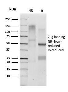
 SDS-PAGE
(SDS-PAGE Analysis Purified RCAS1 Recombinant Rabbit Monoclonal Antibody (EBAG9/7033R). Confirmation of Purity and Integrity of Antibody.)
SDS-PAGE
(SDS-PAGE Analysis Purified RCAS1 Recombinant Rabbit Monoclonal Antibody (EBAG9/7033R). Confirmation of Purity and Integrity of Antibody.)
RCAS1/Estrogen Receptor Binding Site Associated, Antigen 9, Monoclonal Antibody (Cat# AAA216030)
 FCM/FACS (Flow Cytometry)
(Flow Cytometric Analysis of PFA-fixed Raji cells. ZMYM3 Mouse Monoclonal Antibody (PCRP-ZMYM3-2F10) followed by goat anti-mouse IgG-CF488 (blue); unstained cells (red).)
FCM/FACS (Flow Cytometry)
(Flow Cytometric Analysis of PFA-fixed Raji cells. ZMYM3 Mouse Monoclonal Antibody (PCRP-ZMYM3-2F10) followed by goat anti-mouse IgG-CF488 (blue); unstained cells (red).)
ZMYM3, Monoclonal Antibody (Cat# AAA216033)
 IHC (Immunohistochemistry)
(Formalin-fixed, paraffin-embedded human lymph node stained with CD4 Recombinant Mouse Monoclonal Antibody (rCD4/3930).)
IHC (Immunohistochemistry)
(Formalin-fixed, paraffin-embedded human lymph node stained with CD4 Recombinant Mouse Monoclonal Antibody (rCD4/3930).)
CD4, Monoclonal Antibody (Cat# AAA216034)
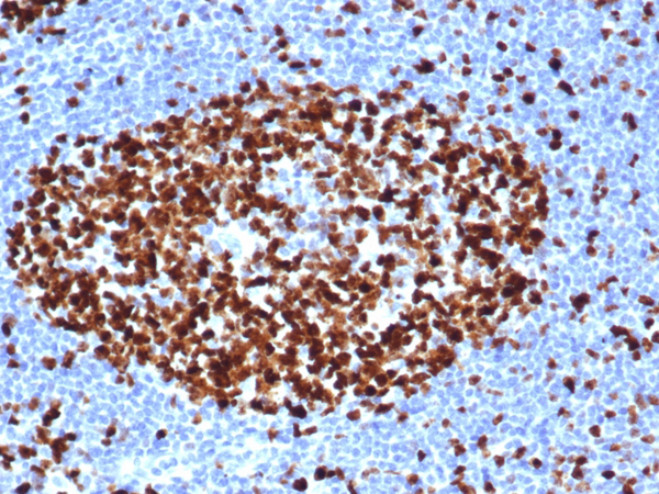
 IHC (Immunohistochemistry)
(Formalin-fixed, paraffin-embedded human lymph node stained with CD5 Recombinant Rabbit Monoclonal Antibody (C5/4561R).)
IHC (Immunohistochemistry)
(Formalin-fixed, paraffin-embedded human lymph node stained with CD5 Recombinant Rabbit Monoclonal Antibody (C5/4561R).)
CD5, Monoclonal Antibody (Cat# AAA216037)
 IHC (Immunohistochemistry)
(Formalin-fixed, paraffin-embedded human tonsil stained with CD6 Recombinant Rabbit Monoclonal Antibody (C6/7022R).)
IHC (Immunohistochemistry)
(Formalin-fixed, paraffin-embedded human tonsil stained with CD6 Recombinant Rabbit Monoclonal Antibody (C6/7022R).)
CD6, Monoclonal Antibody (Cat# AAA216040)
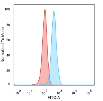
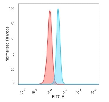
 FCM/FACS (Flow Cytometry)
(Flow Cytometric Analysis of PFA-fixed HeLa cells. RPS6KA5/MSK1 Mouse Monoclonal Antibody (PCRP-RPS6KA5-1A8) followed by goat anti-mouse IgG-CF488 (blue); isotype control (red).)
FCM/FACS (Flow Cytometry)
(Flow Cytometric Analysis of PFA-fixed HeLa cells. RPS6KA5/MSK1 Mouse Monoclonal Antibody (PCRP-RPS6KA5-1A8) followed by goat anti-mouse IgG-CF488 (blue); isotype control (red).)
RPS6KA5/MSK1, Monoclonal Antibody (Cat# AAA216043)
Predicted to react with Mouse and Rat.
 IHC (Immunohistochemistry)
(Formalin-fixed, paraffin-embedded human tonsil stained with CD20 Rabbit Recombinant Monoclonal Antibody (IGEL/4524R).)
IHC (Immunohistochemistry)
(Formalin-fixed, paraffin-embedded human tonsil stained with CD20 Rabbit Recombinant Monoclonal Antibody (IGEL/4524R).)
CD20/MS4A1, Monoclonal Antibody (Cat# AAA216049)
Expected to react with Monkey, Baboon, Gorilla and Chimpanzee.
 WB (Western Blot)
(Western Blot Analysis of Raji cell lysate using CD20 Rabbit Recombinant Monoclonal Antibody (IGEL/6850R).)
WB (Western Blot)
(Western Blot Analysis of Raji cell lysate using CD20 Rabbit Recombinant Monoclonal Antibody (IGEL/6850R).)
CD20/MS4A1, Monoclonal Antibody (Cat# AAA216050)
Expected to react with Monkey, Baboon, Gorilla and Chimpanzee.
 SDS-PAGE
(SDS-PAGE Analysis Purified CD20 Recombinant Rabbit Monoclonal Antibody (C5/7015R). Confirmation of Purity and Integrity of Antibody.)
SDS-PAGE
(SDS-PAGE Analysis Purified CD20 Recombinant Rabbit Monoclonal Antibody (C5/7015R). Confirmation of Purity and Integrity of Antibody.)
CD20/MS4A1, Monoclonal Antibody (Cat# AAA216051)
Expected to react with Monkey, Baboon, Gorilla and Chimpanzee.
 IHC (Immunohistochemistry)
(IHC analysis of formalin-fixed, paraffin-embedded human tonsil using rBLCAM/6749 at 2ug/ml. HIER: Tris/EDTA, pH9. 0, 45min. Inset: PBS instead of primary antibody, secondary negative control.)
IHC (Immunohistochemistry)
(IHC analysis of formalin-fixed, paraffin-embedded human tonsil using rBLCAM/6749 at 2ug/ml. HIER: Tris/EDTA, pH9. 0, 45min. Inset: PBS instead of primary antibody, secondary negative control.)
CD22/BL-CAM, Monoclonal Antibody (Cat# AAA216053)
 IHC (Immunohistochemistry)
(Formalin-fixed, paraffin-embedded human tonsil stained with CD27-Monospecific Mouse Monoclonal Antibody (LPFS2/4176).)
IHC (Immunohistochemistry)
(Formalin-fixed, paraffin-embedded human tonsil stained with CD27-Monospecific Mouse Monoclonal Antibody (LPFS2/4176).)
CD27, Monoclonal Antibody (Cat# AAA216056)
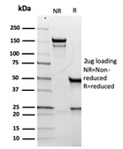
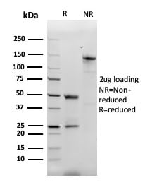
 SDS-PAGE
(SDS-PAGE Analysis Purified CD33 Mouse Monoclonal Antibody (SIGLEC3/3600). Confirmation of Integrity and Purity of Antibody.)
SDS-PAGE
(SDS-PAGE Analysis Purified CD33 Mouse Monoclonal Antibody (SIGLEC3/3600). Confirmation of Integrity and Purity of Antibody.)
CD33/SIGLEC3, Monoclonal Antibody (Cat# AAA216063)
 IHC (Immunohistochemistry)
(IHC analysis of formalin-fixed, paraffin-embedded human lung adenocarcinoma. NAPSA/4400R at 2ug/ml in PBS for 30min RT. HIER: Tris/EDTA, pH9. 0, 45min. 2 degree : HRP-polymer, 30min. DAB, 5min.)
IHC (Immunohistochemistry)
(IHC analysis of formalin-fixed, paraffin-embedded human lung adenocarcinoma. NAPSA/4400R at 2ug/ml in PBS for 30min RT. HIER: Tris/EDTA, pH9. 0, 45min. 2 degree : HRP-polymer, 30min. DAB, 5min.)
Napsin A, Monoclonal Antibody (Cat# AAA216065)
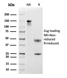
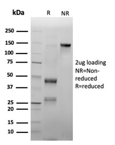
 SDS-PAGE
(SDS-PAGE Analysis Purified CD47 Recombinant Rabbit Monoclonal Antibody (CD47/6362R). Confirmation of Integrity and Purity of Antibody.)
SDS-PAGE
(SDS-PAGE Analysis Purified CD47 Recombinant Rabbit Monoclonal Antibody (CD47/6362R). Confirmation of Integrity and Purity of Antibody.)
CD47/IAP, Monoclonal Antibody (Cat# AAA216072)
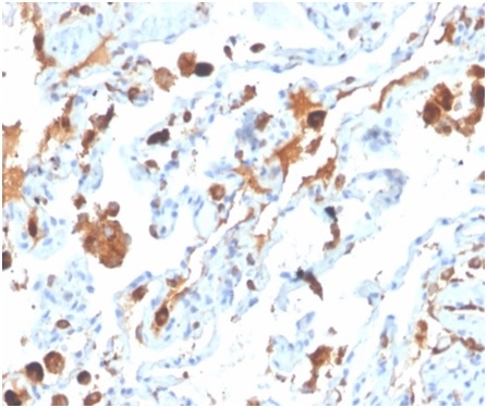
 IHC (Immunohistochemistry)
(Formalin-fixed, paraffin-embedded human tonsil stained with CD48 Mouse Monoclonal Antibody (CD48/4785).)
IHC (Immunohistochemistry)
(Formalin-fixed, paraffin-embedded human tonsil stained with CD48 Mouse Monoclonal Antibody (CD48/4785).)
CD48, Monoclonal Antibody (Cat# AAA216076)
 WB (Western Blot)
(Western blot analysis of Raji cell lysate using CD48 Monospecific Mouse Monoclonal Antibody (CD48/4787).)
WB (Western Blot)
(Western blot analysis of Raji cell lysate using CD48 Monospecific Mouse Monoclonal Antibody (CD48/4787).)
CD48, Monoclonal Antibody (Cat# AAA216078)
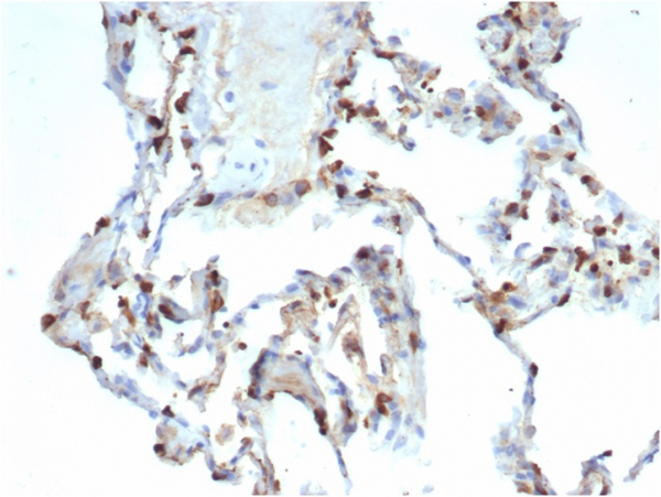
 IHC (Immunohistochemistry)
(Formalin-fixed, paraffin-embedded human tonsil stained with CD79a Rabbit Recombinant Monoclonal Antibody (IGA/3939R).)
IHC (Immunohistochemistry)
(Formalin-fixed, paraffin-embedded human tonsil stained with CD79a Rabbit Recombinant Monoclonal Antibody (IGA/3939R).)
CD79a, Monoclonal Antibody (Cat# AAA216079)
 FCM/FACS (Flow Cytometry)
(Flow Cytometric Analysis of PFA-fixed HeLa cells. Y14/RBM8A Mouse Monoclonal Antibody (PCRP-RBM8A-1B4) followed by goat anti-mouse IgG-CF488 (blue); unstained cells (red).)
FCM/FACS (Flow Cytometry)
(Flow Cytometric Analysis of PFA-fixed HeLa cells. Y14/RBM8A Mouse Monoclonal Antibody (PCRP-RBM8A-1B4) followed by goat anti-mouse IgG-CF488 (blue); unstained cells (red).)
Y14/RBM8A, Monoclonal Antibody (Cat# AAA216081)
 IHC (Immunohistochemistry)
(Formalin-fixed, paraffin-embedded human kidney. Endogenous biotin stained with Biotin Mouse Monoclonal Antibody (BTN399).)
IHC (Immunohistochemistry)
(Formalin-fixed, paraffin-embedded human kidney. Endogenous biotin stained with Biotin Mouse Monoclonal Antibody (BTN399).)
Biotin (Vitamin B7 or Vitamin H), Monoclonal Antibody (Cat# AAA216085)
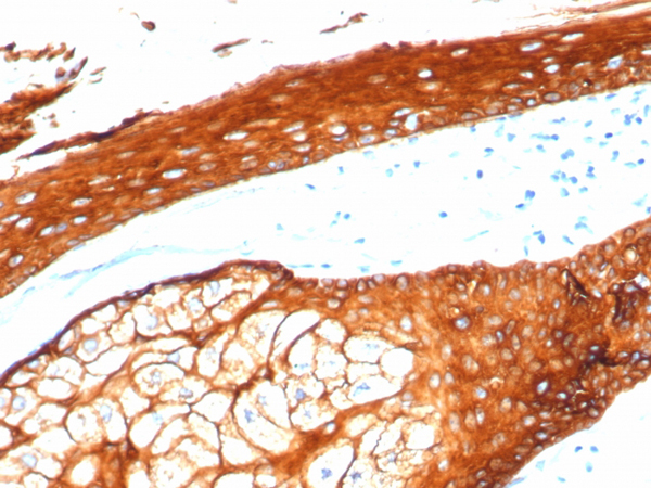
 IHC (Immunohistochemistry)
(Formalin-fixed, paraffin-embedded human skin stained with CK Type I Recombinant Mouse Monoclonal Antibody (rKRTL/6616).)
IHC (Immunohistochemistry)
(Formalin-fixed, paraffin-embedded human skin stained with CK Type I Recombinant Mouse Monoclonal Antibody (rKRTL/6616).)
Cytokeratin, Type I, Monoclonal Antibody (Cat# AAA216089)
 IHC (Immunohistochemistry)
(Formalin-fixed, paraffin-embedded human skin stained with CK Type II Recombinant Rabbit Monoclonal Antibody (KRTH/4392R).)
IHC (Immunohistochemistry)
(Formalin-fixed, paraffin-embedded human skin stained with CK Type II Recombinant Rabbit Monoclonal Antibody (KRTH/4392R).)
Cytokeratin, Type II, Monoclonal Antibody (Cat# AAA216094)
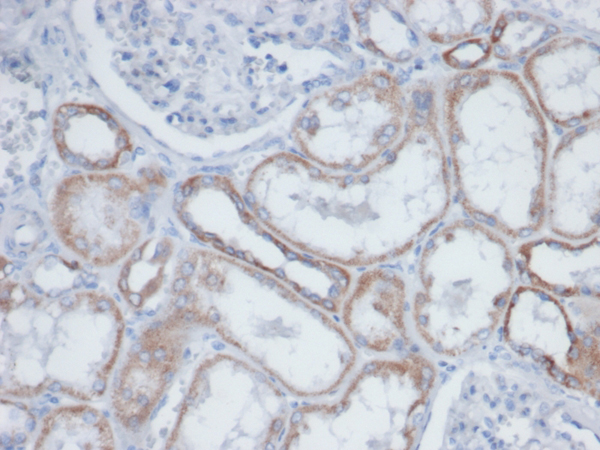
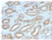
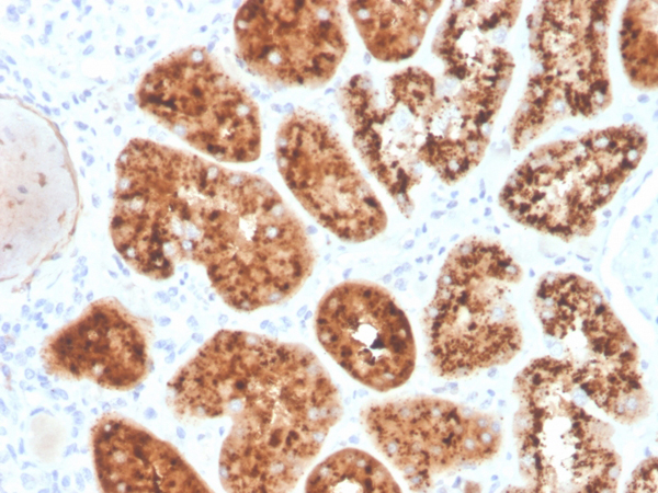 IHC (Immunohistochemistry)
(Formalin-fixed, paraffin-embedded human kidney stained with RBP4 Mouse Monoclonal Antibody (RBP4/4045).)
IHC (Immunohistochemistry)
(Formalin-fixed, paraffin-embedded human kidney stained with RBP4 Mouse Monoclonal Antibody (RBP4/4045).)
RBP4/Retinol Binding Protein 4, Monoclonal Antibody (Cat# AAA215882)
 IHC (Immunohistochemistry)
(Formalin-fixed, paraffin-embedded human mantle cell lymphoma stained Cyclin D1 Recombinant Rabbit Monoclonal Antibody (CCND1/3370R).)
IHC (Immunohistochemistry)
(Formalin-fixed, paraffin-embedded human mantle cell lymphoma stained Cyclin D1 Recombinant Rabbit Monoclonal Antibody (CCND1/3370R).)
Cyclin D1, Monoclonal Antibody (Cat# AAA215888)
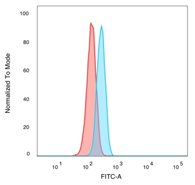
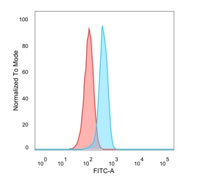
 FCM/FACS (Flow Cytometry)
(Flow Cytometric Analysis of PFA-fixed HeLa cells. NF-kB p65/RELA Mouse Monoclonal Antibody (PCRP-RELA-2B6) followed by goat anti-mouse IgG-CF488 (blue); isotype control (red).)
FCM/FACS (Flow Cytometry)
(Flow Cytometric Analysis of PFA-fixed HeLa cells. NF-kB p65/RELA Mouse Monoclonal Antibody (PCRP-RELA-2B6) followed by goat anti-mouse IgG-CF488 (blue); isotype control (red).)
NF-kB p65/RELA, Monoclonal Antibody (Cat# AAA215891)
Predicted to react with Rat.
 FCM/FACS (Flow Cytometry)
(Flow Cytometric Analysis of PFA-fixed HeLa cells. BCL6 Mouse Monoclonal Antibody (PCRP-BCL6-1E2) followed by goat anti-mouse IgG-CF488 (blue); unstained cells (red).)
FCM/FACS (Flow Cytometry)
(Flow Cytometric Analysis of PFA-fixed HeLa cells. BCL6 Mouse Monoclonal Antibody (PCRP-BCL6-1E2) followed by goat anti-mouse IgG-CF488 (blue); unstained cells (red).)
BCL6, Monoclonal Antibody (Cat# AAA215896)
 IHC (Immunohistochemistry)
(Formalin-fixed, paraffin-embedded human lymph node stained with RXRB Mouse Monoclonal Antibody (PCRP-RXRB-2B6).)
IHC (Immunohistochemistry)
(Formalin-fixed, paraffin-embedded human lymph node stained with RXRB Mouse Monoclonal Antibody (PCRP-RXRB-2B6).)
RXRB, Monoclonal Antibody (Cat# AAA215899)
 SDS-PAGE
(SDS-PAGE Analysis Purified MCP2/CCL8 Mouse Monoclonal Antibody (CCL8/3312). Confirmation of Purity and Integrity of Antibody.)
SDS-PAGE
(SDS-PAGE Analysis Purified MCP2/CCL8 Mouse Monoclonal Antibody (CCL8/3312). Confirmation of Purity and Integrity of Antibody.)
Monocyte Chemotactic Protein 2 (MCP2)/CCL8, Monoclonal Antibody (Cat# AAA215907)
 SDS-PAGE
(SDS-PAGE Analysis Purified MCP2/CCL8 Mouse Monoclonal Antibody (CCL8/3683). Confirmation of Purity and Integrity of Antibody.)
SDS-PAGE
(SDS-PAGE Analysis Purified MCP2/CCL8 Mouse Monoclonal Antibody (CCL8/3683). Confirmation of Purity and Integrity of Antibody.)
Monocyte Chemotactic Protein 2 (MCP2)/CCL8, Monoclonal Antibody (Cat# AAA215908)
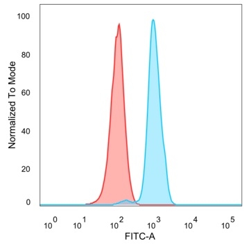
 IHC (Immunohistochemistry)
(Formalin-fixed, paraffin-embedded human lung stained with Surfactant Protein D Mouse Monoclonal Antibody (SFTPD/4363).)
IHC (Immunohistochemistry)
(Formalin-fixed, paraffin-embedded human lung stained with Surfactant Protein D Mouse Monoclonal Antibody (SFTPD/4363).)
Pulmonary Surfactant-Associated Protein D (SFTPD), Monoclonal Antibody (Cat# AAA215915)
 IHC (Immunohistochemistry)
(Formalin-fixed, paraffin-embedded human lung carcinoma stained with Surfactant Protein D Recombinant Rabbit Monoclonal (SFTPD/7086R).)
IHC (Immunohistochemistry)
(Formalin-fixed, paraffin-embedded human lung carcinoma stained with Surfactant Protein D Recombinant Rabbit Monoclonal (SFTPD/7086R).)
Pulmonary Surfactant-Associated Protein D (SFTPD), Monoclonal Antibody (Cat# AAA215918)
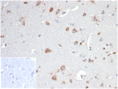
 IHC (Immunohistochemistry)
(Formalin-fixed, paraffin-embedded human testis stained with INI-1 Mouse Monoclonal Antibody (SMARCB1/3984) at 2ug/ml. Inset: PBS instead of primary antibody, secondary negative control.)
IHC (Immunohistochemistry)
(Formalin-fixed, paraffin-embedded human testis stained with INI-1 Mouse Monoclonal Antibody (SMARCB1/3984) at 2ug/ml. Inset: PBS instead of primary antibody, secondary negative control.)
Integrase interactor 1 (INI-1)/SNF5/SMARCB1, Monoclonal Antibody (Cat# AAA215921)
 IHC (Immunohistochemistry)
(Formalin-fixed, paraffin-embedded human sarcoma stained with INI-1 Recombinant Rabbit Monoclonal Antibody (SMARCB1/4587R))
IHC (Immunohistochemistry)
(Formalin-fixed, paraffin-embedded human sarcoma stained with INI-1 Recombinant Rabbit Monoclonal Antibody (SMARCB1/4587R))
Integrase interactor 1 (INI-1)/SNF5/SMARCB1, Monoclonal Antibody (Cat# AAA215922)
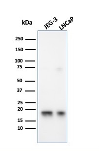 WB (Western Blot)
(Western Blot Analysis of JEG-3 and LNCaP cell lysates using Superoxide Dismutase 1 Mouse Monoclonal Antibody (SOD1/3926).)
WB (Western Blot)
(Western Blot Analysis of JEG-3 and LNCaP cell lysates using Superoxide Dismutase 1 Mouse Monoclonal Antibody (SOD1/3926).)
Superoxide Dismutase 1 (SOD1), Monoclonal Antibody (Cat# AAA215930)
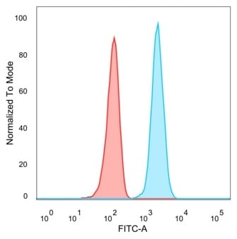
 FCM/FACS (Flow Cytometry)
(Flow Cytometric Analysis of PFA-fixed MCF-7 cells using Superoxide Dismutase 1 Mouse Monoclonal Antibody (SOD1/4331) followed by goat anti-mouse IgG-CF488 (blue); isotype control (red).)
FCM/FACS (Flow Cytometry)
(Flow Cytometric Analysis of PFA-fixed MCF-7 cells using Superoxide Dismutase 1 Mouse Monoclonal Antibody (SOD1/4331) followed by goat anti-mouse IgG-CF488 (blue); isotype control (red).)
Superoxide Dismutase 1 (SOD1), Monoclonal Antibody (Cat# AAA215932)
 IHC (Immunohistochemistry)
(Formalin-fixed, paraffin-embedded human tonsil stained with CD43 Recombinant Rabbit Monoclonal Antibody (SPN/6562R). Inset: PBS instead of primary antibody, secondary negative control.)
IHC (Immunohistochemistry)
(Formalin-fixed, paraffin-embedded human tonsil stained with CD43 Recombinant Rabbit Monoclonal Antibody (SPN/6562R). Inset: PBS instead of primary antibody, secondary negative control.)
CD43, Monoclonal Antibody (Cat# AAA215942)
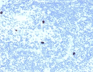
 IHC (Immunohiostchemistry)
Osteopontin (OSP)/Secreted Phosphoprotein 1 (SPP1), Monoclonal Antibody (Cat# AAA215943)
 FCM/FACS (Flow Cytometry)
(Flow cytometric analysis of PFA-fixed HeLa cells. STAT5A Mouse Monoclonal Antibody (PCRP-STAT5A-1A9) followed by goat anti-mouse IgG-CF488 (blue); isotype control (red).)
FCM/FACS (Flow Cytometry)
(Flow cytometric analysis of PFA-fixed HeLa cells. STAT5A Mouse Monoclonal Antibody (PCRP-STAT5A-1A9) followed by goat anti-mouse IgG-CF488 (blue); isotype control (red).)
STAT5A, Monoclonal Antibody (Cat# AAA215948)
Predicted to work with Mouse and Rat.
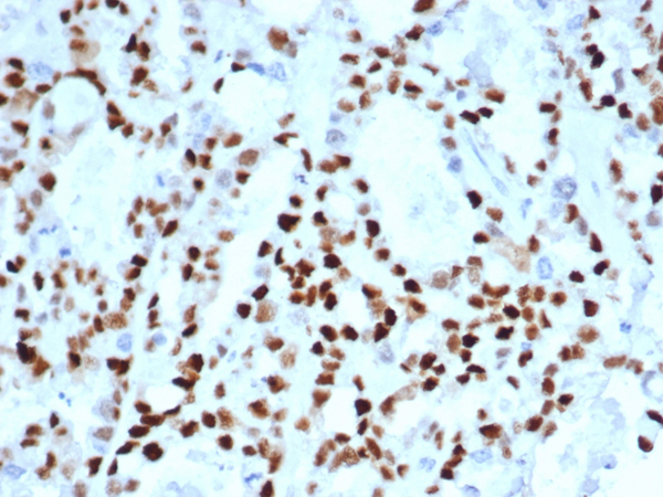
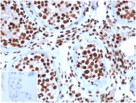
 IHC (Immunohistochemistry)
(Formalin-fixed, paraffin-embedded human tonsil stained with Ki67 Recombinant Mouse Monoclonal Antibody (rMKI67/6499).)
IHC (Immunohistochemistry)
(Formalin-fixed, paraffin-embedded human tonsil stained with Ki67 Recombinant Mouse Monoclonal Antibody (rMKI67/6499).)
Ki-67, Monoclonal Antibody (Cat# AAA215805)
 IHC (Immunohistochemistry)
(Formalin-fixed, paraffin-embedded human tonsil stained with Ki67 Recombinant Rabbit Monoclonal Antibody (MKI67/4947R).)
IHC (Immunohistochemistry)
(Formalin-fixed, paraffin-embedded human tonsil stained with Ki67 Recombinant Rabbit Monoclonal Antibody (MKI67/4947R).)
Ki-67, Monoclonal Antibody (Cat# AAA215808)
 IHC (Immunohistochemistry)
(Formalin-fixed, paraffin-embedded human tonsil stained with Ki67 Recombinant Rabbit Monoclonal Antibody (MKI67/6517R).)
IHC (Immunohistochemistry)
(Formalin-fixed, paraffin-embedded human tonsil stained with Ki67 Recombinant Rabbit Monoclonal Antibody (MKI67/6517R).)
Ki-67, Monoclonal Antibody (Cat# AAA215809)
 SDS-PAGE
(SDS-PAGE Analysis Purified MPO Recombinant Mouse Monoclonal Antibody (rMPO/6904). Confirmation of Integrity and Purity of Antibody.)
SDS-PAGE
(SDS-PAGE Analysis Purified MPO Recombinant Mouse Monoclonal Antibody (rMPO/6904). Confirmation of Integrity and Purity of Antibody.)
Myeloperoxidase/MPO, Monoclonal Antibody (Cat# AAA215814)
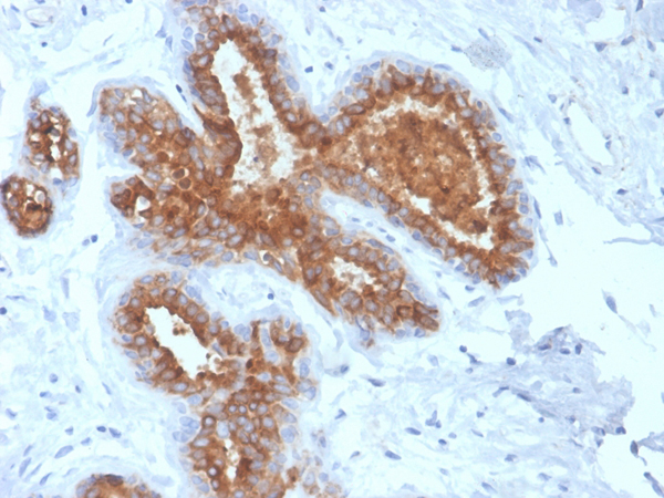 IHC (Immunohistochemistry)
(Formalin-fixed, paraffin-embedded human breast carcinoma stained with MUC-1/EMA Recombinant Mouse Monoclonal Antibody (rMUC1/4418).)
IHC (Immunohistochemistry)
(Formalin-fixed, paraffin-embedded human breast carcinoma stained with MUC-1/EMA Recombinant Mouse Monoclonal Antibody (rMUC1/4418).)
MUC1/CA15-3/EMA/CD227, Monoclonal Antibody (Cat# AAA215818)
What are Monoclonal Antibodies?
Monoclonal antibodies are specialized laboratory-produced proteins developed for binding to specific biological antigens or other molecular targets. Since they come from a single cell (or clone), they are especially consistent and accurate in the data they are involved in producing.
This type of antibody material has been shown to be a powerful tool in finding and subsequently destroying harmful cells in an organism, such as those found in cancers or various autoimmune diseases. This makes them excellent aids in medical testing and research, which is why they are so widely used.
AAA Biotech offers a comprehensive range of high-quality monoclonal antibodies that perform effectively in various laboratory tests, including (amongst others) ELISA, western blotting, immunohistochemistry, and flow cytometry. All of the products in our catalog are thoroughly quality tested to make sure that they are reliable and will consistently perform well in your research.
What Are The Uses of Monoclonal Antibodies
Monoclonal antibodies are used in many lab tests, including (amongst others) ELISA, western blotting, immunohistochemistry, and flow cytometry.
ELISA is a test that helps detect a specific substance/analyte in a sample. It uses antibodies (often monoclonal) bound to a solid surface (such as the well of a microplate) to “capture” the substance/analyte in the sample and immobilize it so that the detection antibody component can then bind to it and produce a signal, which can then be measured.
Western blotting identifies specific proteins in a sample. The sample is first separated on a gel, and then antibodies are applied that will typically bind to the target, which will all be localized to a single band in a lane.
Immunohistochemistry helps locate specific proteins in cells or tissue samples using antibodies.
Flow cytometry looks at and sorts cells. It uses antibodies that are conjugated to reporter molecules called “fluorophores”, which, under special lights, emit light themselves, which can then be measured by a detector instrument.
How Monoclonal Antibodies Are Used as Medicine?
Please note that all of the products listed in AAA Biotech’s also known as AAA Bio or AAABio catalog are strictly for research-use only (RUO).
Monoclonal antibodies can also be used as therapeutic/medical treatments, particularly in the context of cancers. They are designed to find and bind to specific cells or proteins, helping the immune system recognize and attack the cancer. These treatments work in different ways, such as:
- Radioimmunotherapy attaches a small amount of radioactive molecule to the antibody, so it delivers the radiation directly to the cancer cells that the antibody is specifically binding to.
- Antibody-directed enzyme prodrug therapy uses antibodies that are specifically bound to special enzymes. These enzymes activate a harmless drug in the body and turn it into a cancer-killing drug only near the cancer cells—this helps avoid harming healthy cells.
- Immunoliposomes are tiny “bubbles” filled with medicine/drug and coated with antibodies. They carry the drug straight to the cancer cells.
Why Buy Monoclonal Antibodies From Us?
At AAA Biotech, we provide high-performance monoclonal antibodies designed to support a wide range of research needs.
1. Validated for Versatile Applications
The antibodies in our catalog are extensively validated and compatible with multiple techniques, including (but not limited to) ELISA, flow cytometry (FC), immunocytochemistry (ICC), immunofluorescence (IF), immunohistochemistry (IHC), immunoprecipitation (IP), and western blotting (WB).
2. Wide Selection & Specialized Options
We offer antibodies for common and rare species, that are available in various conjugated forms, and also in recombinant formats. Essentially, there is almost anything one might need to meet their experimental model’s requirements.
3. High-Quality Proteins
Our proteins meet high purity standards—90% or more as confirmed by SDS-PAGE. Many are available with tags like His, Flag, GST, or MBP, and we also supply native and biologically active proteins for functional studies.
Frequently Asked Questions
1. Are your monoclonal antibodies validated for specific applications?
Yes, our antibodies are tested and validated for use in methods such as ELISA, western blot, IHC, flow cytometry, and more. Refer to specific product pages or datasheets for individual product information.
2. How do I choose the right monoclonal antibody for my application?
Review the product details directly for application validation, species reactivity, and target information. You may also contact our support team at any time for help.
3. How quickly can I receive my order?
Most orders are processed and shipped within 1–3 business days, depending on product availability and your shipping location.