Filters
▼Clonality
▼Type
▼Reactivity
▼Gene Name
▼Isotype
▼Host
▼Application
▼Clone
▼Monoclonal Antibodies
Get accurate results in your research with our Monoclonal Antibodies, which are specially made to target exactly what you require for your research, and will produce consistent, reliable performance in lab tests.
Viewing 9650-9700 of 27597 product results
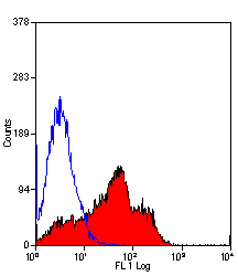
 FCM/FACS (Flow Cytometry)
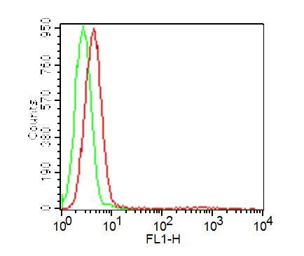
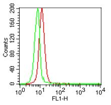
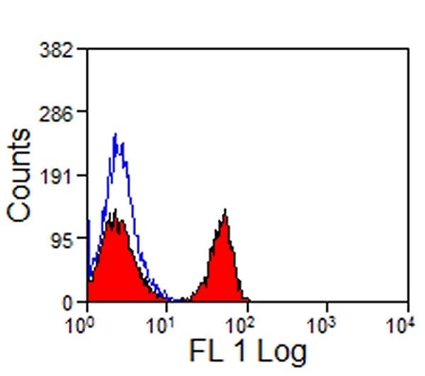
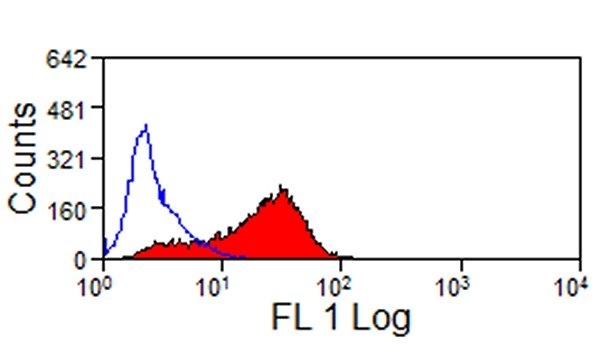
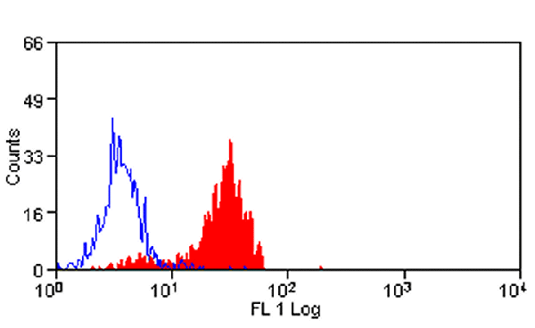
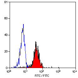
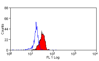
(Fig: 1 Cell surface flow analysis of FITC-conjugated Rat IgG2a isotype control in Ramos cell line using 0.5 ug/10^6 cells (Clone: 2A3). Green represents cell only; red represents Rat IgG2a isotype control conjugated with FITC.)
FCM/FACS (Flow Cytometry)
(Fig: 1 Cell surface flow analysis of FITC-conjugated Rat IgG2a isotype control in Ramos cell line using 0.5 ug/10^6 cells (Clone: 2A3). Green represents cell only; red represents Rat IgG2a isotype control conjugated with FITC.)
IgG2a, Monoclonal Isotype Control (Cat# AAA78386)
Affinity Chromatography
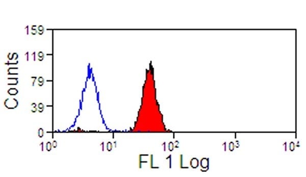
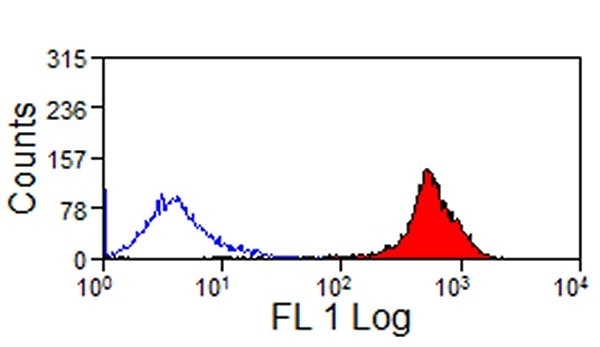
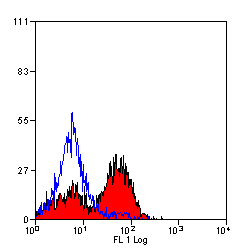
 FCM/FACS (Flow Cytometry)
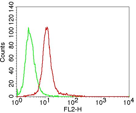
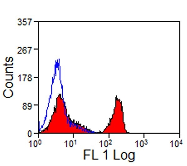
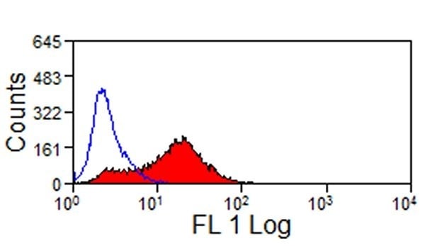
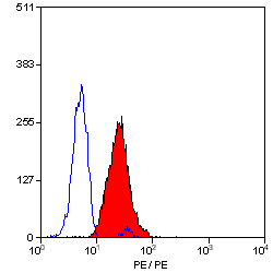
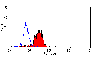
(Figure-2: Intra cellular Flow analysis of Caspase-14 in A431 cells using 0.5 ug/10^6 cells of Caspase-14 antibody (Clone: ABM1B24). Green represents isotype control; red represents anti-Caspase-14 antibody. Goat anti-mouse PE conjugate was used as secondary antibody.)
FCM/FACS (Flow Cytometry)
(Figure-2: Intra cellular Flow analysis of Caspase-14 in A431 cells using 0.5 ug/10^6 cells of Caspase-14 antibody (Clone: ABM1B24). Green represents isotype control; red represents anti-Caspase-14 antibody. Goat anti-mouse PE conjugate was used as secondary antibody.)
Caspase-14, Monoclonal Antibody (Cat# AAA78401)
Protein G Chromatography
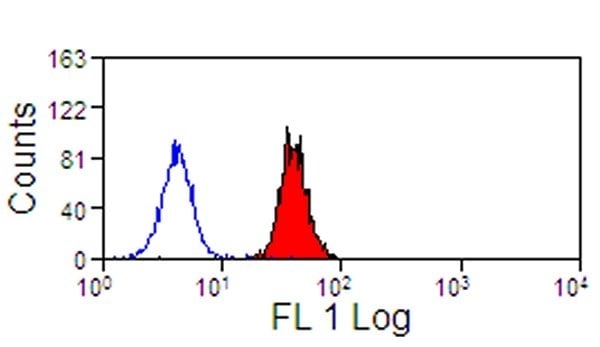
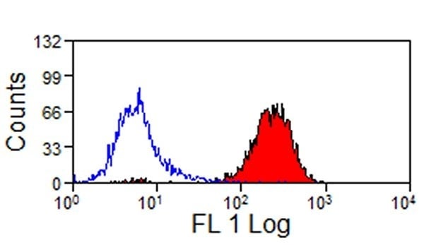
 FCM/FACS (Flow Cytometry)
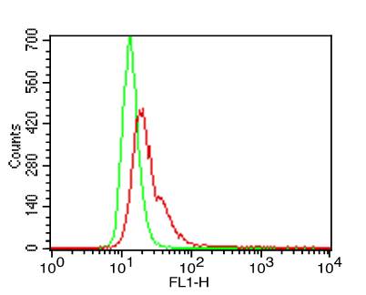
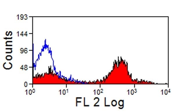
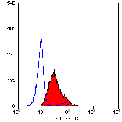
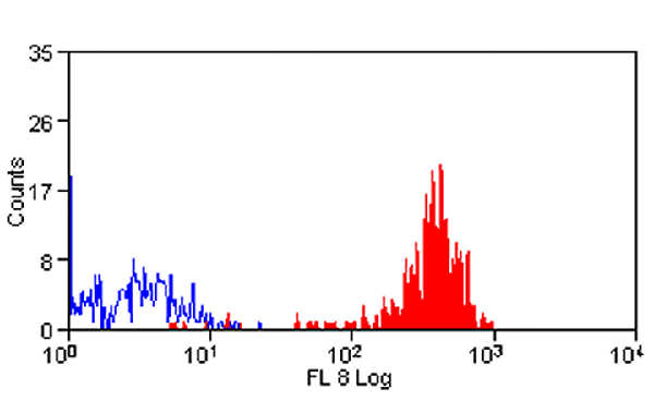
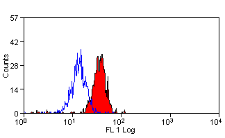
(Fig-2: Intracellular flow analysis of hTLR6 FITC conjugated in hPBMC using 0.5 ug/10^6 cells of hTLR6-FITC conjugated antibody (Clone: ABM1B50). Green represents isotype control and red represent hTLR6-FITC conjugated.)
FCM/FACS (Flow Cytometry)
(Fig-2: Intracellular flow analysis of hTLR6 FITC conjugated in hPBMC using 0.5 ug/10^6 cells of hTLR6-FITC conjugated antibody (Clone: ABM1B50). Green represents isotype control and red represent hTLR6-FITC conjugated.)
TLR6, Monoclonal Antibody (Cat# AAA78405)
Protein G Chromatography
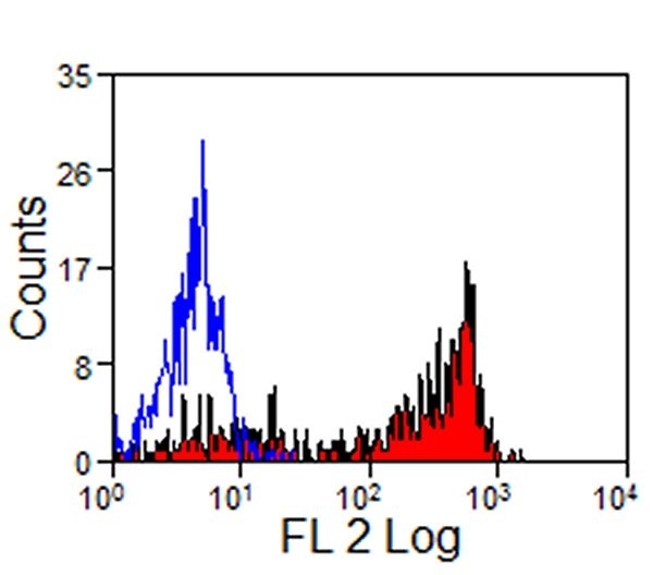
 FCM/FACS (Flow Cytometry)
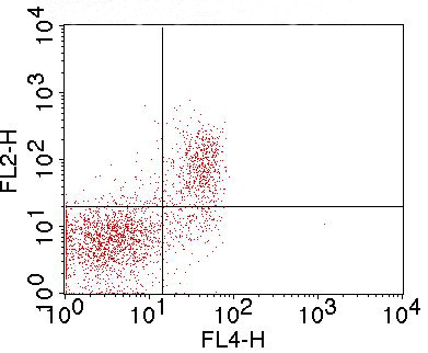
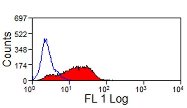
(Figure-2: Cell surface flowcytometric staining of mGITR (Clone: DTA-1) in mouse Splenocytes. Mouse Splenocytes were first stained with Anti-mCD3 APC conjugated, mCD3 APC positive cells were gated and further analyzed for mGITR using 0.5 ug/10^6. Goat anti-Rat PE secondary antibody was used.)
FCM/FACS (Flow Cytometry)
(Figure-2: Cell surface flowcytometric staining of mGITR (Clone: DTA-1) in mouse Splenocytes. Mouse Splenocytes were first stained with Anti-mCD3 APC conjugated, mCD3 APC positive cells were gated and further analyzed for mGITR using 0.5 ug/10^6. Goat anti-Rat PE secondary antibody was used.)
GITR, Monoclonal Antibody (Cat# AAA78419)
Protein G Chromatography
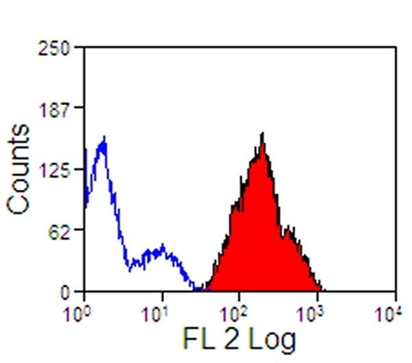
 FCM/FACS (Flow Cytometry)
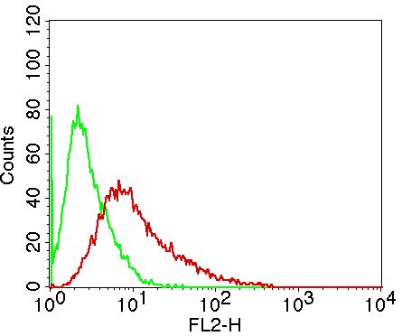
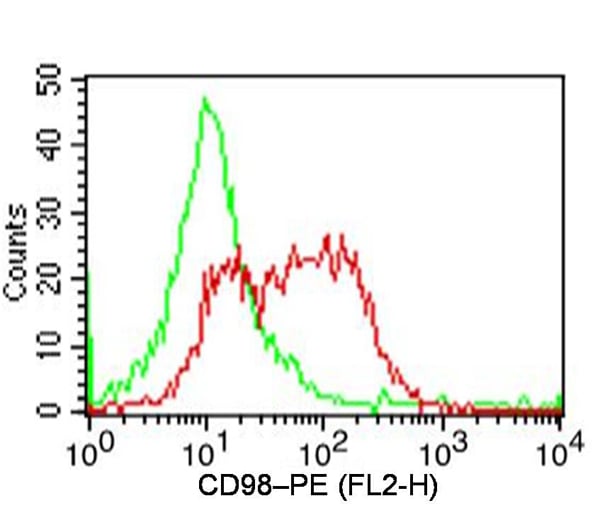
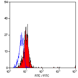
(Fig-2: Cell surface flow analysis of hCD98 on human PBMCs (Monocytes gated) using 1 ug/ 10^6 cells. Green represents isotype control; red represents anti-hCD98 antibody (10-4168). Goat anti-mouse PE conjugated secondary antibody was used.. (Cells were incubated with primary antibody for 45 min. then washed twice with PBS by centrifuging at 1100 rpm for 5 min, followed by 30 min incubation with conjugated secondary antibody. Data acquisition was done after washing twice with PBS as mentioned above).)
FCM/FACS (Flow Cytometry)
(Fig-2: Cell surface flow analysis of hCD98 on human PBMCs (Monocytes gated) using 1 ug/ 10^6 cells. Green represents isotype control; red represents anti-hCD98 antibody (10-4168). Goat anti-mouse PE conjugated secondary antibody was used.. (Cells were incubated with primary antibody for 45 min. then washed twice with PBS by centrifuging at 1100 rpm for 5 min, followed by 30 min incubation with conjugated secondary antibody. Data acquisition was done after washing twice with PBS as mentioned above).)
hCD98, Monoclonal Antibody (Cat# AAA78423)
Protein G Chromatography
 WB (Western Blot)
(The image below shows that anti-human IL-17A antibody clone 7F10 reacts with human 293 cell expressed human IL-17A antigen (100ng) in Western Blot. The blot was stained with HRP substrate 4-Chloro-1-naphthol)
WB (Western Blot)
(The image below shows that anti-human IL-17A antibody clone 7F10 reacts with human 293 cell expressed human IL-17A antigen (100ng) in Western Blot. The blot was stained with HRP substrate 4-Chloro-1-naphthol)
IL-17A, Monoclonal Antibody (Cat# AAA77970)
Triiodothyronine, Monoclonal Antibody (Cat# AAA78055)
C-reactive protein (cCRP), Monoclonal Antibody (Cat# AAA78096)
Purity: Determined by Gel electrophoresis and gel scanning
 Application Data
(Binding of anti-CD75 antibody to human cell lines)
Application Data
(Binding of anti-CD75 antibody to human cell lines)
CD75, Monoclonal Antibody (Cat# AAA78147)
Kanamycin, Monoclonal Antibody (Cat# AAA81755)
 IHC (Immunohiostchemistry)
(Immunohistochemical analysis of paraffin-embedded human kidney, using JAG1 Antibody.)
IHC (Immunohiostchemistry)
(Immunohistochemical analysis of paraffin-embedded human kidney, using JAG1 Antibody.)
JAG1, Monoclonal Antibody (Cat# AAA126875)
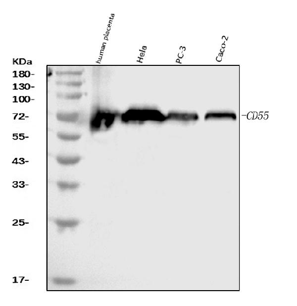
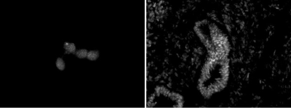
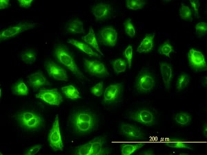
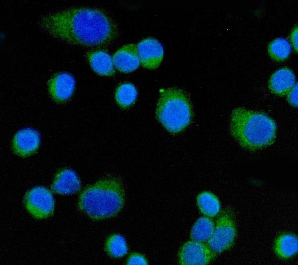 IF (Immunofluorescence)
(Figure 3. IF analysis of CD55 using anti-CD55 antibody (AAA126881).CD55 was detected in an immunocytochemical section of SiHa cells. Enzyme antigen retrieval was performed using IHC enzyme antigen retrieval reagent (AR0022) for 15 mins. The cells were blocked with 10% goat serum. And then incubated with 5 ug/mL mouse anti-CD55 Antibody (AAA126881) overnight at 4 degree C. DyLight488 Conjugated Goat Anti-Mouse IgG was used as secondary antibody at 1:100 dilution and incubated for 30 minutes at 37 degree C. The section was counterstained with DAPI. Visualize using a fluorescence microscope and filter sets appropriate for the label used.)
IF (Immunofluorescence)
(Figure 3. IF analysis of CD55 using anti-CD55 antibody (AAA126881).CD55 was detected in an immunocytochemical section of SiHa cells. Enzyme antigen retrieval was performed using IHC enzyme antigen retrieval reagent (AR0022) for 15 mins. The cells were blocked with 10% goat serum. And then incubated with 5 ug/mL mouse anti-CD55 Antibody (AAA126881) overnight at 4 degree C. DyLight488 Conjugated Goat Anti-Mouse IgG was used as secondary antibody at 1:100 dilution and incubated for 30 minutes at 37 degree C. The section was counterstained with DAPI. Visualize using a fluorescence microscope and filter sets appropriate for the label used.)
CD55, Monoclonal Antibody (Cat# AAA126881)
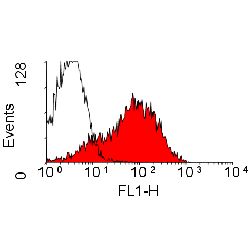
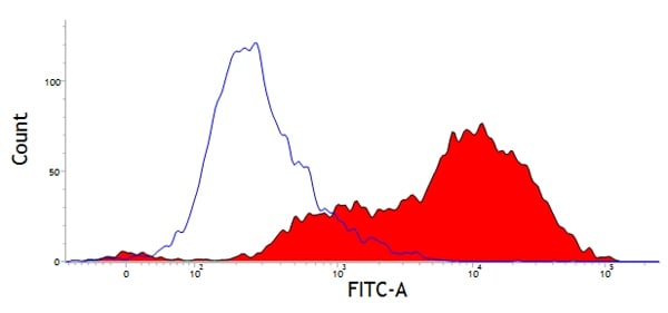
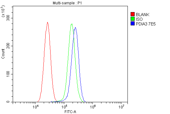 FCM/FACS (Flow Cytometry)
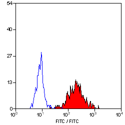
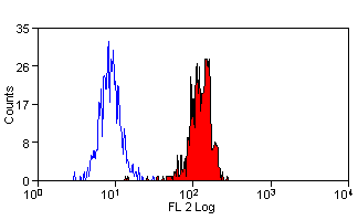
(Figure 3. Flow Cytometry analysis of U87 cells using anti-ERp57 antibody (AAA126893).Overlay histogram showing U87 cells stained with AAA126893 (Blue line). The cells were blocked with 10% normal goat serum. And then incubated with mouse anti-ERp57 Antibody (AAA126893, 1 ug/1x10^6 cells) for 30 min at 20 degree C. DyLight488 conjugated goat anti-mouse IgG was used as secondary antibody for 30 minutes at 20 degree C. Isotype control antibody (Green line) was mouse IgG (1 ug/1x10^6) used under the same conditions. Unlabelled sample (Red line) was also used as a control.)
FCM/FACS (Flow Cytometry)
(Figure 3. Flow Cytometry analysis of U87 cells using anti-ERp57 antibody (AAA126893).Overlay histogram showing U87 cells stained with AAA126893 (Blue line). The cells were blocked with 10% normal goat serum. And then incubated with mouse anti-ERp57 Antibody (AAA126893, 1 ug/1x10^6 cells) for 30 min at 20 degree C. DyLight488 conjugated goat anti-mouse IgG was used as secondary antibody for 30 minutes at 20 degree C. Isotype control antibody (Green line) was mouse IgG (1 ug/1x10^6) used under the same conditions. Unlabelled sample (Red line) was also used as a control.)
ERp57, Monoclonal Antibody (Cat# AAA126893)

 WB (Western Blot)
(Western blot analysis of GPCR/LGR6 expression in HUVEC cell lysate.)
WB (Western Blot)
(Western blot analysis of GPCR/LGR6 expression in HUVEC cell lysate.)
GPCR/LGR6, Monoclonal Antibody (Cat# AAA126947)
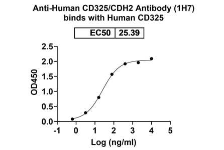 Bioactivity
(Detects CD325/CDH2 in indirect ELISAs.)
Bioactivity
(Detects CD325/CDH2 in indirect ELISAs.)
CD325/CDH2, Monoclonal Antibody (Cat# AAA120168)
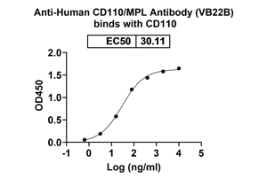 Bioactivity
(Detects CD110/MPL in indirect ELISAs)
Bioactivity
(Detects CD110/MPL in indirect ELISAs)
CD110/MPL, Monoclonal Antibody (Cat# AAA120172)
PAPILLOMAVIRUS 16 ONCOPROTEIN E7, Monoclonal Antibody (Cat# AAA49545)
ACTH, Monoclonal Antibody (Cat# AAA49551)
 Application Data
(Human apolipoprotein E detected with Mouse anti Human apolipoprotein E followed by Rabbit F(ab')2 anti Mouse IgG:HRP)
Application Data
(Human apolipoprotein E detected with Mouse anti Human apolipoprotein E followed by Rabbit F(ab')2 anti Mouse IgG:HRP)
APOLIPOPROTEIN E, Monoclonal Antibody (Cat# AAA50105)
 Application Data
(Human peripheral blood monocytes stained with Mouse anti Human Siglec-9:RPE)
Application Data
(Human peripheral blood monocytes stained with Mouse anti Human Siglec-9:RPE)
SIGLEC-9, Monoclonal Antibody (Cat# AAA50116)
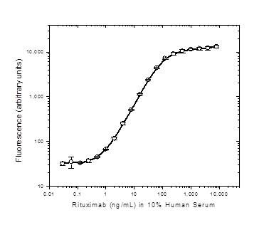
 Application Data
(Detection of Rituximab in direct ELISA. Rituximab was used as the coating reagent followed by Rat anti Rituximab:HRP (AAA50117) as the detection antibody)
Application Data
(Detection of Rituximab in direct ELISA. Rituximab was used as the coating reagent followed by Rat anti Rituximab:HRP (AAA50117) as the detection antibody)
RITUXIMAB, Monoclonal Antibody (Cat# AAA50117)
 Application Data
(Staining of equine peripheral blood lymphocytes with Mouse anti Equine CD44:FITC(AAA50133))
Application Data
(Staining of equine peripheral blood lymphocytes with Mouse anti Equine CD44:FITC(AAA50133))
CD44, Monoclonal Antibody (Cat# AAA50133)
 WB (Western Blot)
(Western blot analysis of HeLa cell nuclear extract (40 ug) probed with CHD5 antibody (AAA50445) at a 2 ug/ml dilution.)
WB (Western Blot)
(Western blot analysis of HeLa cell nuclear extract (40 ug) probed with CHD5 antibody (AAA50445) at a 2 ug/ml dilution.)
CHD5, Monoclonal Antibody (Cat# AAA50445)
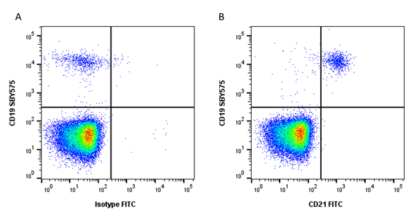 Application Data
(Figure A. StarBright Yellow 575 conjugated Mouse anti Human CD19 and FITC conjugated Mouse IgG1 isotype control . Figure B. StarBright Yellow 575 conjugated Mouse anti Human CD19 and FITC conjugated Mouse anti Human CD21 . All experiments performed on red cell lysed human blood gated on single cell lymphocytes, in the presence of 10% human serum. Data acquired on the ZE5 Cell Analyzer.)
Application Data
(Figure A. StarBright Yellow 575 conjugated Mouse anti Human CD19 and FITC conjugated Mouse IgG1 isotype control . Figure B. StarBright Yellow 575 conjugated Mouse anti Human CD19 and FITC conjugated Mouse anti Human CD21 . All experiments performed on red cell lysed human blood gated on single cell lymphocytes, in the presence of 10% human serum. Data acquired on the ZE5 Cell Analyzer.)
CD44, Monoclonal Antibody (Cat# AAA50483)
 Application Data
(Horse peripheral blood lymphocytes stained with Mouse anti Horse CD4:RPE (AAA50147))
Application Data
(Horse peripheral blood lymphocytes stained with Mouse anti Horse CD4:RPE (AAA50147))
CD4, Monoclonal Antibody (Cat# AAA50147)
CD11c, Monoclonal Antibody (Cat# AAA50152)
 Application Data
(Staining of JAM-C transfected CHO cells with Rat anti Mouse JAM-C:Biotin (AAA50206) followed by Streptavidin:FITC)
Application Data
(Staining of JAM-C transfected CHO cells with Rat anti Mouse JAM-C:Biotin (AAA50206) followed by Streptavidin:FITC)
JAM-C, Monoclonal Antibody (Cat# AAA50206)
 Application Data
(Purified Bovine IgA detected with Mouse anti Bovine/Ovine IgA followed by Rabbit F(ab')2 anti Mouse IgG:HRP)
Application Data
(Purified Bovine IgA detected with Mouse anti Bovine/Ovine IgA followed by Rabbit F(ab')2 anti Mouse IgG:HRP)
IgA, Monoclonal Antibody (Cat# AAA50248)
YELLOW FEVER VIRUS, Monoclonal Antibody (Cat# AAA50578)
 WB (Western Blot)
(Western blot of anti-STAT3 monoclonal antibody against full-length STAT3-His recombinant protein(1).)
WB (Western Blot)
(Western blot of anti-STAT3 monoclonal antibody against full-length STAT3-His recombinant protein(1).)
STAT3, Monoclonal Antibody (Cat# AAA51888)
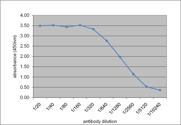
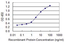 ELISA
(Detection limit for recombinant GST tagged COL1A1 is 0.03 ng/ml as a capture antibody.)
ELISA
(Detection limit for recombinant GST tagged COL1A1 is 0.03 ng/ml as a capture antibody.)
COL1A1 / Collagen I, Monoclonal Antibody (Cat# AAA51905)

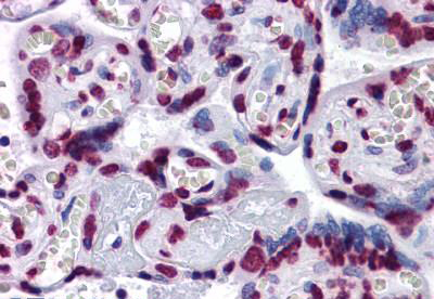
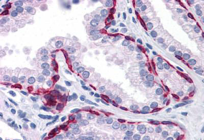
 IHC (Immunohistochemisry)
(Anti-CD69 antibody IHC of human small intestine. Immunohistochemistry of formalin-fixed, paraffin-embedded tissue after heat-induced antigen retrieval. Antibody dilution 1:200.)
IHC (Immunohistochemisry)
(Anti-CD69 antibody IHC of human small intestine. Immunohistochemistry of formalin-fixed, paraffin-embedded tissue after heat-induced antigen retrieval. Antibody dilution 1:200.)
CD69, Monoclonal Antibody (Cat# AAA51928)
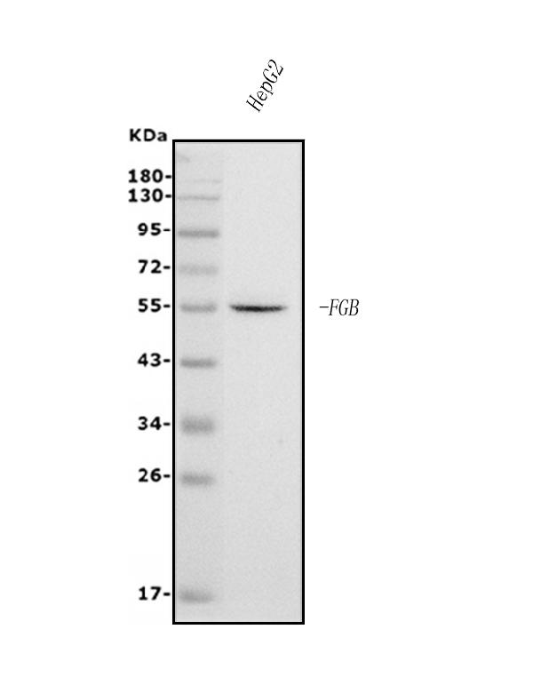
 WB (Western Blot)
(KRTHB1 monoclonal antibody clone 3B10-5B10 Western blot of KRTHB1 expression in HepG2.)
WB (Western Blot)
(KRTHB1 monoclonal antibody clone 3B10-5B10 Western blot of KRTHB1 expression in HepG2.)
KRT81 / KRTHB1 / MLN 137, Monoclonal Antibody (Cat# AAA51939)
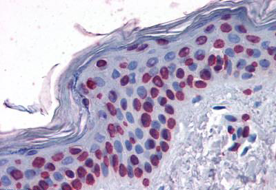
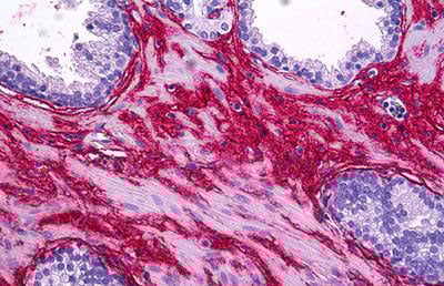
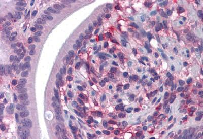
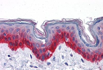
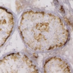
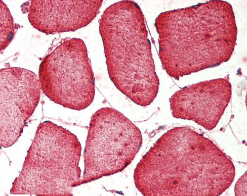
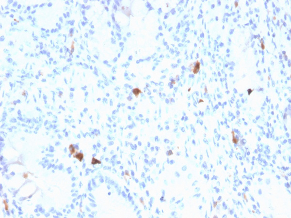
 IHC (Immunohistochemistry)
(Anti-CD147 antibody IHC of human testis. Immunohistochemistry of formalin-fixed, paraffin-embedded tissue after heat-induced antigen retrieval. Antibody concentration 10 ug/ml.)
IHC (Immunohistochemistry)
(Anti-CD147 antibody IHC of human testis. Immunohistochemistry of formalin-fixed, paraffin-embedded tissue after heat-induced antigen retrieval. Antibody concentration 10 ug/ml.)
Basigin / Emmprin / CD147, Monoclonal Antibody (Cat# AAA51783)
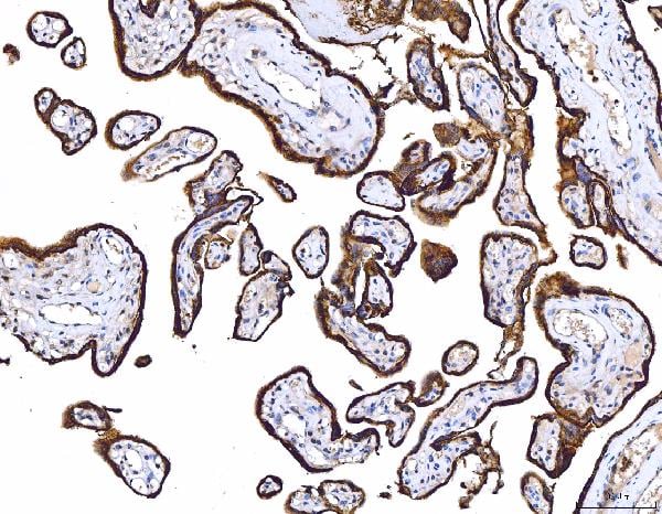
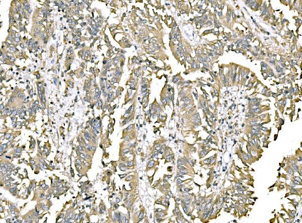
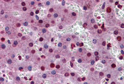
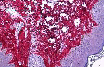
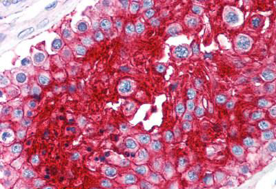
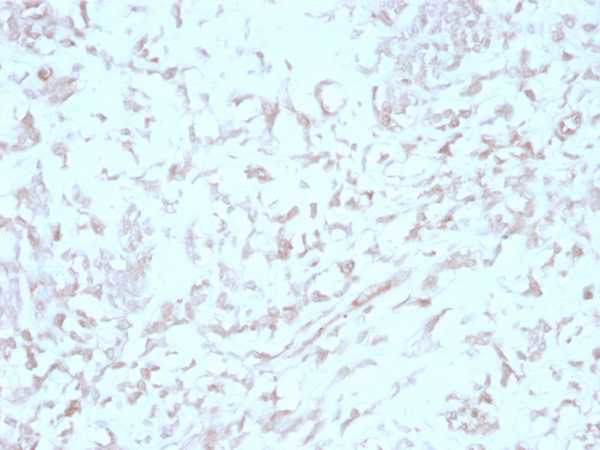
 IHC (Immunohiostchemistry)
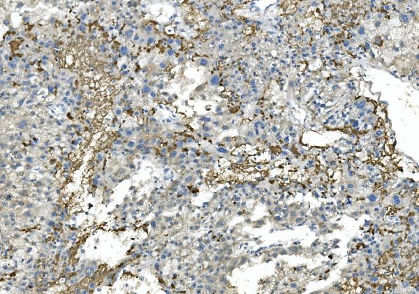
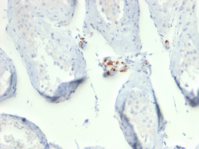
(Figure 2. IHC analysis of Fibrinogen beta chain/FGB using anti-Fibrinogen beta chain/FGB antibody (AAA125915).Fibrinogen beta chain/FGB was detected in paraffin-embedded section of human liver cancer tissue. Heat mediated antigen retrieval was performed in EDTA buffer (pH8. 0, epitope retrieval solution). The tissue section was blocked with 10% goat serum. The tissue section was then incubated with 2μg/ml mouse anti-Fibrinogen beta chain/FGB Antibody (AAA125915) overnight at 4 degree C. Biotinylated goat anti-mouse IgG was used as secondary antibody and incubated for 30 minutes at 37 degree C. The tissue section was developed using Strepavidin-Biotin-Complex (SABC) with DAB as the chromogen.)
IHC (Immunohiostchemistry)
(Figure 2. IHC analysis of Fibrinogen beta chain/FGB using anti-Fibrinogen beta chain/FGB antibody (AAA125915).Fibrinogen beta chain/FGB was detected in paraffin-embedded section of human liver cancer tissue. Heat mediated antigen retrieval was performed in EDTA buffer (pH8. 0, epitope retrieval solution). The tissue section was blocked with 10% goat serum. The tissue section was then incubated with 2μg/ml mouse anti-Fibrinogen beta chain/FGB Antibody (AAA125915) overnight at 4 degree C. Biotinylated goat anti-mouse IgG was used as secondary antibody and incubated for 30 minutes at 37 degree C. The tissue section was developed using Strepavidin-Biotin-Complex (SABC) with DAB as the chromogen.)
Fibrinogen beta chain/FGB, Monoclonal Antibody (Cat# AAA125915)
 Application Data
(Staining of rat spleen cells with Mouse anti Rat CD44)
Application Data
(Staining of rat spleen cells with Mouse anti Rat CD44)
CD44, Monoclonal Antibody (Cat# AAA49429)
 Application Data
(Staining of human peripheral blood granulocytes with Mouse anti Human CD18 (Activation Epitope):RPE)
Application Data
(Staining of human peripheral blood granulocytes with Mouse anti Human CD18 (Activation Epitope):RPE)
CD18, Monoclonal Antibody (Cat# AAA49324)

 Application Data
(Published customer image: T lymphocytes in PBLs following DPV gC DNA vaccination. 3, 5, 7, 14, 28, 42 days after vaccination, the isolated PBLs were stained with monoclonal antibodies against duck CD4 (A), and CD8 (B). The results presented are the mean of all specimens of each group +/- SD.From: Lian B, Cheng A, Wang M, Zhu D, Luo Q, Jia R, Liu F, Han X, Chen X. Induction of immune responses in ducks with a DNA vaccine encoding duck plague virus glycoprotein C. Virol J. 2011 May 10;8:214.)
Application Data
(Published customer image: T lymphocytes in PBLs following DPV gC DNA vaccination. 3, 5, 7, 14, 28, 42 days after vaccination, the isolated PBLs were stained with monoclonal antibodies against duck CD4 (A), and CD8 (B). The results presented are the mean of all specimens of each group +/- SD.From: Lian B, Cheng A, Wang M, Zhu D, Luo Q, Jia R, Liu F, Han X, Chen X. Induction of immune responses in ducks with a DNA vaccine encoding duck plague virus glycoprotein C. Virol J. 2011 May 10;8:214.)
CD8 ALPHA, Monoclonal Antibody (Cat# AAA49383)
C3c, Monoclonal Antibody (Cat# AAA49392)
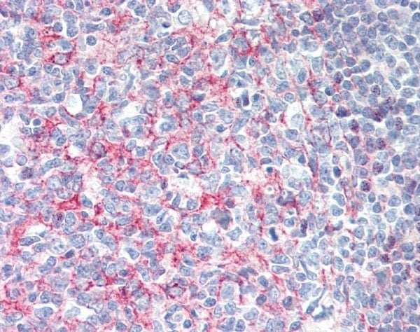
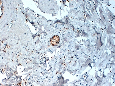
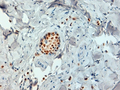
 Application Data
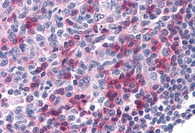
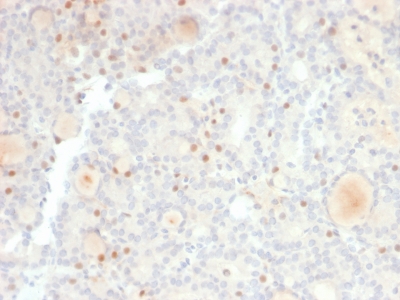
(Human testis stained with Mouse anti Human Inhibin alpha, showing Sertoli cells, Leydig cells and spermatogonia. Formalin fixed paraffin processed tissue)
Application Data
(Human testis stained with Mouse anti Human Inhibin alpha, showing Sertoli cells, Leydig cells and spermatogonia. Formalin fixed paraffin processed tissue)
INHIBIN ALPHA, Monoclonal Antibody (Cat# AAA49722)
 Application Data
(Formalin fixed, paraffin embedded human breast cancer biopsy stained with Mouse anti Human estrogen receptor alpha antibody followed by HRP polymer detection and DAB substrate develoment following heat mediated antigen retrieval using citrate buffer at pH 6.2 (high power))
Application Data
(Formalin fixed, paraffin embedded human breast cancer biopsy stained with Mouse anti Human estrogen receptor alpha antibody followed by HRP polymer detection and DAB substrate develoment following heat mediated antigen retrieval using citrate buffer at pH 6.2 (high power))
ESTROGEN RECEPTOR ALPHA, Monoclonal Antibody (Cat# AAA49614)
 Application Data
(Staining of human peripheral blood monocytes with Mouse anti Human CD63)
Application Data
(Staining of human peripheral blood monocytes with Mouse anti Human CD63)
CD63, Monoclonal Antibody (Cat# AAA49640)
 Application Data
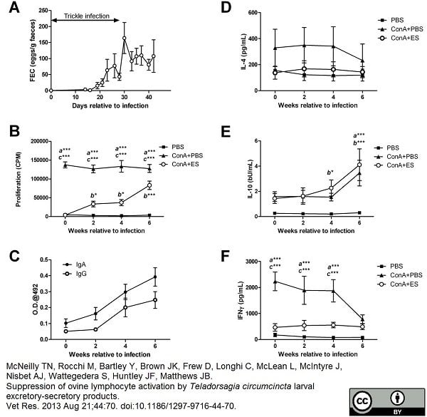
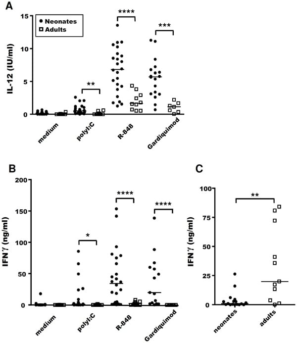
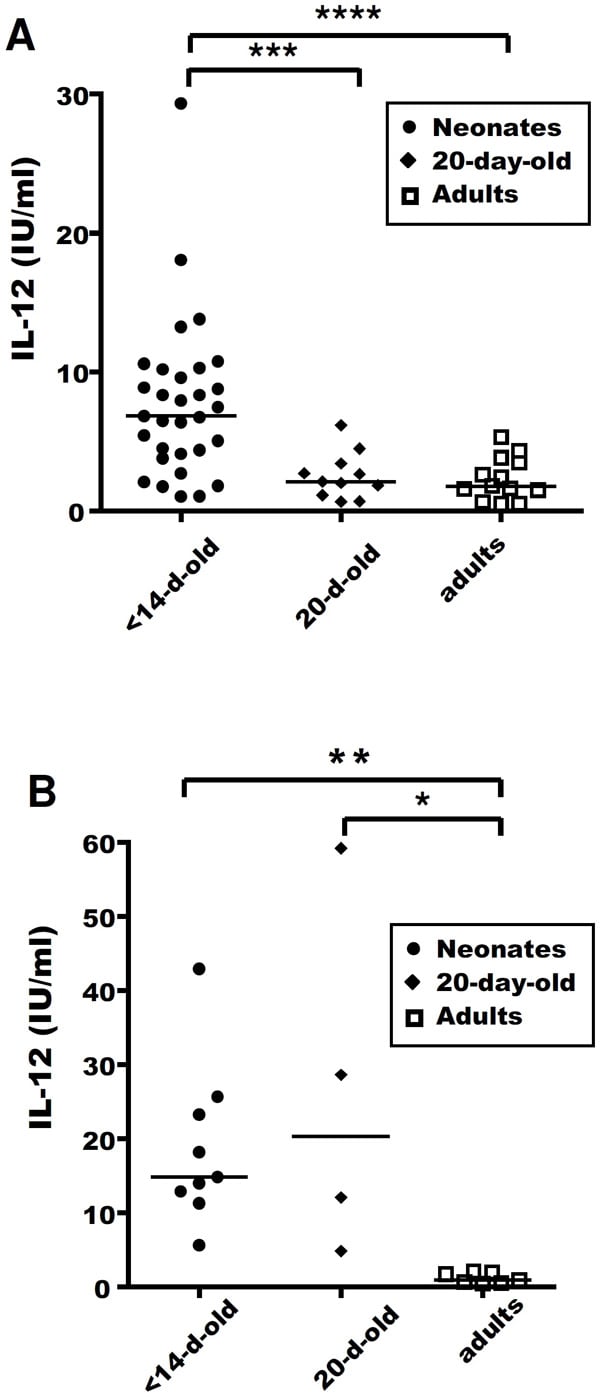
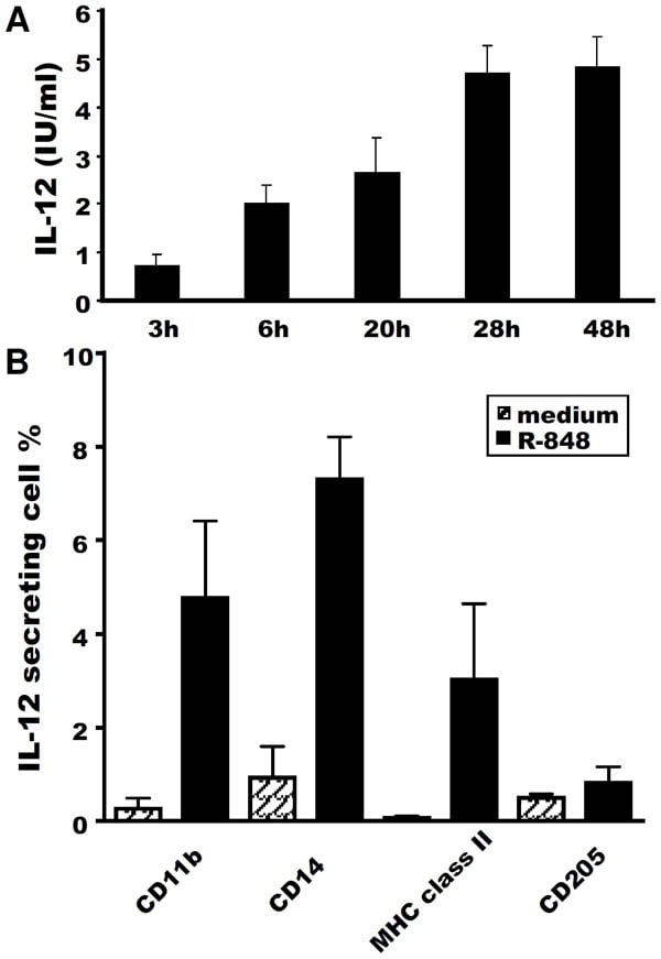
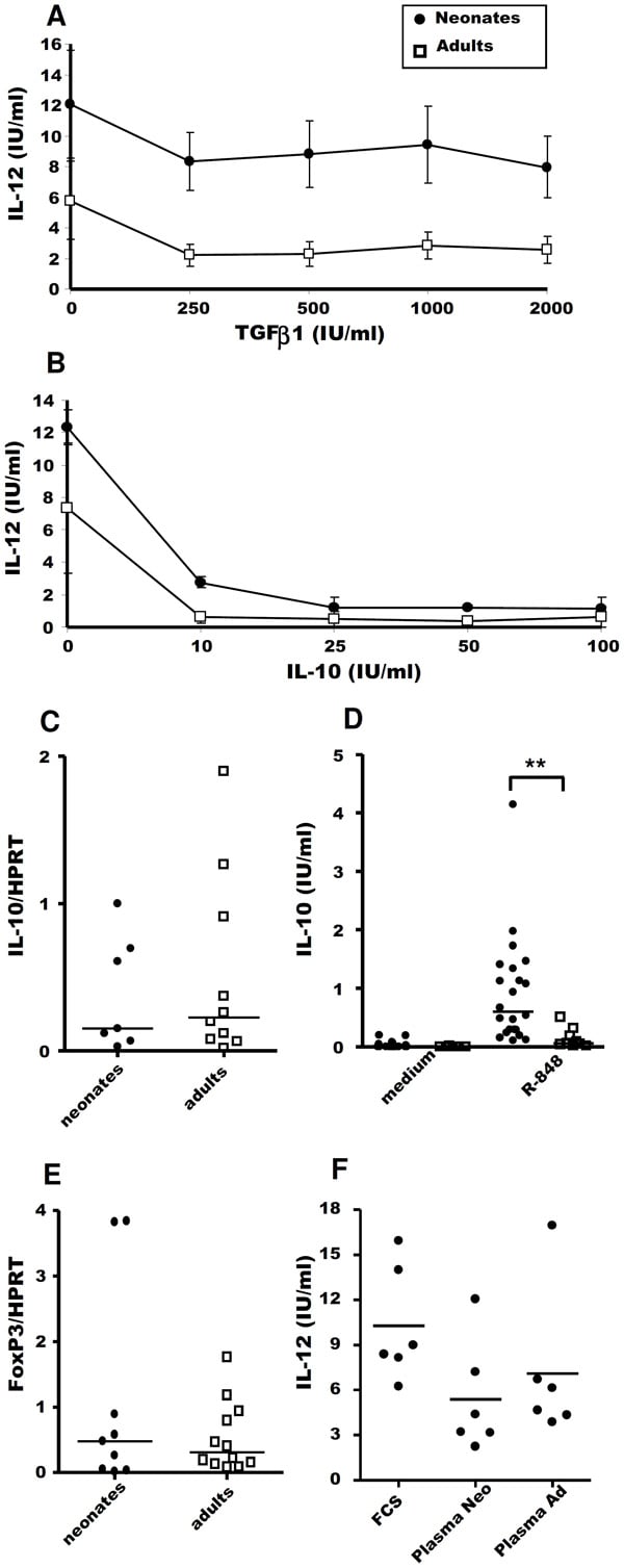
(Published customer image: Roles of TGFbeta1 and IL-10 in regulating IL-12 responses to R-848. MLN cells from neonates (closed circles) and adults (open squares) were cultured in vitro for 48 h, with or without 0.5 ug/ml R-848, in the presence of rhTGFbeta1 (n = 7) (A) or rovIL-10 (n = 5) (B). Supernatants were harvested and ELISA was carried out to assess IL-12 secretion. The mean +/- SEM level of IL-12 secretion is shown (A, B). RNA was extracted and purified from freshly isolated MLN cells. IL-10 mRNA levels were determined by quantitative RT-PCR. Median normalised values are presented for neonates (closed circles) and adults (open squares) (C). MLN cells from neonates (closed circles) and adults (open squares) were stimulated in vitro for 48 h with or without 0.5 ug/ml R-848. Supernatants were harvested and ELISA carried out to assess IL-10 secretion. Medians are indicated. Non-parametric Mann-Whitney tests were used to compare data for neonates and adults: **p=0.001 (D). RNA was extracted and purified from freshly isolated MLN cells. Foxp3 mRNA levels were determined by quantitative RT-PCR. Median normalised values are presented for neonates (closed circles) and adults (open squares) (E). Comparison of the IL-12 response of neonate MLN cells to R-848 stimulation in culture medium supplemented with 10%FCS, 10% neonate autologous plasma or 10% adult plasma. Paired t-test between neonate and adult plasma were non-significant (F).From: Ferret-Bernard S, Remot A, Lacroix-Lamand© S, Metton C, Bernardet N, et al. (2010) Cellular and Molecular Mechanisms Underlying the Strong Neonatal IL-12 Response of Lamb Mesenteric Lymph Node Cells to R-848. PLoS ONE 5(10): e13705.)
Application Data
(Published customer image: Roles of TGFbeta1 and IL-10 in regulating IL-12 responses to R-848. MLN cells from neonates (closed circles) and adults (open squares) were cultured in vitro for 48 h, with or without 0.5 ug/ml R-848, in the presence of rhTGFbeta1 (n = 7) (A) or rovIL-10 (n = 5) (B). Supernatants were harvested and ELISA was carried out to assess IL-12 secretion. The mean +/- SEM level of IL-12 secretion is shown (A, B). RNA was extracted and purified from freshly isolated MLN cells. IL-10 mRNA levels were determined by quantitative RT-PCR. Median normalised values are presented for neonates (closed circles) and adults (open squares) (C). MLN cells from neonates (closed circles) and adults (open squares) were stimulated in vitro for 48 h with or without 0.5 ug/ml R-848. Supernatants were harvested and ELISA carried out to assess IL-10 secretion. Medians are indicated. Non-parametric Mann-Whitney tests were used to compare data for neonates and adults: **p=0.001 (D). RNA was extracted and purified from freshly isolated MLN cells. Foxp3 mRNA levels were determined by quantitative RT-PCR. Median normalised values are presented for neonates (closed circles) and adults (open squares) (E). Comparison of the IL-12 response of neonate MLN cells to R-848 stimulation in culture medium supplemented with 10%FCS, 10% neonate autologous plasma or 10% adult plasma. Paired t-test between neonate and adult plasma were non-significant (F).From: Ferret-Bernard S, Remot A, Lacroix-Lamand© S, Metton C, Bernardet N, et al. (2010) Cellular and Molecular Mechanisms Underlying the Strong Neonatal IL-12 Response of Lamb Mesenteric Lymph Node Cells to R-848. PLoS ONE 5(10): e13705.)
IL-12, Monoclonal Antibody (Cat# AAA49642)
 Application Data
(Staining of human peripheral blood monocytes wtih Mouse anti Human Dectin:FITC)
Application Data
(Staining of human peripheral blood monocytes wtih Mouse anti Human Dectin:FITC)
DECTIN-1, Monoclonal Antibody (Cat# AAA49780)

 Application Data
(Staining of pig peripheral blood lymphocytes with Mouse anti Pig CD52 followed by Rabbit F(ab')2 anti Mouse IgG:FITC)
Application Data
(Staining of pig peripheral blood lymphocytes with Mouse anti Pig CD52 followed by Rabbit F(ab')2 anti Mouse IgG:FITC)
CD52, Monoclonal Antibody (Cat# AAA49808)
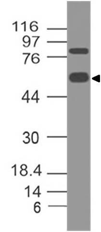
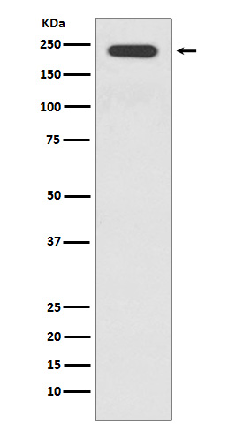
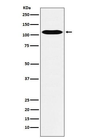
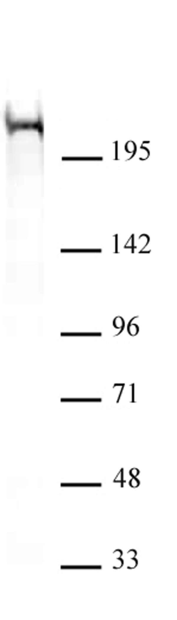
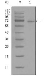
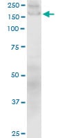
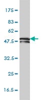
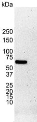
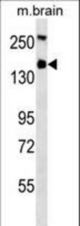 WB (Western Blot)
(TSC1 Antibody western blot of mouse brain tissue lysates (35 ug/lane). The TSC1 antibody detected the TSC1 protein (arrow).)
WB (Western Blot)
(TSC1 Antibody western blot of mouse brain tissue lysates (35 ug/lane). The TSC1 antibody detected the TSC1 protein (arrow).)
Hamartin / TSC1, Monoclonal Antibody (Cat# AAA52562)
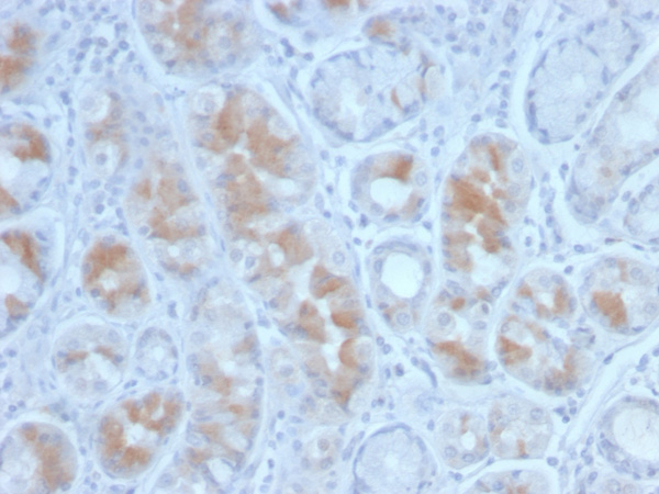
 IHC (Immunohistochemistry)
(Formalin-fixed, paraffin-embedded human liver stained with Alpha-1-Antichymotrypsin Mouse Monoclonal Antibody (SERPINA3/4187).)
IHC (Immunohistochemistry)
(Formalin-fixed, paraffin-embedded human liver stained with Alpha-1-Antichymotrypsin Mouse Monoclonal Antibody (SERPINA3/4187).)
Alpha-1-Antichymotrypsin (SERPINA3), Monoclonal Antibody (Cat# AAA215565)
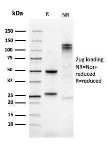 SDS-PAGE
Kindlin-1/KIND1, Monoclonal Antibody (Cat# AAA215463)
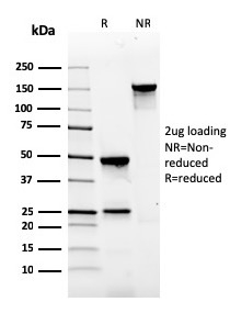 SDS-PAGE
(SDS-PAGE Analysis Purified Gastrin Mouse Monoclonal Antibody (GAST/2634). Confirmation of Integrity and Purity of Antibody.)
SDS-PAGE
(SDS-PAGE Analysis Purified Gastrin Mouse Monoclonal Antibody (GAST/2634). Confirmation of Integrity and Purity of Antibody.)
Gastrin, Monoclonal Antibody (Cat# AAA214981)
 Application Data
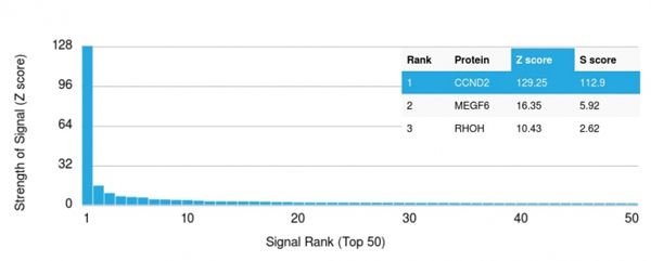
(Analysis of Protein Array containing more than 19,000 full-length human proteins using Cyclin D2 Mouse Recombinant Monoclonal Antibody (CCND2/2620). Z- and S- Score: The Z-score represents the strength of a signal that a monoclonal antibody (MAb) (in combination with a fluorescently-tagged anti-IgG secondary antibody) produces when binding to a particular protein on the HuProtTM array. Z-scores are described in units of standard deviations (SD's) above the mean value of all signals generated on that array. If targets on HuProtTM are arranged in descending order of the Z-score, the S-score is the difference (also in units of SD's) between the Z-score. S-score therefore represents the relative target specificity of a MAb to its intended target. A MAb is considered to specific to its intended target, if the MAb has an S-score of at least 2.5. For example, if a MAb binds to protein X with a Z-score of 43 and to protein Y with a Z-score of 14, then the S-score for the binding of that MAb to protein X is equal to 29.)
Application Data
(Analysis of Protein Array containing more than 19,000 full-length human proteins using Cyclin D2 Mouse Recombinant Monoclonal Antibody (CCND2/2620). Z- and S- Score: The Z-score represents the strength of a signal that a monoclonal antibody (MAb) (in combination with a fluorescently-tagged anti-IgG secondary antibody) produces when binding to a particular protein on the HuProtTM array. Z-scores are described in units of standard deviations (SD's) above the mean value of all signals generated on that array. If targets on HuProtTM are arranged in descending order of the Z-score, the S-score is the difference (also in units of SD's) between the Z-score. S-score therefore represents the relative target specificity of a MAb to its intended target. A MAb is considered to specific to its intended target, if the MAb has an S-score of at least 2.5. For example, if a MAb binds to protein X with a Z-score of 43 and to protein Y with a Z-score of 14, then the S-score for the binding of that MAb to protein X is equal to 29.)
Cyclin D2, Monoclonal Antibody (Cat# AAA214845)
What are Monoclonal Antibodies?
Monoclonal antibodies are specialized laboratory-produced proteins developed for binding to specific biological antigens or other molecular targets. Since they come from a single cell (or clone), they are especially consistent and accurate in the data they are involved in producing.
This type of antibody material has been shown to be a powerful tool in finding and subsequently destroying harmful cells in an organism, such as those found in cancers or various autoimmune diseases. This makes them excellent aids in medical testing and research, which is why they are so widely used.
AAA Biotech offers a comprehensive range of high-quality monoclonal antibodies that perform effectively in various laboratory tests, including (amongst others) ELISA, western blotting, immunohistochemistry, and flow cytometry. All of the products in our catalog are thoroughly quality tested to make sure that they are reliable and will consistently perform well in your research.
What Are The Uses of Monoclonal Antibodies
Monoclonal antibodies are used in many lab tests, including (amongst others) ELISA, western blotting, immunohistochemistry, and flow cytometry.
ELISA is a test that helps detect a specific substance/analyte in a sample. It uses antibodies (often monoclonal) bound to a solid surface (such as the well of a microplate) to “capture” the substance/analyte in the sample and immobilize it so that the detection antibody component can then bind to it and produce a signal, which can then be measured.
Western blotting identifies specific proteins in a sample. The sample is first separated on a gel, and then antibodies are applied that will typically bind to the target, which will all be localized to a single band in a lane.
Immunohistochemistry helps locate specific proteins in cells or tissue samples using antibodies.
Flow cytometry looks at and sorts cells. It uses antibodies that are conjugated to reporter molecules called “fluorophores”, which, under special lights, emit light themselves, which can then be measured by a detector instrument.
How Monoclonal Antibodies Are Used as Medicine?
Please note that all of the products listed in AAA Biotech’s also known as AAA Bio or AAABio catalog are strictly for research-use only (RUO).
Monoclonal antibodies can also be used as therapeutic/medical treatments, particularly in the context of cancers. They are designed to find and bind to specific cells or proteins, helping the immune system recognize and attack the cancer. These treatments work in different ways, such as:
- Radioimmunotherapy attaches a small amount of radioactive molecule to the antibody, so it delivers the radiation directly to the cancer cells that the antibody is specifically binding to.
- Antibody-directed enzyme prodrug therapy uses antibodies that are specifically bound to special enzymes. These enzymes activate a harmless drug in the body and turn it into a cancer-killing drug only near the cancer cells—this helps avoid harming healthy cells.
- Immunoliposomes are tiny “bubbles” filled with medicine/drug and coated with antibodies. They carry the drug straight to the cancer cells.
Why Buy Monoclonal Antibodies From Us?
At AAA Biotech, we provide high-performance monoclonal antibodies designed to support a wide range of research needs.
1. Validated for Versatile Applications
The antibodies in our catalog are extensively validated and compatible with multiple techniques, including (but not limited to) ELISA, flow cytometry (FC), immunocytochemistry (ICC), immunofluorescence (IF), immunohistochemistry (IHC), immunoprecipitation (IP), and western blotting (WB).
2. Wide Selection & Specialized Options
We offer antibodies for common and rare species, that are available in various conjugated forms, and also in recombinant formats. Essentially, there is almost anything one might need to meet their experimental model’s requirements.
3. High-Quality Proteins
Our proteins meet high purity standards—90% or more as confirmed by SDS-PAGE. Many are available with tags like His, Flag, GST, or MBP, and we also supply native and biologically active proteins for functional studies.
Frequently Asked Questions
1. Are your monoclonal antibodies validated for specific applications?
Yes, our antibodies are tested and validated for use in methods such as ELISA, western blot, IHC, flow cytometry, and more. Refer to specific product pages or datasheets for individual product information.
2. How do I choose the right monoclonal antibody for my application?
Review the product details directly for application validation, species reactivity, and target information. You may also contact our support team at any time for help.
3. How quickly can I receive my order?
Most orders are processed and shipped within 1–3 business days, depending on product availability and your shipping location.