Filters
▼Clonality
▼Type
▼Reactivity
▼Gene Name
▼Isotype
▼Host
▼Application
▼Clone
▼Monoclonal Antibodies
Get accurate results in your research with our Monoclonal Antibodies, which are specially made to target exactly what you require for your research, and will produce consistent, reliable performance in lab tests.
Viewing 9750-9800 of 27597 product results
 IHC (Immunohiostchemistry)
(DAB staining on IHCP;Sample: Human Small intestine Tissue; Primary Ab: 10ug/ml Mouse AntiHuman IFNb AntibodySecond Ab: 2ug/mL HRPLinked Caprine AntiMouse IgG Polyclonal Antibody(Catalog: SAA544Mu19))
IHC (Immunohiostchemistry)
(DAB staining on IHCP;Sample: Human Small intestine Tissue; Primary Ab: 10ug/ml Mouse AntiHuman IFNb AntibodySecond Ab: 2ug/mL HRPLinked Caprine AntiMouse IgG Polyclonal Antibody(Catalog: SAA544Mu19))
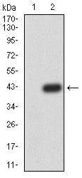
Interferon Beta (IFNb), Monoclonal Antibody (Cat# AAA151508)
 WB (Western Blot)
(Western Blot Sample: Lane1: Rat Serum; Lane2: Rat Spleen lysate Primary Ab: 1ug/ml Mouse AntiMouse b2M Antibody Second Ab: 0.2ug/mL HRPLinked Caprine AntiMouse IgG Polyclonal Antibody (Catalog: SAA544Mu19))
WB (Western Blot)
(Western Blot Sample: Lane1: Rat Serum; Lane2: Rat Spleen lysate Primary Ab: 1ug/ml Mouse AntiMouse b2M Antibody Second Ab: 0.2ug/mL HRPLinked Caprine AntiMouse IgG Polyclonal Antibody (Catalog: SAA544Mu19))
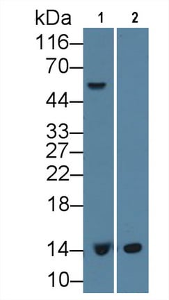
Beta2Microglobulin (b2M), Monoclonal Antibody (Cat# AAA151518)
 WB (Western Blot)
(Western Blot; Sample: Lane1: Human Serum; Lane2: Human Urine; Lane3: Human Lung Lysate; Lane3: Bovine Skeletal muscle Lysate Primary Ab: 2ug/ml Mouse AntiHuman GSN Antibody Second Ab: 0.2ug/mL HRPLinked Caprine AntiMouse IgG Polyclonal Antibody (Catalog: SAA544Mu19))
WB (Western Blot)
(Western Blot; Sample: Lane1: Human Serum; Lane2: Human Urine; Lane3: Human Lung Lysate; Lane3: Bovine Skeletal muscle Lysate Primary Ab: 2ug/ml Mouse AntiHuman GSN Antibody Second Ab: 0.2ug/mL HRPLinked Caprine AntiMouse IgG Polyclonal Antibody (Catalog: SAA544Mu19))
Gelsolin (GSN), Monoclonal Antibody (Cat# AAA151523)
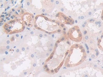
 IHC (Immunohiostchemistry)
(DAB staining on IHCP;Sample: Rat Kidney Tissue; Primary Ab: 30ug/ml Mouse AntiRat PAI2 AntibodySecond Ab: 2ug/mL HRPLinked Caprine AntiMouse IgG Polyclonal Antibody(Catalog: SAA544Mu19))
IHC (Immunohiostchemistry)
(DAB staining on IHCP;Sample: Rat Kidney Tissue; Primary Ab: 30ug/ml Mouse AntiRat PAI2 AntibodySecond Ab: 2ug/mL HRPLinked Caprine AntiMouse IgG Polyclonal Antibody(Catalog: SAA544Mu19))
Plasminogen Activator Inhibitor 2 (PAI2), Monoclonal Antibody (Cat# AAA151553)
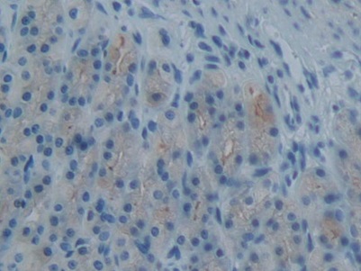
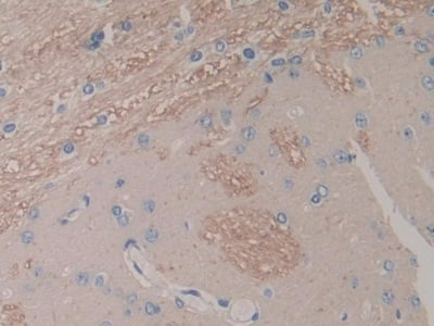
 IHC (Immunohiostchemistry)
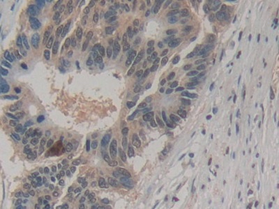
(DAB staining on IHC-P; Samples: Human Cerebrum Tissue; Primary Ab: 20ug/ml Mouse Anti-Human CTSB Antibody Second Ab: 2ug/mL HRP-Linked Caprine Anti-Mouse IgG Polyclonal Antibody)
IHC (Immunohiostchemistry)
(DAB staining on IHC-P; Samples: Human Cerebrum Tissue; Primary Ab: 20ug/ml Mouse Anti-Human CTSB Antibody Second Ab: 2ug/mL HRP-Linked Caprine Anti-Mouse IgG Polyclonal Antibody)
Cathepsin B (CTSB), Monoclonal Antibody (Cat# AAA151129)
 IHC (Immunohistochemistry)
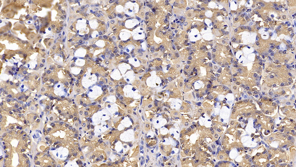
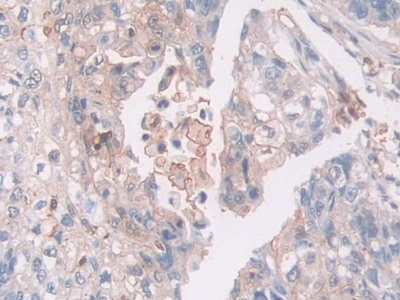
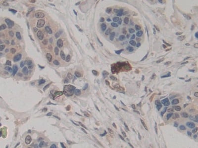
(DAB staining on IHCP;Sample: Human Liver cancer Tissue; Primary Ab: 20ug/ml Mouse AntiHuman AREG AntibodySecond Ab: 2ug/mL HRPLinked Caprine AntiMouse IgG Polyclonal Antibody(Catalog: SAA544Mu19))
IHC (Immunohistochemistry)
(DAB staining on IHCP;Sample: Human Liver cancer Tissue; Primary Ab: 20ug/ml Mouse AntiHuman AREG AntibodySecond Ab: 2ug/mL HRPLinked Caprine AntiMouse IgG Polyclonal Antibody(Catalog: SAA544Mu19))
Amphiregulin (AREG), Monoclonal Antibody (Cat# AAA151428)
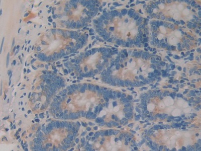 IHC (Immunohistochemisry)
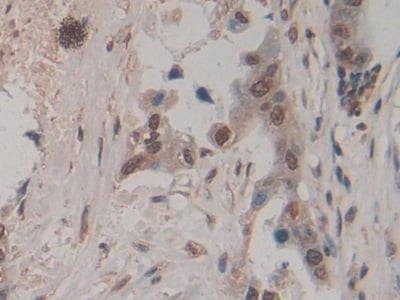
(DAB staining on IHCP;Sample: Rat Intestine Tissue; Primary Ab: 30ug/ml Mouse AntiRat IL1a AntibodySecond Ab: 2ug/mL HRPLinked Caprine AntiMouse IgG Polyclonal Antibody(Catalog: SAA544Mu19))
IHC (Immunohistochemisry)
(DAB staining on IHCP;Sample: Rat Intestine Tissue; Primary Ab: 30ug/ml Mouse AntiRat IL1a AntibodySecond Ab: 2ug/mL HRPLinked Caprine AntiMouse IgG Polyclonal Antibody(Catalog: SAA544Mu19))
Interleukin 1 Alpha (IL1a), Monoclonal Antibody (Cat# AAA151457)

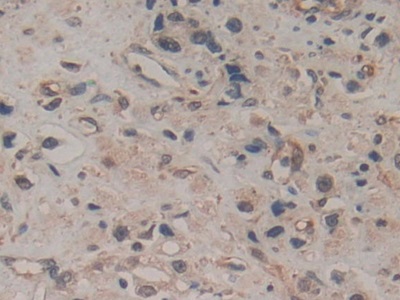
 IHC (Immunohiostchemistry)
(IHC Image)
IHC (Immunohiostchemistry)
(IHC Image)
Matrix Metalloproteinase 13 (MMP13), Monoclonal Antibody (Cat# AAA151478)
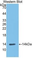
 WB (Western Blot)
(Western Blot; Sample: Bovine Serum Primary Ab: 2ug/mL Mouse Anti-Bovine aFP Antibody Second Ab: 0.2ug/mL HRP-Linked Caprine Anti-Mouse IgG Polyclonal Antibody)
WB (Western Blot)
(Western Blot; Sample: Bovine Serum Primary Ab: 2ug/mL Mouse Anti-Bovine aFP Antibody Second Ab: 0.2ug/mL HRP-Linked Caprine Anti-Mouse IgG Polyclonal Antibody)
Alpha-Fetoprotein (AFP), Monoclonal Antibody (Cat# AAA150792)
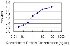 ELISA
(Detection limit for recombinant GST tagged KCNJ5 is 0.03 ng/ml as a capture antibody.)
ELISA
(Detection limit for recombinant GST tagged KCNJ5 is 0.03 ng/ml as a capture antibody.)
KCNJ5/Kir3.4/GIRK4, Monoclonal Antibody (Cat# AAA162334)
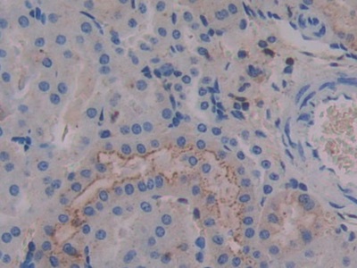
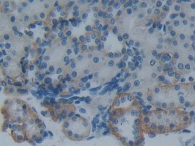
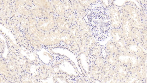
 IHC (Immunohistochemisry)
(DAB staining on IHC-P;Sample: human Kidney Tissue; Primary Ab: 30ug/ml Mouse Anti-human ANTXR2 AntibodySecond Ab: 2ug/mL HRP-Linked Caprine Anti-Mouse IgG Polyclonal Antibody)
IHC (Immunohistochemisry)
(DAB staining on IHC-P;Sample: human Kidney Tissue; Primary Ab: 30ug/ml Mouse Anti-human ANTXR2 AntibodySecond Ab: 2ug/mL HRP-Linked Caprine Anti-Mouse IgG Polyclonal Antibody)
Anthrax Toxin Receptor 2 (ANTXR2), Monoclonal Antibody (Cat# AAA152849)
Prostaglandin E2 (PGE2), Monoclonal Antibody (Cat# AAA152552)
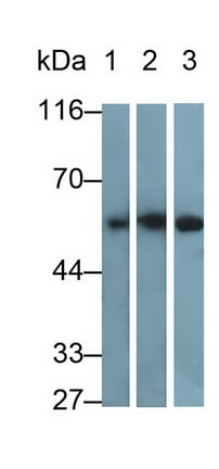
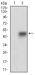
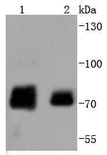
 WB (Western Blot)
(Western Blot; Sample: Lane1: Porcine Heart lysate; Lane2: Porcine Skeletal muscle lysate; Lane3: Rat Skeletal muscle lysatePrimary Ab: 0.2ug/ml Mouse Anti-human Des AntibodySecond Ab: 0.2ug/mL HRP-Linked Caprine Anti-Mouse IgG Polyclonal Antibody)
WB (Western Blot)
(Western Blot; Sample: Lane1: Porcine Heart lysate; Lane2: Porcine Skeletal muscle lysate; Lane3: Rat Skeletal muscle lysatePrimary Ab: 0.2ug/ml Mouse Anti-human Des AntibodySecond Ab: 0.2ug/mL HRP-Linked Caprine Anti-Mouse IgG Polyclonal Antibody)
Desmin (Des), Monoclonal Antibody (Cat# AAA152567)
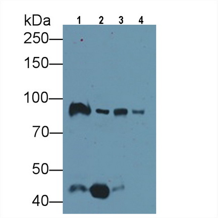
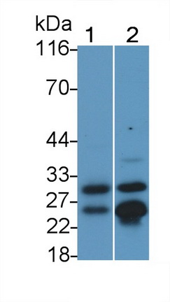
 WB (Western Blot)
(Western Blot; Sample: Lane1: Porcine Skeletal muscle lysate; Lane2: Porcine Esophagus lysate; Lane3: Rat Skeletal muscle lysate; Lane4: Mouse Skeletal muscle lysate Primary Ab: 0.01ug/ml Mouse Anti-human MYH8 Antibody Second Ab: 0.2ug/mL HRP-Linked Caprine Anti-Mouse IgG Polyclonal Antibody)
WB (Western Blot)
(Western Blot; Sample: Lane1: Porcine Skeletal muscle lysate; Lane2: Porcine Esophagus lysate; Lane3: Rat Skeletal muscle lysate; Lane4: Mouse Skeletal muscle lysate Primary Ab: 0.01ug/ml Mouse Anti-human MYH8 Antibody Second Ab: 0.2ug/mL HRP-Linked Caprine Anti-Mouse IgG Polyclonal Antibody)
Myosin Heavy Chain 8, Skeletal Muscle, Perinatal (MYH8), Monoclonal Antibody (Cat# AAA152806)
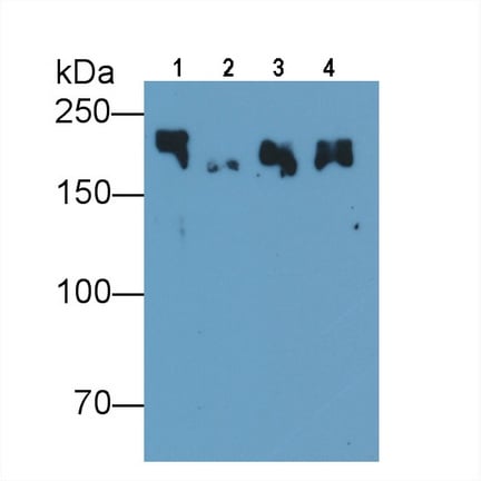
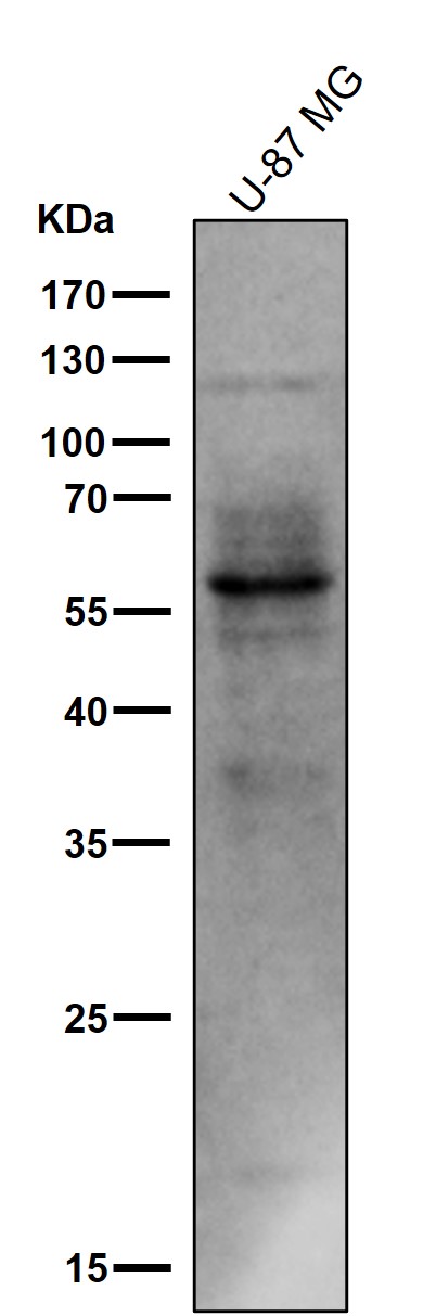
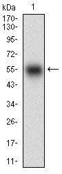
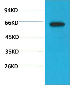
 WB (Western Blot)
(Western Blot; Sample: U87MG cell lysate Primary Ab: 3ug/ml Mouse AntiHuman Reelin Antibody Second Ab: 0.2ug/mL HRPLinked Caprine AntiMouse IgG Polyclonal Antibody (Catalog: SAA544Mu19))
WB (Western Blot)
(Western Blot; Sample: U87MG cell lysate Primary Ab: 3ug/ml Mouse AntiHuman Reelin Antibody Second Ab: 0.2ug/mL HRPLinked Caprine AntiMouse IgG Polyclonal Antibody (Catalog: SAA544Mu19))
Reelin (RELN), Monoclonal Antibody (Cat# AAA151815)
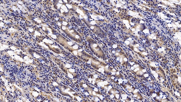
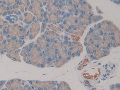
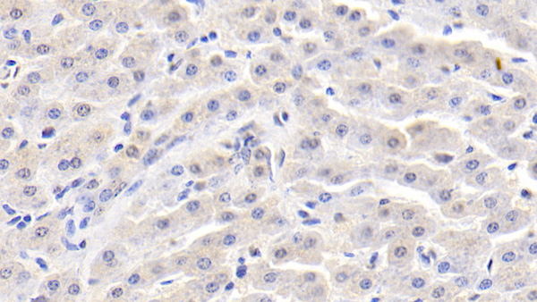
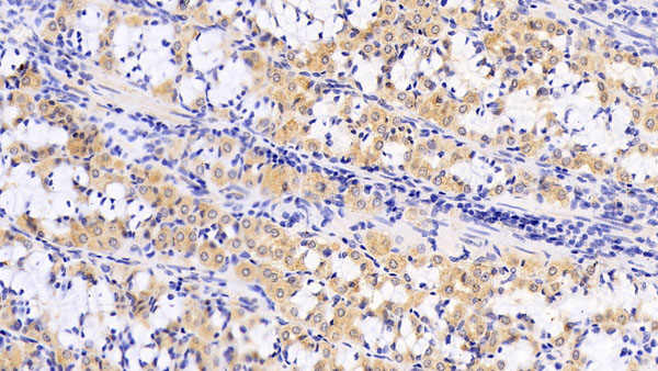
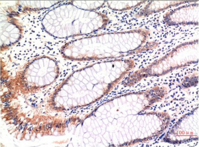
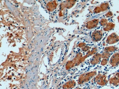 IHC (Immunohiostchemistry)
(DAB staining on IHCP;Sample: Human Stomach Tissue; Primary Ab: 10ug/ml Mouse AntiHuman FGF21 AntibodySecond Ab: 2ug/mL HRPLinked Caprine AntiMouse IgG Polyclonal Antibody(Catalog: SAA544Mu19))
IHC (Immunohiostchemistry)
(DAB staining on IHCP;Sample: Human Stomach Tissue; Primary Ab: 10ug/ml Mouse AntiHuman FGF21 AntibodySecond Ab: 2ug/mL HRPLinked Caprine AntiMouse IgG Polyclonal Antibody(Catalog: SAA544Mu19))
Fibroblast Growth Factor 21 (FGF21), Monoclonal Antibody (Cat# AAA151821)
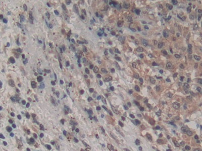
 IHC (Immunohiostchemistry)
(DAB staining on IHCP;Samples: Human Cerebrum Tissue; Primary Ab: 10ug/ml Mouse AntiHuman ZPI AntibodySecond Ab: 2ug/mL HRPLinked Caprine AntiMouse IgG Polyclonal Antibody(Catalog: SAA544Mu19))
IHC (Immunohiostchemistry)
(DAB staining on IHCP;Samples: Human Cerebrum Tissue; Primary Ab: 10ug/ml Mouse AntiHuman ZPI AntibodySecond Ab: 2ug/mL HRPLinked Caprine AntiMouse IgG Polyclonal Antibody(Catalog: SAA544Mu19))
Serpin A10 (SERPINA10), Monoclonal Antibody (Cat# AAA151778)
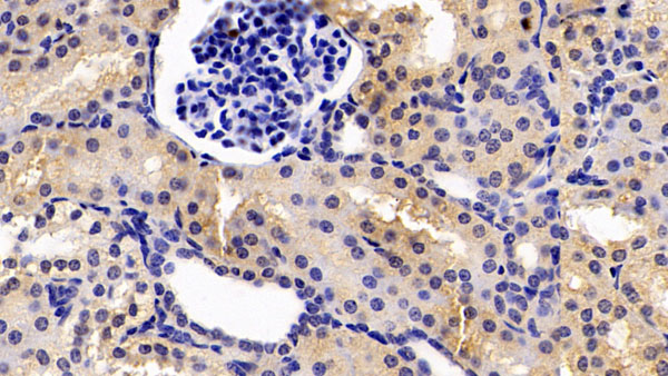
 IHC (Immunohiostchemistry)
(DAB staining on IHCP;Sample: Human Kidney Tissue; Primary Ab: 30ug/ml Mouse AntiHuman SEMA3A AntibodySecond Ab: 2ug/mL HRPLinked Caprine AntiMouse IgG Polyclonal Antibody(Catalog: SAA544Mu19))
IHC (Immunohiostchemistry)
(DAB staining on IHCP;Sample: Human Kidney Tissue; Primary Ab: 30ug/ml Mouse AntiHuman SEMA3A AntibodySecond Ab: 2ug/mL HRPLinked Caprine AntiMouse IgG Polyclonal Antibody(Catalog: SAA544Mu19))
Semaphorin 3A (SEMA3A), Monoclonal Antibody (Cat# AAA151905)
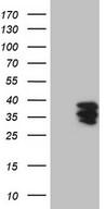 WB (Western Blot)
(Human recombinant protein fragment corresponding to amino acids 38-307 of human ENTPD1 (NP_001767) produced in E Coli.)
WB (Western Blot)
(Human recombinant protein fragment corresponding to amino acids 38-307 of human ENTPD1 (NP_001767) produced in E Coli.)
CD39, Monoclonal Antibody (Cat# AAA162323)
 WB (Western Blot)
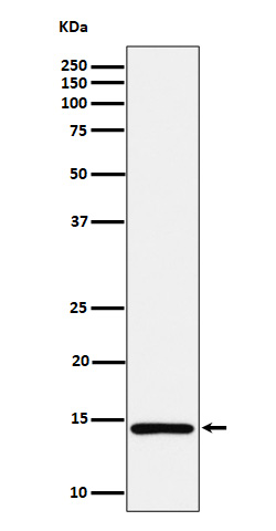
(Western blot analysis of Phospho-Tau (S214) expression in mouse cerebral cortex cell lysate.)
WB (Western Blot)
(Western blot analysis of Phospho-Tau (S214) expression in mouse cerebral cortex cell lysate.)
Tau, Monoclonal Antibody (Cat# AAA128106)
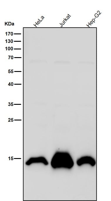 WB (Western Blot)
(All lanes use the Antibody at 1:5K dilution for 1 hour at room temperature.)
WB (Western Blot)
(All lanes use the Antibody at 1:5K dilution for 1 hour at room temperature.)
Histone H2A, Monoclonal Antibody (Cat# AAA128199)
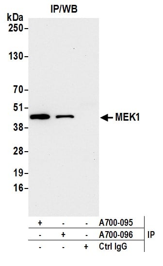
 WB (Western Blot)
(Detection of human MEK1 by western blot. Samples: Whole cell lysate (50 ug) from HeLa, Hep-G2, Jurkat, A-549, RKO, U2OS, and LNCaP cells prepared using NETN lysis buffer. Antibody: Rabbit anti-MEK1 recombinant monoclonal antibody (AAA213574 lot 1) used at 1:1000. Secondary: HRP-conjugated goat anti-rabbit IgG . Detection: Chemiluminescence with an exposure time of 10 seconds. Lower Panel: Rabbit anti-COPB2 .)
WB (Western Blot)
(Detection of human MEK1 by western blot. Samples: Whole cell lysate (50 ug) from HeLa, Hep-G2, Jurkat, A-549, RKO, U2OS, and LNCaP cells prepared using NETN lysis buffer. Antibody: Rabbit anti-MEK1 recombinant monoclonal antibody (AAA213574 lot 1) used at 1:1000. Secondary: HRP-conjugated goat anti-rabbit IgG . Detection: Chemiluminescence with an exposure time of 10 seconds. Lower Panel: Rabbit anti-COPB2 .)
MEK1, Monoclonal Recombinant Antibody (Cat# AAA213574)
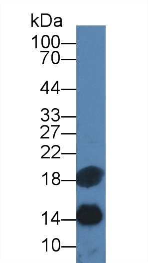
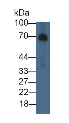
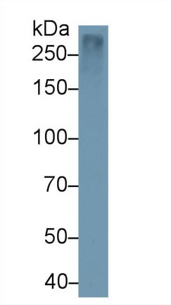
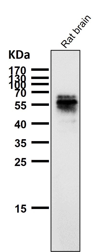
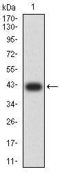
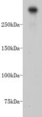
 WB (Western Blot)
(Western Blot: Sample: RecombinantREN,Rat))
WB (Western Blot)
(Western Blot: Sample: RecombinantREN,Rat))
Renin, Monoclonal Antibody (Cat# AAA141288)
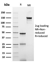 SDS-PAGE
(SDS-PAGE Analysis Purified Anti-Hexa-histidine Recombinant Mouse Monoclonal r6HIS/6423). Confirmation of Integrity and Purity of Antibody.)
SDS-PAGE
(SDS-PAGE Analysis Purified Anti-Hexa-histidine Recombinant Mouse Monoclonal r6HIS/6423). Confirmation of Integrity and Purity of Antibody.)
Hexa-histidine, Monoclonal Antibody (Cat# AAA216086)
 SDS-PAGE
(SDS-PAGE Analysis Purified Gastrin Mouse Monoclonal Antibody (GAST/2634). Confirmation of Integrity and Purity of Antibody.)
SDS-PAGE
(SDS-PAGE Analysis Purified Gastrin Mouse Monoclonal Antibody (GAST/2634). Confirmation of Integrity and Purity of Antibody.)
Gastrin, Monoclonal Antibody (Cat# AAA214981)
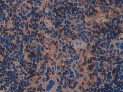
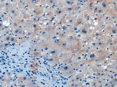
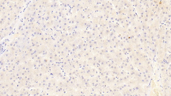
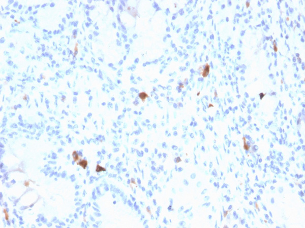
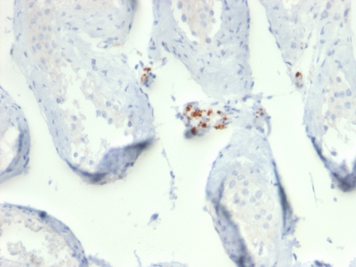
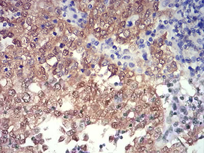
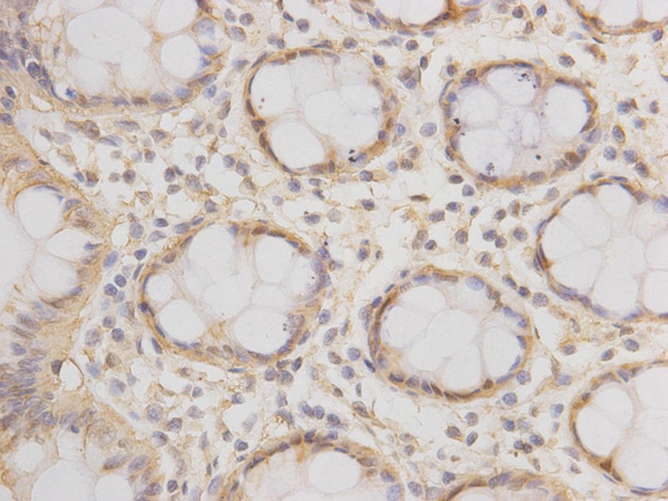
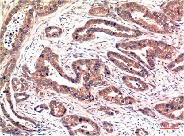
 IHC (Immunohistochemistry)
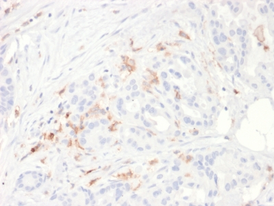
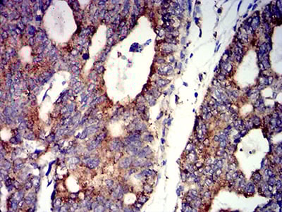
(Formalin-fixed, paraffin-embedded human liver carcinoma in colon stained with B7-H6/NCR3LG1 Mouse Monoclonal Antibody (B7H6/4821).)
IHC (Immunohistochemistry)
(Formalin-fixed, paraffin-embedded human liver carcinoma in colon stained with B7-H6/NCR3LG1 Mouse Monoclonal Antibody (B7H6/4821).)
B7-H6/NCR3LG1 (Natural Killer Cell Cytotoxicity Receptor 3 Ligand 1), Monoclonal Antibody (Cat# AAA215758)
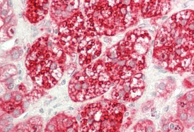
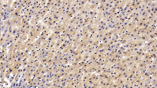
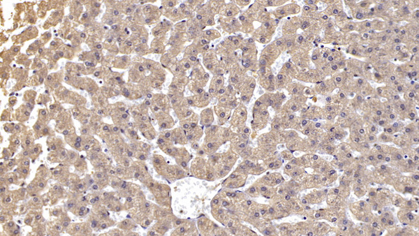
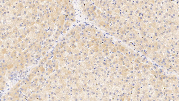
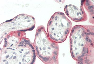
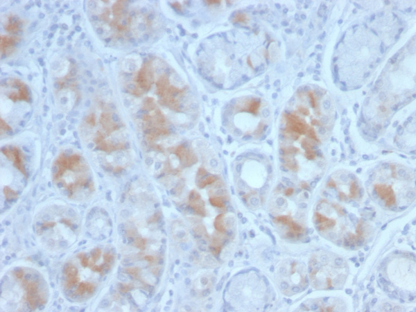
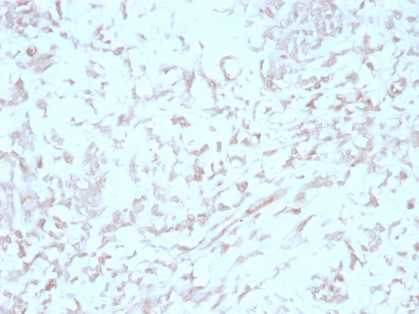
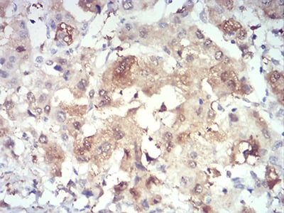
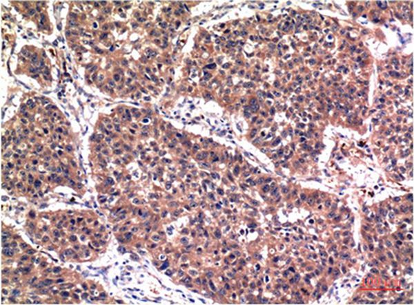
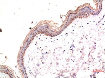
 IHC (Immunohistochemistry)
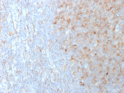
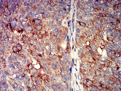
(Formalin-fixed, paraffin-embedded human liver stained with Alpha-1-Antichymotrypsin Mouse Monoclonal Antibody (SERPINA3/4187).)
IHC (Immunohistochemistry)
(Formalin-fixed, paraffin-embedded human liver stained with Alpha-1-Antichymotrypsin Mouse Monoclonal Antibody (SERPINA3/4187).)
Alpha-1-Antichymotrypsin (SERPINA3), Monoclonal Antibody (Cat# AAA215565)
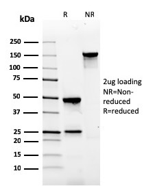
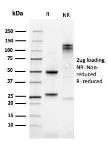 SDS-PAGE
Kindlin-1/KIND1, Monoclonal Antibody (Cat# AAA215463)
 Application Data
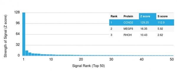
(Analysis of Protein Array containing more than 19,000 full-length human proteins using Cyclin D2 Mouse Recombinant Monoclonal Antibody (CCND2/2620). Z- and S- Score: The Z-score represents the strength of a signal that a monoclonal antibody (MAb) (in combination with a fluorescently-tagged anti-IgG secondary antibody) produces when binding to a particular protein on the HuProtTM array. Z-scores are described in units of standard deviations (SD's) above the mean value of all signals generated on that array. If targets on HuProtTM are arranged in descending order of the Z-score, the S-score is the difference (also in units of SD's) between the Z-score. S-score therefore represents the relative target specificity of a MAb to its intended target. A MAb is considered to specific to its intended target, if the MAb has an S-score of at least 2.5. For example, if a MAb binds to protein X with a Z-score of 43 and to protein Y with a Z-score of 14, then the S-score for the binding of that MAb to protein X is equal to 29.)
Application Data
(Analysis of Protein Array containing more than 19,000 full-length human proteins using Cyclin D2 Mouse Recombinant Monoclonal Antibody (CCND2/2620). Z- and S- Score: The Z-score represents the strength of a signal that a monoclonal antibody (MAb) (in combination with a fluorescently-tagged anti-IgG secondary antibody) produces when binding to a particular protein on the HuProtTM array. Z-scores are described in units of standard deviations (SD's) above the mean value of all signals generated on that array. If targets on HuProtTM are arranged in descending order of the Z-score, the S-score is the difference (also in units of SD's) between the Z-score. S-score therefore represents the relative target specificity of a MAb to its intended target. A MAb is considered to specific to its intended target, if the MAb has an S-score of at least 2.5. For example, if a MAb binds to protein X with a Z-score of 43 and to protein Y with a Z-score of 14, then the S-score for the binding of that MAb to protein X is equal to 29.)
Cyclin D2, Monoclonal Antibody (Cat# AAA214845)
 SDS_PAGE
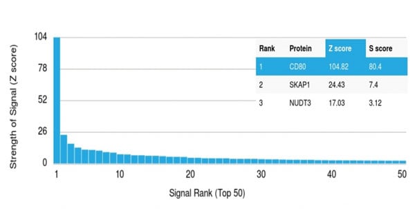
(SDS-PAGE Analysis Purified CD80 Mouse Monoclonal Antibody (C80/2723). Confirmation of Integrity and Purity of Antibody.)
SDS_PAGE
(SDS-PAGE Analysis Purified CD80 Mouse Monoclonal Antibody (C80/2723). Confirmation of Integrity and Purity of Antibody.)
CD80, Monoclonal Antibody (Cat# AAA214850)
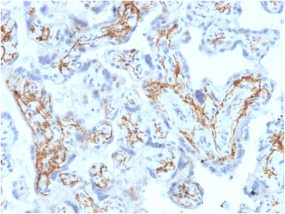
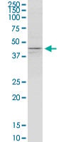
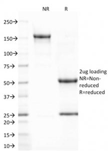
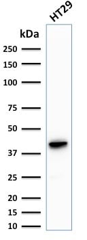
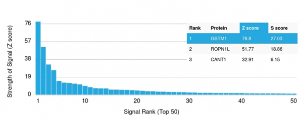 Application Data
(Analysis of Protein Array containing more than 19,000 full-length human proteins using Glutathione S-Transferase Mu1 (GSTM1) Mouse Monoclonal Antibody (CPTC-GSTMu1-3). Z- and S- Score: The Z-score represents the strength of a signal that a monoclonal antibody (MAb) (in combination with a fluorescently-tagged anti-IgG secondary antibody) produces when binding to a particular protein on the HuProtTM array. Z-scores are described in units of standard deviations (SD's) above the mean value of all signals generated on that array. If targets on HuProtTM are arranged in descending order of the Z-score, the S-score is the difference (also in units of SD's) between the Z-score. S-score therefore represents the relative target specificity of a MAb to its intended target. A MAb is considered to specific to its intended target, if the MAb has an S-score of at least 2.5. For example, if a MAb binds to protein X with a Z-score of 43 and to protein Y with a Z-score of 14, then the S-score for the binding of that MAb to protein X is equal to 29.)
Application Data
(Analysis of Protein Array containing more than 19,000 full-length human proteins using Glutathione S-Transferase Mu1 (GSTM1) Mouse Monoclonal Antibody (CPTC-GSTMu1-3). Z- and S- Score: The Z-score represents the strength of a signal that a monoclonal antibody (MAb) (in combination with a fluorescently-tagged anti-IgG secondary antibody) produces when binding to a particular protein on the HuProtTM array. Z-scores are described in units of standard deviations (SD's) above the mean value of all signals generated on that array. If targets on HuProtTM are arranged in descending order of the Z-score, the S-score is the difference (also in units of SD's) between the Z-score. S-score therefore represents the relative target specificity of a MAb to its intended target. A MAb is considered to specific to its intended target, if the MAb has an S-score of at least 2.5. For example, if a MAb binds to protein X with a Z-score of 43 and to protein Y with a Z-score of 14, then the S-score for the binding of that MAb to protein X is equal to 29.)
Glutathione S-Transferase Mu1 (GSTM1), Monoclonal Antibody (Cat# AAA214753)
 Application Data
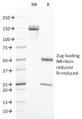
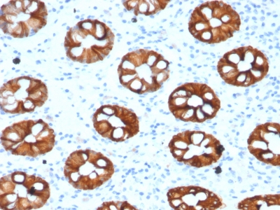
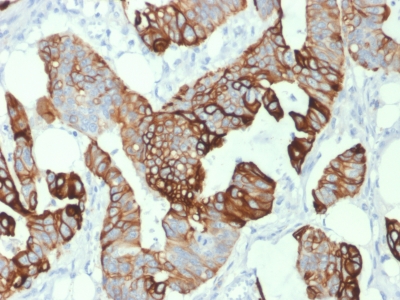
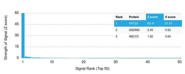
(Analysis of Protein Array containing more than 19,000 full-length human proteins using Cytokeratin 20 (KRT20) Mouse Monoclonal Antibody (KRT20/1993). Z- and S- Score: The Z-score represents the strength of a signal that a monoclonal antibody (MAb) (in combination with a fluorescently-tagged anti-IgG secondary antibody) produces when binding to a particular protein on the HuProtTM array. Z-scores are described in units of standard deviations (SD's) above the mean value of all signals generated on that array. If targets on HuProtTM are arranged in descending order of the Z-score, the S-score is the difference (also in units of SD's) between the Z-score. S-score therefore represents the relative target specificity of a MAb to its intended target. A MAb is considered to specific to its intended target, if the MAb has an S-score of at least 2.5. For example, if a MAb binds to protein X with a Z-score of 43 and to protein Y with a Z-score of 14, then the S-score for the binding of that MAb to protein X is equal to 29.)
Application Data
(Analysis of Protein Array containing more than 19,000 full-length human proteins using Cytokeratin 20 (KRT20) Mouse Monoclonal Antibody (KRT20/1993). Z- and S- Score: The Z-score represents the strength of a signal that a monoclonal antibody (MAb) (in combination with a fluorescently-tagged anti-IgG secondary antibody) produces when binding to a particular protein on the HuProtTM array. Z-scores are described in units of standard deviations (SD's) above the mean value of all signals generated on that array. If targets on HuProtTM are arranged in descending order of the Z-score, the S-score is the difference (also in units of SD's) between the Z-score. S-score therefore represents the relative target specificity of a MAb to its intended target. A MAb is considered to specific to its intended target, if the MAb has an S-score of at least 2.5. For example, if a MAb binds to protein X with a Z-score of 43 and to protein Y with a Z-score of 14, then the S-score for the binding of that MAb to protein X is equal to 29.)
Cytokeratin 20 (KRT20), Monoclonal Antibody (Cat# AAA214795)
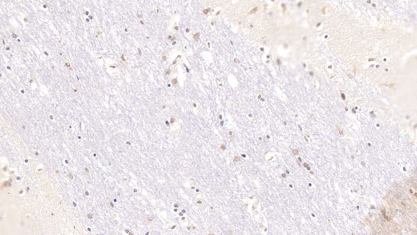
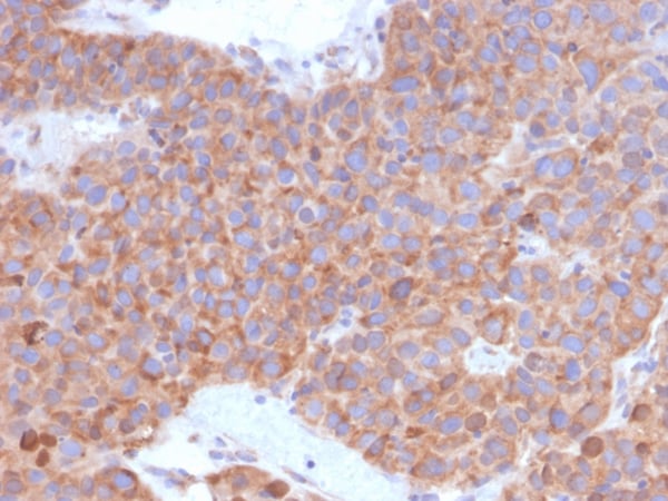
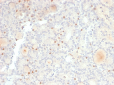
 Application Data
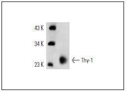
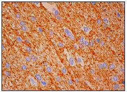
(Immunoperoxidase staining of formalin fixed, paraffin-embedded human cerebral cortex tissue showing neuropil staining.)
Application Data
(Immunoperoxidase staining of formalin fixed, paraffin-embedded human cerebral cortex tissue showing neuropil staining.)
Thy-1, Monoclonal Antibody (Cat# AAA312002)
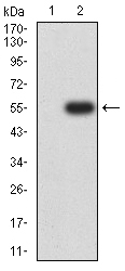
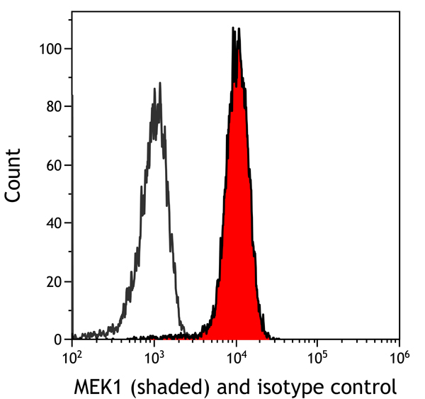
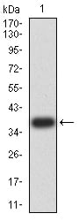
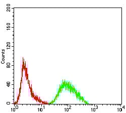
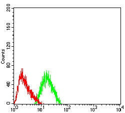 FCM/FACS (Flow Cytometry)
(Flow cytometric analysis of SH-SY5Y cells with CHRNA7 antibody at 1/100 dilution (green) compared with an unlabelled control (cells without incubation with primary antibody; red).)
FCM/FACS (Flow Cytometry)
(Flow cytometric analysis of SH-SY5Y cells with CHRNA7 antibody at 1/100 dilution (green) compared with an unlabelled control (cells without incubation with primary antibody; red).)
CHRNA7, Monoclonal Antibody (Cat# AAA312035)


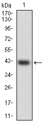
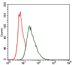
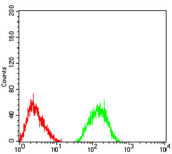 FCM/FACS (Flow Cytometry)
(Flow cytometric analysis of SH-SY5Y cells with GRM7 antibody at 1/100 dilution (green) compared with an unlabelled control (cells without incubation with primary antibody; red).)
FCM/FACS (Flow Cytometry)
(Flow cytometric analysis of SH-SY5Y cells with GRM7 antibody at 1/100 dilution (green) compared with an unlabelled control (cells without incubation with primary antibody; red).)
GRM7, Monoclonal Antibody (Cat# AAA312036)
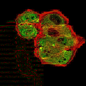
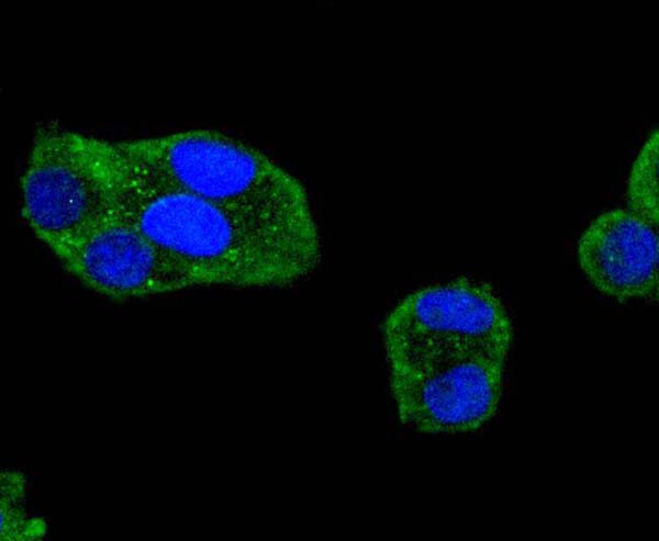
 ICC (Immunocytochemistry)
(ICC staining SMCP (green) in Hela cells. The nuclear counter stain is DAPI (blue). Cells were fixed in paraformaldehyde, permeabilised with 0.25% Triton X100/PBS.)
ICC (Immunocytochemistry)
(ICC staining SMCP (green) in Hela cells. The nuclear counter stain is DAPI (blue). Cells were fixed in paraformaldehyde, permeabilised with 0.25% Triton X100/PBS.)
SMCP, Monoclonal Antibody (Cat# AAA312039)
 FCM/FACS (Flow Cytometry)
(Flow cytometric analysis of Hela cells with PRDM4 antibody at 1/100 dilution (green) compared with an unlabelled control (cells without incubation with primary antibody; red).)
FCM/FACS (Flow Cytometry)
(Flow cytometric analysis of Hela cells with PRDM4 antibody at 1/100 dilution (green) compared with an unlabelled control (cells without incubation with primary antibody; red).)
PRDM4, Monoclonal Antibody (Cat# AAA312060)
 FCM/FACS (Flow Cytometry)
(Flow cytometric analysis of HeLa cells with HSF4 antibody at 1/100 dilution (green) compared with an unlabelled control (cells without incubation with primary antibody; red).)
FCM/FACS (Flow Cytometry)
(Flow cytometric analysis of HeLa cells with HSF4 antibody at 1/100 dilution (green) compared with an unlabelled control (cells without incubation with primary antibody; red).)
HSF4, Monoclonal Antibody (Cat# AAA312082)
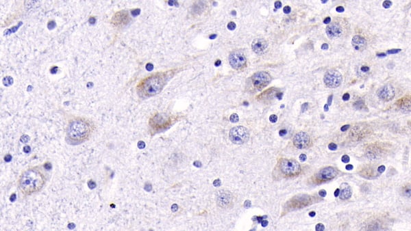
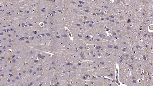
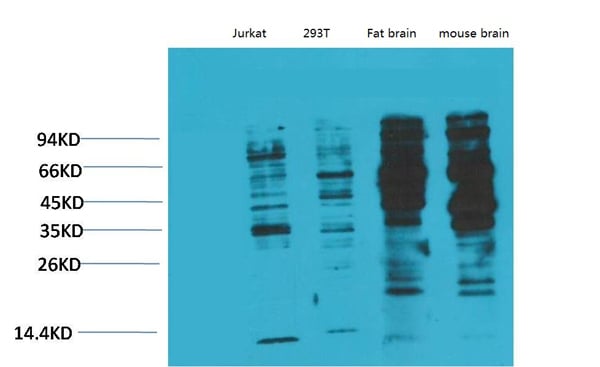
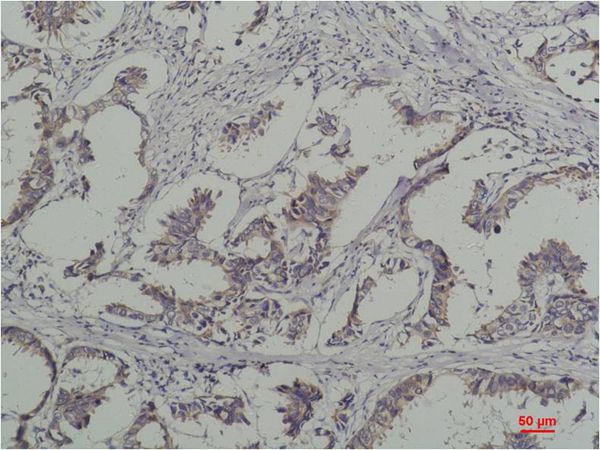
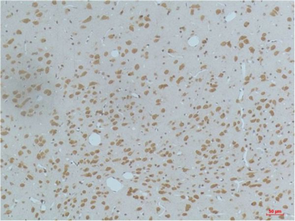
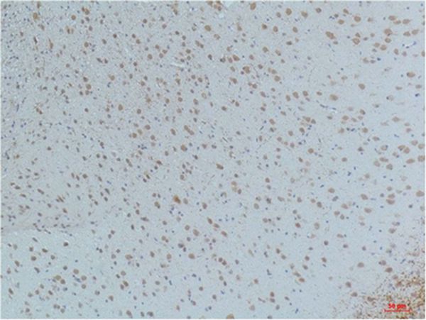
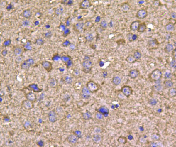 IHC (Immunohistochemistry)
(Immunohistochemical analysis of paraffin-embedded mouse brain tissue using anti-HSP90 beta antibody. Counter stained with hematoxylin.)
IHC (Immunohistochemistry)
(Immunohistochemical analysis of paraffin-embedded mouse brain tissue using anti-HSP90 beta antibody. Counter stained with hematoxylin.)
HSP90 Beta, Monoclonal Antibody (Cat# AAA312087)
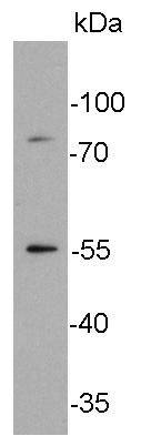
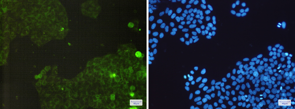
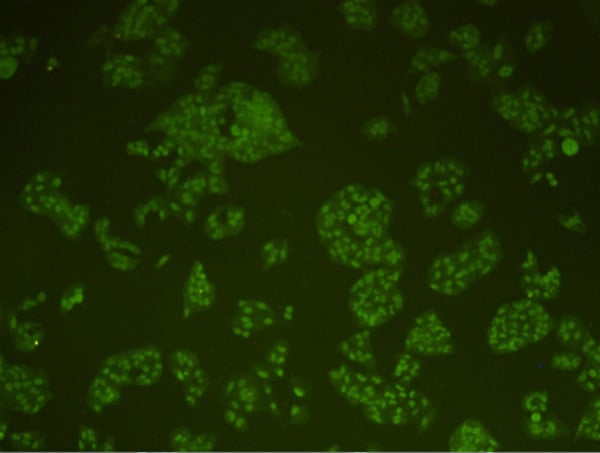 IF (Immunofluorescence)
(Immunofluorescent staining of HepG2 cells using anti-DNA-PKcs Mouse mAb.)
IF (Immunofluorescence)
(Immunofluorescent staining of HepG2 cells using anti-DNA-PKcs Mouse mAb.)
DNA-PKcs/PRKDC, Monoclonal Antibody (Cat# AAA311886)
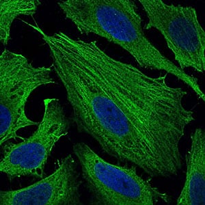
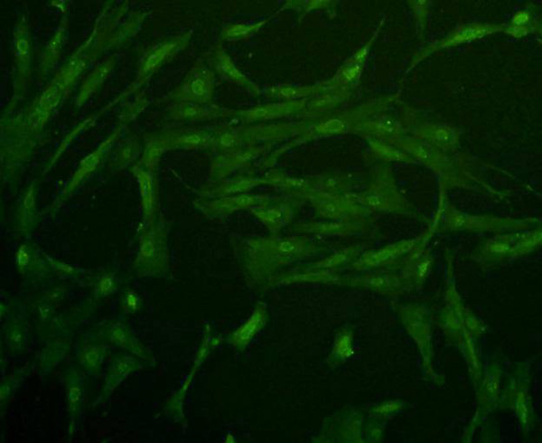 ICC (Immunocytochemistry)
(ICC staining Glut4 in NIH/3T3 cells (green). Cells were fixed in paraformaldehyde, permeabilised with 0.25% Triton X100/PBS.)
ICC (Immunocytochemistry)
(ICC staining Glut4 in NIH/3T3 cells (green). Cells were fixed in paraformaldehyde, permeabilised with 0.25% Triton X100/PBS.)
Glucose Transporter GLUT4, Monoclonal Antibody (Cat# AAA311904)
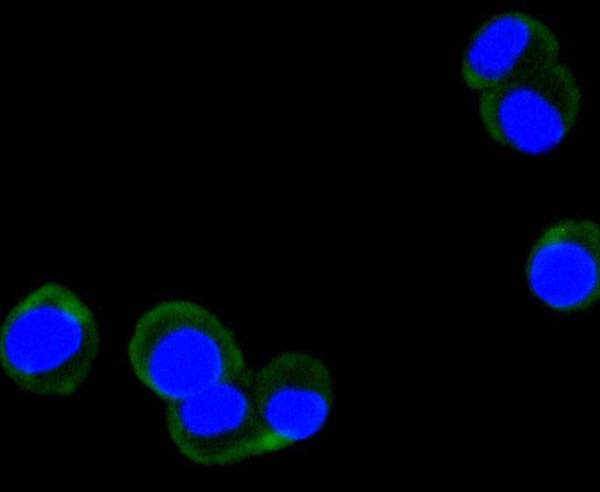
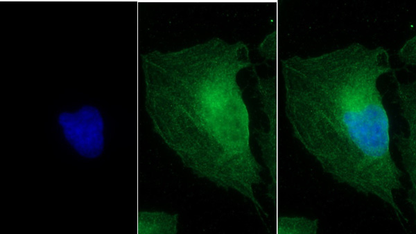
 ICC (Immunocytochemistry)
(ICC staining phospho-FAK (Y397) in NIH/3T3 cells (green). The nuclear counter stain is DAPI (blue). Cells were fixed in paraformaldehyde, permeabilised with 0.25% Triton X100/PBS.)
ICC (Immunocytochemistry)
(ICC staining phospho-FAK (Y397) in NIH/3T3 cells (green). The nuclear counter stain is DAPI (blue). Cells were fixed in paraformaldehyde, permeabilised with 0.25% Triton X100/PBS.)
FAK, Monoclonal Antibody (Cat# AAA311013)
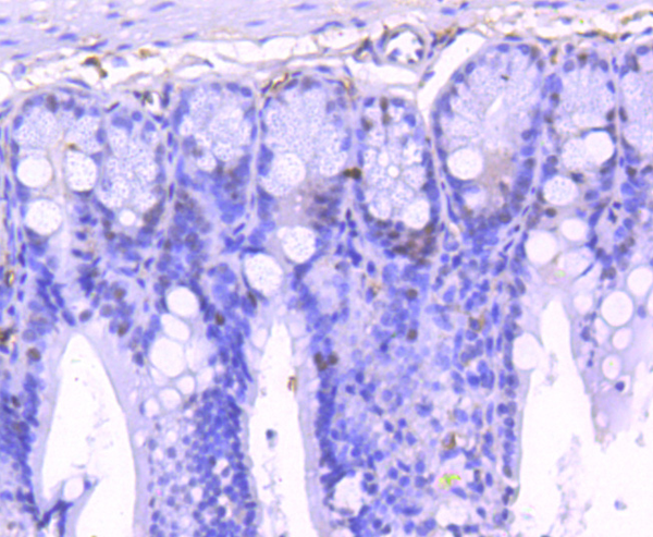 IHC (Immunohistochemisry)
(Immunohistochemical analysis of paraffin-embedded mouse colon tissue using anti-Phospho-STAT1 (S727) antibody. Counter stained with hematoxylin.)
IHC (Immunohistochemisry)
(Immunohistochemical analysis of paraffin-embedded mouse colon tissue using anti-Phospho-STAT1 (S727) antibody. Counter stained with hematoxylin.)
STAT1, Monoclonal Antibody (Cat# AAA311017)
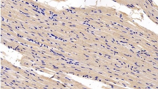
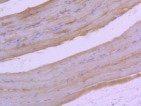 IHC (Immunohiostchemistry)
(Immunohistochemical analysis of paraffin-embedded rat skeletal muscle tissue using anti-Phospho-Raf1 (S259) antibody. Counter stained with hematoxylin.)
IHC (Immunohiostchemistry)
(Immunohistochemical analysis of paraffin-embedded rat skeletal muscle tissue using anti-Phospho-Raf1 (S259) antibody. Counter stained with hematoxylin.)
RAF1, Monoclonal Antibody (Cat# AAA311035)
 IHC (Immunohiostchemistry)
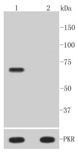
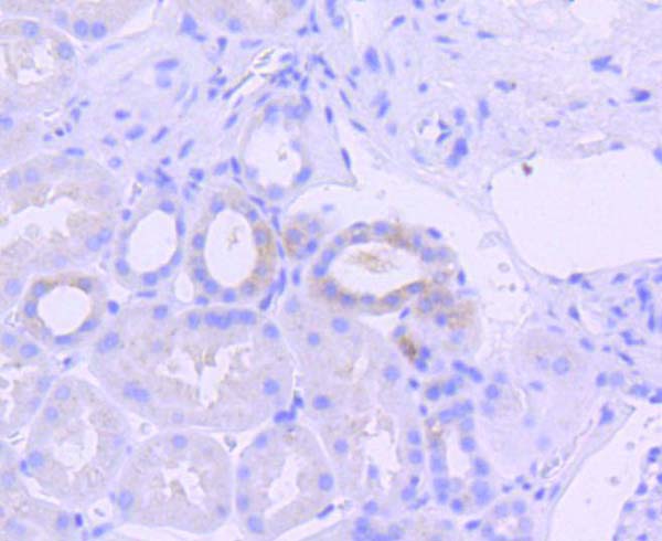
(Immunohistochemical analysis of paraffin-embedded human kidney tissue using anti-Phospho-PKR (T446) antibody. Counter stained with hematoxylin.)
IHC (Immunohiostchemistry)
(Immunohistochemical analysis of paraffin-embedded human kidney tissue using anti-Phospho-PKR (T446) antibody. Counter stained with hematoxylin.)
PKR, Monoclonal Antibody (Cat# AAA311000)
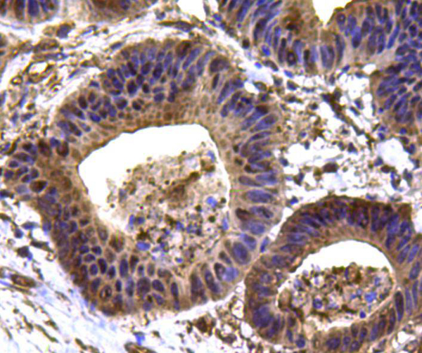
 IHC (Immunohistochemisry)
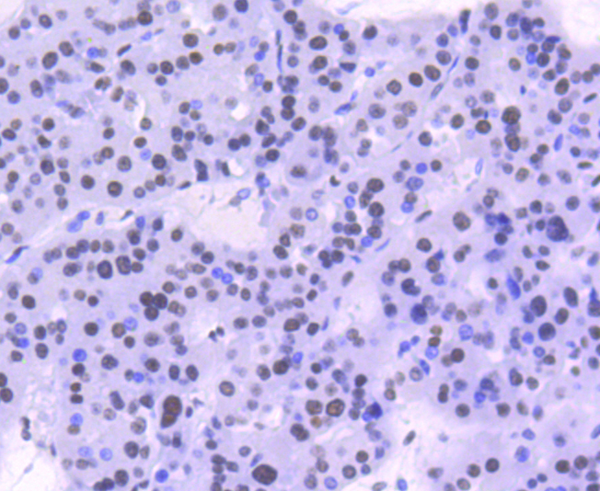
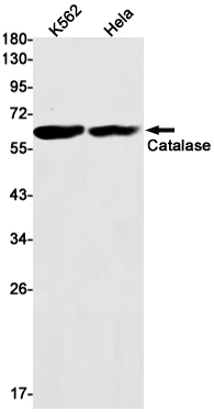
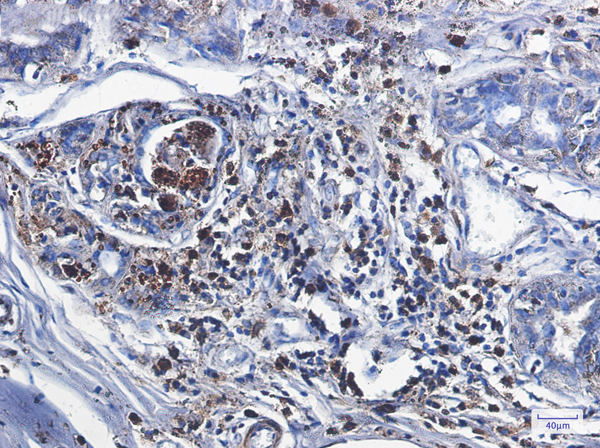
(Immunohistochemistry of Catalase in paraffin-embedded Human breast cancer tissue using Catalase Rabbit mAb at dilution 1/1)
IHC (Immunohistochemisry)
(Immunohistochemistry of Catalase in paraffin-embedded Human breast cancer tissue using Catalase Rabbit mAb at dilution 1/1)
Catalase, Monoclonal Antibody (Cat# AAA314323)
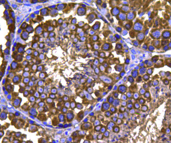
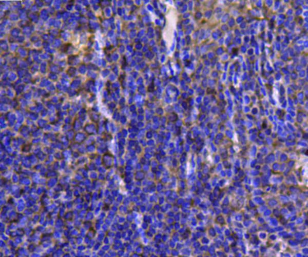
 IHC (Immunohiostchemistry)
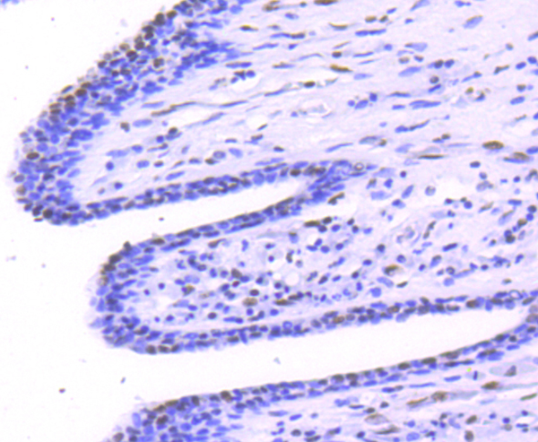
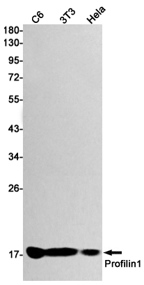
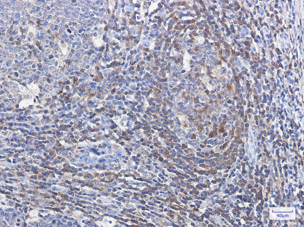
(Immunohistochemistry of Profilin 1 in paraffin-embedded Human tonsil using Profilin 1 Rabbit mAb at dilution 1/100)
IHC (Immunohiostchemistry)
(Immunohistochemistry of Profilin 1 in paraffin-embedded Human tonsil using Profilin 1 Rabbit mAb at dilution 1/100)
Profilin 1, Monoclonal Antibody (Cat# AAA314282)
 IHC (Immunohiostchemistry)
(Immunohistochemical analysis of paraffin-embedded Human Breast Caricnoma using PhosphotyrosineMouse mAb diluted at 1:200.)
IHC (Immunohiostchemistry)
(Immunohistochemical analysis of paraffin-embedded Human Breast Caricnoma using PhosphotyrosineMouse mAb diluted at 1:200.)
Phosphotyrosine, Monoclonal Antibody (Cat# AAA309598)
 IHC (Immunohiostchemistry)
(Immunohistochemical analysis of paraffin-embedded Mouse Brain Tissue using CaMKIIb/ g /&d (Phospho Thr287) Mouse mAb diluted at 1:200.)
IHC (Immunohiostchemistry)
(Immunohistochemical analysis of paraffin-embedded Mouse Brain Tissue using CaMKIIb/ g /&d (Phospho Thr287) Mouse mAb diluted at 1:200.)
CaMKIIbeta/gamma/delta, Monoclonal Antibody (Cat# AAA309599)
 IHC (Immunohistochemistry)
(Immunohistochemical analysis of paraffin-embedded Human Skin Tissue using Phospho-MLKL S358 Mouse mAb diluted at 1:200.)
IHC (Immunohistochemistry)
(Immunohistochemical analysis of paraffin-embedded Human Skin Tissue using Phospho-MLKL S358 Mouse mAb diluted at 1:200.)
Akt, Monoclonal Antibody (Cat# AAA309601)
What are Monoclonal Antibodies?
Monoclonal antibodies are specialized laboratory-produced proteins developed for binding to specific biological antigens or other molecular targets. Since they come from a single cell (or clone), they are especially consistent and accurate in the data they are involved in producing.
This type of antibody material has been shown to be a powerful tool in finding and subsequently destroying harmful cells in an organism, such as those found in cancers or various autoimmune diseases. This makes them excellent aids in medical testing and research, which is why they are so widely used.
AAA Biotech offers a comprehensive range of high-quality monoclonal antibodies that perform effectively in various laboratory tests, including (amongst others) ELISA, western blotting, immunohistochemistry, and flow cytometry. All of the products in our catalog are thoroughly quality tested to make sure that they are reliable and will consistently perform well in your research.
What Are The Uses of Monoclonal Antibodies
Monoclonal antibodies are used in many lab tests, including (amongst others) ELISA, western blotting, immunohistochemistry, and flow cytometry.
ELISA is a test that helps detect a specific substance/analyte in a sample. It uses antibodies (often monoclonal) bound to a solid surface (such as the well of a microplate) to “capture” the substance/analyte in the sample and immobilize it so that the detection antibody component can then bind to it and produce a signal, which can then be measured.
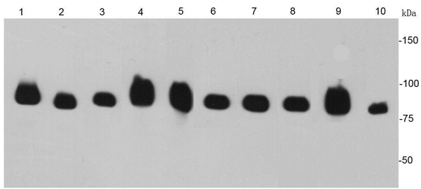
Western blotting identifies specific proteins in a sample. The sample is first separated on a gel, and then antibodies are applied that will typically bind to the target, which will all be localized to a single band in a lane.
Immunohistochemistry helps locate specific proteins in cells or tissue samples using antibodies.
Flow cytometry looks at and sorts cells. It uses antibodies that are conjugated to reporter molecules called “fluorophores”, which, under special lights, emit light themselves, which can then be measured by a detector instrument.
How Monoclonal Antibodies Are Used as Medicine?
Please note that all of the products listed in AAA Biotech’s also known as AAA Bio or AAABio catalog are strictly for research-use only (RUO).
Monoclonal antibodies can also be used as therapeutic/medical treatments, particularly in the context of cancers. They are designed to find and bind to specific cells or proteins, helping the immune system recognize and attack the cancer. These treatments work in different ways, such as:
- Radioimmunotherapy attaches a small amount of radioactive molecule to the antibody, so it delivers the radiation directly to the cancer cells that the antibody is specifically binding to.
- Antibody-directed enzyme prodrug therapy uses antibodies that are specifically bound to special enzymes. These enzymes activate a harmless drug in the body and turn it into a cancer-killing drug only near the cancer cells—this helps avoid harming healthy cells.
- Immunoliposomes are tiny “bubbles” filled with medicine/drug and coated with antibodies. They carry the drug straight to the cancer cells.
Why Buy Monoclonal Antibodies From Us?
At AAA Biotech, we provide high-performance monoclonal antibodies designed to support a wide range of research needs.
1. Validated for Versatile Applications
The antibodies in our catalog are extensively validated and compatible with multiple techniques, including (but not limited to) ELISA, flow cytometry (FC), immunocytochemistry (ICC), immunofluorescence (IF), immunohistochemistry (IHC), immunoprecipitation (IP), and western blotting (WB).
2. Wide Selection & Specialized Options
We offer antibodies for common and rare species, that are available in various conjugated forms, and also in recombinant formats. Essentially, there is almost anything one might need to meet their experimental model’s requirements.
3. High-Quality Proteins
Our proteins meet high purity standards—90% or more as confirmed by SDS-PAGE. Many are available with tags like His, Flag, GST, or MBP, and we also supply native and biologically active proteins for functional studies.
Frequently Asked Questions
1. Are your monoclonal antibodies validated for specific applications?
Yes, our antibodies are tested and validated for use in methods such as ELISA, western blot, IHC, flow cytometry, and more. Refer to specific product pages or datasheets for individual product information.
2. How do I choose the right monoclonal antibody for my application?
Review the product details directly for application validation, species reactivity, and target information. You may also contact our support team at any time for help.
3. How quickly can I receive my order?
Most orders are processed and shipped within 1–3 business days, depending on product availability and your shipping location.