Filters
▼Clonality
▼Type
▼Reactivity
▼Gene Name
▼Isotype
▼Host
▼Application
▼Clone
▼Monoclonal Antibodies
Get accurate results in your research with our Monoclonal Antibodies, which are specially made to target exactly what you require for your research, and will produce consistent, reliable performance in lab tests.
Viewing 9700-9750 of 27597 product results
 ICC (Immunocytochemistry)
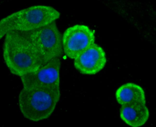
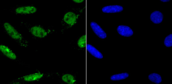
(ICC staining Caspase-6 in SW480 cells (green). The nuclear counter stain is DAPI (blue). Cells were fixed in paraformaldehyde, permeabilised with 0.25% Triton X100/PBS.)
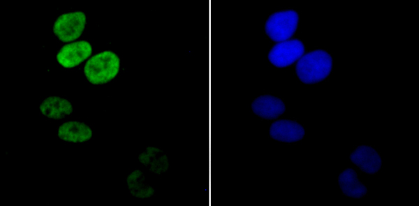
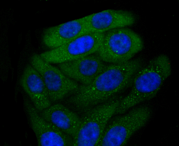
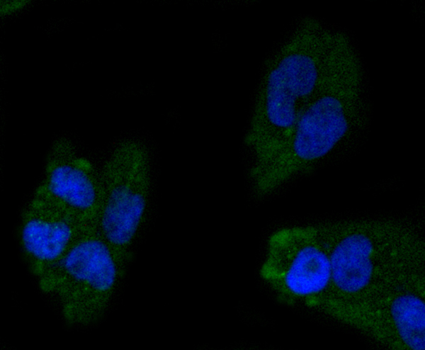
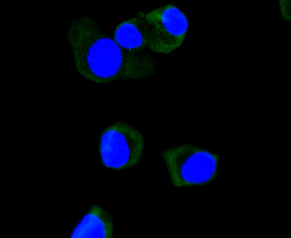
ICC (Immunocytochemistry)
(ICC staining Caspase-6 in SW480 cells (green). The nuclear counter stain is DAPI (blue). Cells were fixed in paraformaldehyde, permeabilised with 0.25% Triton X100/PBS.)
Caspase-6, Monoclonal Antibody (Cat# AAA312253)
 IHC (Immunohiostchemistry)
(Immunohistochemical analysis of paraffin-embedded human tonsil tissue using anti-Aurora B antibody. Counter stained with hematoxylin.)
IHC (Immunohiostchemistry)
(Immunohistochemical analysis of paraffin-embedded human tonsil tissue using anti-Aurora B antibody. Counter stained with hematoxylin.)
Aurora B, Monoclonal Antibody (Cat# AAA312254)
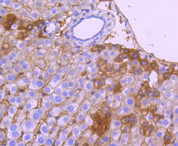
 IHC (Immunohistochemistry)
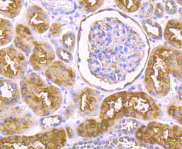
(Immunohistochemical analysis of paraffin-embedded human kidney tissue using anti-P2Y6 antibody. Counter stained with hematoxylin.)
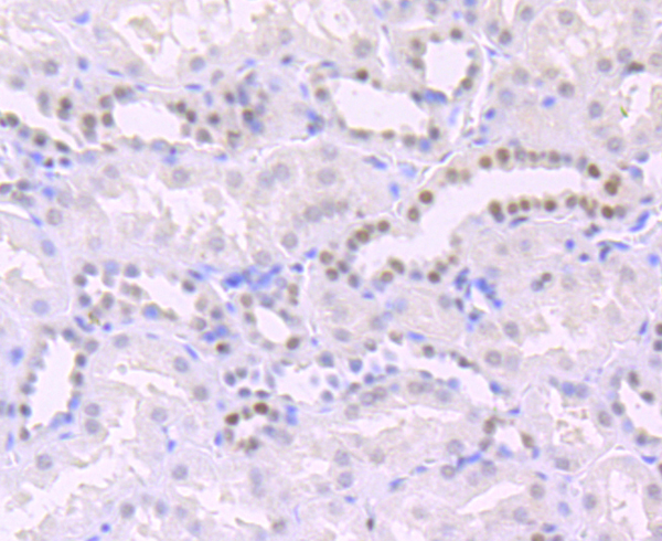
IHC (Immunohistochemistry)
(Immunohistochemical analysis of paraffin-embedded human kidney tissue using anti-P2Y6 antibody. Counter stained with hematoxylin.)
P2Y6, Monoclonal Antibody (Cat# AAA312668)
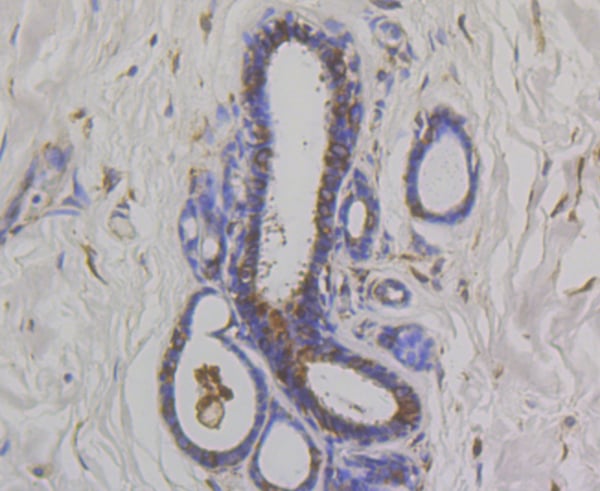
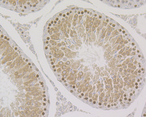
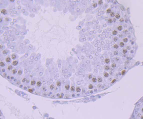
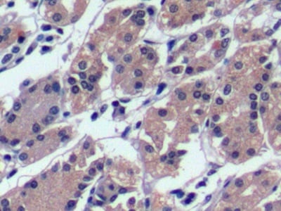
 IHC (Immunohistochemistry)
(Immunohistochemical analysis of paraffin-embedded mouse testis tissue using anti-OS9 antibody. Counter stained with hematoxylin.)
IHC (Immunohistochemistry)
(Immunohistochemical analysis of paraffin-embedded mouse testis tissue using anti-OS9 antibody. Counter stained with hematoxylin.)
OS9, Monoclonal Antibody (Cat# AAA312671)
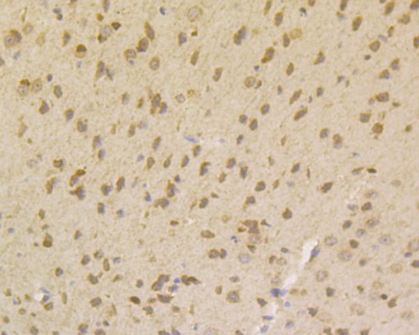 IHC (Immunohistochemisry)
(Immunohistochemical analysis of paraffin-embedded mouse brain tissue using anti-Gemin 1 antibody. Counter stained with hematoxylin.)
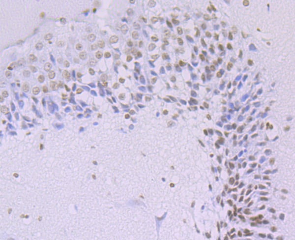
IHC (Immunohistochemisry)
(Immunohistochemical analysis of paraffin-embedded mouse brain tissue using anti-Gemin 1 antibody. Counter stained with hematoxylin.)
Gemin 1, Monoclonal Antibody (Cat# AAA312672)
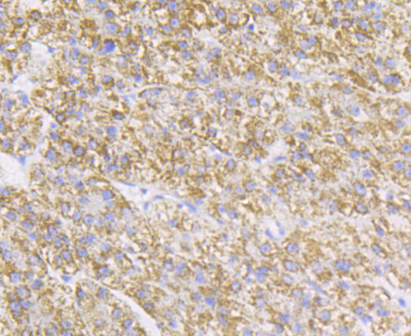
 IHC (Immunohistochemistry)
(Immunohistochemical analysis of paraffin-embedded mouse spleen tissue using anti-NOXA2 antibody. Counter stained with hematoxylin.)
IHC (Immunohistochemistry)
(Immunohistochemical analysis of paraffin-embedded mouse spleen tissue using anti-NOXA2 antibody. Counter stained with hematoxylin.)
NOXA2, Monoclonal Antibody (Cat# AAA312678)
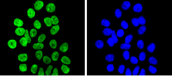
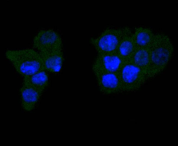
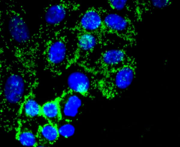
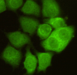
 ICC (Immunocytochemistry)
(ICC staining Histone H3 (di methyl K9) in NIH/3T3 cells (green). The nuclear counter stain is DAPI (blue). Cells were fixed in paraformaldehyde, permeabilised with 0.25% Triton X100/PBS.)
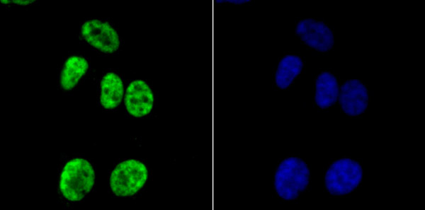
ICC (Immunocytochemistry)
(ICC staining Histone H3 (di methyl K9) in NIH/3T3 cells (green). The nuclear counter stain is DAPI (blue). Cells were fixed in paraformaldehyde, permeabilised with 0.25% Triton X100/PBS.)
Histone H3, Monoclonal Antibody (Cat# AAA312710)
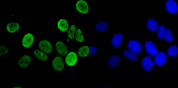
 ICC (Immunocytochemistry)
(ICC staining Histone H3 (acetyl K14) in PC-3M cells (green). The nuclear counter stain is DAPI (blue). Cells were fixed in paraformaldehyde, permeabilised with 0.25% Triton X100/PBS.)
ICC (Immunocytochemistry)
(ICC staining Histone H3 (acetyl K14) in PC-3M cells (green). The nuclear counter stain is DAPI (blue). Cells were fixed in paraformaldehyde, permeabilised with 0.25% Triton X100/PBS.)
Histone H3, Monoclonal Antibody (Cat# AAA312712)
 ICC (Immunocytochemistry)
(ICC staining CXCR3 in HUVEC cells (green). The nuclear counter stain is DAPI (blue). Cells were fixed in paraformaldehyde, permeabilised with 0.25% Triton X100/PBS.)
ICC (Immunocytochemistry)
(ICC staining CXCR3 in HUVEC cells (green). The nuclear counter stain is DAPI (blue). Cells were fixed in paraformaldehyde, permeabilised with 0.25% Triton X100/PBS.)
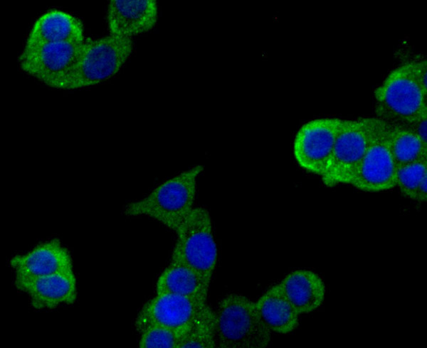
CXCR3, Monoclonal Antibody (Cat# AAA312523)
 FCM/FACS (Flow Cytometry)
(Flow cytometric analysis of LOVO cells with Plexin A1 antibody at 1/50 dilution (red) compared with an unlabelled control (cells without incubation with primary antibody; black). Alexa Fluor 488-conjugated goat anti rabbit IgG was used as the secondary antibody.)
FCM/FACS (Flow Cytometry)
(Flow cytometric analysis of LOVO cells with Plexin A1 antibody at 1/50 dilution (red) compared with an unlabelled control (cells without incubation with primary antibody; black). Alexa Fluor 488-conjugated goat anti rabbit IgG was used as the secondary antibody.)
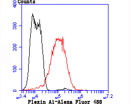
Plexin A1, Monoclonal Antibody (Cat# AAA312565)

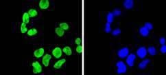
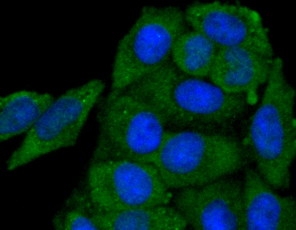
 ICC (Immunocytochemistry)
(ICC staining IKBKE in SK-Br-3 cells (green). The nuclear counter stain is DAPI (blue). Cells were fixed in paraformaldehyde, permeabilised with 0.25% Triton X100/PBS.)
ICC (Immunocytochemistry)
(ICC staining IKBKE in SK-Br-3 cells (green). The nuclear counter stain is DAPI (blue). Cells were fixed in paraformaldehyde, permeabilised with 0.25% Triton X100/PBS.)
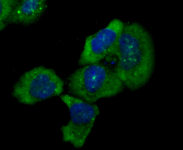
IKBKE, Monoclonal Antibody (Cat# AAA312566)
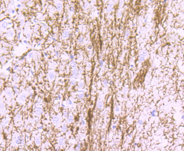
 IHC (Immunohiostchemistry)
(Immunohistochemical analysis of paraffin-embedded mouse brain tissue using anti-Myelin Basic Protein antibody. Counter stained with hematoxylin.)
IHC (Immunohiostchemistry)
(Immunohistochemical analysis of paraffin-embedded mouse brain tissue using anti-Myelin Basic Protein antibody. Counter stained with hematoxylin.)
Myelin Basic Protein, Monoclonal Antibody (Cat# AAA312401)
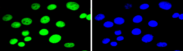
 ICC (Immunocytochemistry)
(ICC staining Apolipoprotein A1 in N2A cells (green). The nuclear counter stain is DAPI (blue). Cells were fixed in paraformaldehyde, permeabilised with 0.25% Triton X100/PBS.)
ICC (Immunocytochemistry)
(ICC staining Apolipoprotein A1 in N2A cells (green). The nuclear counter stain is DAPI (blue). Cells were fixed in paraformaldehyde, permeabilised with 0.25% Triton X100/PBS.)
Apolipoprotein A1, Monoclonal Antibody (Cat# AAA312406)
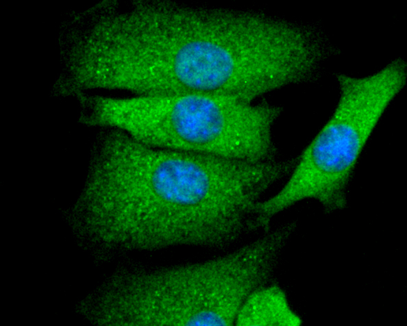
 ICC (Immunocytochemistry)
(ICC staining Fibronectin in NIH/3T3 cells (green). The nuclear counter stain is DAPI (blue). Cells were fixed in paraformaldehyde, permeabilised with 0.25% Triton X100/PBS.)
ICC (Immunocytochemistry)
(ICC staining Fibronectin in NIH/3T3 cells (green). The nuclear counter stain is DAPI (blue). Cells were fixed in paraformaldehyde, permeabilised with 0.25% Triton X100/PBS.)
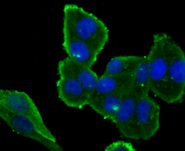
Fibronectin, Monoclonal Antibody (Cat# AAA312408)
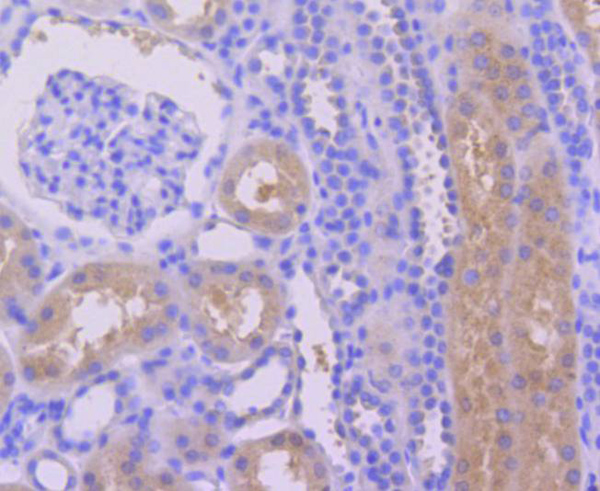
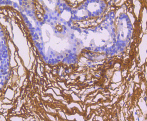
 IHC (Immunohiostchemistry)
(Immunohistochemical analysis of paraffin-embedded mouse kidney tissue using anti-Jagged1 antibody. Counter stained with hematoxylin.)
IHC (Immunohiostchemistry)
(Immunohistochemical analysis of paraffin-embedded mouse kidney tissue using anti-Jagged1 antibody. Counter stained with hematoxylin.)
Jagged1, Monoclonal Antibody (Cat# AAA312423)
 ICC (Immunocytochemistry)
(ICC staining Mad2L1 in SH-SY5Y cells (green). The nuclear counter stain is DAPI (blue). Cells were fixed in paraformaldehyde, permeabilised with 0.25% Triton X100/PBS.)
ICC (Immunocytochemistry)
(ICC staining Mad2L1 in SH-SY5Y cells (green). The nuclear counter stain is DAPI (blue). Cells were fixed in paraformaldehyde, permeabilised with 0.25% Triton X100/PBS.)
Mad2L1, Monoclonal Antibody (Cat# AAA312590)
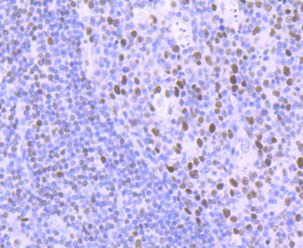
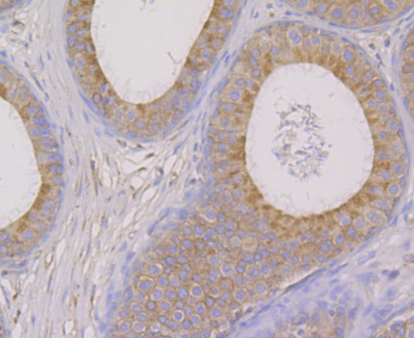
 IHC (Immunohistochemistry)
(Immunohistochemical analysis of paraffin-embedded rat testis tissue using anti-DNA Ligase I antibody. Counter stained with hematoxylin.)
IHC (Immunohistochemistry)
(Immunohistochemical analysis of paraffin-embedded rat testis tissue using anti-DNA Ligase I antibody. Counter stained with hematoxylin.)
DNA Ligase I, Monoclonal Antibody (Cat# AAA312612)
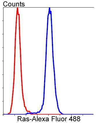 FCM/FACS (Flow Cytometry)
(Flow cytometric analysis of Hela cells with Ras antibody at 1/50 dilution (blue) compared with an unlabelled control (cells without incubation with primary antibody; red). Alexa Fluor 488-conjugated goat anti rabbit IgG was used as the secondary antibody.)
FCM/FACS (Flow Cytometry)
(Flow cytometric analysis of Hela cells with Ras antibody at 1/50 dilution (blue) compared with an unlabelled control (cells without incubation with primary antibody; red). Alexa Fluor 488-conjugated goat anti rabbit IgG was used as the secondary antibody.)
RAS, Monoclonal Antibody (Cat# AAA312133)


 FCM/FACS (Flow Cytometry)
(Flow cytometric analysis of Hela cells with alpha 1 Catenin antibody at 1/50 dilution (red) compared with an unlabelled control (cells without incubation with primary antibody; black). Alexa Fluor 488-conjugated goat anti rabbit IgG was used as the secondary antibody.)
FCM/FACS (Flow Cytometry)
(Flow cytometric analysis of Hela cells with alpha 1 Catenin antibody at 1/50 dilution (red) compared with an unlabelled control (cells without incubation with primary antibody; black). Alexa Fluor 488-conjugated goat anti rabbit IgG was used as the secondary antibody.)
alpha 1 Catenin, Monoclonal Antibody (Cat# AAA312262)
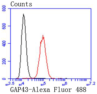 FCM/FACS (Flow Cytometry)
(Flow cytometric analysis of SH-SY-5y cells with GAP43 antibody at 1/50 dilution (red) compared with an unlabelled control (cells without incubation with primary antibody; black). Alexa Fluor 488-conjugated goat anti rabbit IgG was used as the secondary antibody.)
FCM/FACS (Flow Cytometry)
(Flow cytometric analysis of SH-SY-5y cells with GAP43 antibody at 1/50 dilution (red) compared with an unlabelled control (cells without incubation with primary antibody; black). Alexa Fluor 488-conjugated goat anti rabbit IgG was used as the secondary antibody.)
GAP43, Monoclonal Antibody (Cat# AAA312281)
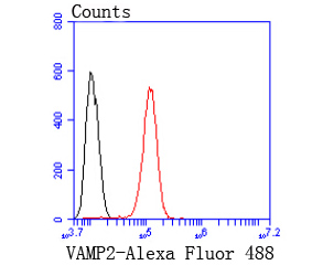 FCM/FACS (Flow Cytometry)
(Flow cytometric analysis of SH-SY5Y cells with VAMP2 antibody at 1/50 dilution (red) compared with an unlabelled control (cells without incubation with primary antibody; black). Alexa Fluor 488-conjugated goat anti rabbit IgG was used as the secondary antibody.)
FCM/FACS (Flow Cytometry)
(Flow cytometric analysis of SH-SY5Y cells with VAMP2 antibody at 1/50 dilution (red) compared with an unlabelled control (cells without incubation with primary antibody; black). Alexa Fluor 488-conjugated goat anti rabbit IgG was used as the secondary antibody.)
VAMP2, Monoclonal Antibody (Cat# AAA312465)
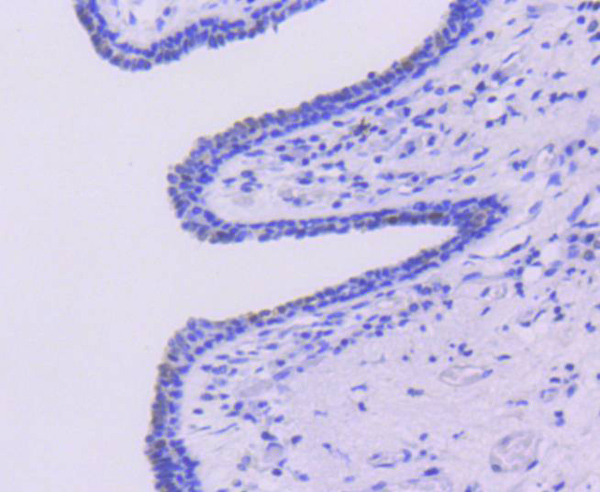
 IHC (Immunohistochemistry)
(Immunohistochemical analysis of paraffin-embedded human uterus tissue using anti-Claudin 5 antibody. Counter stained with hematoxylin.)
IHC (Immunohistochemistry)
(Immunohistochemical analysis of paraffin-embedded human uterus tissue using anti-Claudin 5 antibody. Counter stained with hematoxylin.)
Claudin 5, Monoclonal Antibody (Cat# AAA312470)
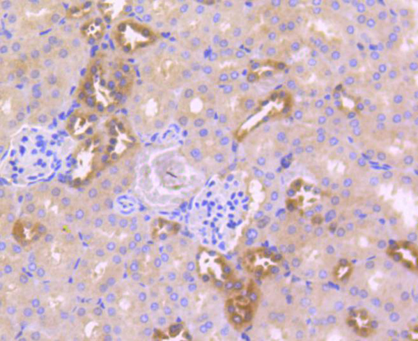
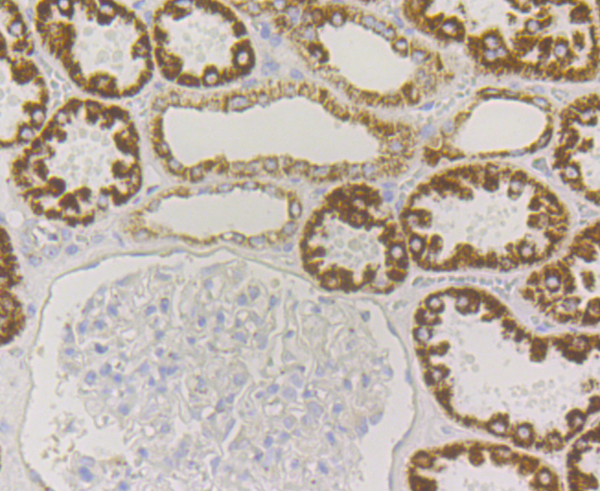
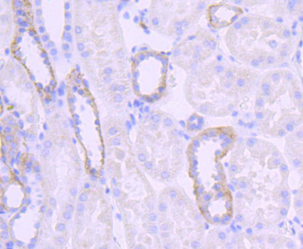
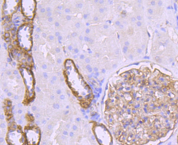
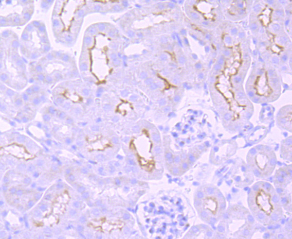 IHC (Immunohistochemisry)
(Immunohistochemical analysis of paraffin-embedded mouse kidney tissue using anti-mGluR1 antibody. Counter stained with hematoxylin.)
IHC (Immunohistochemisry)
(Immunohistochemical analysis of paraffin-embedded mouse kidney tissue using anti-mGluR1 antibody. Counter stained with hematoxylin.)
mGluR1, Monoclonal Antibody (Cat# AAA312478)
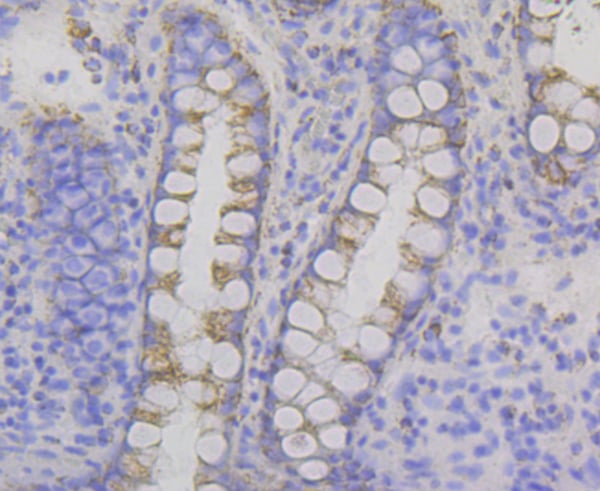
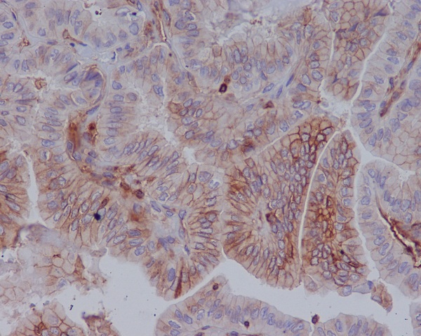
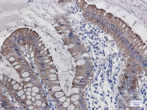
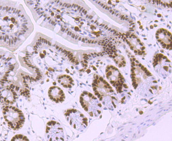 IHC (Immunohistochemistry)
(Immunohistochemical analysis of paraffin-embedded mouse colon tissue using anti-MSH6 antibody. Counter stained with hematoxylin.)
IHC (Immunohistochemistry)
(Immunohistochemical analysis of paraffin-embedded mouse colon tissue using anti-MSH6 antibody. Counter stained with hematoxylin.)
MSH6, Monoclonal Antibody (Cat# AAA312494)
 Application Data
(Dilution: WB: 1:1000 IF: 1:100-200)
Application Data
(Dilution: WB: 1:1000 IF: 1:100-200)
EGFR, Monoclonal Antibody (Cat# AAA293511)
 IHC (Immunohiostchemistry)
(Immunohistochemical analysis of paraffin-embedded human thyroid carcinoma, using MHC class 1 Antibody.)
IHC (Immunohiostchemistry)
(Immunohistochemical analysis of paraffin-embedded human thyroid carcinoma, using MHC class 1 Antibody.)
MHC class 1, Monoclonal Recombinant Antibody (Cat# AAA314902)
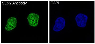 IF (Immunofluorescence)
(Immunofluorescent analysis of NCCIT cells, using SOX2 Antibody .)
IF (Immunofluorescence)
(Immunofluorescent analysis of NCCIT cells, using SOX2 Antibody .)
SOX2, Monoclonal Recombinant Antibody (Cat# AAA314906)
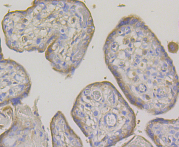
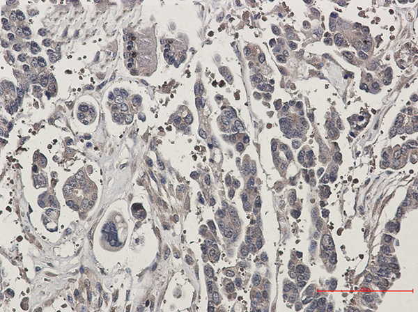
 IHC (Immunohiostchemistry)
(Immunohistochemical analysis of paraffin-embedded human breast cancer, using MRP8 Antibody.)
IHC (Immunohiostchemistry)
(Immunohistochemical analysis of paraffin-embedded human breast cancer, using MRP8 Antibody.)
MRP8, Monoclonal Recombinant Antibody (Cat# AAA314913)
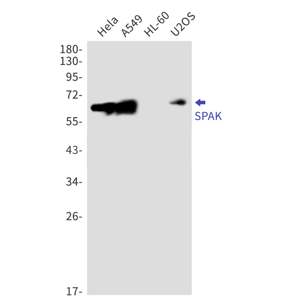
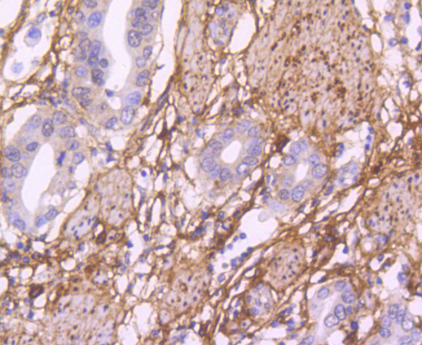
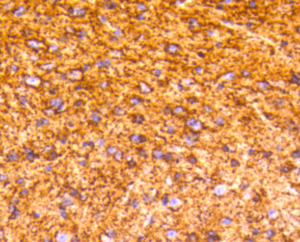
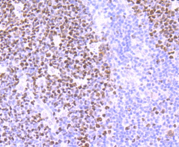
 IHC (Immunohiostchemistry)
(Immunohistochemistry of SPAK in paraffin-embedded Human Cholangiocarcinoma using SPAK Rabbit mAb at dilution 1/50)
IHC (Immunohiostchemistry)
(Immunohistochemistry of SPAK in paraffin-embedded Human Cholangiocarcinoma using SPAK Rabbit mAb at dilution 1/50)
SPAK, Monoclonal Antibody (Cat# AAA314573)
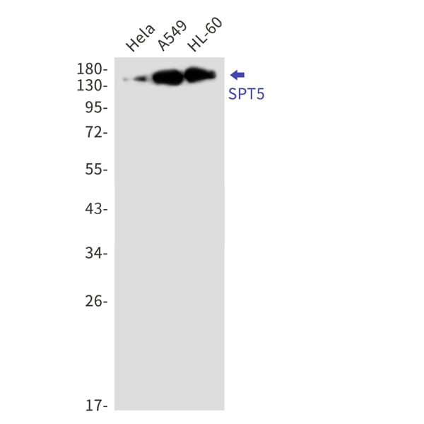
 IHC (Immunohiostchemistry)
(Immunohistochemistry of SPT5 in paraffin-embedded Human breast cancer tissue using SPT5 Rabbit mAb at dilution 1/50)
IHC (Immunohiostchemistry)
(Immunohistochemistry of SPT5 in paraffin-embedded Human breast cancer tissue using SPT5 Rabbit mAb at dilution 1/50)
SPT5, Monoclonal Antibody (Cat# AAA314574)
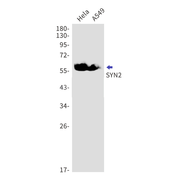
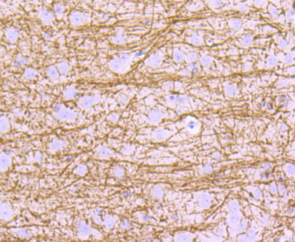
 IHC (Immunohiostchemistry)
(Immunohistochemistry of SYN2 in paraffin-embedded Human Brain using SYN2 Rabbit mAb at dilution 1/50)
IHC (Immunohiostchemistry)
(Immunohistochemistry of SYN2 in paraffin-embedded Human Brain using SYN2 Rabbit mAb at dilution 1/50)
Synapsin 2, Monoclonal Antibody (Cat# AAA314577)

 IF (Immunofluorescence)
(Immunofluorescent analysis of A673 cells, using ErbB4 (HER4) Antibody .)
IF (Immunofluorescence)
(Immunofluorescent analysis of A673 cells, using ErbB4 (HER4) Antibody .)
ErbB4 (HER4), Monoclonal Recombinant Antibody (Cat# AAA314997)
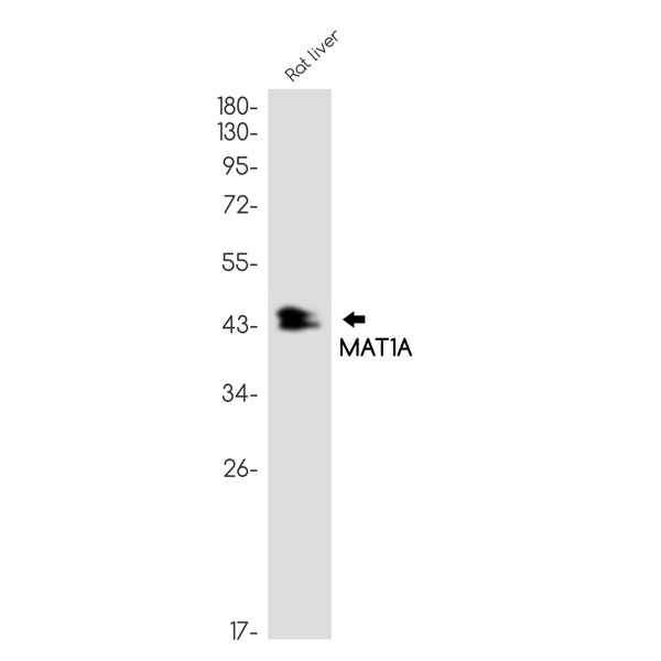
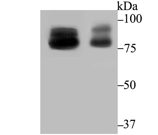
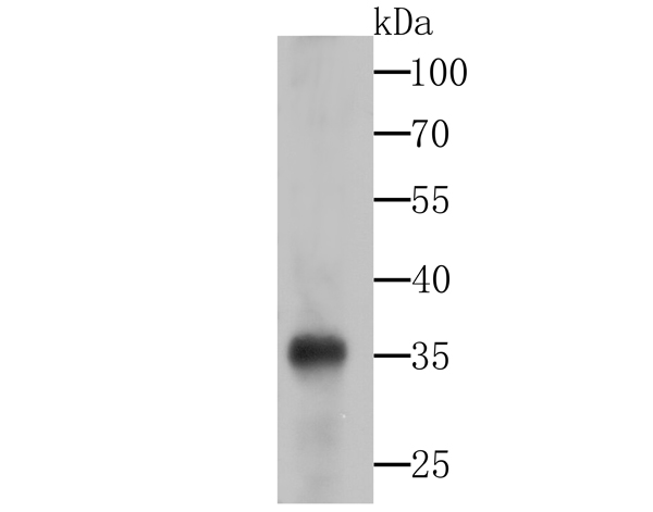
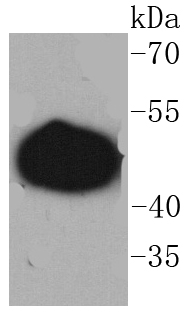
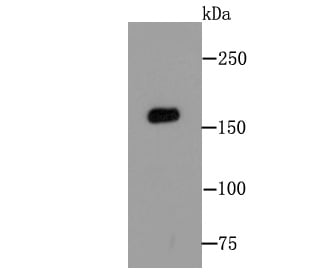
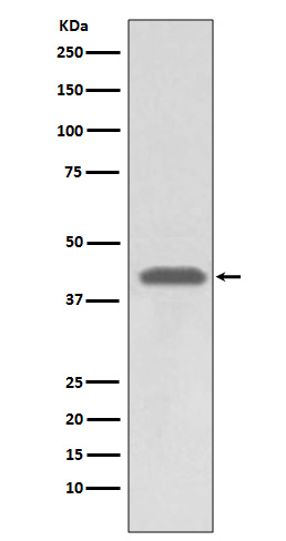
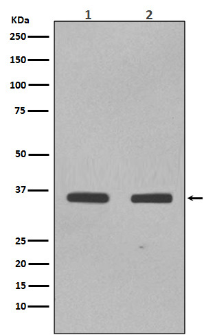
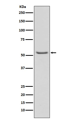
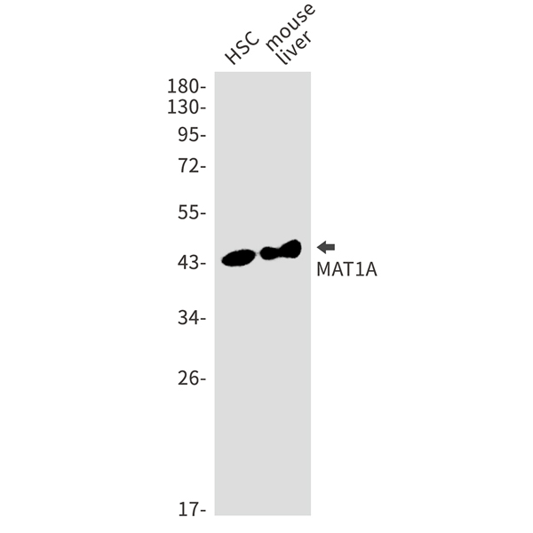 WB (Western Blot)
(Western blot analysis of MAT1A in HSC, mouse liver lysates using MAT1A antibody.)
WB (Western Blot)
(Western blot analysis of MAT1A in HSC, mouse liver lysates using MAT1A antibody.)
MAT1A, Monoclonal Antibody (Cat# AAA314782)
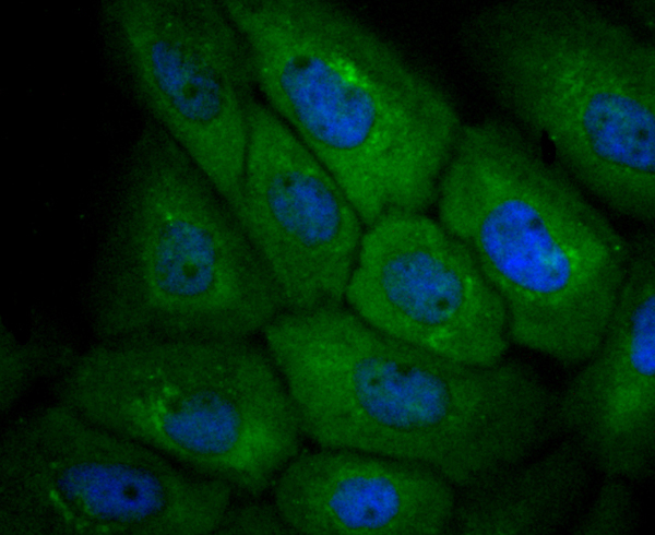
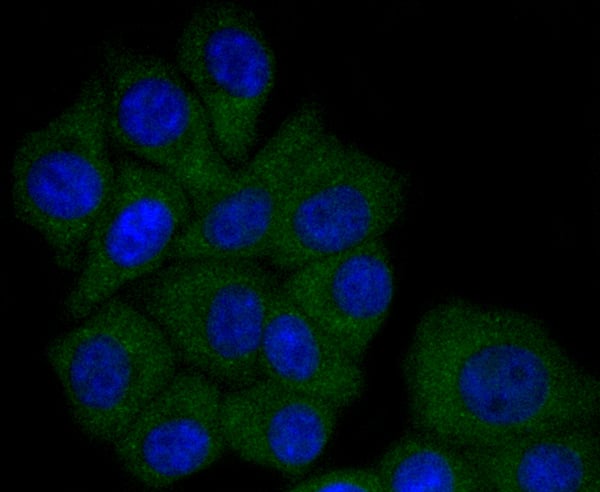
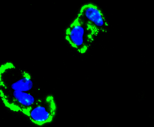
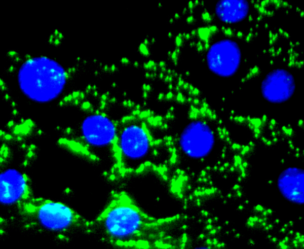
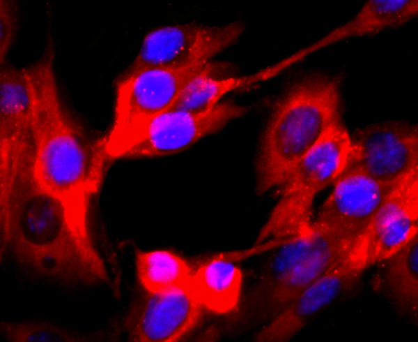
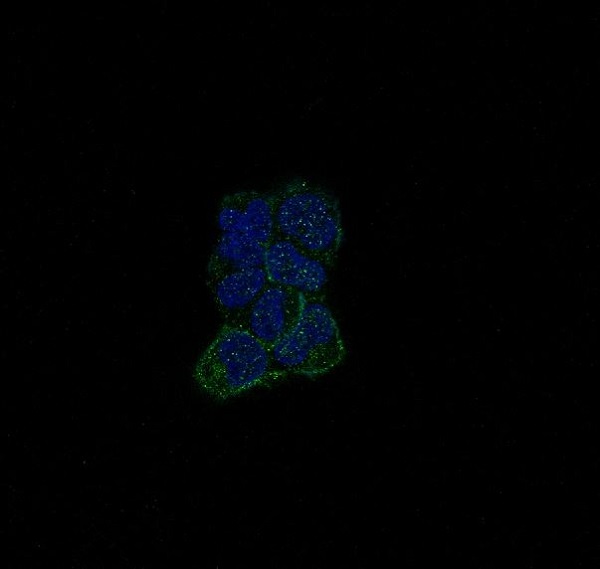
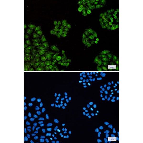
 ICC (Immunocytochemistry)
(Immunocytochemistry analysis of PFKP(green) in Hela using PFKP antibody,and DAPI(blue))
ICC (Immunocytochemistry)
(Immunocytochemistry analysis of PFKP(green) in Hela using PFKP antibody,and DAPI(blue))
PFKP, Monoclonal Antibody (Cat# AAA314791)

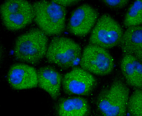
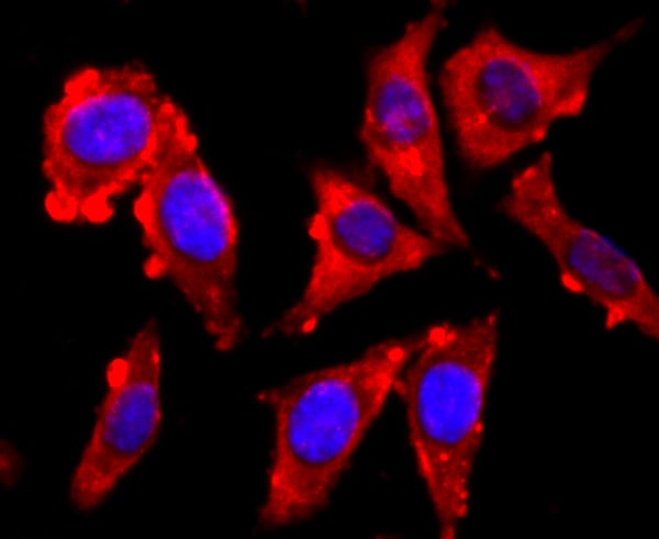
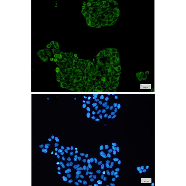
 ICC (Immunocytochemistry)
(Immunocytochemistry analysis of TTF2(green) in Hela using TTF2 antibody,and DAPI(blue))
ICC (Immunocytochemistry)
(Immunocytochemistry analysis of TTF2(green) in Hela using TTF2 antibody,and DAPI(blue))
Transcription Termination Factor 2, Monoclonal Antibody (Cat# AAA314810)
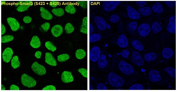 IF (Immunofluorescence)
(Immunofluorescent analysis of A549 cells treated with TGF?? , using Phospho-Smad3 (S423 + S425) Antibody.)
IF (Immunofluorescence)
(Immunofluorescent analysis of A549 cells treated with TGF?? , using Phospho-Smad3 (S423 + S425) Antibody.)
Smad3, Monoclonal Recombinant Antibody (Cat# AAA314843)

 IHC (Immunohiostchemistry)
(Immunohistochemistry analysis of paraffin-embedded Human breast cancer using BTF3 antibody.High-pressure and temperature Sodium Citrate pH 6.0 was used for antigen retrieval.)
IHC (Immunohiostchemistry)
(Immunohistochemistry analysis of paraffin-embedded Human breast cancer using BTF3 antibody.High-pressure and temperature Sodium Citrate pH 6.0 was used for antigen retrieval.)
BTF3, Monoclonal Antibody (Cat# AAA314759)
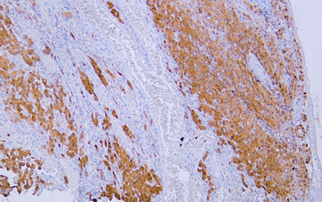
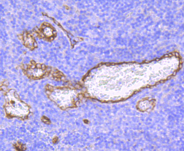
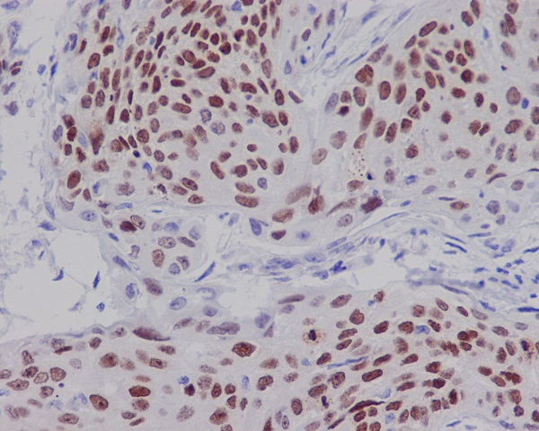
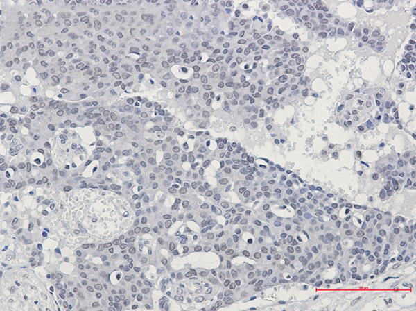
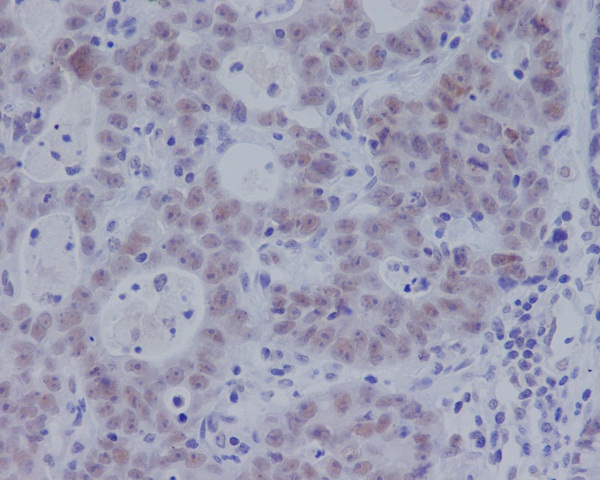
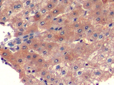
 IHC (Immunohistochemistry)
(Human testis tissue was stained with anti-Calretinin(ABT-Calret 1) antibody.)
IHC (Immunohistochemistry)
(Human testis tissue was stained with anti-Calretinin(ABT-Calret 1) antibody.)
Calretinin, Monoclonal Antibody (Cat# AAA320702)
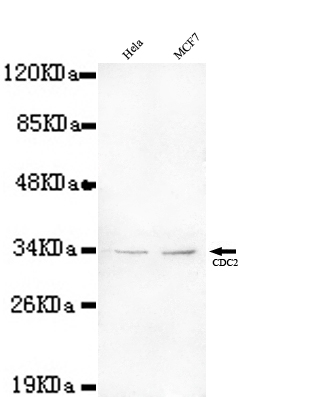
 Application Data
(HeLa cells using anti- CDC2 antibody diluted 1:50)
Application Data
(HeLa cells using anti- CDC2 antibody diluted 1:50)
CDC2, Monoclonal Antibody (Cat# AAA300808)
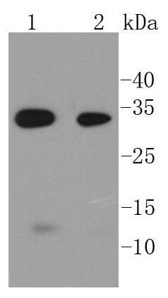
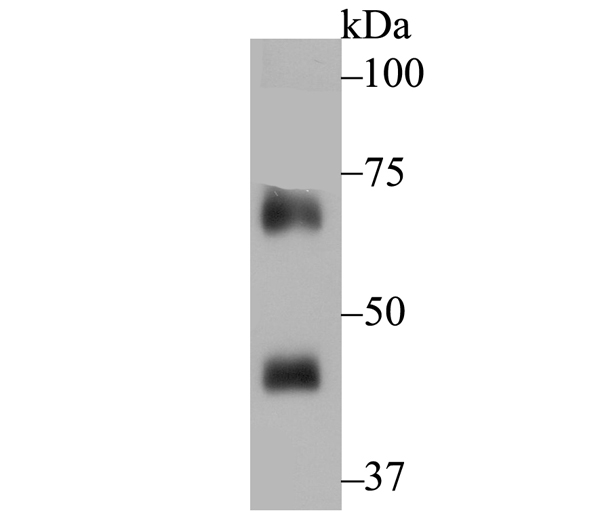
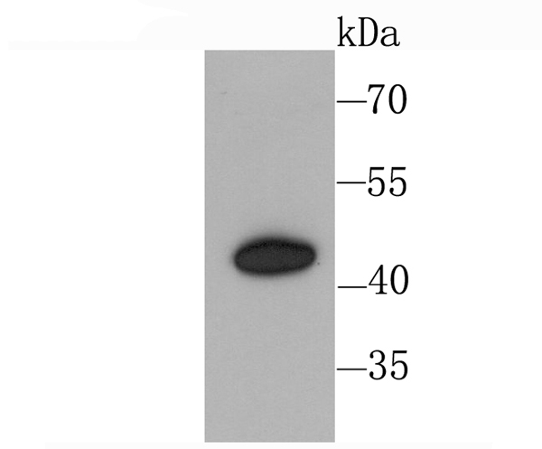
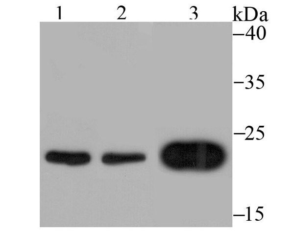
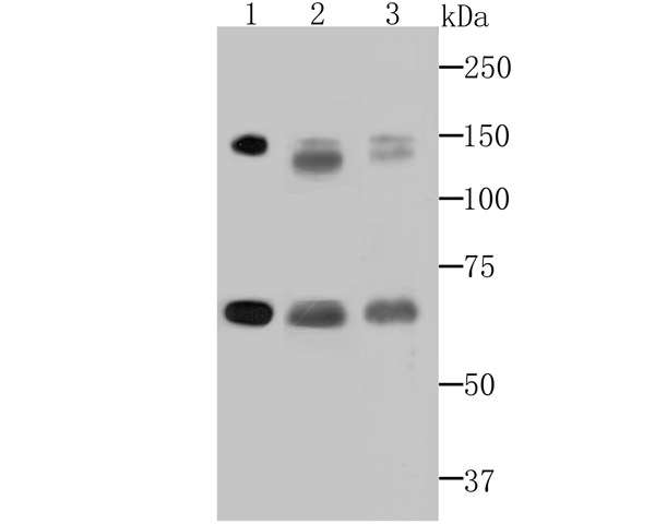
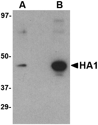 WB (Western Blot)
(Western blot analysis of (A) 5 ng and (B) 25 ng of recombinant HA1 with Hemagglutinin antibody at 1 ug/mL.)
WB (Western Blot)
(Western blot analysis of (A) 5 ng and (B) 25 ng of recombinant HA1 with Hemagglutinin antibody at 1 ug/mL.)
Hemagglutinin, Monoclonal Antibody (Cat# AAA300830)

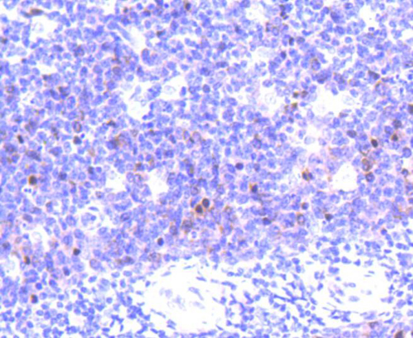
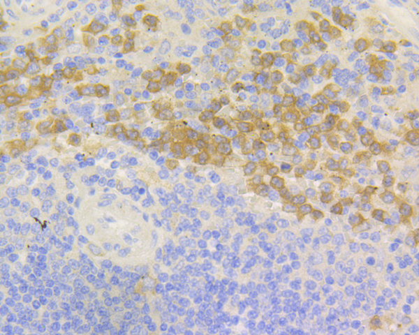
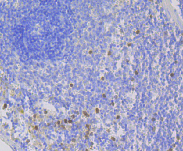
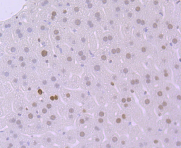
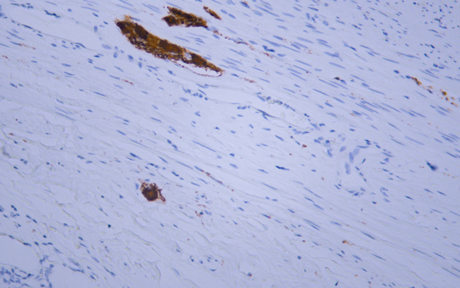
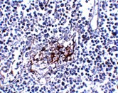 IHC (Immunohiostchemistry)
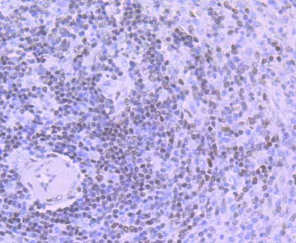
(Immunohistochemistry of IL-33 in human lymph node tissue with IL-33 antibody at 5 ug/mL.)
IHC (Immunohiostchemistry)
(Immunohistochemistry of IL-33 in human lymph node tissue with IL-33 antibody at 5 ug/mL.)
IL-33, Monoclonal Antibody (Cat# AAA300835)
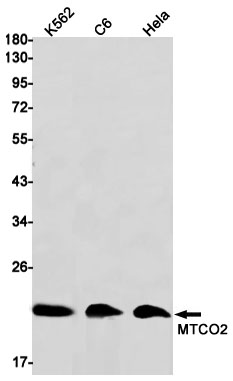
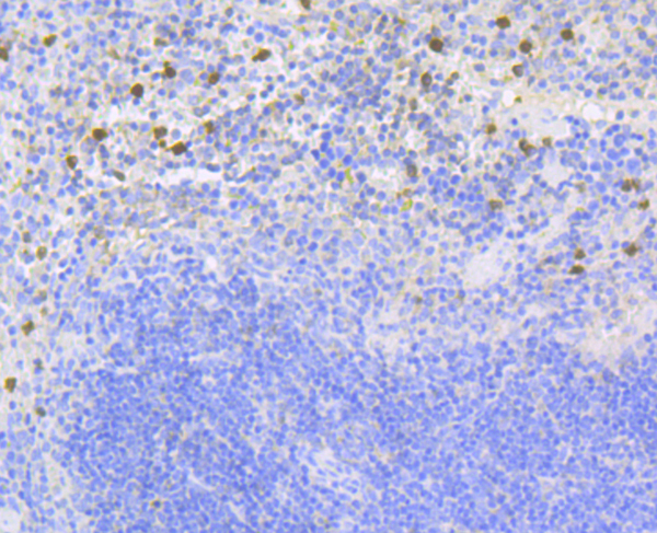
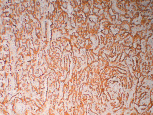
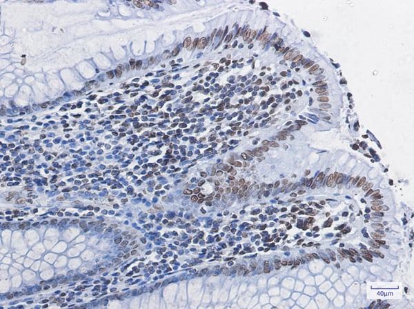
 IHC (Immunohiostchemistry)
(Immunohistochemistry of MTCO2 in paraffin-embedded Human tonsil using MTCO2 Rabbit mAb at dilution 1/50)
IHC (Immunohiostchemistry)
(Immunohistochemistry of MTCO2 in paraffin-embedded Human tonsil using MTCO2 Rabbit mAb at dilution 1/50)
MTCO2, Monoclonal Antibody (Cat# AAA314415)
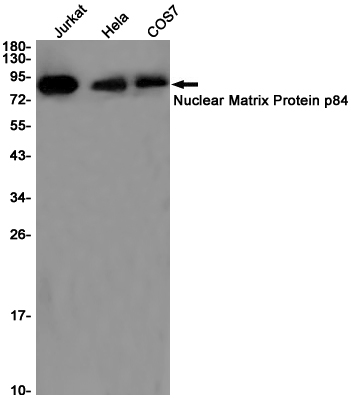
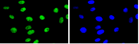
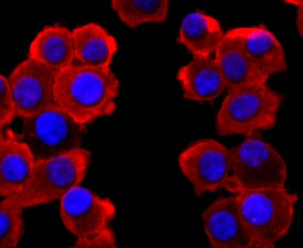
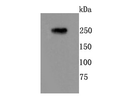
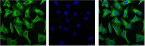
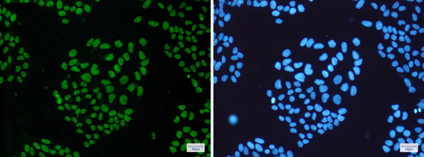 IF (Immunofluorescence)
(Immunofluorescence of Nuclear Matrix Protein p84(green) in Hela cells using Nuclear Matrix Protein p84 Rabbit mAb at dilution 1/50, and DAPI(blue))
IF (Immunofluorescence)
(Immunofluorescence of Nuclear Matrix Protein p84(green) in Hela cells using Nuclear Matrix Protein p84 Rabbit mAb at dilution 1/50, and DAPI(blue))
THO Complex Subunit 1, Monoclonal Antibody (Cat# AAA314426)
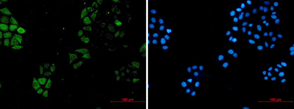
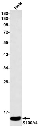
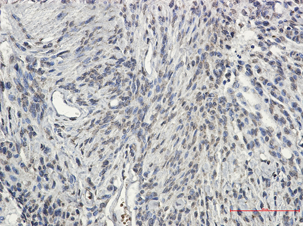
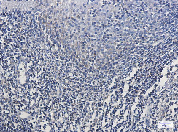
 IHC (Immunohistochemisry)
(Immunohistochemistry of S100A4 in paraffin-embedded Human lung cancer tissue using S100A4 Rabbit mAb at dilution 1/50)
IHC (Immunohistochemisry)
(Immunohistochemistry of S100A4 in paraffin-embedded Human lung cancer tissue using S100A4 Rabbit mAb at dilution 1/50)
S100A4, Monoclonal Antibody (Cat# AAA314454)
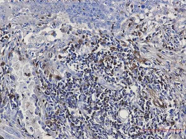
 IHC (Immunohiostchemistry)
(Immunohistochemistry of SCIN in paraffin-embedded Human colon cancer tissue using SCIN Rabbit mAb at dilution 1/50)
IHC (Immunohiostchemistry)
(Immunohistochemistry of SCIN in paraffin-embedded Human colon cancer tissue using SCIN Rabbit mAb at dilution 1/50)
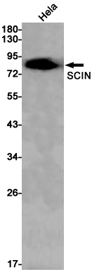
SCIN, Monoclonal Antibody (Cat# AAA314455)
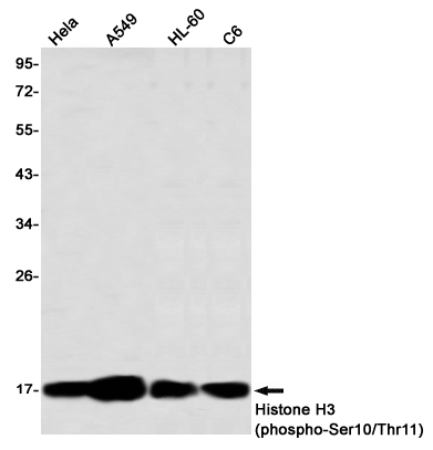
 IHC (Immunohiostchemistry)
(Immunohistochemistry of Histone H3 (phospho-Ser10/Thr11) in paraffin-embedded Human lung cancer tissue using Histone H3 (phospho-Ser10/Thr11) Rabbit mAb at dilution 1/50)
IHC (Immunohiostchemistry)
(Immunohistochemistry of Histone H3 (phospho-Ser10/Thr11) in paraffin-embedded Human lung cancer tissue using Histone H3 (phospho-Ser10/Thr11) Rabbit mAb at dilution 1/50)
Histone H3, Monoclonal Antibody (Cat# AAA314502)
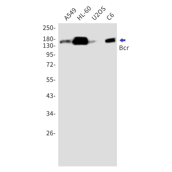
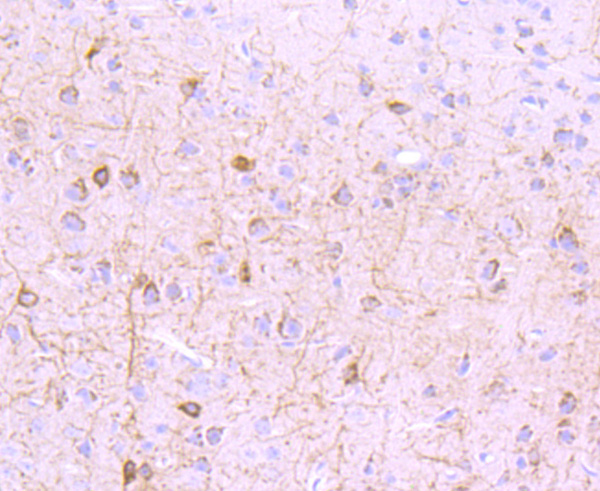
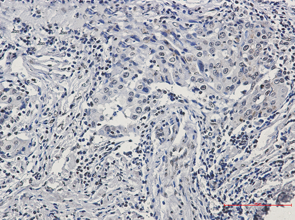
 IHC (Immunohiostchemistry)
(Immunohistochemistry of Bcr in paraffin-embedded Human Brain using Bcr Rabbit mAb at dilution 1/50)
IHC (Immunohiostchemistry)
(Immunohistochemistry of Bcr in paraffin-embedded Human Brain using Bcr Rabbit mAb at dilution 1/50)
Bcr, Monoclonal Antibody (Cat# AAA314510)
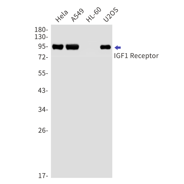
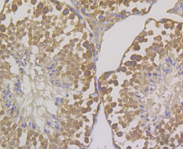
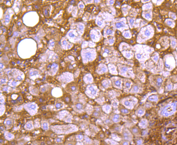
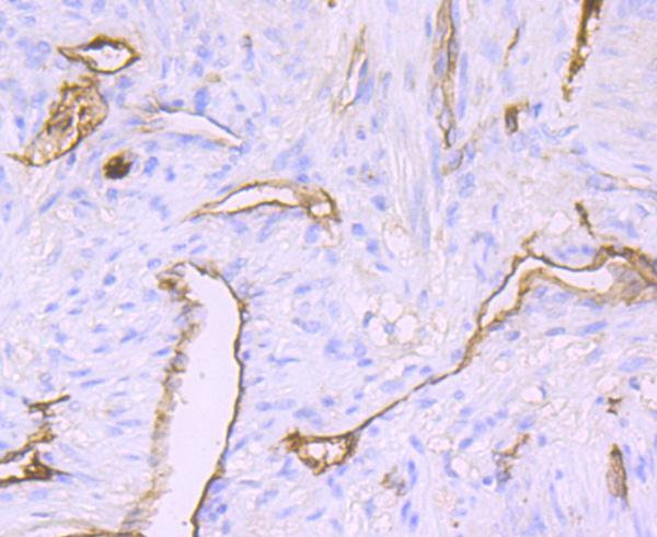
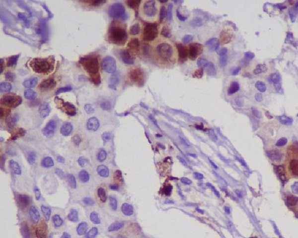
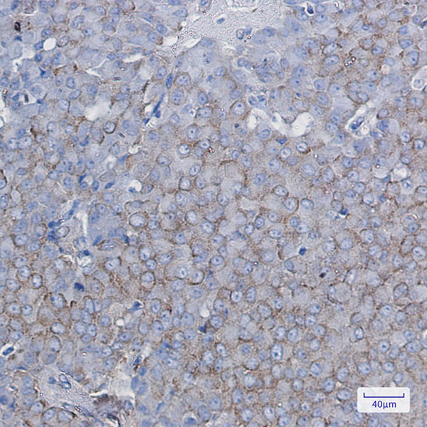
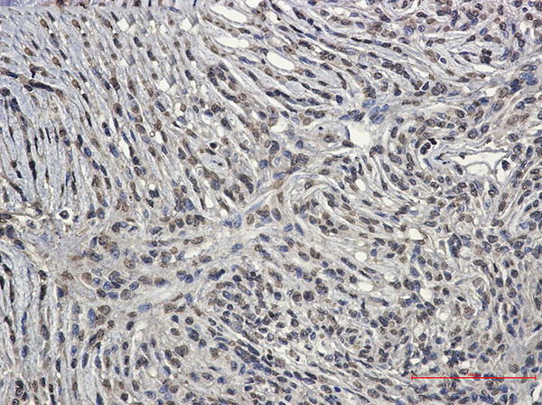
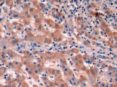
 IHC (Immunohiostchemistry)
(Immunohistochemical of IGF1 Receptor in Human lung cancer tissue using IGF1 Receptor antibody at dilution 1/50)
IHC (Immunohiostchemistry)
(Immunohistochemical of IGF1 Receptor in Human lung cancer tissue using IGF1 Receptor antibody at dilution 1/50)
IGF1 Receptor, Monoclonal Antibody (Cat# AAA314537)
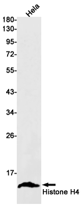
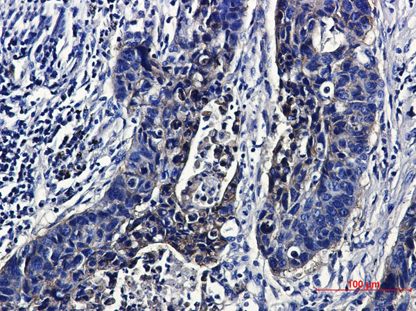
 IHC (Immunohiostchemistry)
(Immunohistochemistry of Histone H4 in paraffin-embedded Human colon cancer tissue using Histone H4 Rabbit mAb at dilution 1/100)
IHC (Immunohiostchemistry)
(Immunohistochemistry of Histone H4 in paraffin-embedded Human colon cancer tissue using Histone H4 Rabbit mAb at dilution 1/100)
Histone H4, Monoclonal Antibody (Cat# AAA314376)
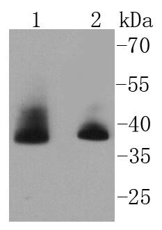
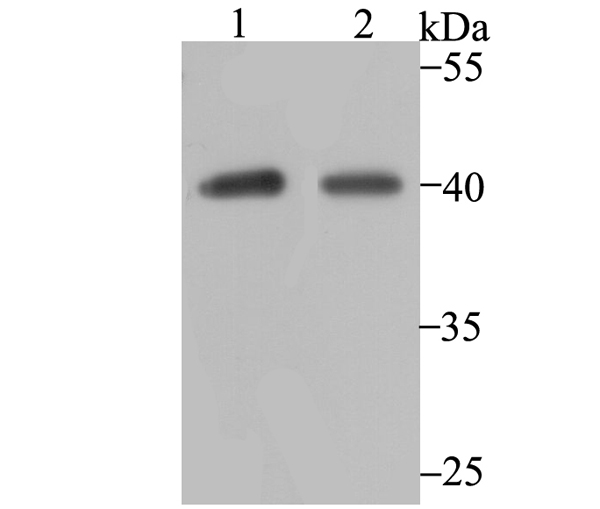
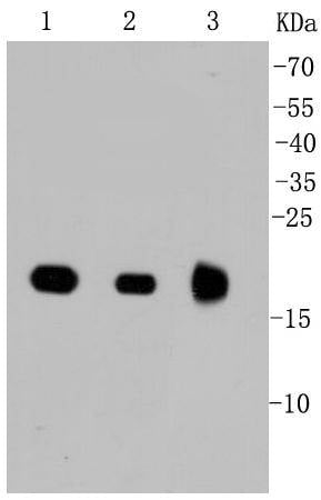
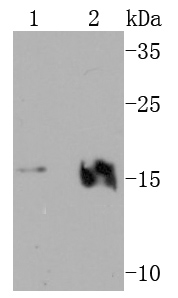
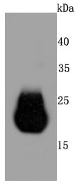
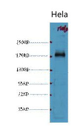
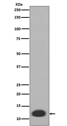
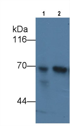 WB (Western Blot)
(Western Blot; Sample: Lane1: Human Liver lysate; Lane2: Human Lung lysate Primary Ab: 3ug/ml Mouse AntiHuman aFP Antibody Second Ab: 0.2ug/mL HRPLinked Caprine AntiMouse IgG Polyclonal Antibody (Catalog: SAA544Mu19))
WB (Western Blot)
(Western Blot; Sample: Lane1: Human Liver lysate; Lane2: Human Lung lysate Primary Ab: 3ug/ml Mouse AntiHuman aFP Antibody Second Ab: 0.2ug/mL HRPLinked Caprine AntiMouse IgG Polyclonal Antibody (Catalog: SAA544Mu19))
AlphaFetoprotein (AFP), Monoclonal Antibody (Cat# AAA151495)
What are Monoclonal Antibodies?
Monoclonal antibodies are specialized laboratory-produced proteins developed for binding to specific biological antigens or other molecular targets. Since they come from a single cell (or clone), they are especially consistent and accurate in the data they are involved in producing.
This type of antibody material has been shown to be a powerful tool in finding and subsequently destroying harmful cells in an organism, such as those found in cancers or various autoimmune diseases. This makes them excellent aids in medical testing and research, which is why they are so widely used.
AAA Biotech offers a comprehensive range of high-quality monoclonal antibodies that perform effectively in various laboratory tests, including (amongst others) ELISA, western blotting, immunohistochemistry, and flow cytometry. All of the products in our catalog are thoroughly quality tested to make sure that they are reliable and will consistently perform well in your research.
What Are The Uses of Monoclonal Antibodies
Monoclonal antibodies are used in many lab tests, including (amongst others) ELISA, western blotting, immunohistochemistry, and flow cytometry.
ELISA is a test that helps detect a specific substance/analyte in a sample. It uses antibodies (often monoclonal) bound to a solid surface (such as the well of a microplate) to “capture” the substance/analyte in the sample and immobilize it so that the detection antibody component can then bind to it and produce a signal, which can then be measured.
Western blotting identifies specific proteins in a sample. The sample is first separated on a gel, and then antibodies are applied that will typically bind to the target, which will all be localized to a single band in a lane.
Immunohistochemistry helps locate specific proteins in cells or tissue samples using antibodies.
Flow cytometry looks at and sorts cells. It uses antibodies that are conjugated to reporter molecules called “fluorophores”, which, under special lights, emit light themselves, which can then be measured by a detector instrument.
How Monoclonal Antibodies Are Used as Medicine?
Please note that all of the products listed in AAA Biotech’s also known as AAA Bio or AAABio catalog are strictly for research-use only (RUO).
Monoclonal antibodies can also be used as therapeutic/medical treatments, particularly in the context of cancers. They are designed to find and bind to specific cells or proteins, helping the immune system recognize and attack the cancer. These treatments work in different ways, such as:
- Radioimmunotherapy attaches a small amount of radioactive molecule to the antibody, so it delivers the radiation directly to the cancer cells that the antibody is specifically binding to.
- Antibody-directed enzyme prodrug therapy uses antibodies that are specifically bound to special enzymes. These enzymes activate a harmless drug in the body and turn it into a cancer-killing drug only near the cancer cells—this helps avoid harming healthy cells.
- Immunoliposomes are tiny “bubbles” filled with medicine/drug and coated with antibodies. They carry the drug straight to the cancer cells.
Why Buy Monoclonal Antibodies From Us?
At AAA Biotech, we provide high-performance monoclonal antibodies designed to support a wide range of research needs.
1. Validated for Versatile Applications
The antibodies in our catalog are extensively validated and compatible with multiple techniques, including (but not limited to) ELISA, flow cytometry (FC), immunocytochemistry (ICC), immunofluorescence (IF), immunohistochemistry (IHC), immunoprecipitation (IP), and western blotting (WB).
2. Wide Selection & Specialized Options
We offer antibodies for common and rare species, that are available in various conjugated forms, and also in recombinant formats. Essentially, there is almost anything one might need to meet their experimental model’s requirements.
3. High-Quality Proteins
Our proteins meet high purity standards—90% or more as confirmed by SDS-PAGE. Many are available with tags like His, Flag, GST, or MBP, and we also supply native and biologically active proteins for functional studies.
Frequently Asked Questions
1. Are your monoclonal antibodies validated for specific applications?
Yes, our antibodies are tested and validated for use in methods such as ELISA, western blot, IHC, flow cytometry, and more. Refer to specific product pages or datasheets for individual product information.
2. How do I choose the right monoclonal antibody for my application?
Review the product details directly for application validation, species reactivity, and target information. You may also contact our support team at any time for help.
3. How quickly can I receive my order?
Most orders are processed and shipped within 1–3 business days, depending on product availability and your shipping location.