Filters
▼Clonality
▼Type
▼Reactivity
▼Gene Name
▼Isotype
▼Host
▼Application
▼Clone
▼Monoclonal Antibodies
Get accurate results in your research with our Monoclonal Antibodies, which are specially made to target exactly what you require for your research, and will produce consistent, reliable performance in lab tests.
Viewing 9600-9650 of 27597 product results
 WB (Western Blot)
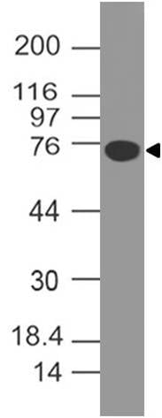
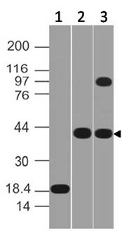
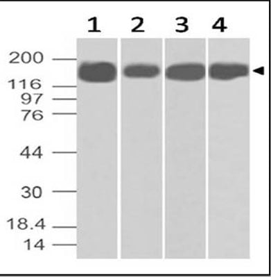
(Fig-4: Western blot analysis of B7-H4. Anti-B7-H4 antibody (Clone: ABM53A6) was used at 2 ug/ml on (1) HCT-116, (2) PC3, (3) Kato 111, (4) C2C12 and (5) RAW Lysates.)
WB (Western Blot)
(Fig-4: Western blot analysis of B7-H4. Anti-B7-H4 antibody (Clone: ABM53A6) was used at 2 ug/ml on (1) HCT-116, (2) PC3, (3) Kato 111, (4) C2C12 and (5) RAW Lysates.)
NALE B7-H4, Monoclonal Antibody (Cat# AAA78562)
 Application Data
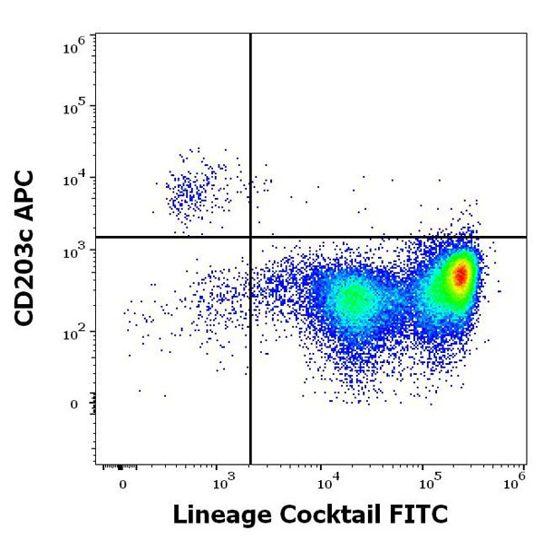
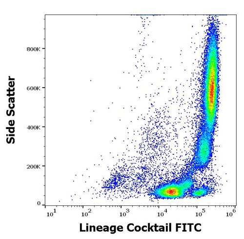
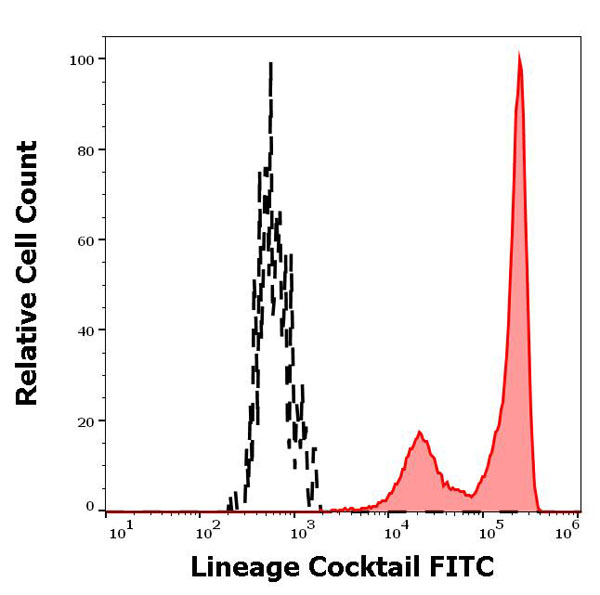
(Figure-3: Separation of human target population cells (red-filled) from basophils (black-dashed) in flow cytometry analysis (surface staining) of human peripheral whole blood stained using Lineage Cocktail FITC (20 ul cocktail/100 ul of peripheral whole blood).)
Application Data
(Figure-3: Separation of human target population cells (red-filled) from basophils (black-dashed) in flow cytometry analysis (surface staining) of human peripheral whole blood stained using Lineage Cocktail FITC (20 ul cocktail/100 ul of peripheral whole blood).)
Lineage Cocktail (CD3/CD14/CD16/CD19/CD20/CD56), Monoclonal Assay Kit (Cat# AAA78592)
 Application Data
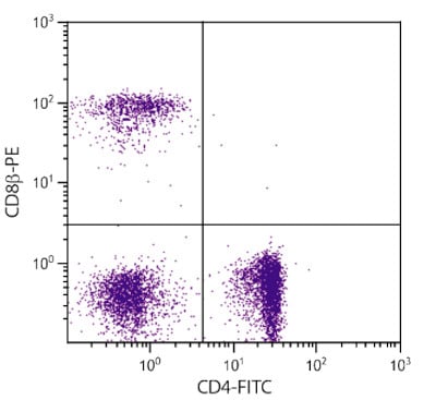
(Chicken peripheral blood lymphocytes were stained with Mouse Anti-Chicken CD8Β-PE and Mouse Anti-Chicken CD4-FITC .)
Application Data
(Chicken peripheral blood lymphocytes were stained with Mouse Anti-Chicken CD8Β-PE and Mouse Anti-Chicken CD4-FITC .)
CD8 beta, Monoclonal Antibody (Cat# AAA78705)
c-Kit, Monoclonal Antibody (Cat# AAA78707)
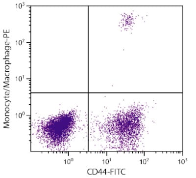 Application Data
(Chicken peripheral blood monocytes were stained with Mouse Anti-chicken Monocyte/Macrophage-PE (AAA78709) and Mouse Anti-Chicken CD44-FITC .)
Application Data
(Chicken peripheral blood monocytes were stained with Mouse Anti-chicken Monocyte/Macrophage-PE (AAA78709) and Mouse Anti-Chicken CD44-FITC .)
Monocyte/Macrophage, Monoclonal Antibody (Cat# AAA78709)
Mouse Anti-Human IgM (u chain specific), Monoclonal Secondary Antibody (Cat# AAA78712)
Mouse Anti-Human IgG4 (gamma 4 chain specific), Monoclonal Secondary Antibody (Cat# AAA78734)
 IHC (Immunohistochemistry)
(High grade squamous intraepithelial lesion of the uterine cervix stained with mouse anti-p16 (clone R15-A) monoclonal antibody, shows significant nuclear and cytoplasmic positivity of target cells. Formalin fixed, paraffin embedded human tissues (4um sections) stained according to related datasheet.)
IHC (Immunohistochemistry)
(High grade squamous intraepithelial lesion of the uterine cervix stained with mouse anti-p16 (clone R15-A) monoclonal antibody, shows significant nuclear and cytoplasmic positivity of target cells. Formalin fixed, paraffin embedded human tissues (4um sections) stained according to related datasheet.)
p16, Monoclonal Antibody (Cat# AAA79022)
Rat Anti-Mouse IgG2b, Monoclonal Secondary Antibody (Cat# AAA78878)
Mouse IgM Isotype Control, Monoclonal Isotype Control (Cat# AAA78884)
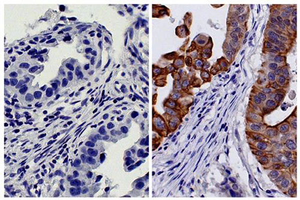 IHC (Immunohistochemistry)
(Paraffin embedded human gastric cancer tissue was stained with Mouse IgG2b-HRP isotype control and Mouse Anti-Cytokeratin 18-HRP followed by DAB and hematoxylin)
IHC (Immunohistochemistry)
(Paraffin embedded human gastric cancer tissue was stained with Mouse IgG2b-HRP isotype control and Mouse Anti-Cytokeratin 18-HRP followed by DAB and hematoxylin)
Mouse IgG2b Isotype Control, Monoclonal Isotype Control (Cat# AAA78892)
 WB (Western Blot)
(Western blot analysis of different tissue and cell lysates using mouse mAb to GAP43, MO22171, dilution 1:2,000, in green: [1] protein standard (red), [2] rat brain, [3] rat spinal cord, [4] mouse brain, [5] mouse spinal cord, [6] C6 cells, [7] SH-SY5Y cells. Single band at 43 kDa mark correspond to GAP43 protein.)
WB (Western Blot)
(Western blot analysis of different tissue and cell lysates using mouse mAb to GAP43, MO22171, dilution 1:2,000, in green: [1] protein standard (red), [2] rat brain, [3] rat spinal cord, [4] mouse brain, [5] mouse spinal cord, [6] C6 cells, [7] SH-SY5Y cells. Single band at 43 kDa mark correspond to GAP43 protein.)
GAP43, Monoclonal Antibody (Cat# AAA76611)
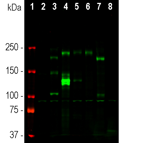 WB (Western Blot)
(Western blot analysis of different tissue and cell lysates using mouse mAb to ankyrin 3, MO22185, dilution 1:2,000 in green: [1] protein standard (red), [2] NIH-3T3, [3] C6, [4] HEK293, [5] HeLa, [6] SH-SY5Y cells, [7] rat brain, and [8] mouse brain lysates.)
WB (Western Blot)
(Western blot analysis of different tissue and cell lysates using mouse mAb to ankyrin 3, MO22185, dilution 1:2,000 in green: [1] protein standard (red), [2] NIH-3T3, [3] C6, [4] HEK293, [5] HeLa, [6] SH-SY5Y cells, [7] rat brain, and [8] mouse brain lysates.)
Ankyrin 3, Monoclonal Antibody (Cat# AAA76629)
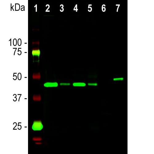
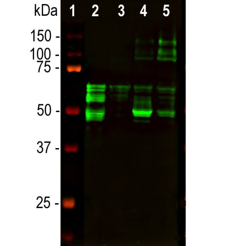 WB (Western Blot)
(Western blot analysis of tissue lysates using mouse mAb to MAP-tau, MO22191, dilution 1:2,000 in green: [1] protein standard (red), [2] rat brain, [3] rat spinal cord, [4] mouse brain, [5] mouse spinal cord. Tau protein is expressed as up to 9 different isoforms of different molecular weight, and so appears as multiple closely spaced bands in the region of the blot from 48kDa to 67kDa with some higher molecular weight "big tau" Isoforms.)
WB (Western Blot)
(Western blot analysis of tissue lysates using mouse mAb to MAP-tau, MO22191, dilution 1:2,000 in green: [1] protein standard (red), [2] rat brain, [3] rat spinal cord, [4] mouse brain, [5] mouse spinal cord. Tau protein is expressed as up to 9 different isoforms of different molecular weight, and so appears as multiple closely spaced bands in the region of the blot from 48kDa to 67kDa with some higher molecular weight "big tau" Isoforms.)
Tau, Monoclonal Antibody (Cat# AAA76633)
 WB (Western Blot)
(Western blot analysis of various HEK293 cell lysates using mouse mAb to mCherry, dilution 1:5,000, in green. [1] protein standard, [2] untransfected HEK293 lysate, [3] lysate of HEK293 cells transfected with mCherry-HA construct, and [4] lysate of HEK293 cells transfected with an acGFP construct.)
WB (Western Blot)
(Western blot analysis of various HEK293 cell lysates using mouse mAb to mCherry, dilution 1:5,000, in green. [1] protein standard, [2] untransfected HEK293 lysate, [3] lysate of HEK293 cells transfected with mCherry-HA construct, and [4] lysate of HEK293 cells transfected with an acGFP construct.)
mCherry, Monoclonal Antibody (Cat# AAA76634)
 IHC (Immunohistochemisry)
(Immunohistochemistry analysis of formalin-fixed, paraffin-embedded breast cancer tissue stained with 2ug/ml anti-OPN 53.)
IHC (Immunohistochemisry)
(Immunohistochemistry analysis of formalin-fixed, paraffin-embedded breast cancer tissue stained with 2ug/ml anti-OPN 53.)
Osteopontin, Monoclonal Antibody (Cat# AAA76990)
 Application Data
(Figure: Staining of endogenous NLRP3/NALP3 in epithelial layer of human tonsil (frozen section) using NLRP3/NALP3 (human), mAb (Nalpy3-b) . Method: 5uM frozen section of tissue are dried and fixed with aceton. Tissue is washed with PBS and incubated with NLRP3/NALP3 (human), mAb (Nalpy3-b). Picture courtesy of Dr. Alain Kummer, University Medical Center Utrecht, The Netherlands.)
Application Data
(Figure: Staining of endogenous NLRP3/NALP3 in epithelial layer of human tonsil (frozen section) using NLRP3/NALP3 (human), mAb (Nalpy3-b) . Method: 5uM frozen section of tissue are dried and fixed with aceton. Tissue is washed with PBS and incubated with NLRP3/NALP3 (human), mAb (Nalpy3-b). Picture courtesy of Dr. Alain Kummer, University Medical Center Utrecht, The Netherlands.)
NLRP3/NALP3, Monoclonal Antibody (Cat# AAA76991)
 Application Data
6xHIS Tag, Monoclonal Antibody (Cat# AAA76814)
STX2, Monoclonal Antibody (Cat# AAA76827)
Progesterone, Monoclonal Antibody (Cat# AAA76837)
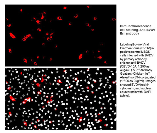
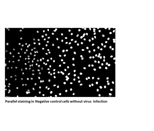 Application Data
BVDV, Monoclonal Antibody (Cat# AAA76850)
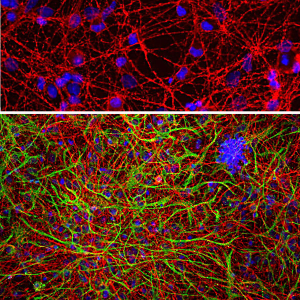
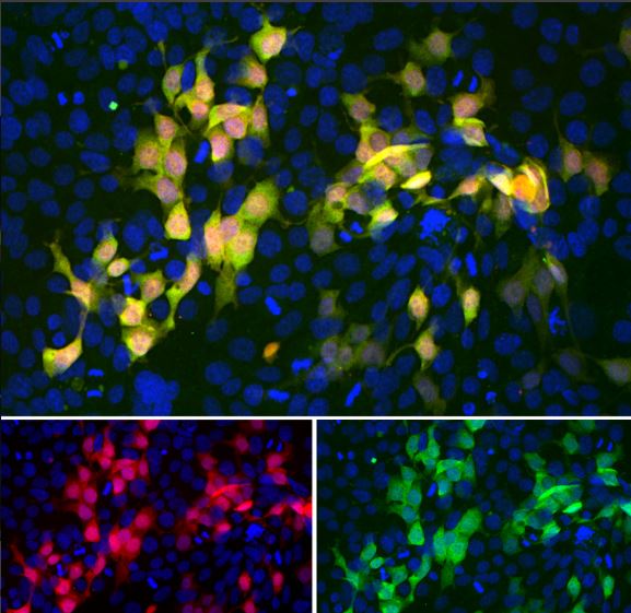
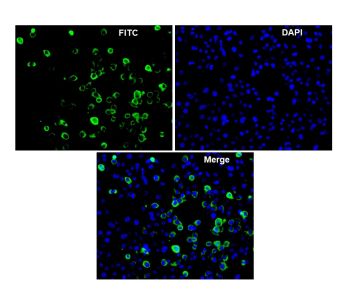
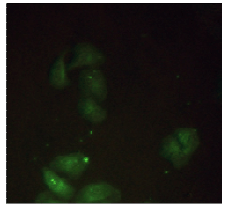
 IF (Immunofluorescence)
Epididymis Protein 4 (HE4), Monoclonal Antibody (Cat# AAA76863)
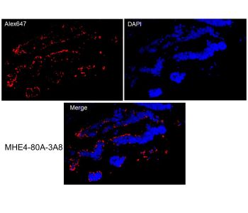
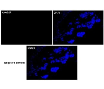
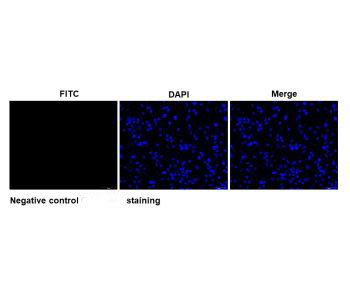 IF (Immunofluorescence)
Calicivirus, Monoclonal Antibody (Cat# AAA76868)
CLIMP-63, Monoclonal Antibody (Cat# AAA77058)
NT-proBNP, Monoclonal Antibody (Cat# AAA77059)
Zika Virus (ZIKV-NS1), Monoclonal Antibody (Cat# AAA77066)
Purified
Immunodeficiency Virus 1 p24 Ab, Monoclonal Antibody (Cat# AAA77069)
Creatine Kinase MB (CK-MB), Monoclonal Antibody (Cat# AAA77070)
Cytomegalovirus (CMV), Monoclonal Antibody (Cat# AAA77097)
CBD, Monoclonal Antibody (Cat# AAA79335)
 FCM/FACS (Flow Cytometry)
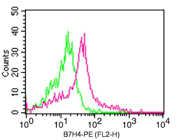
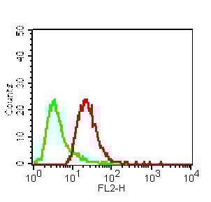
(Fig-2: Cell Surface flow analysis of hCD86 in Raji using 1 ug/10^6 cells. Green represents isotype control ; red represents anti-hCD86 antibody. Goat anti-mouse PE conjugated secondary antibody was used.)
FCM/FACS (Flow Cytometry)
(Fig-2: Cell Surface flow analysis of hCD86 in Raji using 1 ug/10^6 cells. Green represents isotype control ; red represents anti-hCD86 antibody. Goat anti-mouse PE conjugated secondary antibody was used.)
B7-2/CD86, Monoclonal Antibody (Cat# AAA78269)
Protein G Chromatography
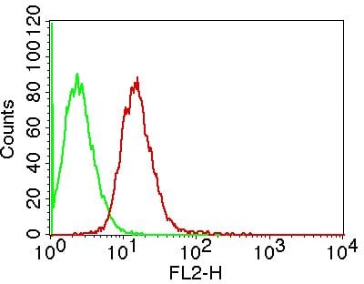
 FCM/FACS (Flow Cytometry)
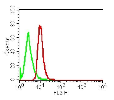
(Fig-1: Cell Surface flow analysis of CD13 in PBMC (Lymphocyte gated) using 1 ug/10^6 cells. Green represents isotype control ; red represents anti-CD13 antibody. Goat anti-mouse PE conjugated secondary antibody was used.)
FCM/FACS (Flow Cytometry)
(Fig-1: Cell Surface flow analysis of CD13 in PBMC (Lymphocyte gated) using 1 ug/10^6 cells. Green represents isotype control ; red represents anti-CD13 antibody. Goat anti-mouse PE conjugated secondary antibody was used.)
CD13, Monoclonal Antibody (Cat# AAA78270)
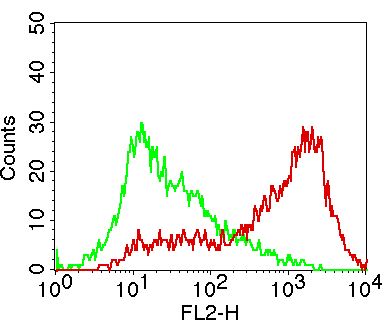
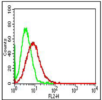
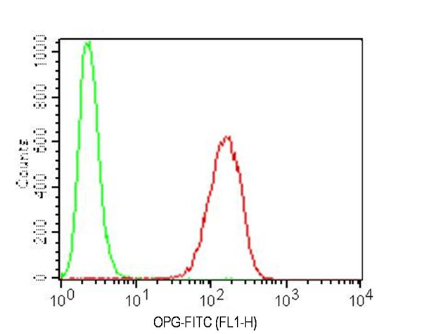 FCM/FACS (Flow Cytometry)
(Fig-2: Intracellular Flow analysis of OPG antibody on Ramos cells using 0.5 ug/ 10^6 cells of anti-OPG antibody (ABM10D2). Green represents isotype control; red represents anti-OPG antibody. Goat anti-mouse FITC conjugate was used as secondary antibody. (Cells were fixed with 4% paraformaldehyde for 10 min and washed with PBS by centrifuging at 1100 for 5 min followed by permeabilization for 20 min and washed again as mentioned above. Then cell were incubated with primary antibody for 45 min. and after washing the cells twice in PBS, incubated with conjugated secondary antibody for 30 min. Data acquisition was done after washing twice with PBS as mentioned above).)
FCM/FACS (Flow Cytometry)
(Fig-2: Intracellular Flow analysis of OPG antibody on Ramos cells using 0.5 ug/ 10^6 cells of anti-OPG antibody (ABM10D2). Green represents isotype control; red represents anti-OPG antibody. Goat anti-mouse FITC conjugate was used as secondary antibody. (Cells were fixed with 4% paraformaldehyde for 10 min and washed with PBS by centrifuging at 1100 for 5 min followed by permeabilization for 20 min and washed again as mentioned above. Then cell were incubated with primary antibody for 45 min. and after washing the cells twice in PBS, incubated with conjugated secondary antibody for 30 min. Data acquisition was done after washing twice with PBS as mentioned above).)
OPG, Monoclonal Antibody (Cat# AAA78280)
Protein G Chromatography
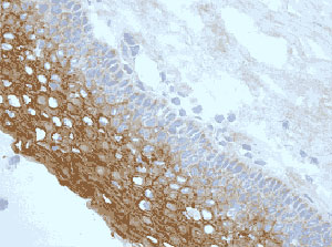
 IHC (Immunohistochemistry)
(Fig-1: Immunohistochemical analysis of PAPP-A in human Placenta tissue using PAPP-A antibody (Clone: ABM4C62) at 5 ug/ml.)
IHC (Immunohistochemistry)
(Fig-1: Immunohistochemical analysis of PAPP-A in human Placenta tissue using PAPP-A antibody (Clone: ABM4C62) at 5 ug/ml.)
PAPP-A, Monoclonal Antibody (Cat# AAA78282)
Protein G Chromatography
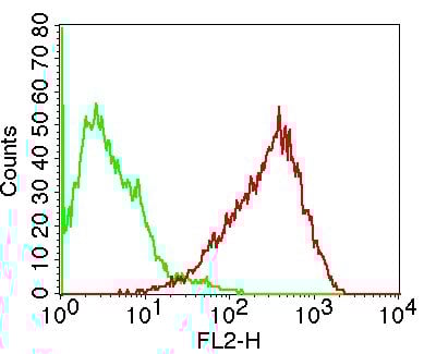
 FCM/FACS (Flow Cytometry)
(Figure-3: Intra cellular flow analysis of Msi-1 on HeLa cells using 2 ug/10^6 cells of Msi-1 antibody (Clone: ABM5D50). Green represents isotype control; red represents anti-Msi-1 antibody. Goat anti-mouse PE conjugate was used as secondary antibody.)
FCM/FACS (Flow Cytometry)
(Figure-3: Intra cellular flow analysis of Msi-1 on HeLa cells using 2 ug/10^6 cells of Msi-1 antibody (Clone: ABM5D50). Green represents isotype control; red represents anti-Msi-1 antibody. Goat anti-mouse PE conjugate was used as secondary antibody.)
MSi-1, Monoclonal Antibody (Cat# AAA78436)
Protein G Chromatography
 FCM/FACS (Flow Cytometry)
(Figure-2: Intracellular flowcytometric analysis of MAVS in MCF-7 cell line at 0.5 ug/10^6 cells using Anti-MAVS antibody. Green represent isotype control and red represent Anti-MAVS antibody. Goat Anti-mouse PE conjugated was used as secondary antibody.)
FCM/FACS (Flow Cytometry)
(Figure-2: Intracellular flowcytometric analysis of MAVS in MCF-7 cell line at 0.5 ug/10^6 cells using Anti-MAVS antibody. Green represent isotype control and red represent Anti-MAVS antibody. Goat Anti-mouse PE conjugated was used as secondary antibody.)
MAVS, Monoclonal Antibody (Cat# AAA78481)

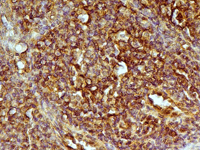 IHC (Immunohiostchemistry)
(Figure-2: Immunohistochemical analysis of CD24 in Human Tonsil tissue using Anti-CD24 antibody (Clone: ML5) at 5 ug/ml.)
IHC (Immunohiostchemistry)
(Figure-2: Immunohistochemical analysis of CD24 in Human Tonsil tissue using Anti-CD24 antibody (Clone: ML5) at 5 ug/ml.)
CD24, Monoclonal Antibody (Cat# AAA78486)
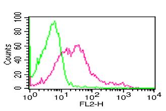
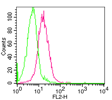 FCM/FACS (Flow Cytometry)
(Figure-1: Flow analysis of Raji cells. 2 ug of Antibody was used. Green mIgG1 isotype control, Red OCIL antibody.)
FCM/FACS (Flow Cytometry)
(Figure-1: Flow analysis of Raji cells. 2 ug of Antibody was used. Green mIgG1 isotype control, Red OCIL antibody.)
OCIL, Monoclonal Antibody (Cat# AAA78501)
IL-1beta, Monoclonal Antibody (Cat# AAA77897)
Does not show any cross reaction with recombinant human IL-1alpha, recombinant murine IL-1alpha or IL-1beta.
 FCM/FACS (Flow Cytometry)
(Above is the histogram of Hela cells stained with anti-Human B2MG mAb D3A2 and fluorescence labelled secondary antibody. Black line represents histogram of Hela cells stained with isotype control, anti-TB 38Kd antigen mAb B12F8)
FCM/FACS (Flow Cytometry)
(Above is the histogram of Hela cells stained with anti-Human B2MG mAb D3A2 and fluorescence labelled secondary antibody. Black line represents histogram of Hela cells stained with isotype control, anti-TB 38Kd antigen mAb B12F8)
Beta 2 Microglobulin, Monoclonal Antibody (Cat# AAA77900)
SAA, Monoclonal Antibody (Cat# AAA77908)
Does not show any cross-reaction with other human cytokines or growth factors tested such as IL-1beta, IL-8, MCAF, TGF-beta and EGF.
HCV NS-3, Monoclonal Antibody (Cat# AAA77912)
No cross reaction was found with synthetic recombinant core protein C + envelope protein M (residues 1-142 on HCV polyprotein), synthetic core protein C (residues 1 -61), synthetic NS-3 protein (residues 1378-
SAA, Monoclonal Antibody (Cat# AAA77917)
Does not show any cross-reaction with other human cytokines or growth factors tested such as IL-1beta, IL-8, MCAF, TGF-beta and EGF.
TSH, Monoclonal Antibody (Cat# AAA77924)
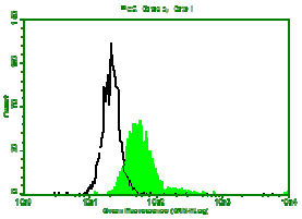
 Application Data
(Binding of anti-CD38/FITC to human Raji cells)
Application Data
(Binding of anti-CD38/FITC to human Raji cells)
CD38, Monoclonal Antibody (Cat# AAA78175)
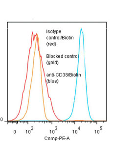
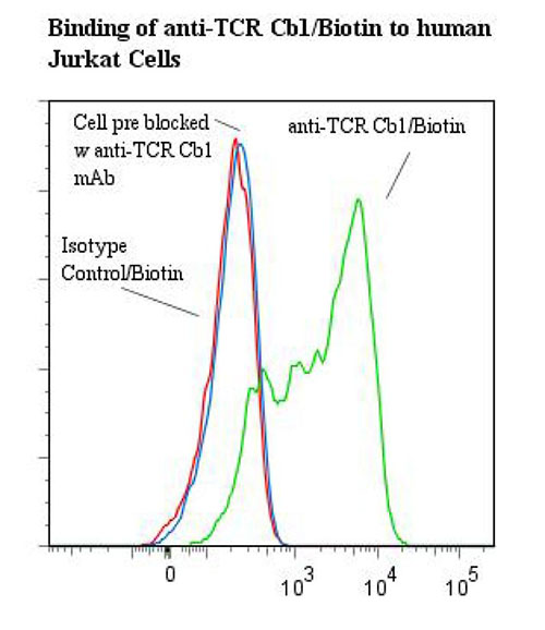 Application Data
TCR C beta 1, Monoclonal Antibody (Cat# AAA78183)
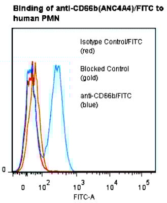 FCM/FACS (Flow Cytometry)
CD66b (CEACAM8), Monoclonal Antibody (Cat# AAA78197)
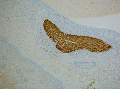
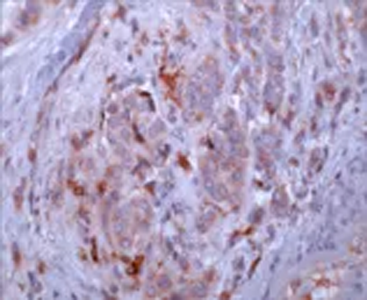
 IHC (Immunohiostchemistry)
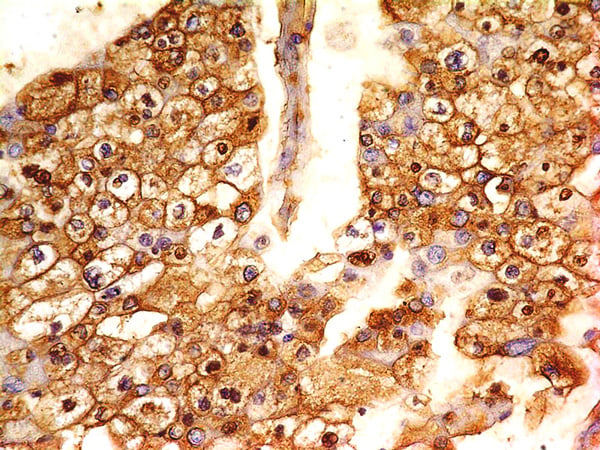
(Immunohistochemical analysis of Arginase-1 in human liver tissue using Arginase-1 antibody (Clone: ABM4B35) at 5 ug/ml.)
IHC (Immunohiostchemistry)
(Immunohistochemical analysis of Arginase-1 in human liver tissue using Arginase-1 antibody (Clone: ABM4B35) at 5 ug/ml.)
Arginase-1, Monoclonal Antibody (Cat# AAA78236)
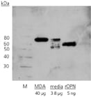
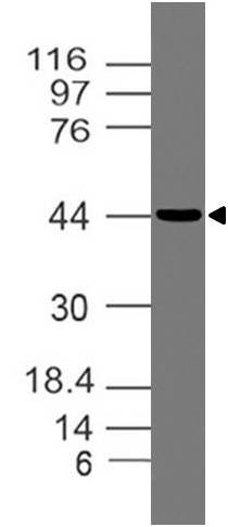
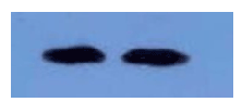
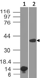
 WB (Western Blot)
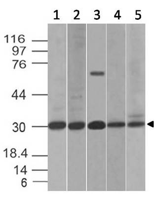
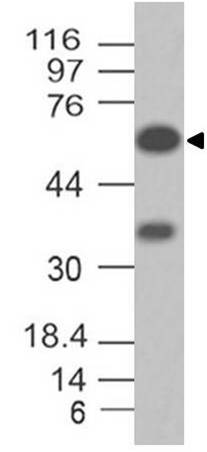
(Expression analysis of Topo II alpha. Anti-Topo II alpha antibody (Clone: ABM48B4) was tested at 1 ug/ml on (1) HT-29, (2) 293, (3) A549 and (4) Raji Lysates.)
WB (Western Blot)
(Expression analysis of Topo II alpha. Anti-Topo II alpha antibody (Clone: ABM48B4) was tested at 1 ug/ml on (1) HT-29, (2) 293, (3) A549 and (4) Raji Lysates.)
Topo II alpha, Monoclonal Antibody (Cat# AAA78248)
What are Monoclonal Antibodies?
Monoclonal antibodies are specialized laboratory-produced proteins developed for binding to specific biological antigens or other molecular targets. Since they come from a single cell (or clone), they are especially consistent and accurate in the data they are involved in producing.
This type of antibody material has been shown to be a powerful tool in finding and subsequently destroying harmful cells in an organism, such as those found in cancers or various autoimmune diseases. This makes them excellent aids in medical testing and research, which is why they are so widely used.
AAA Biotech offers a comprehensive range of high-quality monoclonal antibodies that perform effectively in various laboratory tests, including (amongst others) ELISA, western blotting, immunohistochemistry, and flow cytometry. All of the products in our catalog are thoroughly quality tested to make sure that they are reliable and will consistently perform well in your research.
What Are The Uses of Monoclonal Antibodies
Monoclonal antibodies are used in many lab tests, including (amongst others) ELISA, western blotting, immunohistochemistry, and flow cytometry.
ELISA is a test that helps detect a specific substance/analyte in a sample. It uses antibodies (often monoclonal) bound to a solid surface (such as the well of a microplate) to “capture” the substance/analyte in the sample and immobilize it so that the detection antibody component can then bind to it and produce a signal, which can then be measured.
Western blotting identifies specific proteins in a sample. The sample is first separated on a gel, and then antibodies are applied that will typically bind to the target, which will all be localized to a single band in a lane.
Immunohistochemistry helps locate specific proteins in cells or tissue samples using antibodies.
Flow cytometry looks at and sorts cells. It uses antibodies that are conjugated to reporter molecules called “fluorophores”, which, under special lights, emit light themselves, which can then be measured by a detector instrument.
How Monoclonal Antibodies Are Used as Medicine?
Please note that all of the products listed in AAA Biotech’s also known as AAA Bio or AAABio catalog are strictly for research-use only (RUO).
Monoclonal antibodies can also be used as therapeutic/medical treatments, particularly in the context of cancers. They are designed to find and bind to specific cells or proteins, helping the immune system recognize and attack the cancer. These treatments work in different ways, such as:
- Radioimmunotherapy attaches a small amount of radioactive molecule to the antibody, so it delivers the radiation directly to the cancer cells that the antibody is specifically binding to.
- Antibody-directed enzyme prodrug therapy uses antibodies that are specifically bound to special enzymes. These enzymes activate a harmless drug in the body and turn it into a cancer-killing drug only near the cancer cells—this helps avoid harming healthy cells.
- Immunoliposomes are tiny “bubbles” filled with medicine/drug and coated with antibodies. They carry the drug straight to the cancer cells.
Why Buy Monoclonal Antibodies From Us?
At AAA Biotech, we provide high-performance monoclonal antibodies designed to support a wide range of research needs.
1. Validated for Versatile Applications
The antibodies in our catalog are extensively validated and compatible with multiple techniques, including (but not limited to) ELISA, flow cytometry (FC), immunocytochemistry (ICC), immunofluorescence (IF), immunohistochemistry (IHC), immunoprecipitation (IP), and western blotting (WB).
2. Wide Selection & Specialized Options
We offer antibodies for common and rare species, that are available in various conjugated forms, and also in recombinant formats. Essentially, there is almost anything one might need to meet their experimental model’s requirements.
3. High-Quality Proteins
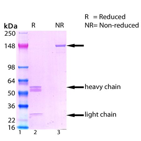
Our proteins meet high purity standards—90% or more as confirmed by SDS-PAGE. Many are available with tags like His, Flag, GST, or MBP, and we also supply native and biologically active proteins for functional studies.
Frequently Asked Questions
1. Are your monoclonal antibodies validated for specific applications?
Yes, our antibodies are tested and validated for use in methods such as ELISA, western blot, IHC, flow cytometry, and more. Refer to specific product pages or datasheets for individual product information.
2. How do I choose the right monoclonal antibody for my application?
Review the product details directly for application validation, species reactivity, and target information. You may also contact our support team at any time for help.
3. How quickly can I receive my order?
Most orders are processed and shipped within 1–3 business days, depending on product availability and your shipping location.