Filters
▼Clonality
▼Type
▼Reactivity
▼Gene Name
▼Isotype
▼Host
▼Application
▼Clone
▼Monoclonal Antibodies
Get accurate results in your research with our Monoclonal Antibodies, which are specially made to target exactly what you require for your research, and will produce consistent, reliable performance in lab tests.
Viewing 9400-9450 of 27597 product results
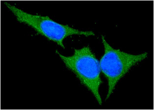
 IF (Immunofluorescence)
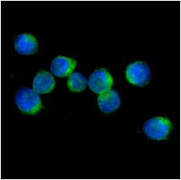
(ICC/IF analysis of IL32 in Jurkat cells line, stained with DAPI (Blue) for nucleus staining and monoclonal anti-human IL32 antibody (1:100) with goat anti-mouse IgG-Alexa fluor 488 conjugate (Green).)
IF (Immunofluorescence)
(ICC/IF analysis of IL32 in Jurkat cells line, stained with DAPI (Blue) for nucleus staining and monoclonal anti-human IL32 antibody (1:100) with goat anti-mouse IgG-Alexa fluor 488 conjugate (Green).)
IL-32, Monoclonal Antibody (Cat# AAA48064)
 IF (Immunofluorescence)
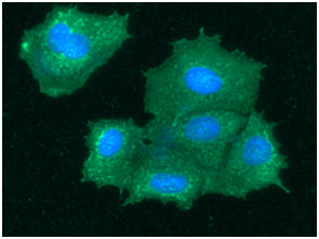
(ICC/IF analysis of BHMT in Hep3B cells line, stained with DAPI (Blue) for nucleus staining and monoclonal anti-human BHMT antibody (1:100) with goat anti-mouse IgG-Alexa fluor 488 conjugate (Green).)
IF (Immunofluorescence)
(ICC/IF analysis of BHMT in Hep3B cells line, stained with DAPI (Blue) for nucleus staining and monoclonal anti-human BHMT antibody (1:100) with goat anti-mouse IgG-Alexa fluor 488 conjugate (Green).)
BHMT, Monoclonal Antibody (Cat# AAA47998)
 IF (Immunofluorescence)
(ICC/IF analysis of BID in HeLa cells line, stained with DAPI (Blue) for nucleus staining and monoclonal anti-human BID antibody (1:100) with goat anti-mouse IgG-Alexa fluor 488 conjugate (Green).)
IF (Immunofluorescence)
(ICC/IF analysis of BID in HeLa cells line, stained with DAPI (Blue) for nucleus staining and monoclonal anti-human BID antibody (1:100) with goat anti-mouse IgG-Alexa fluor 488 conjugate (Green).)
BID, Monoclonal Antibody (Cat# AAA48038)

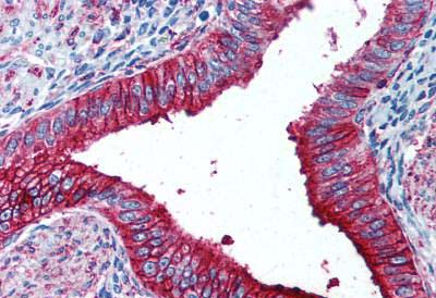
 Application Data
(Detection of ENaC alpha (green) in paraffinembedded, formalin-fixed rat kidney at 10ug/ml.)
Application Data
(Detection of ENaC alpha (green) in paraffinembedded, formalin-fixed rat kidney at 10ug/ml.)
ENaC alpha, Monoclonal Antibody (Cat# AAA47854)
 Application Data
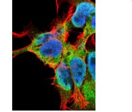
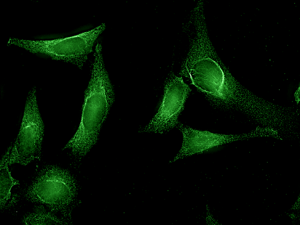
(Detection of FGF13 in neuroblastoma cell line SK-N-BE at 10ug/ml: DAPI (blue) nuclear stain, Texas Red F actin stain, ATTO 488 (green) FGF13 stain.)
Application Data
(Detection of FGF13 in neuroblastoma cell line SK-N-BE at 10ug/ml: DAPI (blue) nuclear stain, Texas Red F actin stain, ATTO 488 (green) FGF13 stain.)
Fibroblast Growth Factor 13 (FGF13), Monoclonal Antibody (Cat# AAA47861)
 Application Data
(Detection of QKI-5 in neuroblastoma cell line SKN-BE at 10ug/ml: DAPI (blue) nuclear stain, Texas Red F actin stain, ATTO 488 (green) QKI-5 stain.)
Application Data
(Detection of QKI-5 in neuroblastoma cell line SKN-BE at 10ug/ml: DAPI (blue) nuclear stain, Texas Red F actin stain, ATTO 488 (green) QKI-5 stain.)
Pan-QKI, Monoclonal Antibody (Cat# AAA47867)
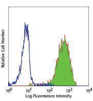
 FCM/FACS (Flow Cytometry)
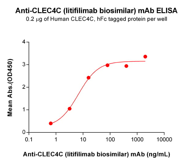
(Figure 1. ELISA plate pre-coated by 2 ug/mL (100 uL/well) Human CLEC4C Protein, hFc Tag (PME100756) can bind Anti-CLEC4C (litifilimab biosimilar) mAb (AAA47600) in a linear range of 0.64–80.00 ng/mL. In order to specifically detect AAA47600, mouse anti-human Fab-specific antibody was used as detection antibody.)
FCM/FACS (Flow Cytometry)
(Figure 1. ELISA plate pre-coated by 2 ug/mL (100 uL/well) Human CLEC4C Protein, hFc Tag (PME100756) can bind Anti-CLEC4C (litifilimab biosimilar) mAb (AAA47600) in a linear range of 0.64–80.00 ng/mL. In order to specifically detect AAA47600, mouse anti-human Fab-specific antibody was used as detection antibody.)
CLEC4C, Monoclonal Antibody (Cat# AAA47600)
Human Procollagen Type I C-Peptide (PIP), Monoclonal Antibody (Cat# AAA47689)
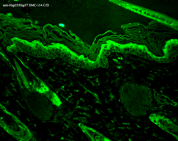

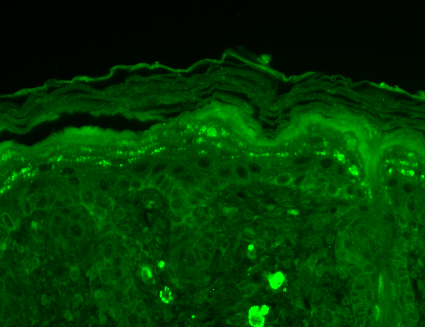
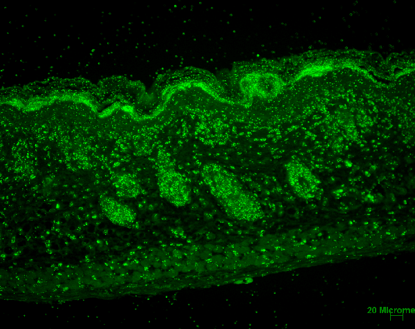
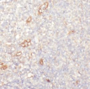
 IHC (Immunohistochemistry)
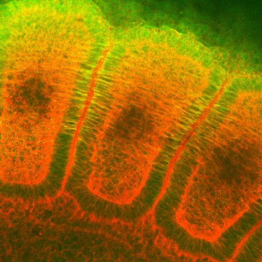
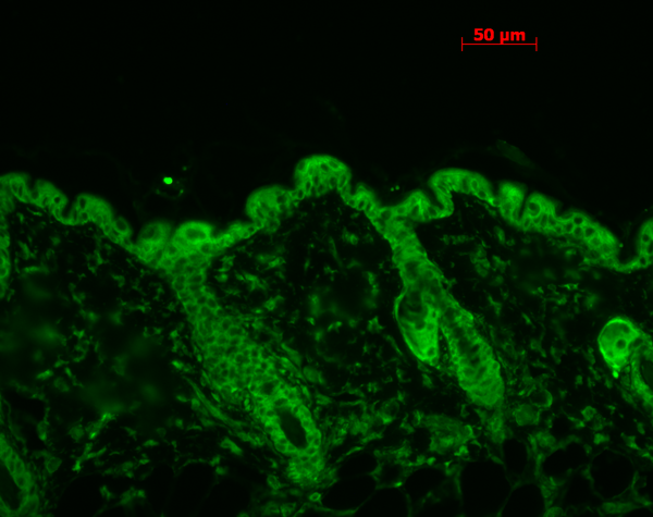
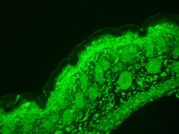
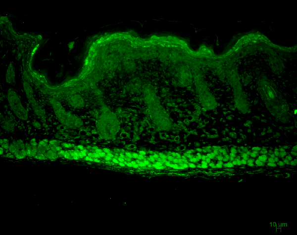
(Immunohistochemistry analysis using Mouse Anti-Hsp27 Monoclonal Antibody, Clone 8A7. Tissue: backskin. Species: Mouse. Fixation: Bouin's Fixative and paraffin-embedded. Primary Antibody: Mouse Anti-Hsp27 Monoclonal Antibody at 1:100 for 1 hour at RT. Secondary Antibody: FITC Goat Anti-Mouse (green) at 1:50 for 1 hour at RT. Localization: Epidermis.)
IHC (Immunohistochemistry)
(Immunohistochemistry analysis using Mouse Anti-Hsp27 Monoclonal Antibody, Clone 8A7. Tissue: backskin. Species: Mouse. Fixation: Bouin's Fixative and paraffin-embedded. Primary Antibody: Mouse Anti-Hsp27 Monoclonal Antibody at 1:100 for 1 hour at RT. Secondary Antibody: FITC Goat Anti-Mouse (green) at 1:50 for 1 hour at RT. Localization: Epidermis.)
Hsp25/Hsp27, Monoclonal Antibody (Cat# AAA102855)

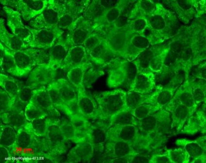
 IHC (Immunohistochemistry)
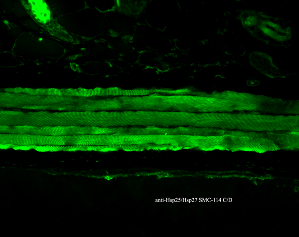
(Immunohistochemistry analysis using Mouse Anti-Hsp90 Monoclonal Antibody, Clone 4F3.E8. Tissue: backskin. Species: Mouse. Fixation: Bouin's Fixative and paraffin-embedded. Primary Antibody: Mouse Anti-Hsp90 Monoclonal Antibody at 1:100 for 1 hour at RT. Secondary Antibody: FITC Goat Anti-Mouse (green) at 1:50 for 1 hour at RT.)
IHC (Immunohistochemistry)
(Immunohistochemistry analysis using Mouse Anti-Hsp90 Monoclonal Antibody, Clone 4F3.E8. Tissue: backskin. Species: Mouse. Fixation: Bouin's Fixative and paraffin-embedded. Primary Antibody: Mouse Anti-Hsp90 Monoclonal Antibody at 1:100 for 1 hour at RT. Secondary Antibody: FITC Goat Anti-Mouse (green) at 1:50 for 1 hour at RT.)
Hsp90, Monoclonal Antibody (Cat# AAA102868)


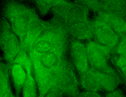
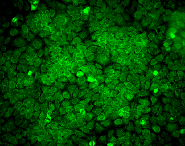
 ICC (Immunocytochemistry)
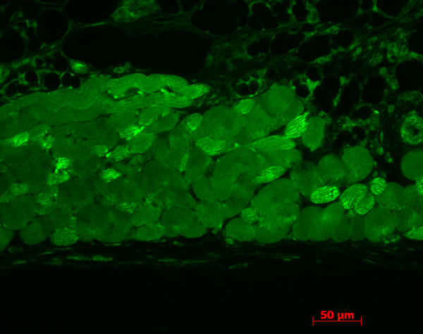
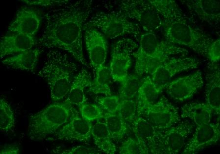
(Immunocytochemistry/Immunofluorescence analysis using Mouse Anti-CD74 Monoclonal Antibody, Clone PIN 1.1. Tissue: HaCaT cells. Species: Human. Fixation: Cold 100% methanol for 10 minutes at -20 degree C. Primary Antibody: Mouse Anti-CD74 Monoclonal Antibody at 1:100 for 1 hour at RT. Secondary Antibody: FITC Goat Anti-Mouse (green) at 1:50 for 1 hour at RT. Localization: Cytoplasmic Staining.)
ICC (Immunocytochemistry)
(Immunocytochemistry/Immunofluorescence analysis using Mouse Anti-CD74 Monoclonal Antibody, Clone PIN 1.1. Tissue: HaCaT cells. Species: Human. Fixation: Cold 100% methanol for 10 minutes at -20 degree C. Primary Antibody: Mouse Anti-CD74 Monoclonal Antibody at 1:100 for 1 hour at RT. Secondary Antibody: FITC Goat Anti-Mouse (green) at 1:50 for 1 hour at RT. Localization: Cytoplasmic Staining.)
CD74, Monoclonal Antibody (Cat# AAA102873)
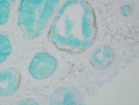
 IHC (Immunohistochemisry)
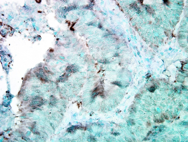
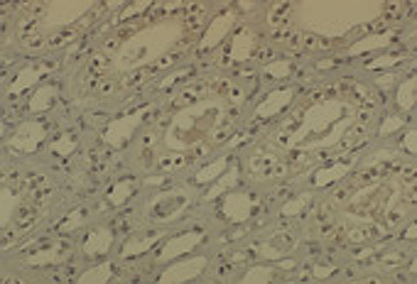
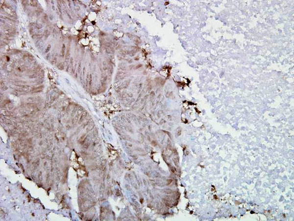
(Immunohistochemistry analysis using Mouse Anti-DNMT1 Monoclonal Antibody, Clone 60B1220.1. Tissue: medullar kidney tissue. Species: Mouse. Primary Antibody: Mouse Anti-DNMT1 Monoclonal Antibody at 1:1000. Secondary Antibody: HRP/DAB Detection System: Biotinylated Goat Anti-Mouse, Streptavidin Peroxidase, DAB Chromogen (brown). Counterstain: Mayer Hematoxylin (purple/blue) nuclear stain.)
IHC (Immunohistochemisry)
(Immunohistochemistry analysis using Mouse Anti-DNMT1 Monoclonal Antibody, Clone 60B1220.1. Tissue: medullar kidney tissue. Species: Mouse. Primary Antibody: Mouse Anti-DNMT1 Monoclonal Antibody at 1:1000. Secondary Antibody: HRP/DAB Detection System: Biotinylated Goat Anti-Mouse, Streptavidin Peroxidase, DAB Chromogen (brown). Counterstain: Mayer Hematoxylin (purple/blue) nuclear stain.)
DNMT1, Monoclonal Antibody (Cat# AAA102895)

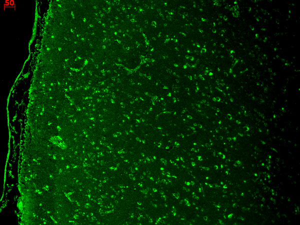
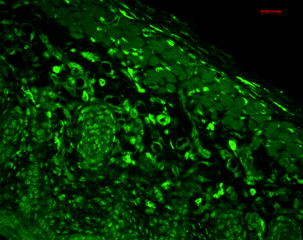
 IHC (Immunohiostchemistry)
(Immunohistochemistry analysis using Mouse Anti-CaV1.3 Calcium Channel Monoclonal Antibody, Clone S48A-9. Tissue: hippocampus. Species: Human. Fixation: Bouin's Fixative and paraffin-embedded. Primary Antibody: Mouse Anti-CaV1.3 Calcium Channel Monoclonal Antibody at 1:1000 for 1 hour at RT. Secondary Antibody: FITC Goat Anti-Mouse (green) at 1:50 for 1 hour at RT.)
IHC (Immunohiostchemistry)
(Immunohistochemistry analysis using Mouse Anti-CaV1.3 Calcium Channel Monoclonal Antibody, Clone S48A-9. Tissue: hippocampus. Species: Human. Fixation: Bouin's Fixative and paraffin-embedded. Primary Antibody: Mouse Anti-CaV1.3 Calcium Channel Monoclonal Antibody at 1:1000 for 1 hour at RT. Secondary Antibody: FITC Goat Anti-Mouse (green) at 1:50 for 1 hour at RT.)
Cav1.3, Monoclonal Antibody (Cat# AAA102902)
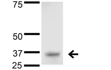
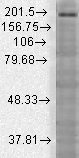
 WB (Western Blot)
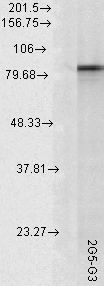
(Western Blot analysis of Rat tissue lysate showing detection of Hsp90 alpha protein using Mouse Anti-Hsp90 alpha Monoclonal Antibody, Clone 2G5.G3. Load: 15 ug. Block: 1.5% BSA for 30 minutes at RT. Primary Antibody: Mouse Anti-Hsp90 alpha Monoclonal Antibody at 1:1000 for 2 hours at RT. Secondary Antibody: Sheep Anti-Mouse IgG: HRP for 1 hour at RT.)
WB (Western Blot)
(Western Blot analysis of Rat tissue lysate showing detection of Hsp90 alpha protein using Mouse Anti-Hsp90 alpha Monoclonal Antibody, Clone 2G5.G3. Load: 15 ug. Block: 1.5% BSA for 30 minutes at RT. Primary Antibody: Mouse Anti-Hsp90 alpha Monoclonal Antibody at 1:1000 for 2 hours at RT. Secondary Antibody: Sheep Anti-Mouse IgG: HRP for 1 hour at RT.)
Hsp90 alpha, Monoclonal Antibody (Cat# AAA102904)

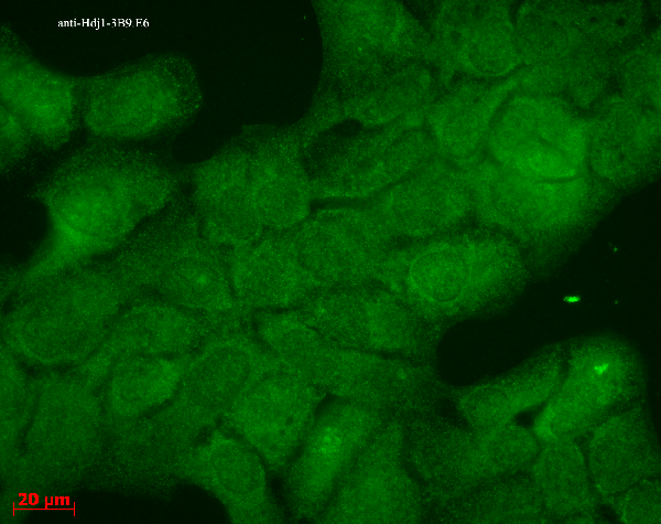
 WB (Western Blot)
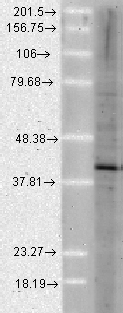
(Western Blot analysis of Human Cell lysates showing detection of Hsp40 protein using Mouse Anti-Hsp40 Monoclonal Antibody, Clone 3B9.E6. Load: 15 ug. Block: 1.5% BSA for 30 minutes at RT. Primary Antibody: Mouse Anti-Hsp40 Monoclonal Antibody at 1:1000 for 2 hours at RT. Secondary Antibody: Sheep Anti-Mouse IgG: HRP for 1 hour at RT.)
WB (Western Blot)
(Western Blot analysis of Human Cell lysates showing detection of Hsp40 protein using Mouse Anti-Hsp40 Monoclonal Antibody, Clone 3B9.E6. Load: 15 ug. Block: 1.5% BSA for 30 minutes at RT. Primary Antibody: Mouse Anti-Hsp40 Monoclonal Antibody at 1:1000 for 2 hours at RT. Secondary Antibody: Sheep Anti-Mouse IgG: HRP for 1 hour at RT.)
Hsp40 (Hdj1), Monoclonal Antibody (Cat# AAA103029)
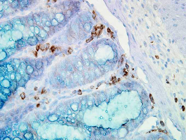
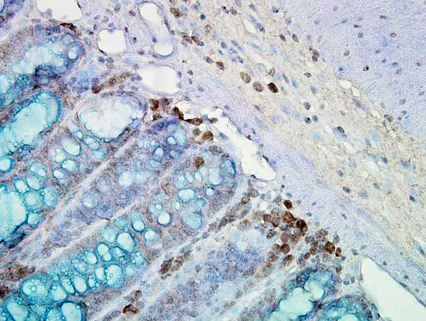 IHC (Immunohiostchemistry)
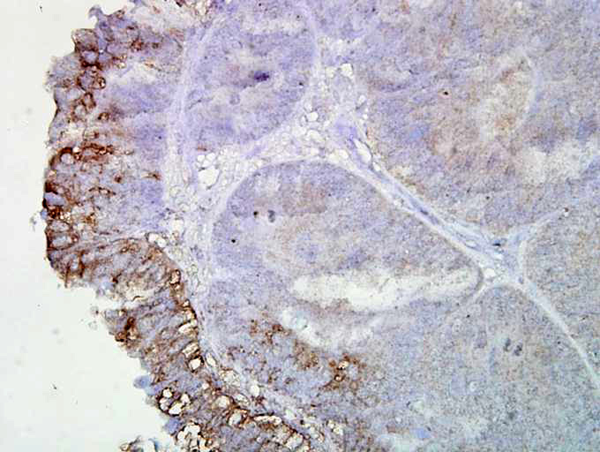
(Immunohistochemistry analysis using Mouse Anti-Hsp90 Monoclonal Antibody, Clone AC-16. Tissue: inflamed colon. Species: Mouse. Fixation: Formalin. Primary Antibody: Mouse Anti-Hsp90 Monoclonal Antibody at 1:2000 for 12 hours at 4 degree C. Secondary Antibody: Biotin Goat Anti-Mouse at 1:2000 for 1 hour at RT. Counterstain: Mayer Hematoxylin (purple/blue) nuclear stain at 200 ul for 2 minutes at RT. Localization: Inflammatory cells. Magnification: 40x. Mostly inflammatory cells, some mucosa.)
IHC (Immunohiostchemistry)
(Immunohistochemistry analysis using Mouse Anti-Hsp90 Monoclonal Antibody, Clone AC-16. Tissue: inflamed colon. Species: Mouse. Fixation: Formalin. Primary Antibody: Mouse Anti-Hsp90 Monoclonal Antibody at 1:2000 for 12 hours at 4 degree C. Secondary Antibody: Biotin Goat Anti-Mouse at 1:2000 for 1 hour at RT. Counterstain: Mayer Hematoxylin (purple/blue) nuclear stain at 200 ul for 2 minutes at RT. Localization: Inflammatory cells. Magnification: 40x. Mostly inflammatory cells, some mucosa.)
Hsp90, Monoclonal Antibody (Cat# AAA103052)
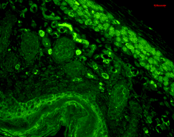
 IHC (Immunohiostchemistry)
(Immunohistochemistry analysis using Mouse Anti-CaV1.3 Calcium Channel Monoclonal Antibody, Clone S48A-9. Tissue: hippocampus. Species: Human. Fixation: Bouin's Fixative and paraffin-embedded. Primary Antibody: Mouse Anti-CaV1.3 Calcium Channel Monoclonal Antibody at 1:1000 for 1 hour at RT. Secondary Antibody: FITC Goat Anti-Mouse (green) at 1:50 for 1 hour at RT.)
IHC (Immunohiostchemistry)
(Immunohistochemistry analysis using Mouse Anti-CaV1.3 Calcium Channel Monoclonal Antibody, Clone S48A-9. Tissue: hippocampus. Species: Human. Fixation: Bouin's Fixative and paraffin-embedded. Primary Antibody: Mouse Anti-CaV1.3 Calcium Channel Monoclonal Antibody at 1:1000 for 1 hour at RT. Secondary Antibody: FITC Goat Anti-Mouse (green) at 1:50 for 1 hour at RT.)
Cav1.3, Monoclonal Antibody (Cat# AAA103072)

 ICC (Immunocytochemistry)
(Immunocytochemistry/Immunofluorescence analysis using Mouse Anti-CD74 Monoclonal Antibody, Clone PIN 1.1. Tissue: HaCaT cells. Species: Human. Fixation: Cold 100% methanol for 10 minutes at -20 degree C. Primary Antibody: Mouse Anti-CD74 Monoclonal Antibody at 1:100 for 1 hour at RT. Secondary Antibody: FITC Goat Anti-Mouse (green) at 1:50 for 1 hour at RT. Localization: Cytoplasmic Staining.)
ICC (Immunocytochemistry)
(Immunocytochemistry/Immunofluorescence analysis using Mouse Anti-CD74 Monoclonal Antibody, Clone PIN 1.1. Tissue: HaCaT cells. Species: Human. Fixation: Cold 100% methanol for 10 minutes at -20 degree C. Primary Antibody: Mouse Anti-CD74 Monoclonal Antibody at 1:100 for 1 hour at RT. Secondary Antibody: FITC Goat Anti-Mouse (green) at 1:50 for 1 hour at RT. Localization: Cytoplasmic Staining.)
CD74, Monoclonal Antibody (Cat# AAA102935)


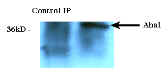
 WB (Western Blot)
(Western Blot analysis of Human Cell lysates showing detection of Aha1 protein using Rat Anti-Aha1 Monoclonal Antibody, Clone 25F2.D10. Load: 15 ug. Block: 1.5% BSA for 30 minutes at RT. Primary Antibody: Rat Anti-Aha1 Monoclonal Antibody at 1:1000 for 2 hours at RT. Secondary Antibody: Sheep Anti-Mouse IgG: HRP for 1 hour at RT.)
WB (Western Blot)
(Western Blot analysis of Human Cell lysates showing detection of Aha1 protein using Rat Anti-Aha1 Monoclonal Antibody, Clone 25F2.D10. Load: 15 ug. Block: 1.5% BSA for 30 minutes at RT. Primary Antibody: Rat Anti-Aha1 Monoclonal Antibody at 1:1000 for 2 hours at RT. Secondary Antibody: Sheep Anti-Mouse IgG: HRP for 1 hour at RT.)
Aha1, Monoclonal Antibody (Cat# AAA102953)
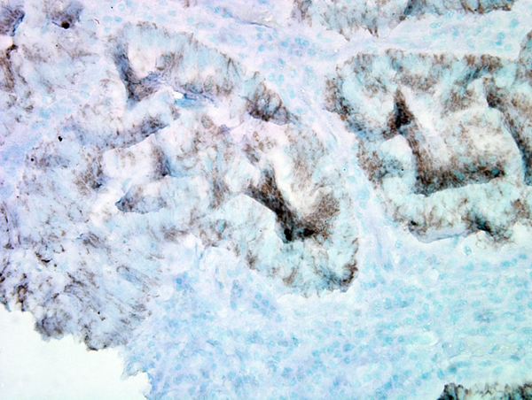
 IHC (Immunohistochemisry)
(Immunohistochemistry analysis using Mouse Anti-Hsp90 alpha Monoclonal Antibody, Clone K41009. Tissue: inflamed colon. Species: Mouse. Fixation: Formalin. Primary Antibody: Mouse Anti-Hsp90 alpha Monoclonal Antibody at 1:5000 for 12 hours at 4 degree C. Secondary Antibody: Biotin Goat Anti-Mouse at 1:2000 for 1 hour at RT. Counterstain: Mayer Hematoxylin (purple/blue) nuclear stain at 200 ul for 2 minutes at RT. Localization: Inflammatory cells. Magnification: 40x. Inflammatory cells.)
IHC (Immunohistochemisry)
(Immunohistochemistry analysis using Mouse Anti-Hsp90 alpha Monoclonal Antibody, Clone K41009. Tissue: inflamed colon. Species: Mouse. Fixation: Formalin. Primary Antibody: Mouse Anti-Hsp90 alpha Monoclonal Antibody at 1:5000 for 12 hours at 4 degree C. Secondary Antibody: Biotin Goat Anti-Mouse at 1:2000 for 1 hour at RT. Counterstain: Mayer Hematoxylin (purple/blue) nuclear stain at 200 ul for 2 minutes at RT. Localization: Inflammatory cells. Magnification: 40x. Inflammatory cells.)
Hsp90 alpha, Monoclonal Antibody (Cat# AAA102965)
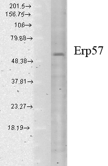

 ICC (Immunocytochemistry)
(Immunocytochemistry/Immunofluorescence analysis using Mouse Anti-Erp57 Monoclonal Antibody, Clone Map.ERP57. Tissue: HaCaT cells. Species: Human. Fixation: Cold 100% methanol for 10 minutes at -20 degree C. Primary Antibody: Mouse Anti-Erp57 Monoclonal Antibody at 1:100 for 1 hour at RT. Secondary Antibody: FITC Goat Anti-Mouse (green) at 1:50 for 1 hour at RT. Localization: Cytoplasmic and perinuclear staining.)
ICC (Immunocytochemistry)
(Immunocytochemistry/Immunofluorescence analysis using Mouse Anti-Erp57 Monoclonal Antibody, Clone Map.ERP57. Tissue: HaCaT cells. Species: Human. Fixation: Cold 100% methanol for 10 minutes at -20 degree C. Primary Antibody: Mouse Anti-Erp57 Monoclonal Antibody at 1:100 for 1 hour at RT. Secondary Antibody: FITC Goat Anti-Mouse (green) at 1:50 for 1 hour at RT. Localization: Cytoplasmic and perinuclear staining.)
Erp57 (Grp58), Monoclonal Antibody (Cat# AAA103197)
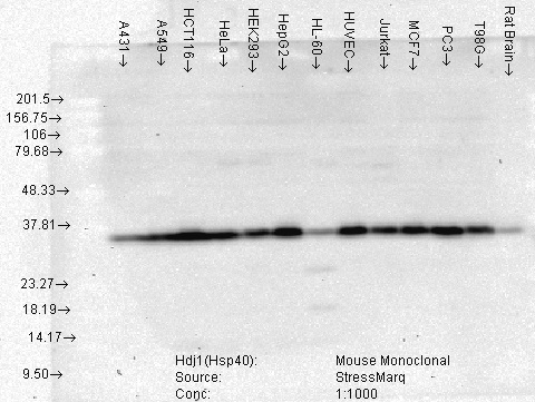 WB (Western Blot)
(Western Blot analysis of Human Cell lysates showing detection of Hsp40 protein using Mouse Anti-Hsp40 Monoclonal Antibody, Clone 3B9.E6. Load: 15 ug. Block: 1.5% BSA for 30 minutes at RT. Primary Antibody: Mouse Anti-Hsp40 Monoclonal Antibody at 1:1000 for 2 hours at RT. Secondary Antibody: Sheep Anti-Mouse IgG: HRP for 1 hour at RT.)
WB (Western Blot)
(Western Blot analysis of Human Cell lysates showing detection of Hsp40 protein using Mouse Anti-Hsp40 Monoclonal Antibody, Clone 3B9.E6. Load: 15 ug. Block: 1.5% BSA for 30 minutes at RT. Primary Antibody: Mouse Anti-Hsp40 Monoclonal Antibody at 1:1000 for 2 hours at RT. Secondary Antibody: Sheep Anti-Mouse IgG: HRP for 1 hour at RT.)
Hsp40 (Hdj1), Monoclonal Antibody (Cat# AAA103212)
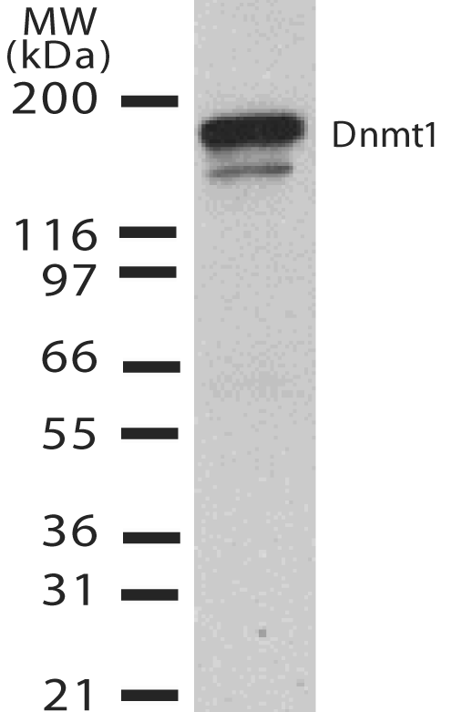
 WB (Western Blot)
(Western Blot analysis of Rat brain membrane lysate showing detection of SHANK3 protein using Mouse Anti-SHANK3 Monoclonal Antibody, Clone S69-46. Load: 15 ug. Block: 1.5% BSA for 30 minutes at RT. Primary Antibody: Mouse Anti-SHANK3 Monoclonal Antibody at 1:1000 for 2 hours at RT. Secondary Antibody: Sheep Anti-Mouse IgG: HRP for 1 hour at RT.)
WB (Western Blot)
(Western Blot analysis of Rat brain membrane lysate showing detection of SHANK3 protein using Mouse Anti-SHANK3 Monoclonal Antibody, Clone S69-46. Load: 15 ug. Block: 1.5% BSA for 30 minutes at RT. Primary Antibody: Mouse Anti-SHANK3 Monoclonal Antibody at 1:1000 for 2 hours at RT. Secondary Antibody: Sheep Anti-Mouse IgG: HRP for 1 hour at RT.)
SHANK3, Monoclonal Antibody (Cat# AAA103252)

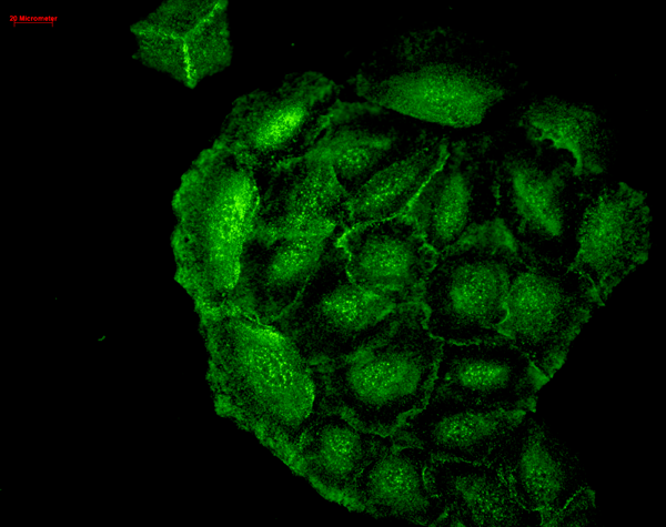 ICC (Immunocytochemistry)
(Immunocytochemistry/Immunofluorescence analysis using Mouse Anti-HO-1 Monoclonal Antibody, Clone 1F12-A6. Tissue: HaCaT cells. Species: Human. Fixation: Cold 100% methanol for 10 minutes at -20 degree C. Primary Antibody: Mouse Anti-HO-1 Monoclonal Antibody at 1:100 for 1 hour at RT. Secondary Antibody: FITC Goat Anti-Mouse (green) at 1:50 for 1 hour at RT. Localization: Cell-cell border staining in epidermis, punctuate nuclear staining.)
ICC (Immunocytochemistry)
(Immunocytochemistry/Immunofluorescence analysis using Mouse Anti-HO-1 Monoclonal Antibody, Clone 1F12-A6. Tissue: HaCaT cells. Species: Human. Fixation: Cold 100% methanol for 10 minutes at -20 degree C. Primary Antibody: Mouse Anti-HO-1 Monoclonal Antibody at 1:100 for 1 hour at RT. Secondary Antibody: FITC Goat Anti-Mouse (green) at 1:50 for 1 hour at RT. Localization: Cell-cell border staining in epidermis, punctuate nuclear staining.)
HO-1, Monoclonal Antibody (Cat# AAA103264)
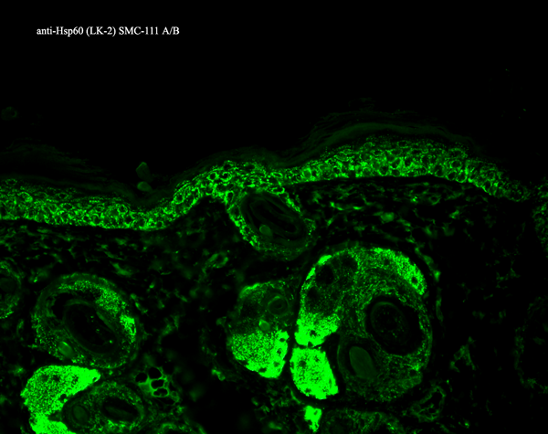
 WB (Western Blot)
(Western Blot analysis of Human Heat Shocked cervical cancer cell line (HeLa) lysate showing detection of Hsp60 protein using Mouse Anti-Hsp60 Monoclonal Antibody, Clone LK-2. Load: 15 ug. Block: 1.5% BSA for 30 minutes at RT. Primary Antibody: Mouse Anti-Hsp60 Monoclonal Antibody at 1:1000 for 2 hours at RT. Secondary Antibody: Sheep Anti-Mouse IgG: HRP for 1 hour at RT.)
WB (Western Blot)
(Western Blot analysis of Human Heat Shocked cervical cancer cell line (HeLa) lysate showing detection of Hsp60 protein using Mouse Anti-Hsp60 Monoclonal Antibody, Clone LK-2. Load: 15 ug. Block: 1.5% BSA for 30 minutes at RT. Primary Antibody: Mouse Anti-Hsp60 Monoclonal Antibody at 1:1000 for 2 hours at RT. Secondary Antibody: Sheep Anti-Mouse IgG: HRP for 1 hour at RT.)
Hsp60, Monoclonal Antibody (Cat# AAA103326)

 ICC (Immunocytochemistry)
(Immunocytochemistry/Immunofluorescence analysis using Mouse Anti-CD74 Monoclonal Antibody, Clone PIN 1.1. Tissue: HaCaT cells. Species: Human. Fixation: Cold 100% methanol for 10 minutes at -20 degree C. Primary Antibody: Mouse Anti-CD74 Monoclonal Antibody at 1:100 for 1 hour at RT. Secondary Antibody: FITC Goat Anti-Mouse (green) at 1:50 for 1 hour at RT. Localization: Cytoplasmic Staining.)
ICC (Immunocytochemistry)
(Immunocytochemistry/Immunofluorescence analysis using Mouse Anti-CD74 Monoclonal Antibody, Clone PIN 1.1. Tissue: HaCaT cells. Species: Human. Fixation: Cold 100% methanol for 10 minutes at -20 degree C. Primary Antibody: Mouse Anti-CD74 Monoclonal Antibody at 1:100 for 1 hour at RT. Secondary Antibody: FITC Goat Anti-Mouse (green) at 1:50 for 1 hour at RT. Localization: Cytoplasmic Staining.)
CD74, Monoclonal Antibody (Cat# AAA103119)

 ICC (Immunocytochemistry)
(Immunocytochemistry/Immunofluorescence analysis using Mouse Anti-Erp57 Monoclonal Antibody, Clone Map.ERP57. Tissue: HaCaT cells. Species: Human. Fixation: Cold 100% methanol for 10 minutes at -20 degree C. Primary Antibody: Mouse Anti-Erp57 Monoclonal Antibody at 1:100 for 1 hour at RT. Secondary Antibody: FITC Goat Anti-Mouse (green) at 1:50 for 1 hour at RT. Localization: Cytoplasmic and perinuclear staining.)
ICC (Immunocytochemistry)
(Immunocytochemistry/Immunofluorescence analysis using Mouse Anti-Erp57 Monoclonal Antibody, Clone Map.ERP57. Tissue: HaCaT cells. Species: Human. Fixation: Cold 100% methanol for 10 minutes at -20 degree C. Primary Antibody: Mouse Anti-Erp57 Monoclonal Antibody at 1:100 for 1 hour at RT. Secondary Antibody: FITC Goat Anti-Mouse (green) at 1:50 for 1 hour at RT. Localization: Cytoplasmic and perinuclear staining.)
Erp57 (Grp58), Monoclonal Antibody (Cat# AAA103367)

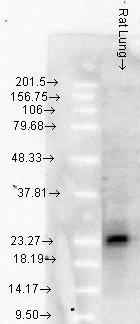
 WB (Western Blot)
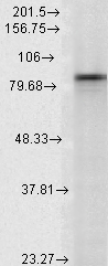
(Western Blot analysis of Rat tissue lysate showing detection of Hsp90 alpha protein using Mouse Anti-Hsp90 alpha Monoclonal Antibody, Clone 2G5.G3. Load: 15 ug. Block: 1.5% BSA for 30 minutes at RT. Primary Antibody: Mouse Anti-Hsp90 alpha Monoclonal Antibody at 1:1000 for 2 hours at RT. Secondary Antibody: Sheep Anti-Mouse IgG: HRP for 1 hour at RT.)
WB (Western Blot)
(Western Blot analysis of Rat tissue lysate showing detection of Hsp90 alpha protein using Mouse Anti-Hsp90 alpha Monoclonal Antibody, Clone 2G5.G3. Load: 15 ug. Block: 1.5% BSA for 30 minutes at RT. Primary Antibody: Mouse Anti-Hsp90 alpha Monoclonal Antibody at 1:1000 for 2 hours at RT. Secondary Antibody: Sheep Anti-Mouse IgG: HRP for 1 hour at RT.)
Hsp90 alpha, Monoclonal Antibody (Cat# AAA103395)

 WB (Western Blot)
(Western Blot analysis of Human Cell lysates showing detection of Aha1 protein using Rat Anti-Aha1 Monoclonal Antibody, Clone 25F2.D10. Load: 15 ug. Block: 1.5% BSA for 30 minutes at RT. Primary Antibody: Rat Anti-Aha1 Monoclonal Antibody at 1:1000 for 2 hours at RT. Secondary Antibody: Sheep Anti-Mouse IgG: HRP for 1 hour at RT.)
WB (Western Blot)
(Western Blot analysis of Human Cell lysates showing detection of Aha1 protein using Rat Anti-Aha1 Monoclonal Antibody, Clone 25F2.D10. Load: 15 ug. Block: 1.5% BSA for 30 minutes at RT. Primary Antibody: Rat Anti-Aha1 Monoclonal Antibody at 1:1000 for 2 hours at RT. Secondary Antibody: Sheep Anti-Mouse IgG: HRP for 1 hour at RT.)
Aha1, Monoclonal Antibody (Cat# AAA102839)


 IHC (Immunohistochemisry)
(Immunohistochemistry analysis using Mouse Anti-DNMT1 Monoclonal Antibody, Clone 60B1220.1. Tissue: medullar kidney tissue. Species: Mouse. Primary Antibody: Mouse Anti-DNMT1 Monoclonal Antibody at 1:1000. Secondary Antibody: HRP/DAB Detection System: Biotinylated Goat Anti-Mouse, Streptavidin Peroxidase, DAB Chromogen (brown). Counterstain: Mayer Hematoxylin (purple/blue) nuclear stain.)
IHC (Immunohistochemisry)
(Immunohistochemistry analysis using Mouse Anti-DNMT1 Monoclonal Antibody, Clone 60B1220.1. Tissue: medullar kidney tissue. Species: Mouse. Primary Antibody: Mouse Anti-DNMT1 Monoclonal Antibody at 1:1000. Secondary Antibody: HRP/DAB Detection System: Biotinylated Goat Anti-Mouse, Streptavidin Peroxidase, DAB Chromogen (brown). Counterstain: Mayer Hematoxylin (purple/blue) nuclear stain.)
DNMT1, Monoclonal Antibody (Cat# AAA102848)
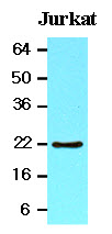
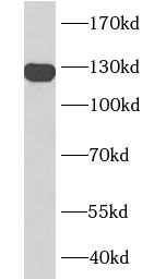 WB (Western Blot)
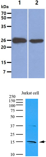
(Jurkat cells were subjected to SDS PAGE followed by western blot with AAA102736 (CD31 antibody) at dilution of 1:1000)
WB (Western Blot)
(Jurkat cells were subjected to SDS PAGE followed by western blot with AAA102736 (CD31 antibody) at dilution of 1:1000)
CD31, Monoclonal Antibody (Cat# AAA102736)
Protein A+G purification
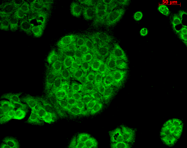
 Application Data
(Staining of A431 cells with Rat anti Human EGF Receptor)
Application Data
(Staining of A431 cells with Rat anti Human EGF Receptor)
EGF R, Monoclonal Antibody (Cat# AAA49481)
 Application Data
(Staining of JAM-C transfected CHO cells with Rat anti Mouse JAM-C:Biotin followed by Streptavidin:FITC)
Application Data
(Staining of JAM-C transfected CHO cells with Rat anti Mouse JAM-C:Biotin followed by Streptavidin:FITC)
JAM-C, Monoclonal Antibody (Cat# AAA50226)
 Application Data
(Human peripheral blood lymphocytes stained with Mouse anti Human CD16: FITC)
Application Data
(Human peripheral blood lymphocytes stained with Mouse anti Human CD16: FITC)
CD16, Monoclonal Antibody (Cat# AAA50238)
 Application Data
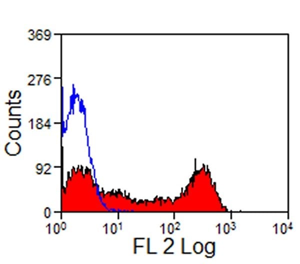
(Staining of bovine peripheral blood lymphocytes with Mouse anti Bovine CD32:RPE)
Application Data
(Staining of bovine peripheral blood lymphocytes with Mouse anti Bovine CD32:RPE)
CD32, Monoclonal Antibody (Cat# AAA49805)
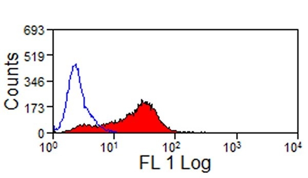
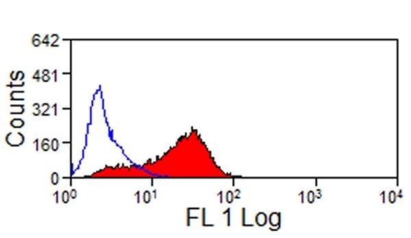
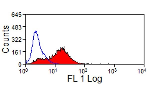
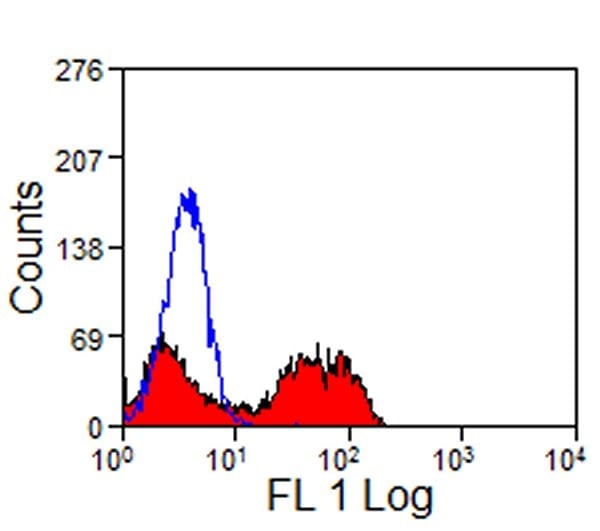
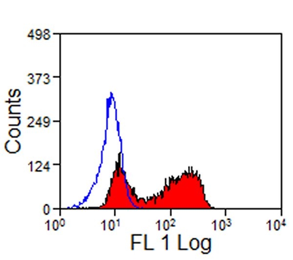
 FCM/FACS (Flow Cytometry)
(Human peripheral blood granulocytes stained with purified HI98, followed by anti-mouse IgGs FITC)
FCM/FACS (Flow Cytometry)
(Human peripheral blood granulocytes stained with purified HI98, followed by anti-mouse IgGs FITC)
SSEA-1 / Lewis x / CD15, Monoclonal Antibody (Cat# AAA51797)
West Nile Virus Envelope Protein, Monoclonal Antibody (Cat# AAA57958)
STREP A, Monoclonal Antibody (Cat# AAA58024)
Free Lambda Light Chain, Monoclonal Antibody (Cat# AAA58028)
Zika Virus Nonstructural Protein (NS1), Monoclonal Antibody (Cat# AAA58034)
Neuron Specific Enolase (NSE) gamma, Monoclonal Antibody (Cat# AAA57812)
Clostridium botulinum Toxin B (a.a. 1278-1291), Monoclonal Antibody (Cat# AAA57919)
C. botulinum Toxin B: 100%
C. botulinum Toxin A: 0%
C. botulinum Toxin C: 0%
C. botulinum Toxin E: 0%
C. botulinum Toxin F (1272-1280): 0%
Hepatitis B Surface Antigen (HBsAg) (ad/ay), Monoclonal Antibody (Cat# AAA57921)
Thyroid Stimulating Hormone (TSH) intact, Monoclonal Antibody (Cat# AAA57738)
Pregnancy Associated Plasma Protein A (PAPP-A) proMBP subunit, Monoclonal Antibody (Cat# AAA57739)
Hemoglobin, Monoclonal Antibody (Cat# AAA57773)
Transferrin, Monoclonal Antibody (Cat# AAA57781)
pro-Brain Natriuretic Peptide, N-terminal (NT-proBNP) (a.a. 61-76), Monoclonal Antibody (Cat# AAA57792)
COVID 19 Nucleocapsid (NP) Coronavirus, Monoclonal Antibody (Cat# AAA58068)
HbsAg, Monoclonal Antibody (Cat# AAA58098)
What are Monoclonal Antibodies?
Monoclonal antibodies are specialized laboratory-produced proteins developed for binding to specific biological antigens or other molecular targets. Since they come from a single cell (or clone), they are especially consistent and accurate in the data they are involved in producing.
This type of antibody material has been shown to be a powerful tool in finding and subsequently destroying harmful cells in an organism, such as those found in cancers or various autoimmune diseases. This makes them excellent aids in medical testing and research, which is why they are so widely used.
AAA Biotech offers a comprehensive range of high-quality monoclonal antibodies that perform effectively in various laboratory tests, including (amongst others) ELISA, western blotting, immunohistochemistry, and flow cytometry. All of the products in our catalog are thoroughly quality tested to make sure that they are reliable and will consistently perform well in your research.
What Are The Uses of Monoclonal Antibodies
Monoclonal antibodies are used in many lab tests, including (amongst others) ELISA, western blotting, immunohistochemistry, and flow cytometry.
ELISA is a test that helps detect a specific substance/analyte in a sample. It uses antibodies (often monoclonal) bound to a solid surface (such as the well of a microplate) to “capture” the substance/analyte in the sample and immobilize it so that the detection antibody component can then bind to it and produce a signal, which can then be measured.
Western blotting identifies specific proteins in a sample. The sample is first separated on a gel, and then antibodies are applied that will typically bind to the target, which will all be localized to a single band in a lane.
Immunohistochemistry helps locate specific proteins in cells or tissue samples using antibodies.
Flow cytometry looks at and sorts cells. It uses antibodies that are conjugated to reporter molecules called “fluorophores”, which, under special lights, emit light themselves, which can then be measured by a detector instrument.
How Monoclonal Antibodies Are Used as Medicine?
Please note that all of the products listed in AAA Biotech’s also known as AAA Bio or AAABio catalog are strictly for research-use only (RUO).
Monoclonal antibodies can also be used as therapeutic/medical treatments, particularly in the context of cancers. They are designed to find and bind to specific cells or proteins, helping the immune system recognize and attack the cancer. These treatments work in different ways, such as:
- Radioimmunotherapy attaches a small amount of radioactive molecule to the antibody, so it delivers the radiation directly to the cancer cells that the antibody is specifically binding to.
- Antibody-directed enzyme prodrug therapy uses antibodies that are specifically bound to special enzymes. These enzymes activate a harmless drug in the body and turn it into a cancer-killing drug only near the cancer cells—this helps avoid harming healthy cells.
- Immunoliposomes are tiny “bubbles” filled with medicine/drug and coated with antibodies. They carry the drug straight to the cancer cells.
Why Buy Monoclonal Antibodies From Us?
At AAA Biotech, we provide high-performance monoclonal antibodies designed to support a wide range of research needs.
1. Validated for Versatile Applications
The antibodies in our catalog are extensively validated and compatible with multiple techniques, including (but not limited to) ELISA, flow cytometry (FC), immunocytochemistry (ICC), immunofluorescence (IF), immunohistochemistry (IHC), immunoprecipitation (IP), and western blotting (WB).
2. Wide Selection & Specialized Options
We offer antibodies for common and rare species, that are available in various conjugated forms, and also in recombinant formats. Essentially, there is almost anything one might need to meet their experimental model’s requirements.
3. High-Quality Proteins
Our proteins meet high purity standards—90% or more as confirmed by SDS-PAGE. Many are available with tags like His, Flag, GST, or MBP, and we also supply native and biologically active proteins for functional studies.
Frequently Asked Questions
1. Are your monoclonal antibodies validated for specific applications?
Yes, our antibodies are tested and validated for use in methods such as ELISA, western blot, IHC, flow cytometry, and more. Refer to specific product pages or datasheets for individual product information.
2. How do I choose the right monoclonal antibody for my application?
Review the product details directly for application validation, species reactivity, and target information. You may also contact our support team at any time for help.
3. How quickly can I receive my order?
Most orders are processed and shipped within 1–3 business days, depending on product availability and your shipping location.