Filters
▼Clonality
▼Type
▼Reactivity
▼Gene Name
▼Isotype
▼Host
▼Application
▼Clone
▼Monoclonal Antibodies
Get accurate results in your research with our Monoclonal Antibodies, which are specially made to target exactly what you require for your research, and will produce consistent, reliable performance in lab tests.
Viewing 9300-9350 of 27597 product results
 SDS-PAGE
(SDS-PAGE Analysis of Purified Neurofilament Mouse Recombinant Monoclonal Antibody (rNF421).)
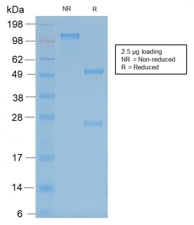
SDS-PAGE
(SDS-PAGE Analysis of Purified Neurofilament Mouse Recombinant Monoclonal Antibody (rNF421).)
Neurofilament (NF-H), Monoclonal Antibody (Cat# AAA214464)
 SDS-PAGE
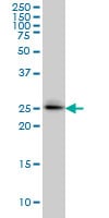
(SDS-PAGE Analysis Purified DAXX Mouse Monoclonal Antibody (PCRP-DAXX-6A8). Confirmation of Purity and Integrity of Antibody.)
SDS-PAGE
(SDS-PAGE Analysis Purified DAXX Mouse Monoclonal Antibody (PCRP-DAXX-6A8). Confirmation of Purity and Integrity of Antibody.)
DAXX, Monoclonal Antibody (Cat# AAA215589)
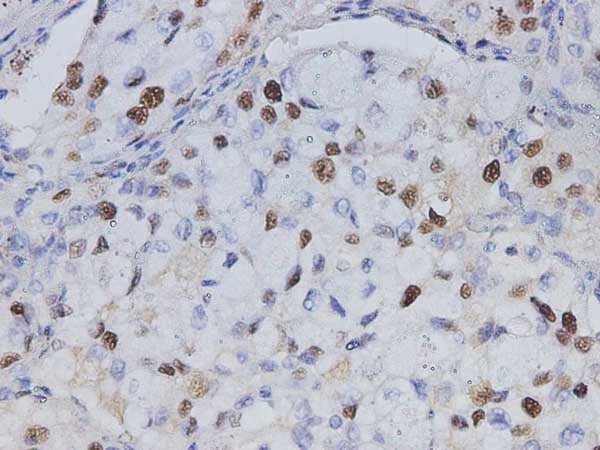
 IHC (Immunohistochemistry)
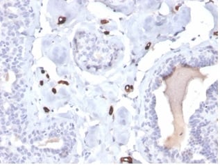
(Formalin-fixed, paraffin-embedded human breast carcinoma stained with FABP4 Mouse Monoclonal Antibody (FABP4/4422).)
IHC (Immunohistochemistry)
(Formalin-fixed, paraffin-embedded human breast carcinoma stained with FABP4 Mouse Monoclonal Antibody (FABP4/4422).)
Fatty Acid Binding Protein 4 (FABP4), Monoclonal Antibody (Cat# AAA215631)
 IHC (Immunohistochemistry)
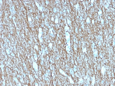
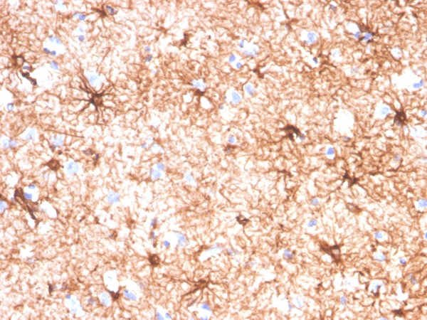
(Formalin-fixed, paraffin-embedded human cerebellum stained with GFAP Mouse Monoclonal Antibody (GFAP/4450).)
IHC (Immunohistochemistry)
(Formalin-fixed, paraffin-embedded human cerebellum stained with GFAP Mouse Monoclonal Antibody (GFAP/4450).)
GFAP, Monoclonal Antibody (Cat# AAA215678)
 IF (Immunofluorescence)
(Immunofluorescent analysis of HEK293 cells with anti-beta Tubulin monoclonal antibody at dilution of 1:4000.)
IF (Immunofluorescence)
(Immunofluorescent analysis of HEK293 cells with anti-beta Tubulin monoclonal antibody at dilution of 1:4000.)
beta Tubulin, Monoclonal Antibody (Cat# AAA178664)
CD132/IL2RG, Monoclonal Antibody (Cat# AAA120171)
 SDS-PAGE
(SDS PAGE for EBV/HHV4 gB/BALF4 Antibody (CL55))
SDS-PAGE
(SDS PAGE for EBV/HHV4 gB/BALF4 Antibody (CL55))
EBV/HHV4 gB/BALF4, Monoclonal Recombinant Antibody (Cat# AAA120342)
Protein A or G purified from cell culture supernatant.
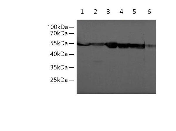
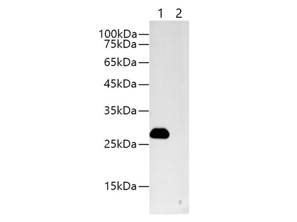
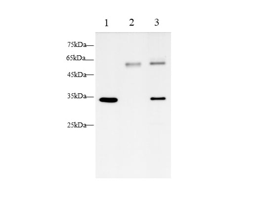
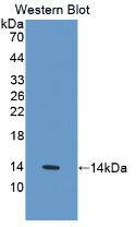
 WB (Western Blot)
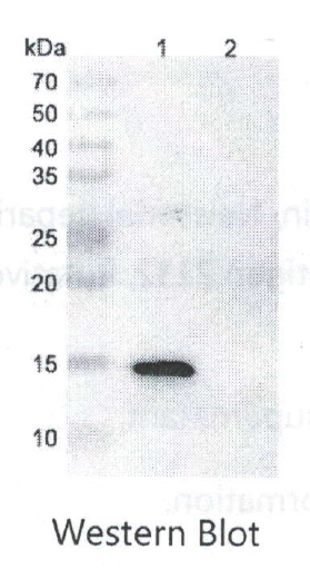
(Various lysates were subjected to SDS-PAGE followed by western blot with Neisseria meningiditis NHBA/ Dextranase antibody AAA120372 at 1 ug/mL.Lane 1: Neisseria meningitidis NHBA/Dextranase transfected HEK293 cell lysate.Lane 2: Non-transfeted HEK293 cell lysate.Second AB: Goat Anti-Human IgG H&L Polyclonal antibody, HRPat 0.1 ug/mL.Predict MW: 14 kDaObserved MW: 14 kDa)
WB (Western Blot)
(Various lysates were subjected to SDS-PAGE followed by western blot with Neisseria meningiditis NHBA/ Dextranase antibody AAA120372 at 1 ug/mL.Lane 1: Neisseria meningitidis NHBA/Dextranase transfected HEK293 cell lysate.Lane 2: Non-transfeted HEK293 cell lysate.Second AB: Goat Anti-Human IgG H&L Polyclonal antibody, HRPat 0.1 ug/mL.Predict MW: 14 kDaObserved MW: 14 kDa)
NHBA/Dextranase, Monoclonal Recombinant Antibody (Cat# AAA120372)
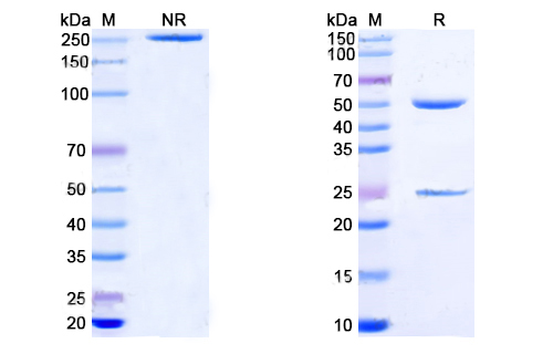
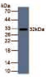
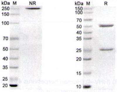 SDS-PAGE
(SDS-PAGE for EBV/HHV-2 gL/BKRF2 Antibody (E1D1))
SDS-PAGE
(SDS-PAGE for EBV/HHV-2 gL/BKRF2 Antibody (E1D1))
EBV/HHV-4 gL/BKRF2, Monoclonal Recombinant Antibody (Cat# AAA120242)
GLP1R, Monoclonal Recombinant Antibody (Cat# AAA120258)

 IHC (Immunohiostchemistry)
(Immunohistochemistry of STAT5b in paraffin-embedded Human breast cancer tissue using STAT5b Rabbit mAb at dilution 1:50)
IHC (Immunohiostchemistry)
(Immunohistochemistry of STAT5b in paraffin-embedded Human breast cancer tissue using STAT5b Rabbit mAb at dilution 1:50)
STAT5b, Monoclonal Antibody (Cat# AAA178824)
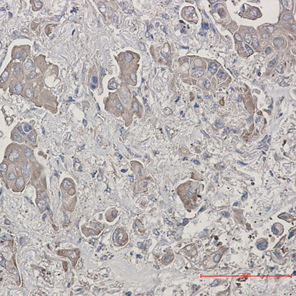 IHC (Immunohiostchemistry)
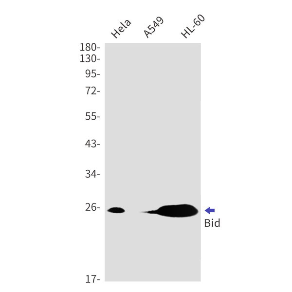
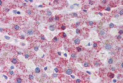
(Immunohistochemistry of Bid in paraffin-embedded liver cancer tissue using Bid Rabbit mAb at dilution 1:50)
IHC (Immunohiostchemistry)
(Immunohistochemistry of Bid in paraffin-embedded liver cancer tissue using Bid Rabbit mAb at dilution 1:50)
Bid, Monoclonal Antibody (Cat# AAA178834)
 IHC (Immunohiostchemistry)
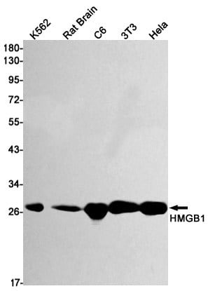
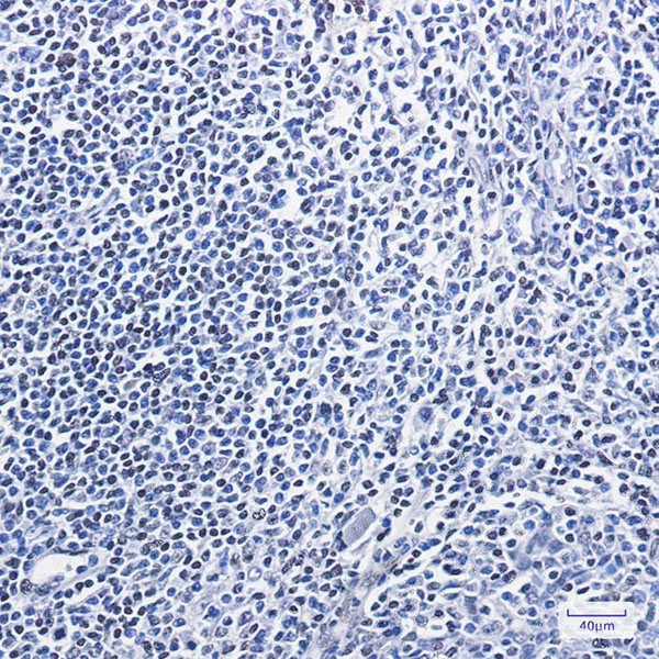
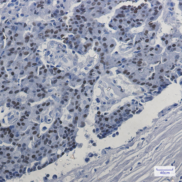
(Immunohistochemistry of HMGB1 in paraffin-embedded Human tonsil using HMGB1 Rabbit mAb at dilution 1:100)
IHC (Immunohiostchemistry)
(Immunohistochemistry of HMGB1 in paraffin-embedded Human tonsil using HMGB1 Rabbit mAb at dilution 1:100)
HMGB1, Monoclonal Antibody (Cat# AAA178772)
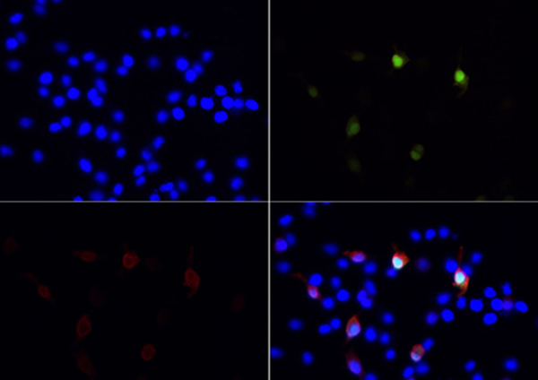
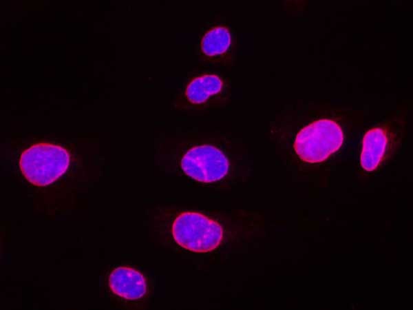
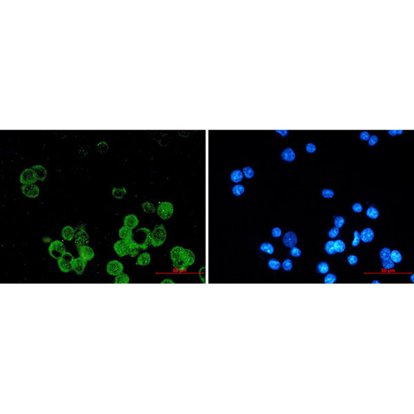 IF (Immunofluorescence)
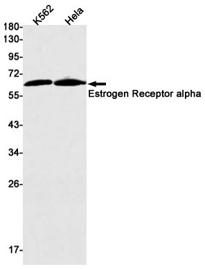
(Immunofluorescence of Estrogen Receptor alpha (green) in MCF-7 using Estrogen Receptor alpha antibody at dilution 1:20, and DAPI(blue))
IF (Immunofluorescence)
(Immunofluorescence of Estrogen Receptor alpha (green) in MCF-7 using Estrogen Receptor alpha antibody at dilution 1:20, and DAPI(blue))
Estrogen Receptor alpha, Monoclonal Antibody (Cat# AAA178794)
 IHC (Immunohiostchemistry)
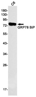
(Immunohistochemistry of GRP78 BiP in paraffin-embedded Human breast cancer tissue using GRP78 BiP Rabbit mAb at dilution 1:50)
IHC (Immunohiostchemistry)
(Immunohistochemistry of GRP78 BiP in paraffin-embedded Human breast cancer tissue using GRP78 BiP Rabbit mAb at dilution 1:50)
GRP78 BiP, Monoclonal Antibody (Cat# AAA178799)
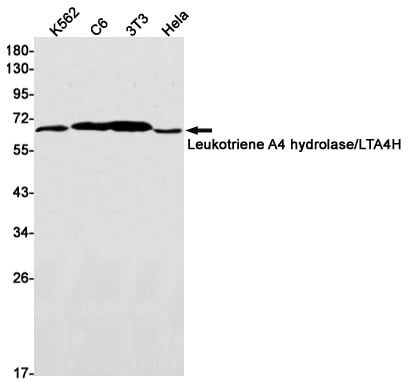
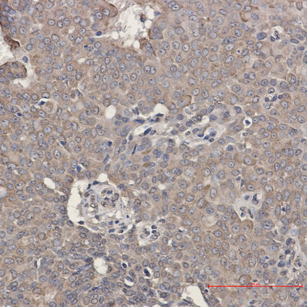
 IHC (Immunohiostchemistry)
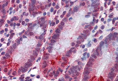
(Immunohistochemistry of Leukotriene A4 hydrolase in paraffin-embedded Human tonsil using Leukotriene A4 hydrolase Rabbit mAb at dilution 1:50)
IHC (Immunohiostchemistry)
(Immunohistochemistry of Leukotriene A4 hydrolase in paraffin-embedded Human tonsil using Leukotriene A4 hydrolase Rabbit mAb at dilution 1:50)
Leukotriene A4 Hydrolase, Monoclonal Antibody (Cat# AAA178803)
 IF (Immunofluorescence)
(Immunofluorescent analysis of 293F cells transfected with Myc-GFP, using anti-Myc-Tag Monoclonal Antibody at 1:500 dilution)
IF (Immunofluorescence)
(Immunofluorescent analysis of 293F cells transfected with Myc-GFP, using anti-Myc-Tag Monoclonal Antibody at 1:500 dilution)
Myc-Tag, Monoclonal Antibody (Cat# AAA178015)
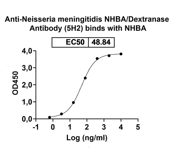
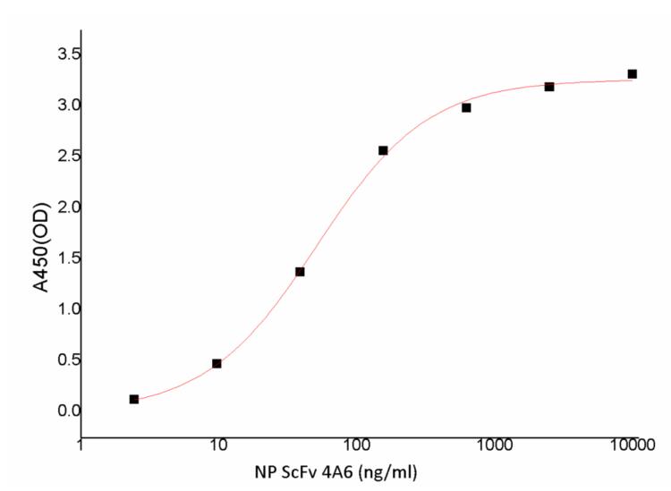 Application Data
(Immobilized 2019-nCoV Nucleocapsid Protein at 5.0ug/ml (100uL/well) can bind Recombinant anti-SARS-CoV2-NP ScFv (4A6), the EC50 is less than 51.29ng/ml.)
Application Data
(Immobilized 2019-nCoV Nucleocapsid Protein at 5.0ug/ml (100uL/well) can bind Recombinant anti-SARS-CoV2-NP ScFv (4A6), the EC50 is less than 51.29ng/ml.)
COVID 19 Nucleocapsid (NP) ScFv Coronavirus, Monoclonal Antibody (Cat# AAA176988)
 IF (Immunofluorescence)
(Immunofluorescence analysis of U-2 OS cells using Lamin B1 Monoclonal Antibody at dilution of 1:500.)
IF (Immunofluorescence)
(Immunofluorescence analysis of U-2 OS cells using Lamin B1 Monoclonal Antibody at dilution of 1:500.)
Lamin B1, Monoclonal Antibody (Cat# AAA179737)
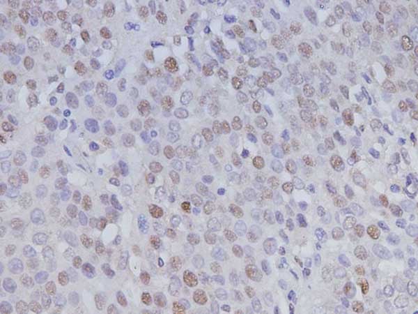 IHC (Immunohistochemistry)
(Immunohistochemistry of paraffin-embedded Human ovary cancer using PCNA Monoclonal Antibody at dilution of 1:200.)
IHC (Immunohistochemistry)
(Immunohistochemistry of paraffin-embedded Human ovary cancer using PCNA Monoclonal Antibody at dilution of 1:200.)
PCNA, Monoclonal Antibody (Cat# AAA179739)
 FCM/FACS (Flow Cytometry)
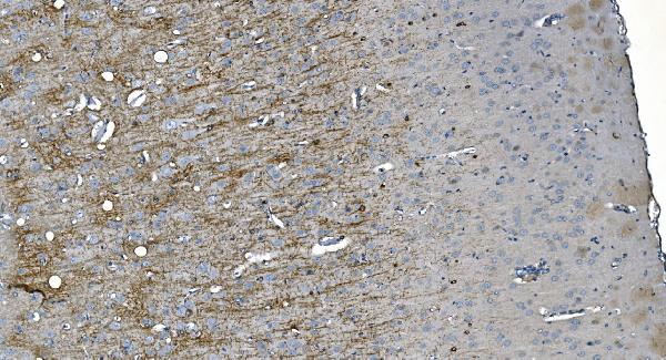
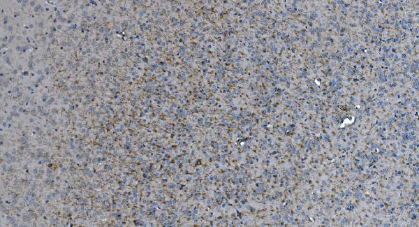
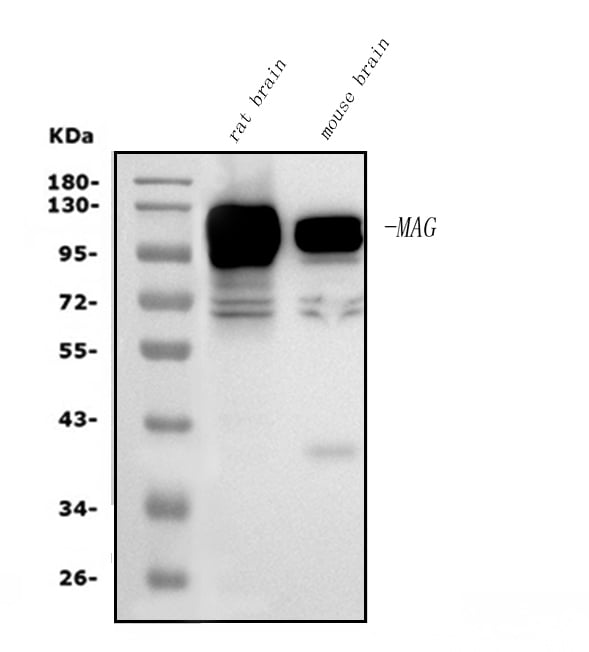
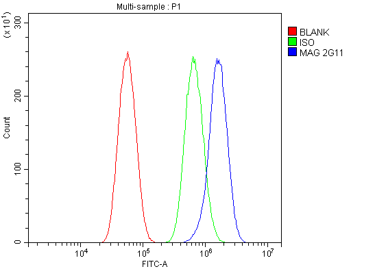
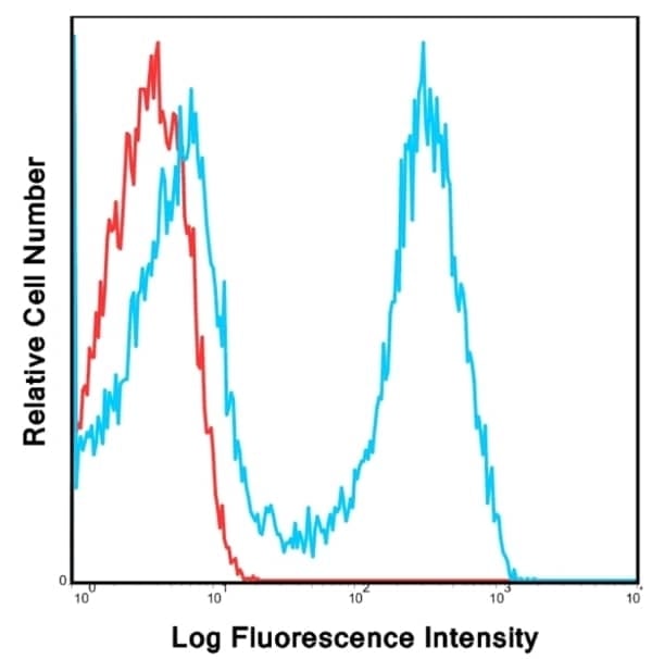
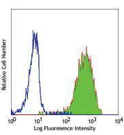
(Figure 4. Flow Cytometry analysis of U87 cells using anti-MAG antibody (AAA125925).Overlay histogram showing U87 cells stained with AAA125925 (Blue line). The cells were blocked with 10% normal goat serum. And then incubated with mouse anti- MAG Antibody (AAA125925, 1μg/1x106 cells) for 30 min at 20 degree C. DyLight®488 conjugated goat anti-mouse IgG (BA1126, 5-10μg/1x106 cells) was used as secondary antibody for 30 minutes at 20 degree C. Isotype control antibody (Green line) was mouse IgG (1μg/1x106) used under the same conditions. Unlabelled sample (Red line) was also used as a control.)
FCM/FACS (Flow Cytometry)
(Figure 4. Flow Cytometry analysis of U87 cells using anti-MAG antibody (AAA125925).Overlay histogram showing U87 cells stained with AAA125925 (Blue line). The cells were blocked with 10% normal goat serum. And then incubated with mouse anti- MAG Antibody (AAA125925, 1μg/1x106 cells) for 30 min at 20 degree C. DyLight®488 conjugated goat anti-mouse IgG (BA1126, 5-10μg/1x106 cells) was used as secondary antibody for 30 minutes at 20 degree C. Isotype control antibody (Green line) was mouse IgG (1μg/1x106) used under the same conditions. Unlabelled sample (Red line) was also used as a control.)
MAG, Monoclonal Antibody (Cat# AAA125925)
 FCM/FACS (Flow Cytometry)
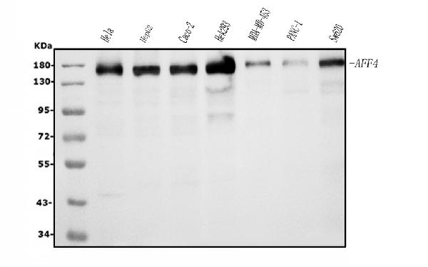
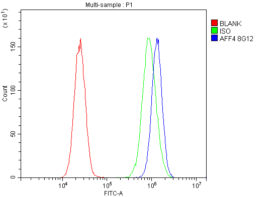
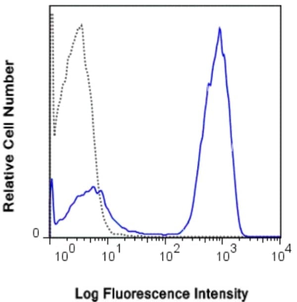
(Figure 2. Flow Cytometry analysis of 293T cells using anti-AFF4 antibody (AAA125931).Overlay histogram showing 293T cells stained with AAA125931 (Blue line). The cells were blocked with 10% normal goat serum. And then incubated with mouse anti- AFF4 Antibody (AAA125931, 1μg/1x106 cells) for 30 min at 20 degree C. DyLight®488 conjugated goat anti-mouse IgG (BA1126, 5-10μg/1x106 cells) was used as secondary antibody for 30 minutes at 20 degree C. Isotype control antibody (Green line) was mouse IgG (1μg/1x106) used under the same conditions. Unlabelled sample (Red line) was also used as a control.)
FCM/FACS (Flow Cytometry)
(Figure 2. Flow Cytometry analysis of 293T cells using anti-AFF4 antibody (AAA125931).Overlay histogram showing 293T cells stained with AAA125931 (Blue line). The cells were blocked with 10% normal goat serum. And then incubated with mouse anti- AFF4 Antibody (AAA125931, 1μg/1x106 cells) for 30 min at 20 degree C. DyLight®488 conjugated goat anti-mouse IgG (BA1126, 5-10μg/1x106 cells) was used as secondary antibody for 30 minutes at 20 degree C. Isotype control antibody (Green line) was mouse IgG (1μg/1x106) used under the same conditions. Unlabelled sample (Red line) was also used as a control.)
AFF4, Monoclonal Antibody (Cat# AAA125931)
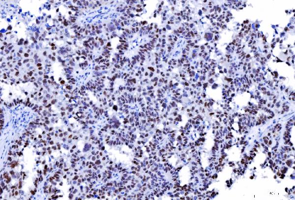
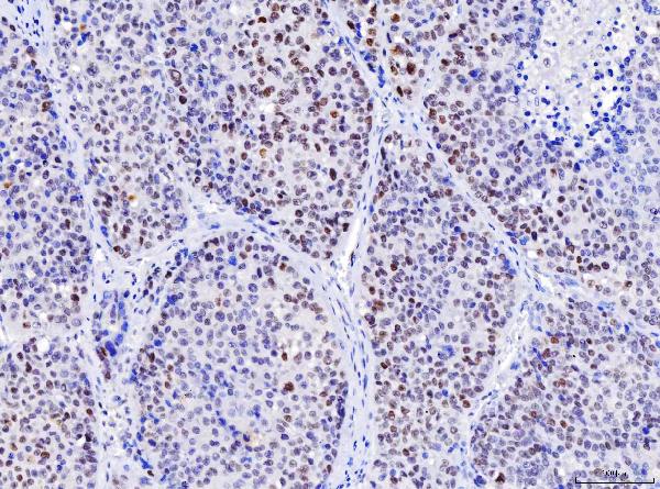
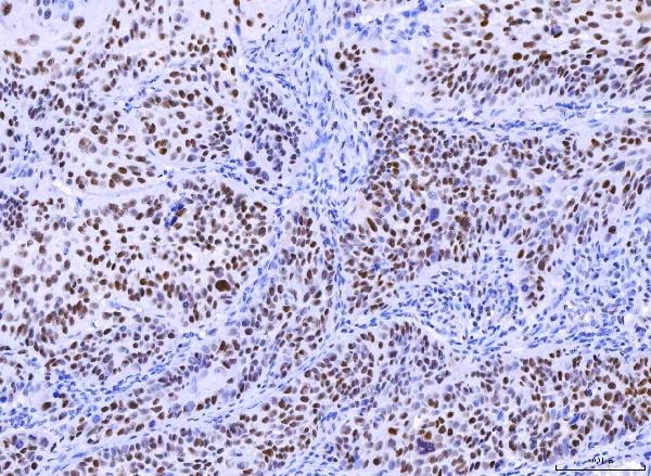
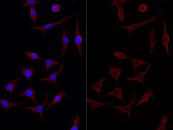
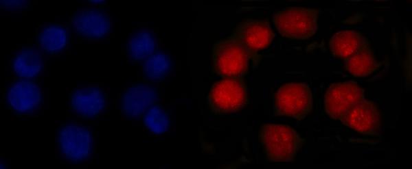 IF (Immunofluorescence)
(Figure 5. IF analysis of MSH2 using anti-MSH2 antibody (AAA126861).MSH2 was detected in an immunocytochemical section of Caco-2 cells. Enzyme antigen retrieval was performed using IHC enzyme antigen retrieval reagent (AR0022) for 15 mins. The cells were blocked with 10% goat serum. And then incubated with 5 ug/mL mouse anti-MSH2 Antibody (AAA126861) overnight at 4 degree C. DyLight594 Conjugated Goat Anti-Mouse IgG (BA1141) was used as secondary antibody at 1:100 dilution and incubated for 30 minutes at 37 degree C. The section was counterstained with DAPI. Visualize using a fluorescence microscope and filter sets appropriate for the label used.)
IF (Immunofluorescence)
(Figure 5. IF analysis of MSH2 using anti-MSH2 antibody (AAA126861).MSH2 was detected in an immunocytochemical section of Caco-2 cells. Enzyme antigen retrieval was performed using IHC enzyme antigen retrieval reagent (AR0022) for 15 mins. The cells were blocked with 10% goat serum. And then incubated with 5 ug/mL mouse anti-MSH2 Antibody (AAA126861) overnight at 4 degree C. DyLight594 Conjugated Goat Anti-Mouse IgG (BA1141) was used as secondary antibody at 1:100 dilution and incubated for 30 minutes at 37 degree C. The section was counterstained with DAPI. Visualize using a fluorescence microscope and filter sets appropriate for the label used.)
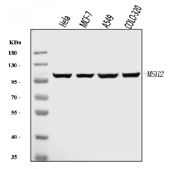
MSH2, Monoclonal Antibody (Cat# AAA126861)
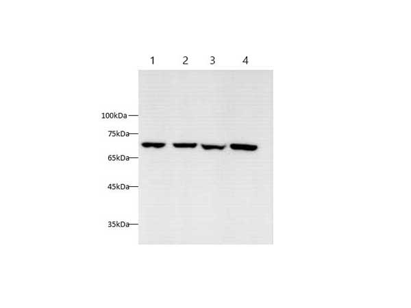
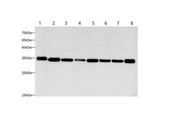
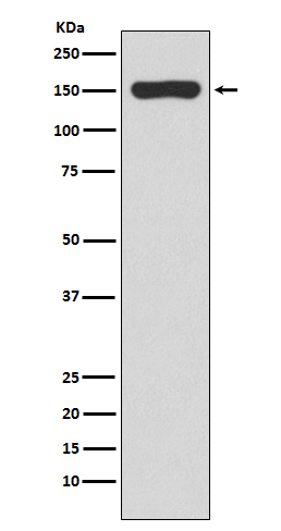 WB (Western Blot)
(Western blot analysis of RNA polymerase beta expression in E Coli lysate.)
WB (Western Blot)
(Western blot analysis of RNA polymerase beta expression in E Coli lysate.)
RNA polymerase beta, Monoclonal Antibody (Cat# AAA126960)
CD3, Monoclonal Antibody (Cat# AAA49585)
 Application Data
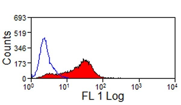
(Staining of rat spleen cells with Mouse anti Rat CD45:Alexa Fluor 488)
Application Data
(Staining of rat spleen cells with Mouse anti Rat CD45:Alexa Fluor 488)
CD45, Monoclonal Antibody (Cat# AAA49689)
 Application Data
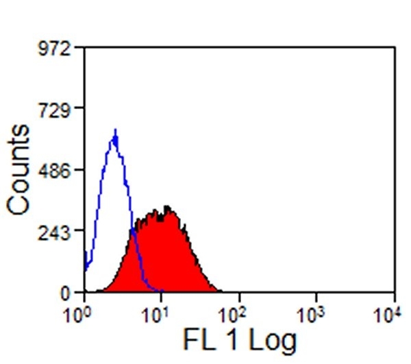
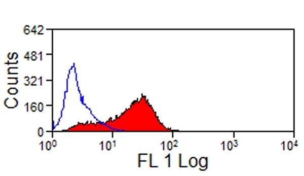
(Staining of bovine peripheral blood lymphocytes with Mouse anti Bovine CD8: FITC)
Application Data
(Staining of bovine peripheral blood lymphocytes with Mouse anti Bovine CD8: FITC)
CD8, Monoclonal Antibody (Cat# AAA49710)
 Application Data
(Staining of CD223 transfected cells with Rat anti Mouse CD223:RPE)
Application Data
(Staining of CD223 transfected cells with Rat anti Mouse CD223:RPE)
CD223, Monoclonal Antibody (Cat# AAA49368)
 Application Data
(Staining of A431 cells with Rat anti Human EGF Receptor)
Application Data
(Staining of A431 cells with Rat anti Human EGF Receptor)
EGF R, Monoclonal Antibody (Cat# AAA49481)
 Application Data
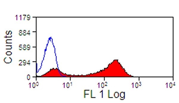
(Staining of bovine peripheral blood monocytes with Mouse anti Bovine CD14)
Application Data
(Staining of bovine peripheral blood monocytes with Mouse anti Bovine CD14)
CD14, Monoclonal Antibody (Cat# AAA49752)
 Application Data
(Staining of human peripheral blood granulocytes with Mouse anti Human Cytochrome b-245 Light Chain. Permeabilised with Leucoperm)
Application Data
(Staining of human peripheral blood granulocytes with Mouse anti Human Cytochrome b-245 Light Chain. Permeabilised with Leucoperm)
CYTOCHROME B245, Monoclonal Antibody (Cat# AAA49770)
 Application Data
(Staining of bovine peripheral blood lymphocytes with Mouse anti Bovine CD32:RPE)
Application Data
(Staining of bovine peripheral blood lymphocytes with Mouse anti Bovine CD32:RPE)
CD32, Monoclonal Antibody (Cat# AAA49805)
 Application Data
(Tim-3 transfected LY5178Y cells stained with Rat anti Mouse TIM-3:Low Endotoxin)
Application Data
(Tim-3 transfected LY5178Y cells stained with Rat anti Mouse TIM-3:Low Endotoxin)
TIM-3, Monoclonal Antibody (Cat# AAA50131)
 Application Data
(Staining of equine peripheral blood granulocytes with Mouse anti Horse CD13:RPE(AAA50197))
Application Data
(Staining of equine peripheral blood granulocytes with Mouse anti Horse CD13:RPE(AAA50197))
CD13, Monoclonal Antibody (Cat# AAA50197)
 Application Data
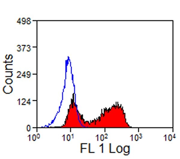
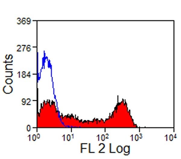
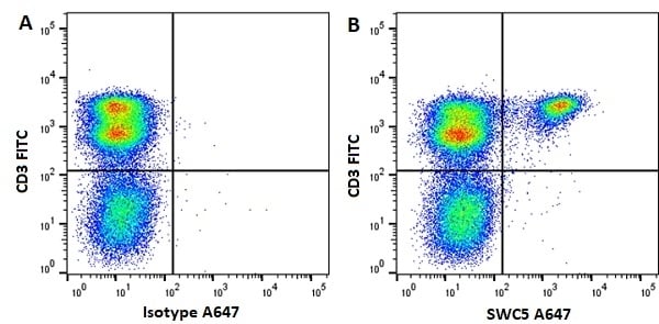
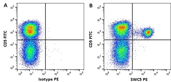
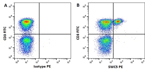
(FigureA.FITC conjugated Mouse anti Pig CD3 and RPE conjugated Mouse IgG1 isotype control . Figure B. FITC conjugated Mouse anti Pig CD3 and RPE conjugated Mouse anti Pig SWC5 . All experiments performed on red cell lysed porcine blood gated on lymphocytes in the presence of 10% pig serum. Data acquired on the ZE5 Cell Analyzer.)
Application Data
(FigureA.FITC conjugated Mouse anti Pig CD3 and RPE conjugated Mouse IgG1 isotype control . Figure B. FITC conjugated Mouse anti Pig CD3 and RPE conjugated Mouse anti Pig SWC5 . All experiments performed on red cell lysed porcine blood gated on lymphocytes in the presence of 10% pig serum. Data acquired on the ZE5 Cell Analyzer.)
SWC5, Monoclonal Antibody (Cat# AAA50391)
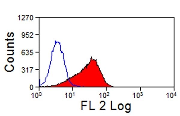
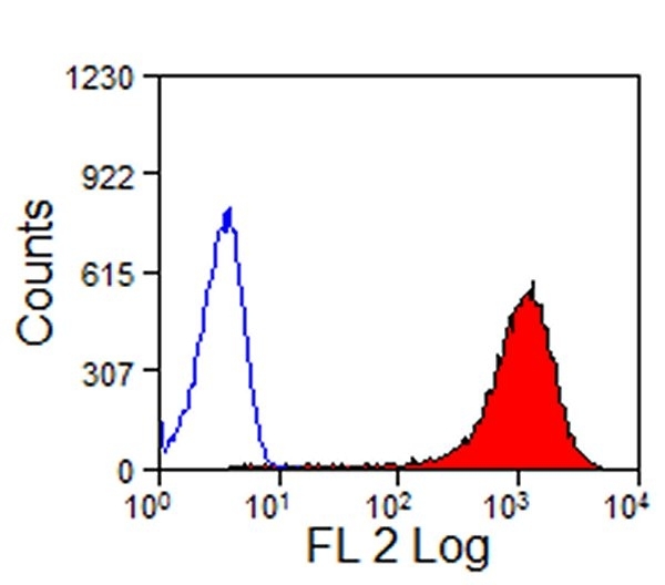
 Application Data
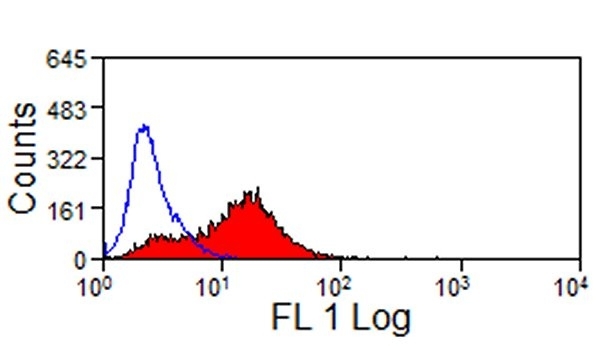
(Surface staining of human peripheral blood lymphocytes with Mouse anti Human CD3:RPE (AAA50422) (Blue) with a relevant isotype control (Grey))
Application Data
(Surface staining of human peripheral blood lymphocytes with Mouse anti Human CD3:RPE (AAA50422) (Blue) with a relevant isotype control (Grey))
CD3, Monoclonal Antibody (Cat# AAA50422)
>90 % by SDS PAGE
 Application Data
(Staining of JAM-C transfected CHO cells with Rat anti Mouse JAM-C:Biotin followed by Streptavidin:FITC)
Application Data
(Staining of JAM-C transfected CHO cells with Rat anti Mouse JAM-C:Biotin followed by Streptavidin:FITC)
JAM-C, Monoclonal Antibody (Cat# AAA50226)
 Application Data
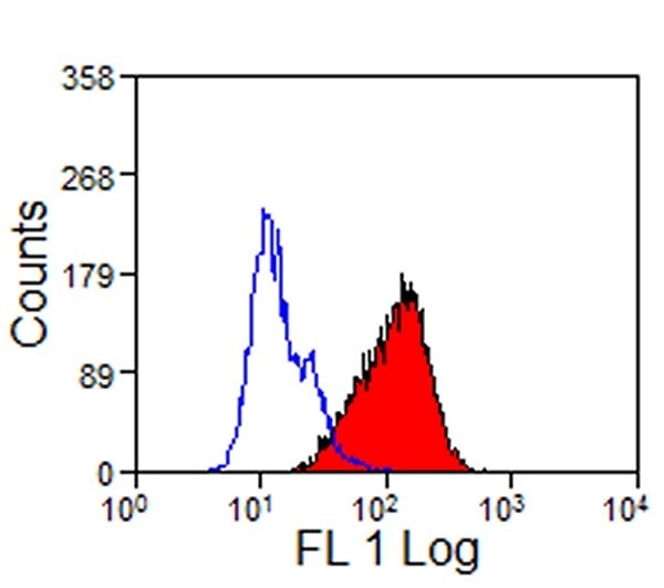
(Human peripheral blood lymphocytes stained with Mouse anti Human CD16: FITC)
Application Data
(Human peripheral blood lymphocytes stained with Mouse anti Human CD16: FITC)
CD16, Monoclonal Antibody (Cat# AAA50238)
CD172a, Monoclonal Antibody (Cat# AAA50316)
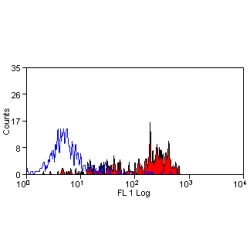
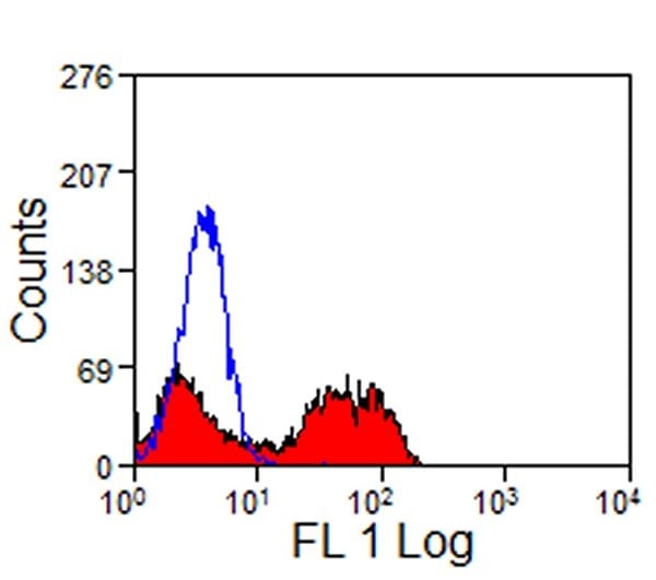
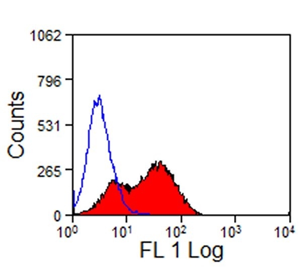
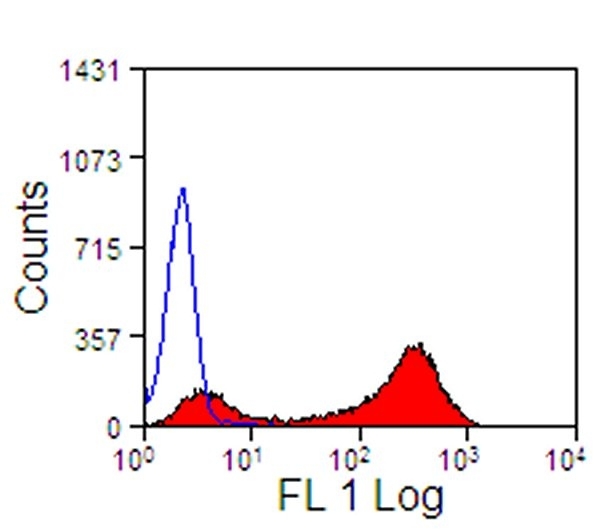
 FCM/FACS (Flow Cytometry)
(Human peripheral blood granulocytes stained with purified HI98, followed by anti-mouse IgGs FITC)
FCM/FACS (Flow Cytometry)
(Human peripheral blood granulocytes stained with purified HI98, followed by anti-mouse IgGs FITC)
SSEA-1 / Lewis x / CD15, Monoclonal Antibody (Cat# AAA51797)
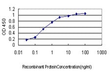 ELISA
(Detection limit for recombinant GST tagged SPSB2 is approximately 0.03 ng/ml as a capture antibody.)
ELISA
(Detection limit for recombinant GST tagged SPSB2 is approximately 0.03 ng/ml as a capture antibody.)
SPSB2, Monoclonal Antibody (Cat# AAA51873)
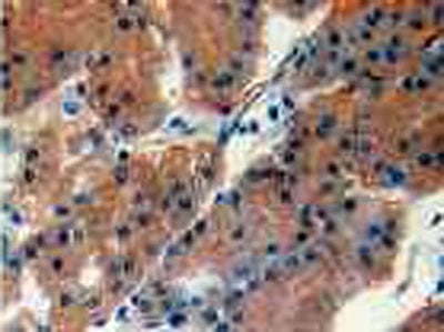
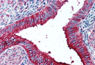
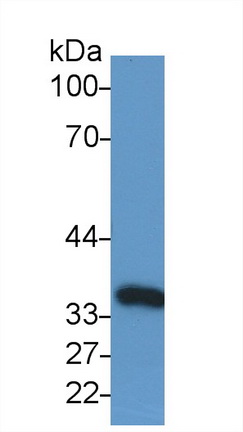
 IHC (Immunohistochemistry)
(DAB staining on IHC-P;Samples: Human Colorectal cancer Tissue;Primary Ab: 30ug/ml Mouse Anti-Human IL33 AntibodySecond Ab: 2ug/mL HRP-Linked Caprine Anti-Mouse IgG Polyclonal Antibody)
IHC (Immunohistochemistry)
(DAB staining on IHC-P;Samples: Human Colorectal cancer Tissue;Primary Ab: 30ug/ml Mouse Anti-Human IL33 AntibodySecond Ab: 2ug/mL HRP-Linked Caprine Anti-Mouse IgG Polyclonal Antibody)
Interleukin 33, Monoclonal Antibody (Cat# AAA144656)
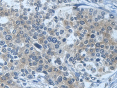 IHC (Immunohistochemisry)
(DAB staining on IHC-P; Samples: Human Prostate cancer Tissue))
IHC (Immunohistochemisry)
(DAB staining on IHC-P; Samples: Human Prostate cancer Tissue))
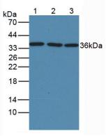
Glyceraldehyde-3-Phosphate Dehydrogenase (GAPDH), Monoclonal Antibody (Cat# AAA145975)
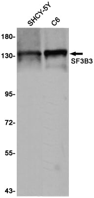
 ICC (Immunocytochemistry)
(Immunocytochemistry of SF3B3(green) in Hela cells using SF3B3 Rabbit mAb at dilution 1/50, and DAPI(blue))
ICC (Immunocytochemistry)
(Immunocytochemistry of SF3B3(green) in Hela cells using SF3B3 Rabbit mAb at dilution 1/50, and DAPI(blue))
SF3B3, Monoclonal Antibody (Cat# AAA314456)
 ICC (Immunocytochemistry)
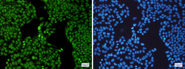
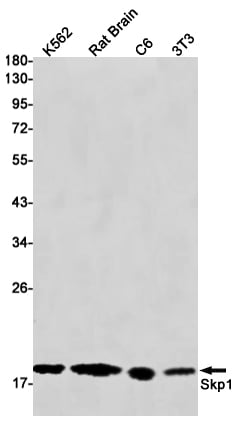
(Immunocytochemistry of Skp1 (green) in MCF-7 using Skp1 antibody at dilution 1/20, and DAPI(blue))
ICC (Immunocytochemistry)
(Immunocytochemistry of Skp1 (green) in MCF-7 using Skp1 antibody at dilution 1/20, and DAPI(blue))
SKP1, Monoclonal Antibody (Cat# AAA314460)
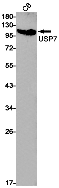
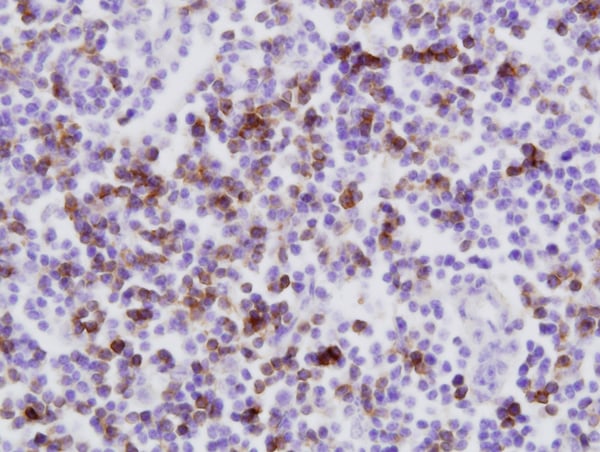
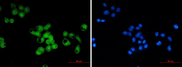
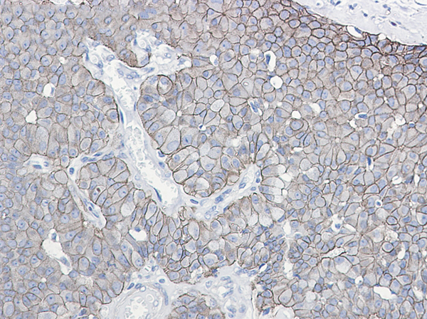
 IHC (Immunohiostchemistry)
(Immunohistochemistry of HAUSP/USP7 in paraffin-embedded Human lung cancer tissue using HAUSP/USP7 Rabbit mAb at dilution 1/20)
IHC (Immunohiostchemistry)
(Immunohistochemistry of HAUSP/USP7 in paraffin-embedded Human lung cancer tissue using HAUSP/USP7 Rabbit mAb at dilution 1/20)
USP7, Monoclonal Antibody (Cat# AAA314481)

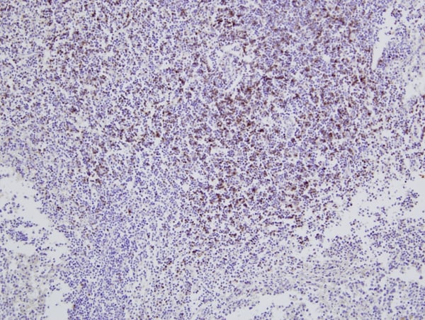
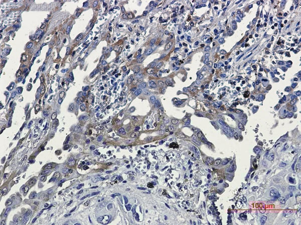 IHC (Immunohiostchemistry)
(Immunohistochemical of RSK3 in Human lung cancer tissue using RSK3 antibody at dilution 1/20)
IHC (Immunohiostchemistry)
(Immunohistochemical of RSK3 in Human lung cancer tissue using RSK3 antibody at dilution 1/20)
RSK3, Monoclonal Antibody (Cat# AAA314492)
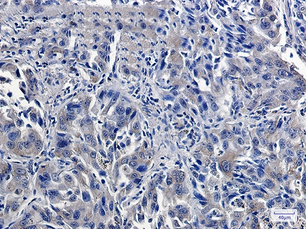
 IHC (Immunohiostchemistry)
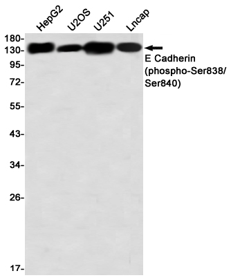
(Immunohistochemistry of E Cadherin (phospho-Ser838/Ser840) in paraffin-embedded Human breast cancer tissue using E Cadherin (phospho-Ser838/Ser840) Rabbit mAb at dilution 1/50)
IHC (Immunohiostchemistry)
(Immunohistochemistry of E Cadherin (phospho-Ser838/Ser840) in paraffin-embedded Human breast cancer tissue using E Cadherin (phospho-Ser838/Ser840) Rabbit mAb at dilution 1/50)
E Cadherin, Monoclonal Antibody (Cat# AAA314499)
 ICC (Immunocytochemistry)
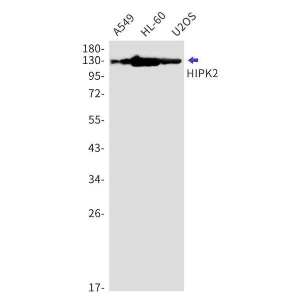
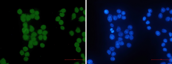
(Immunocytochemistry of HIPK2 (green) in hela using HIPK2 Rabbit mAb at dilution 1/50, and DAPI(blue))
ICC (Immunocytochemistry)
(Immunocytochemistry of HIPK2 (green) in hela using HIPK2 Rabbit mAb at dilution 1/50, and DAPI(blue))
HIPK2, Monoclonal Antibody (Cat# AAA314532)
 ICC (Immunocytochemistry)
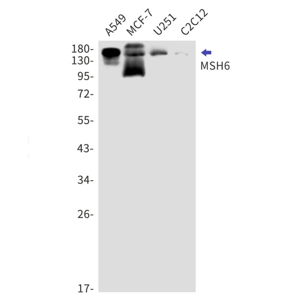
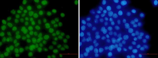
(Immunocytochemistry of MSH6 (green) in hela using MSH6 Rabbit mAb at dilution 1/50, and DAPI(blue))
ICC (Immunocytochemistry)
(Immunocytochemistry of MSH6 (green) in hela using MSH6 Rabbit mAb at dilution 1/50, and DAPI(blue))
MSH6, Monoclonal Antibody (Cat# AAA314551)
What are Monoclonal Antibodies?
Monoclonal antibodies are specialized laboratory-produced proteins developed for binding to specific biological antigens or other molecular targets. Since they come from a single cell (or clone), they are especially consistent and accurate in the data they are involved in producing.
This type of antibody material has been shown to be a powerful tool in finding and subsequently destroying harmful cells in an organism, such as those found in cancers or various autoimmune diseases. This makes them excellent aids in medical testing and research, which is why they are so widely used.
AAA Biotech offers a comprehensive range of high-quality monoclonal antibodies that perform effectively in various laboratory tests, including (amongst others) ELISA, western blotting, immunohistochemistry, and flow cytometry. All of the products in our catalog are thoroughly quality tested to make sure that they are reliable and will consistently perform well in your research.
What Are The Uses of Monoclonal Antibodies
Monoclonal antibodies are used in many lab tests, including (amongst others) ELISA, western blotting, immunohistochemistry, and flow cytometry.
ELISA is a test that helps detect a specific substance/analyte in a sample. It uses antibodies (often monoclonal) bound to a solid surface (such as the well of a microplate) to “capture” the substance/analyte in the sample and immobilize it so that the detection antibody component can then bind to it and produce a signal, which can then be measured.
Western blotting identifies specific proteins in a sample. The sample is first separated on a gel, and then antibodies are applied that will typically bind to the target, which will all be localized to a single band in a lane.
Immunohistochemistry helps locate specific proteins in cells or tissue samples using antibodies.
Flow cytometry looks at and sorts cells. It uses antibodies that are conjugated to reporter molecules called “fluorophores”, which, under special lights, emit light themselves, which can then be measured by a detector instrument.
How Monoclonal Antibodies Are Used as Medicine?
Please note that all of the products listed in AAA Biotech’s also known as AAA Bio or AAABio catalog are strictly for research-use only (RUO).
Monoclonal antibodies can also be used as therapeutic/medical treatments, particularly in the context of cancers. They are designed to find and bind to specific cells or proteins, helping the immune system recognize and attack the cancer. These treatments work in different ways, such as:
- Radioimmunotherapy attaches a small amount of radioactive molecule to the antibody, so it delivers the radiation directly to the cancer cells that the antibody is specifically binding to.
- Antibody-directed enzyme prodrug therapy uses antibodies that are specifically bound to special enzymes. These enzymes activate a harmless drug in the body and turn it into a cancer-killing drug only near the cancer cells—this helps avoid harming healthy cells.
- Immunoliposomes are tiny “bubbles” filled with medicine/drug and coated with antibodies. They carry the drug straight to the cancer cells.
Why Buy Monoclonal Antibodies From Us?
At AAA Biotech, we provide high-performance monoclonal antibodies designed to support a wide range of research needs.
1. Validated for Versatile Applications
The antibodies in our catalog are extensively validated and compatible with multiple techniques, including (but not limited to) ELISA, flow cytometry (FC), immunocytochemistry (ICC), immunofluorescence (IF), immunohistochemistry (IHC), immunoprecipitation (IP), and western blotting (WB).
2. Wide Selection & Specialized Options
We offer antibodies for common and rare species, that are available in various conjugated forms, and also in recombinant formats. Essentially, there is almost anything one might need to meet their experimental model’s requirements.
3. High-Quality Proteins
Our proteins meet high purity standards—90% or more as confirmed by SDS-PAGE. Many are available with tags like His, Flag, GST, or MBP, and we also supply native and biologically active proteins for functional studies.
Frequently Asked Questions
1. Are your monoclonal antibodies validated for specific applications?
Yes, our antibodies are tested and validated for use in methods such as ELISA, western blot, IHC, flow cytometry, and more. Refer to specific product pages or datasheets for individual product information.
2. How do I choose the right monoclonal antibody for my application?
Review the product details directly for application validation, species reactivity, and target information. You may also contact our support team at any time for help.
3. How quickly can I receive my order?
Most orders are processed and shipped within 1–3 business days, depending on product availability and your shipping location.