Filters
▼Clonality
▼Type
▼Reactivity
▼Gene Name
▼Isotype
▼Host
▼Application
▼Clone
▼Monoclonal Antibodies
Get accurate results in your research with our Monoclonal Antibodies, which are specially made to target exactly what you require for your research, and will produce consistent, reliable performance in lab tests.
Viewing 9200-9250 of 27597 product results
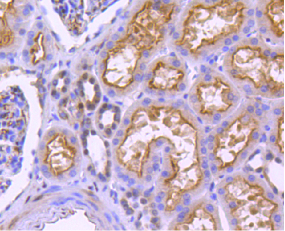
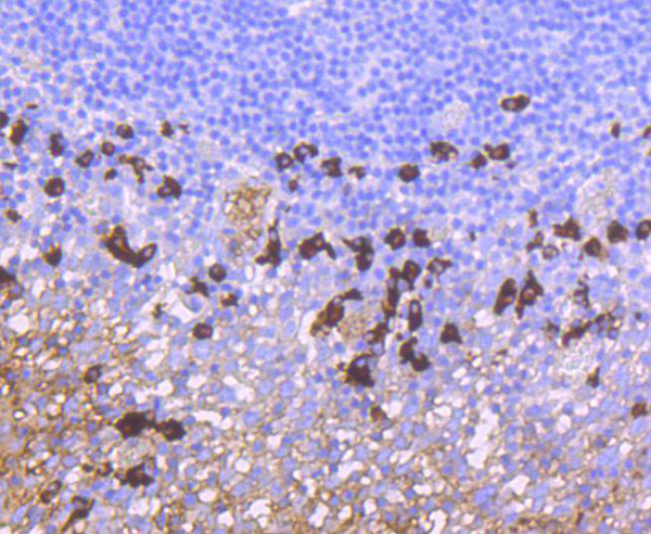
 IHC (Immunohiostchemistry)
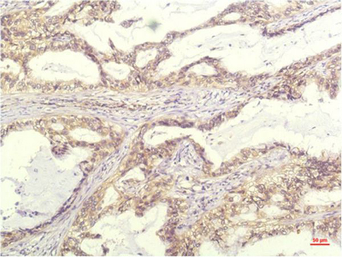
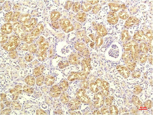
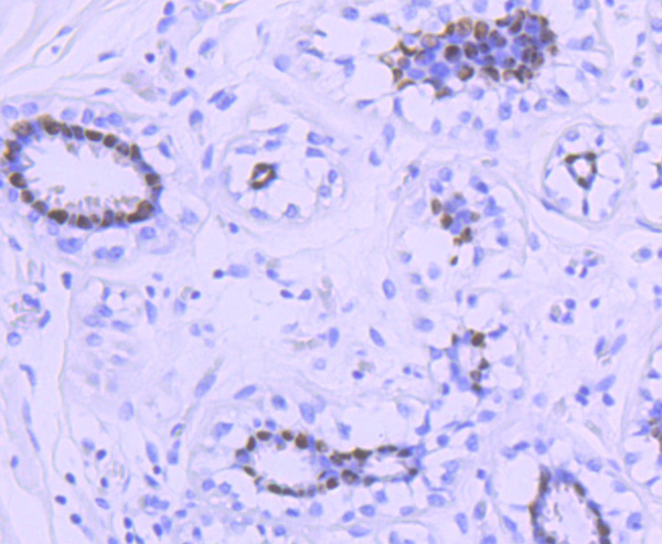
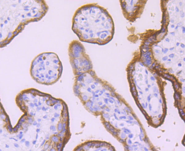
(Immunohistochemical analysis of paraffin-embedded Human Kidney Tissue using IL-8 Mouse mAb diluted at 1:200.)
IHC (Immunohiostchemistry)
(Immunohistochemical analysis of paraffin-embedded Human Kidney Tissue using IL-8 Mouse mAb diluted at 1:200.)
IL-8, Monoclonal Antibody (Cat# AAA309476)
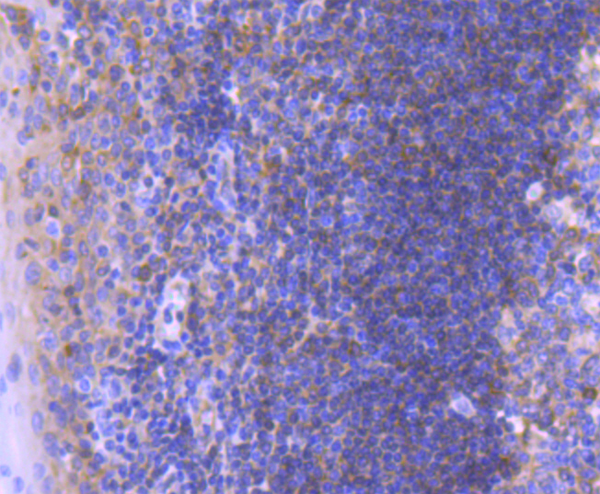
 IHC (Immunohiostchemistry)
(Immunohistochemical analysis of paraffin-embedded Human Stomach Tissue using Acetyl Histone H3 K9 Mouse mAb diluted at 1:200.)
IHC (Immunohiostchemistry)
(Immunohistochemical analysis of paraffin-embedded Human Stomach Tissue using Acetyl Histone H3 K9 Mouse mAb diluted at 1:200.)
H3, Monoclonal Antibody (Cat# AAA310241)
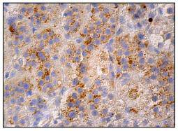
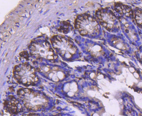
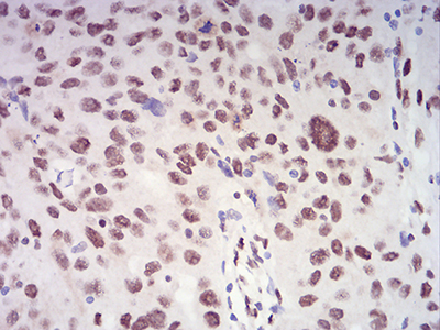
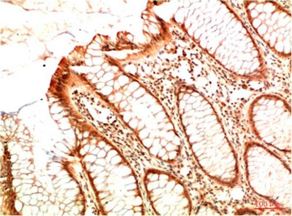 IHC (Immunohistochemisry)
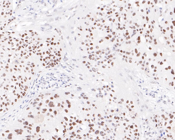
(Immunohistochemical analysis of paraffin-embedded Human Colon Carcinoma Tissue using Acetyl P53 (K382) Mouse mAb diluted at 1:200.)
IHC (Immunohistochemisry)
(Immunohistochemical analysis of paraffin-embedded Human Colon Carcinoma Tissue using Acetyl P53 (K382) Mouse mAb diluted at 1:200.)
Acetyl P53, Monoclonal Antibody (Cat# AAA310244)
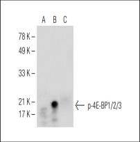
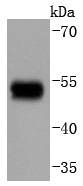
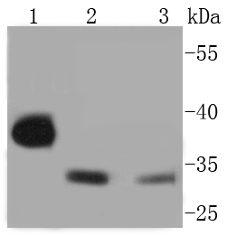
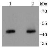
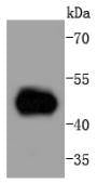
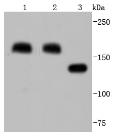
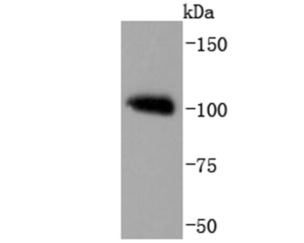
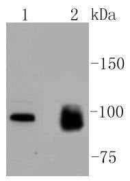
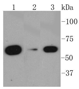
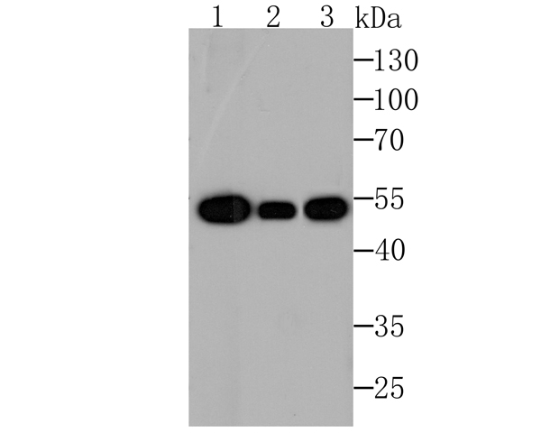
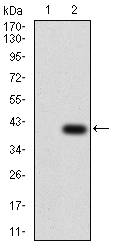
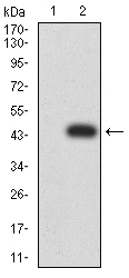
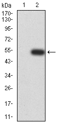
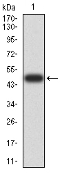
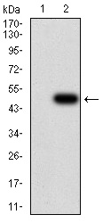
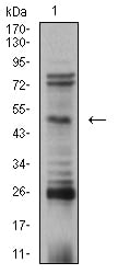
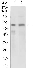
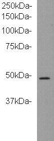
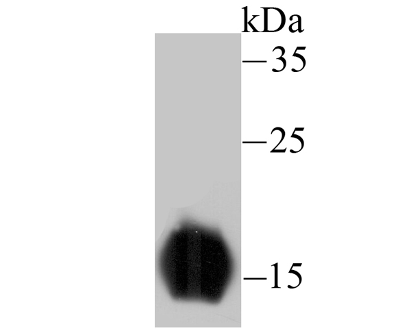
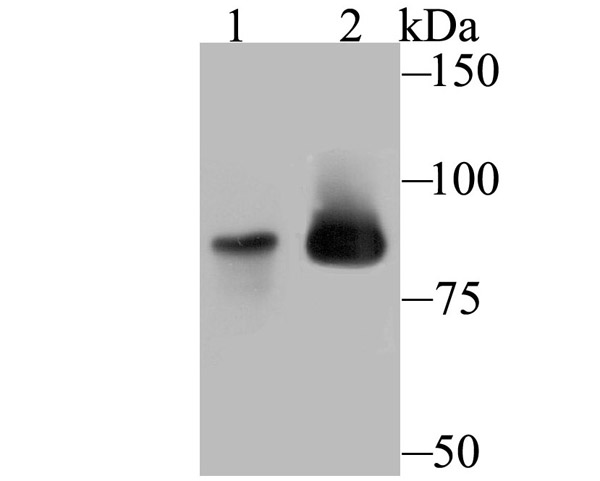
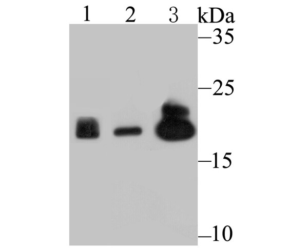
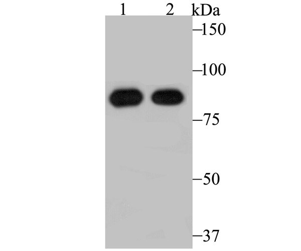
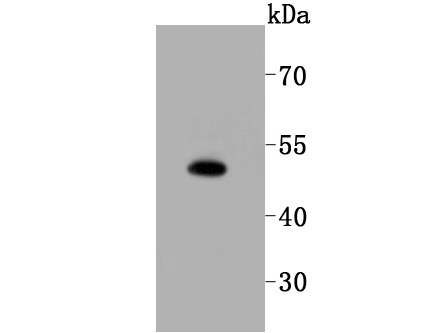
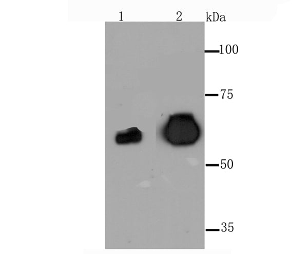
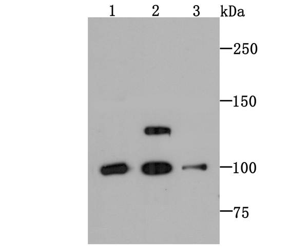
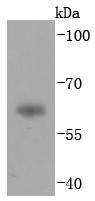
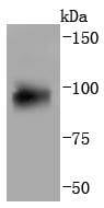
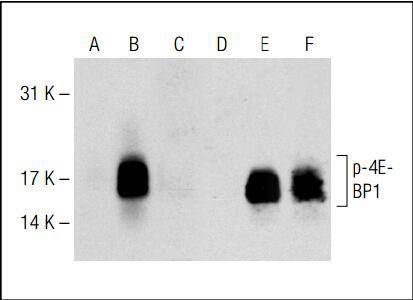 WB (Western Blot)
(Western blot analysis of 4E-BP1 phosphorylation in nontransfected (A, D), untreated human 4E-BP1 transfected (B, E) and lambda protein phosphatase treated human 4E-BP1 transfected (C, F) 293T whole cell lysates. Antibodies tested include p-4E-BP1/2/3 (A, B, C) and 4E-BP1 (D, E, F).)
WB (Western Blot)
(Western blot analysis of 4E-BP1 phosphorylation in nontransfected (A, D), untreated human 4E-BP1 transfected (B, E) and lambda protein phosphatase treated human 4E-BP1 transfected (C, F) 293T whole cell lysates. Antibodies tested include p-4E-BP1/2/3 (A, B, C) and 4E-BP1 (D, E, F).)
4E-BP1/2/3, Monoclonal Antibody (Cat# AAA310988)
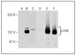
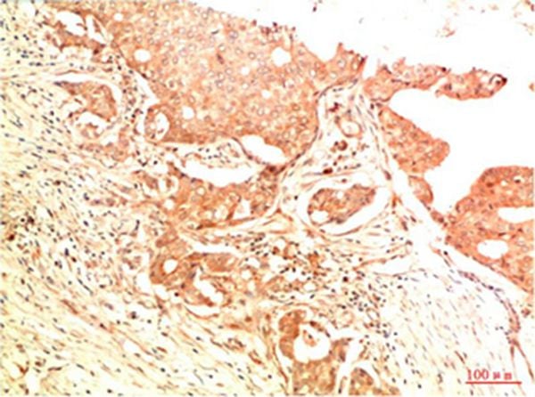
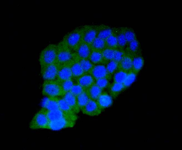
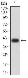
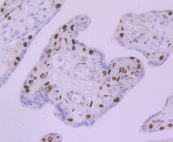
 Application Data
(B.Immunoperoxidase staining of formalin fixed, paraffin-embedded human adrenal gland tissue showing cytoplasmic staining of glandular cells.)
Application Data
(B.Immunoperoxidase staining of formalin fixed, paraffin-embedded human adrenal gland tissue showing cytoplasmic staining of glandular cells.)
FAK, Monoclonal Antibody (Cat# AAA310990)
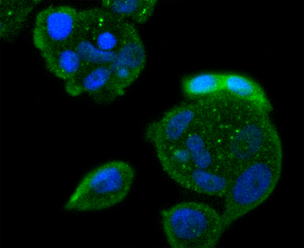
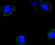
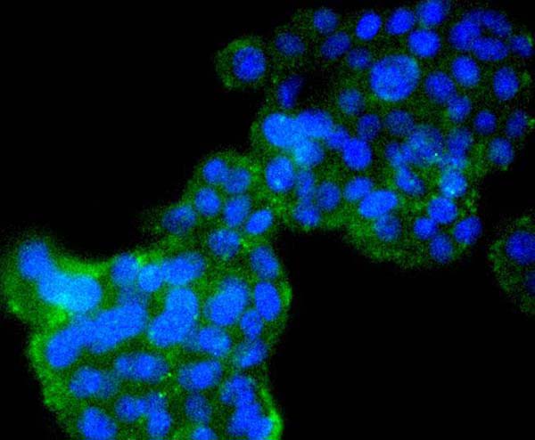
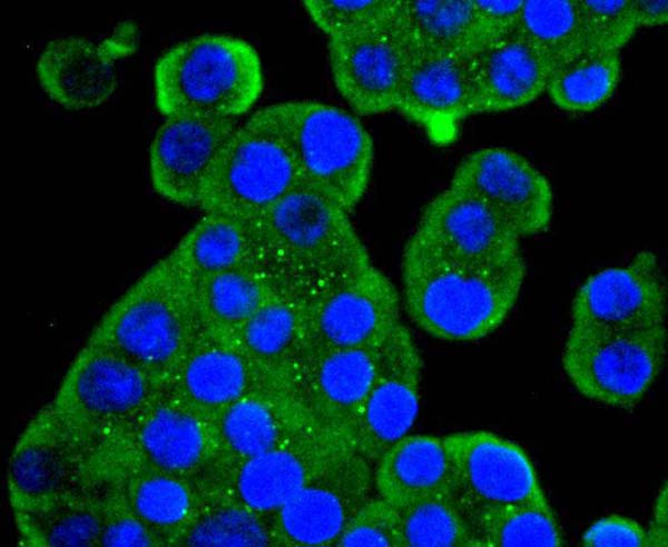
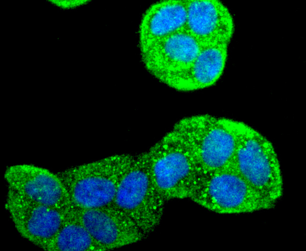
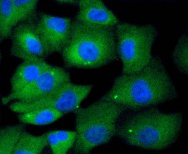
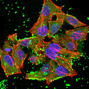
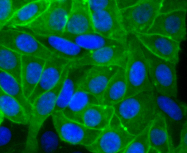
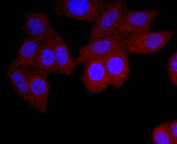
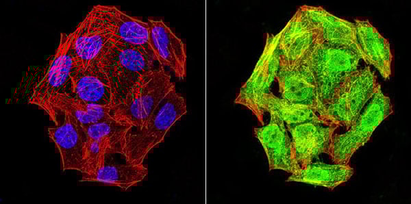 ICC (Immunocytochemistry)
(ICC staining phospho-NLRC4 (Ser-533) (green) and Actin filaments (red) in Hela cells. The nuclear counter stain is DAPI (blue). Cells were fixed in paraformaldehyde, permeabilised with 0.25% Triton X100/PBS.)
ICC (Immunocytochemistry)
(ICC staining phospho-NLRC4 (Ser-533) (green) and Actin filaments (red) in Hela cells. The nuclear counter stain is DAPI (blue). Cells were fixed in paraformaldehyde, permeabilised with 0.25% Triton X100/PBS.)
NLRC4, Monoclonal Antibody (Cat# AAA310993)
 IHC (Immunohiostchemistry)
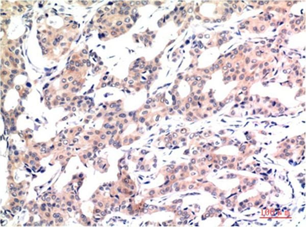
(Immunohistochemical analysis of paraffin-embedded human breast carcinoma tissue using anti-Phospho-GATA3 (S308) antibody. Counter stained with hematoxylin.)
IHC (Immunohiostchemistry)
(Immunohistochemical analysis of paraffin-embedded human breast carcinoma tissue using anti-Phospho-GATA3 (S308) antibody. Counter stained with hematoxylin.)
GATA3, Monoclonal Antibody (Cat# AAA311004)
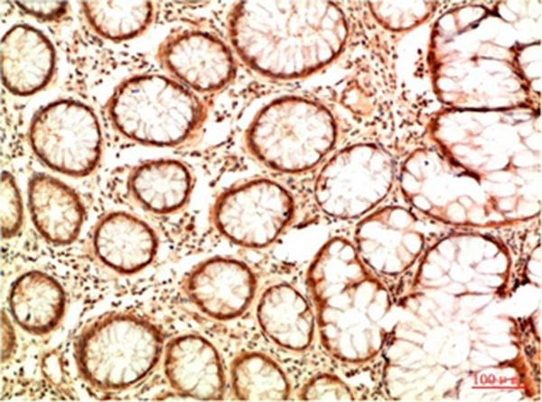
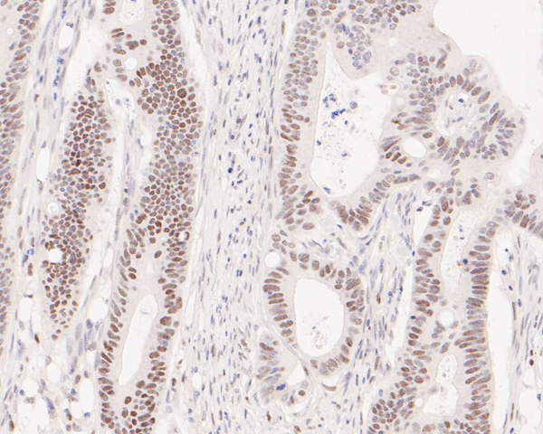
 IHC (Immunohistochemisry)
(Immunohistochemical analysis of paraffin-embedded mouse colon tissue using anti-Phospho-Cdk2 (Y15) antibody. Counter stained with hematoxylin.)
IHC (Immunohistochemisry)
(Immunohistochemical analysis of paraffin-embedded mouse colon tissue using anti-Phospho-Cdk2 (Y15) antibody. Counter stained with hematoxylin.)
CDK2, Monoclonal Antibody (Cat# AAA311024)
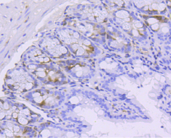
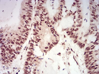
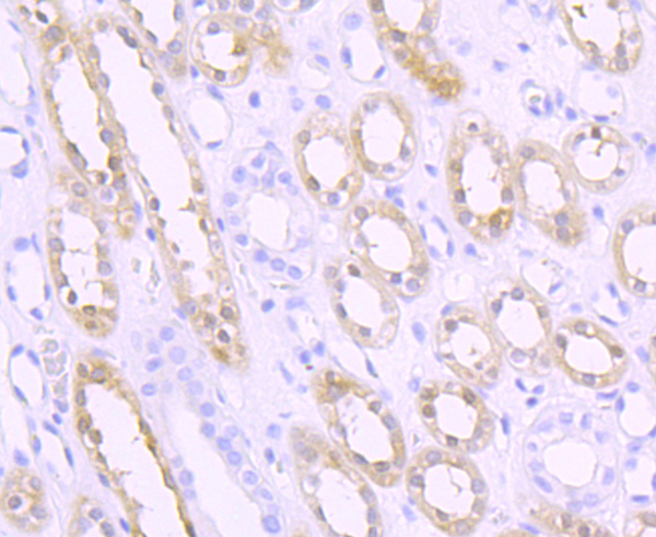
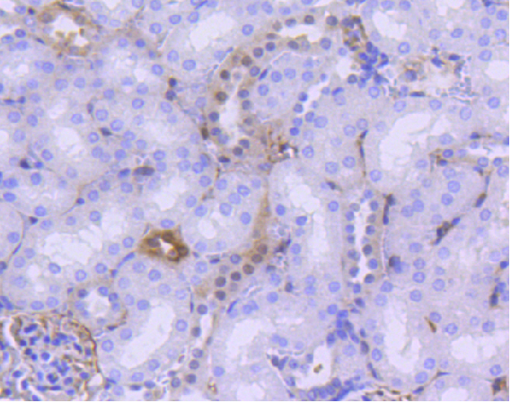 IHC (Immunohistochemisry)
(Immunohistochemical analysis of paraffin-embedded mouse kidney tissue using anti- phospho-YAP1 (S127) antibody. Counter stained with hematoxylin.)
IHC (Immunohistochemisry)
(Immunohistochemical analysis of paraffin-embedded mouse kidney tissue using anti- phospho-YAP1 (S127) antibody. Counter stained with hematoxylin.)
YAP1, Monoclonal Antibody (Cat# AAA311026)
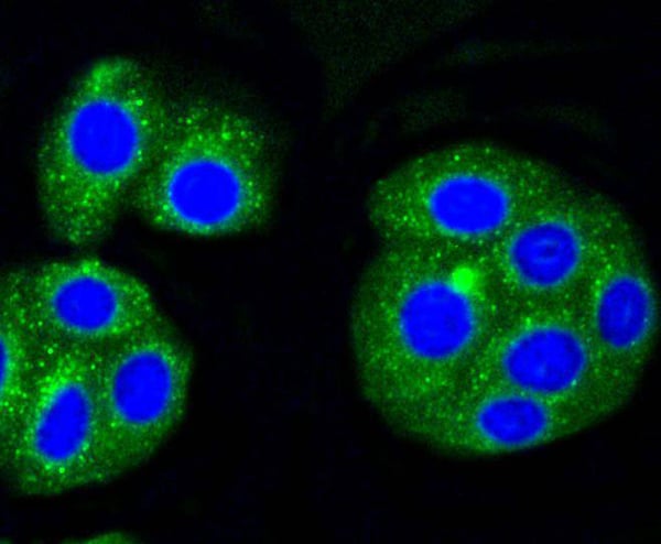

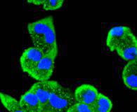
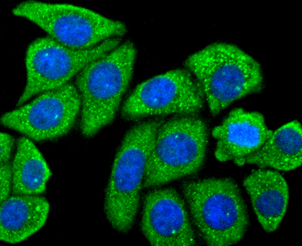
 ICC (Immunocytochemistry)
(ICC staining Alas1 in MCF-7 cells (green). Cells were fixed in paraformaldehyde, permeabilised with 0.25% Triton X100/PBS.)
ICC (Immunocytochemistry)
(ICC staining Alas1 in MCF-7 cells (green). Cells were fixed in paraformaldehyde, permeabilised with 0.25% Triton X100/PBS.)
ALAS1, Monoclonal Antibody (Cat# AAA312141)
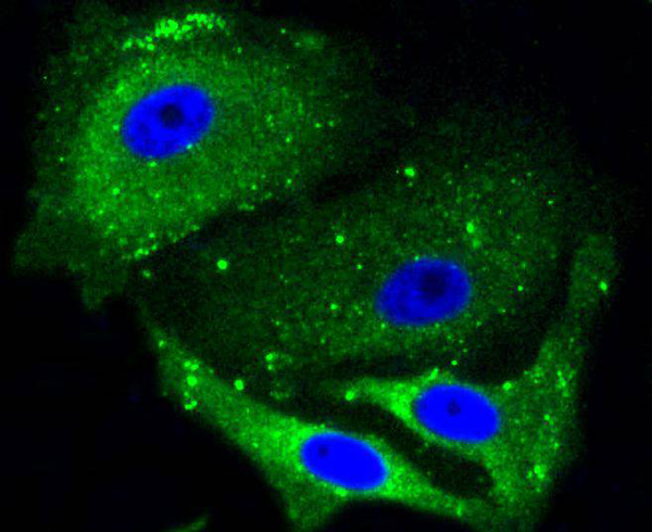
 ICC (Immunocytochemistry)
(ICC staining Bcl10 in SW480 cells (green). The nuclear counter stain is DAPI (blue). Cells were fixed in paraformaldehyde, permeabilised with 0.25% Triton X100/PBS.)
ICC (Immunocytochemistry)
(ICC staining Bcl10 in SW480 cells (green). The nuclear counter stain is DAPI (blue). Cells were fixed in paraformaldehyde, permeabilised with 0.25% Triton X100/PBS.)
BCL10, Monoclonal Antibody (Cat# AAA312306)
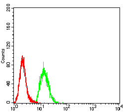
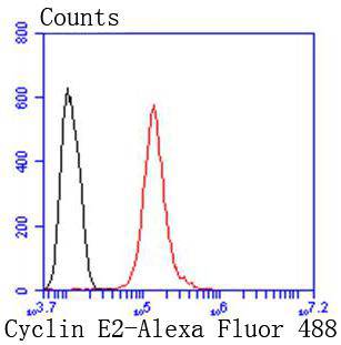 FCM/FACS (Flow Cytometry)
(Flow cytometric analysis of Hela cells with Cyclin E2 antibody at 1/50 dilution (red) compared with an unlabelled control (cells without incubation with primary antibody; black). Alexa Fluor 488-conjugated goat anti rabbit IgG was used as the secondary antibody.)
FCM/FACS (Flow Cytometry)
(Flow cytometric analysis of Hela cells with Cyclin E2 antibody at 1/50 dilution (red) compared with an unlabelled control (cells without incubation with primary antibody; black). Alexa Fluor 488-conjugated goat anti rabbit IgG was used as the secondary antibody.)
Cyclin E2, Monoclonal Antibody (Cat# AAA312321)
 ICC (Immunocytochemistry)
(ICC staining Cyclin B2 in PC-12 cells (green). The nuclear counter stain is DAPI (blue). Cells were fixed in paraformaldehyde, permeabilised with 0.25% Triton X100/PBS.)
ICC (Immunocytochemistry)
(ICC staining Cyclin B2 in PC-12 cells (green). The nuclear counter stain is DAPI (blue). Cells were fixed in paraformaldehyde, permeabilised with 0.25% Triton X100/PBS.)
Cyclin B2, Monoclonal Antibody (Cat# AAA312325)
 ICC (Immunocytochemistry)
(ICC staining MEK5 in SW480 cells (green). The nuclear counter stain is DAPI (blue). Cells were fixed in paraformaldehyde, permeabilised with 0.25% Triton X100/PBS.)
ICC (Immunocytochemistry)
(ICC staining MEK5 in SW480 cells (green). The nuclear counter stain is DAPI (blue). Cells were fixed in paraformaldehyde, permeabilised with 0.25% Triton X100/PBS.)
MEK5, Monoclonal Antibody (Cat# AAA312329)
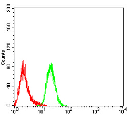
 FCM/FACS (Flow Cytometry)
(Flow cytometric analysis of SH-SY-5Y cells with Tau antibody at 1/50 dilution (red) compared with an unlabelled control (cells without incubation with primary antibody; black). Alexa Fluor 488-conjugated goat anti rabbit IgG was used as the secondary antibody)
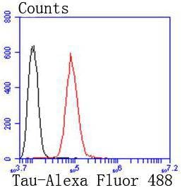
FCM/FACS (Flow Cytometry)
(Flow cytometric analysis of SH-SY-5Y cells with Tau antibody at 1/50 dilution (red) compared with an unlabelled control (cells without incubation with primary antibody; black). Alexa Fluor 488-conjugated goat anti rabbit IgG was used as the secondary antibody)
Tau, Monoclonal Antibody (Cat# AAA312333)
 FCM/FACS (Flow Cytometry)
(Flow cytometric analysis of HepG2 cells with Doublecortin antibody at 1/50 dilution (red) compared with an unlabelled control (cells without incubation with primary antibody; black). Alexa Fluor 488-conjugated goat anti rabbit IgG was used as the secondary antibody)
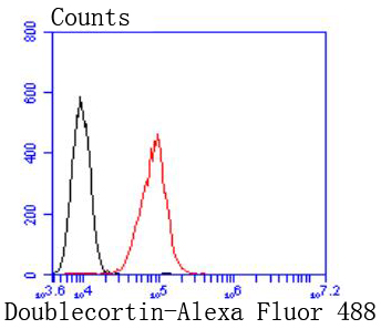
FCM/FACS (Flow Cytometry)
(Flow cytometric analysis of HepG2 cells with Doublecortin antibody at 1/50 dilution (red) compared with an unlabelled control (cells without incubation with primary antibody; black). Alexa Fluor 488-conjugated goat anti rabbit IgG was used as the secondary antibody)
Doublecortin, Monoclonal Antibody (Cat# AAA312394)
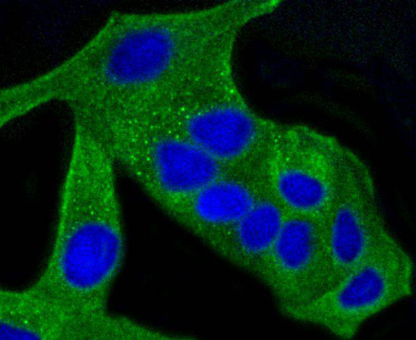
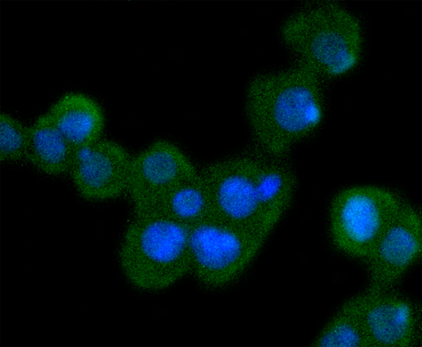
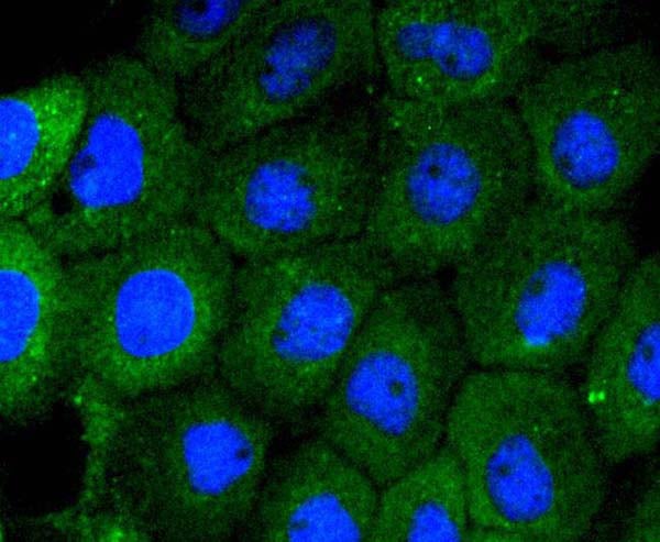
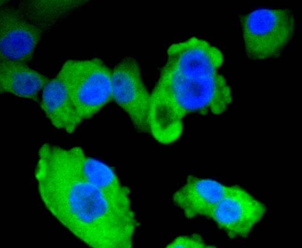
 ICC (Immunocytochemistry)
(ICC staining Mannose Receptor in HepG2 cells (green). The nuclear counter stain is DAPI (blue). Cells were fixed in paraformaldehyde, permeabilised with 0.25% Triton X100/PBS.)
ICC (Immunocytochemistry)
(ICC staining Mannose Receptor in HepG2 cells (green). The nuclear counter stain is DAPI (blue). Cells were fixed in paraformaldehyde, permeabilised with 0.25% Triton X100/PBS.)
Mannose, Monoclonal Antibody (Cat# AAA312397)
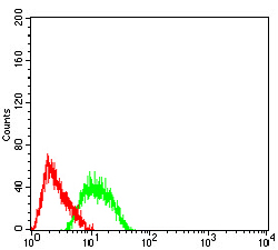
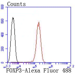 FCM/FACS (Flow Cytometry)
(Flow cytometric analysis of Jurkat cells with FOXP3 antibody at 1/50 dilution (red) compared with an unlabelled control (cells without incubation with primary antibody; black). Alexa Fluor 488-conjugated goat anti rabbit IgG was used as the secondary antibody.)
FCM/FACS (Flow Cytometry)
(Flow cytometric analysis of Jurkat cells with FOXP3 antibody at 1/50 dilution (red) compared with an unlabelled control (cells without incubation with primary antibody; black). Alexa Fluor 488-conjugated goat anti rabbit IgG was used as the secondary antibody.)
FOXP3, Monoclonal Antibody (Cat# AAA312400)
 ICC (Immunocytochemistry)
(ICC staining Progesterone Receptor in MCF-7 cells (green). The nuclear counter stain is DAPI (blue). Cells were fixed in paraformaldehyde, permeabilised with 0.25% Triton X100/PBS.)
ICC (Immunocytochemistry)
(ICC staining Progesterone Receptor in MCF-7 cells (green). The nuclear counter stain is DAPI (blue). Cells were fixed in paraformaldehyde, permeabilised with 0.25% Triton X100/PBS.)
Progesterone, Monoclonal Antibody (Cat# AAA312407)
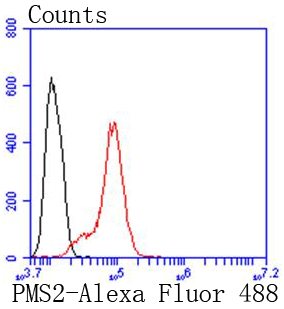
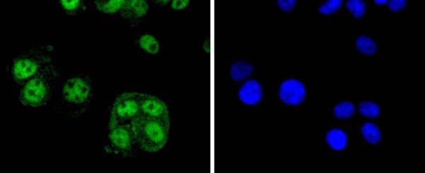
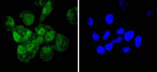
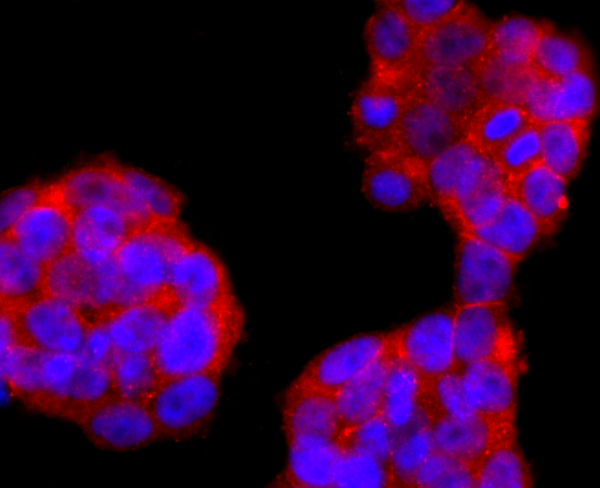
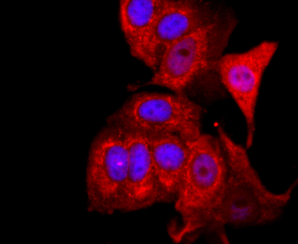
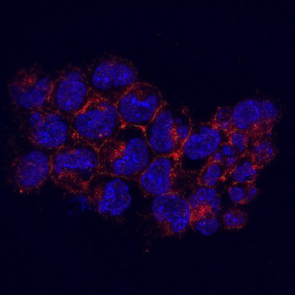
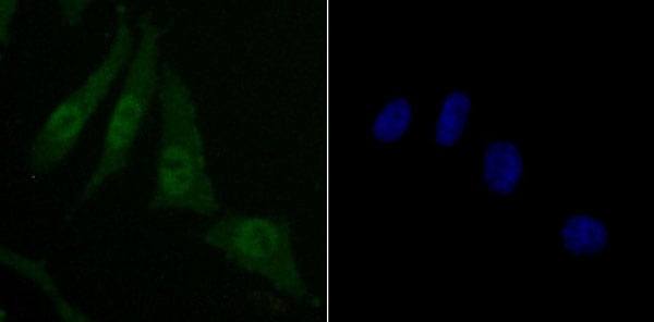
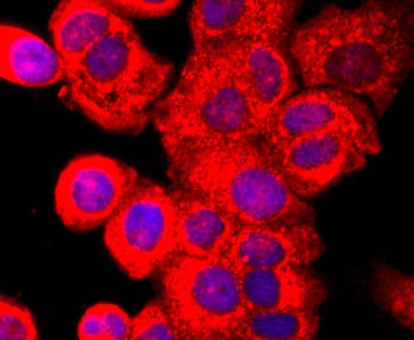
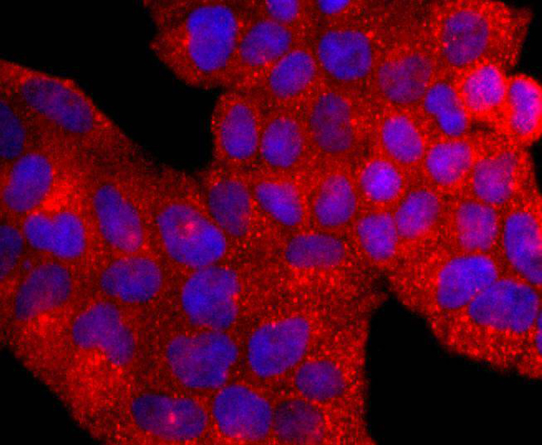
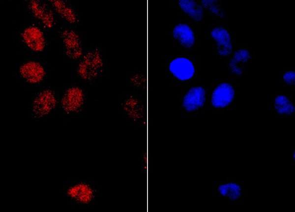 ICC (Immunocytochemistry)
(ICC staining PMS2 in Hela cells (red). Formalin fixed cells were permeabilized with 0.1% Triton X-100 in TBS for 10 minutes at room temperature and blocked with 1% Blocker BSA for 15 minutes at room temperature. Cells were probed with Carbonic anhydrase 2 monoclonal antibody at a dilution of 1:100 for at least 1 hour at room temperature, washed with PBS. Alexa Fluorc 488 Goat anti-Mouse IgG was used as the secondary antibody at 1/100 dilution. The nuclear counter stain is DAPI (blue).)
ICC (Immunocytochemistry)
(ICC staining PMS2 in Hela cells (red). Formalin fixed cells were permeabilized with 0.1% Triton X-100 in TBS for 10 minutes at room temperature and blocked with 1% Blocker BSA for 15 minutes at room temperature. Cells were probed with Carbonic anhydrase 2 monoclonal antibody at a dilution of 1:100 for at least 1 hour at room temperature, washed with PBS. Alexa Fluorc 488 Goat anti-Mouse IgG was used as the secondary antibody at 1/100 dilution. The nuclear counter stain is DAPI (blue).)
PMS2, Monoclonal Antibody (Cat# AAA312175)
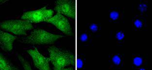
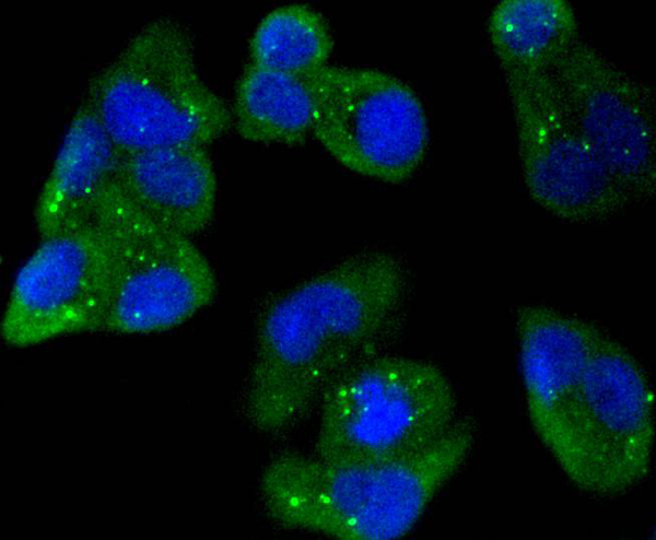
 ICC (Immunocytochemistry)
(ICC staining MKLP1 in A549 cells (green). The nuclear counter stain is DAPI (blue). Cells were fixed in paraformaldehyde, permeabilised with 0.25% Triton X100/PBS.)
ICC (Immunocytochemistry)
(ICC staining MKLP1 in A549 cells (green). The nuclear counter stain is DAPI (blue). Cells were fixed in paraformaldehyde, permeabilised with 0.25% Triton X100/PBS.)
MKLP1, Monoclonal Antibody (Cat# AAA312197)
 FCM/FACS (Flow Cytometry)
(Flow cytometric analysis of Hela cells with AMPK alpha 1 antibody at 1/50 dilution (red) compared with an unlabelled control (cells without incubation with primary antibody; black). Alexa Fluor 488-conjugated goat anti rabbit IgG was used as the secondary antibody.)
FCM/FACS (Flow Cytometry)
(Flow cytometric analysis of Hela cells with AMPK alpha 1 antibody at 1/50 dilution (red) compared with an unlabelled control (cells without incubation with primary antibody; black). Alexa Fluor 488-conjugated goat anti rabbit IgG was used as the secondary antibody.)
AMPK alpha 1, Monoclonal Antibody (Cat# AAA312212)
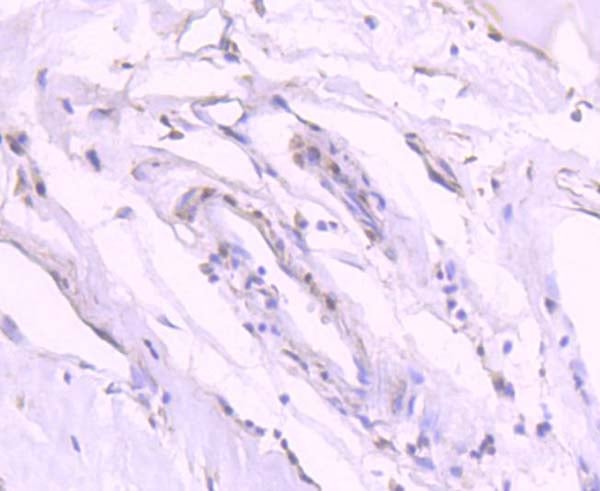
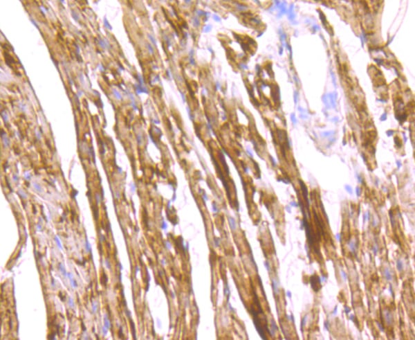
 IHC (Immunohistochemisry)
(Immunohistochemical analysis of paraffin-embedded mouse prostate tissue using anti-Rab5 antibody. Counter stained with hematoxylin.)
IHC (Immunohistochemisry)
(Immunohistochemical analysis of paraffin-embedded mouse prostate tissue using anti-Rab5 antibody. Counter stained with hematoxylin.)
RAB5, Monoclonal Antibody (Cat# AAA312223)


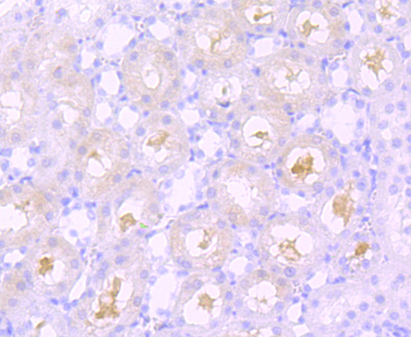
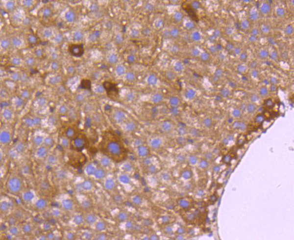 IHC (Immunohistochemistry)
(Immunohistochemical analysis of paraffin-embedded mouse liver tissue using anti-HtrA2 antibody. Counter stained with hematoxylin.)
IHC (Immunohistochemistry)
(Immunohistochemical analysis of paraffin-embedded mouse liver tissue using anti-HtrA2 antibody. Counter stained with hematoxylin.)
HTRA2/Omi, Monoclonal Antibody (Cat# AAA312240)
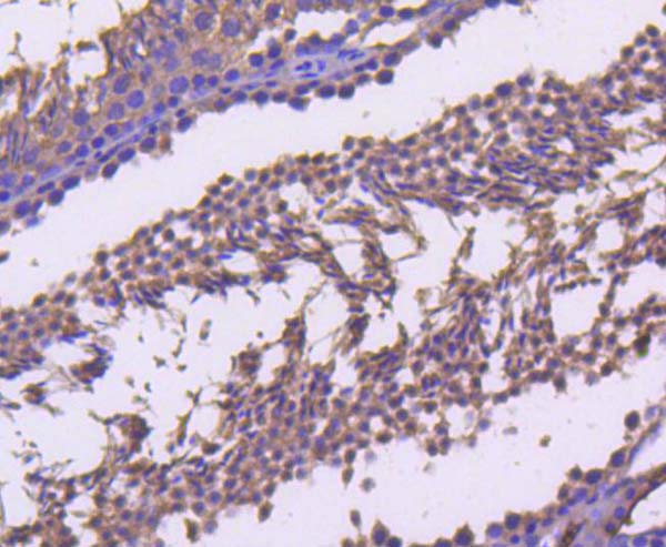
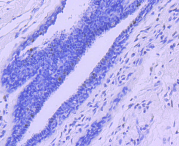
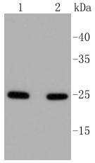
 IHC (Immunohiostchemistry)
(Immunohistochemical analysis of paraffin-embedded mouse fallopian tube tissue using anti-beta Tubulin-HRP in antibody. Counter stained with hematoxylin.)
IHC (Immunohiostchemistry)
(Immunohistochemical analysis of paraffin-embedded mouse fallopian tube tissue using anti-beta Tubulin-HRP in antibody. Counter stained with hematoxylin.)
beta-Tubulin, Monoclonal Antibody (Cat# AAA312019)
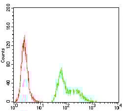
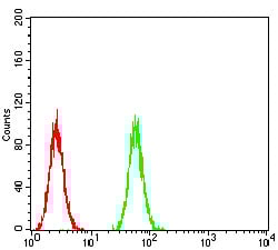 FCM/FACS (Flow Cytometry)
(Flow cytometric analysis of Hela cells with GPCR GPR86 antibody at 1/100 dilution (green) compared with an unlabelled control (cells without incubation with primary antibody; red).)
FCM/FACS (Flow Cytometry)
(Flow cytometric analysis of Hela cells with GPCR GPR86 antibody at 1/100 dilution (green) compared with an unlabelled control (cells without incubation with primary antibody; red).)
GPCR GPR86, Monoclonal Antibody (Cat# AAA312024)
 FCM/FACS (Flow Cytometry)
(Flow cytometric analysis of Hela cells with AP2 alpha antibody at 1/100 dilution (green) compared with an unlabelled control (cells without incubation with primary antibody; red).)
FCM/FACS (Flow Cytometry)
(Flow cytometric analysis of Hela cells with AP2 alpha antibody at 1/100 dilution (green) compared with an unlabelled control (cells without incubation with primary antibody; red).)
TFAP2A, Monoclonal Antibody (Cat# AAA312029)
 FCM/FACS (Flow Cytometry)
(Flow cytometric analysis of Hela cells with KDM3A antibody at 1/100 dilution (green) compared with an unlabelled control (cells without incubation with primary antibody; red).)
FCM/FACS (Flow Cytometry)
(Flow cytometric analysis of Hela cells with KDM3A antibody at 1/100 dilution (green) compared with an unlabelled control (cells without incubation with primary antibody; red).)
KDM3A, Monoclonal Antibody (Cat# AAA312046)
 FCM/FACS (Flow Cytometry)
(Flow cytometric analysis of Hela cells with DHX58 antibody at 1/100 dilution (green) compared with an unlabelled control (cells without incubation with primary antibody; red).)
FCM/FACS (Flow Cytometry)
(Flow cytometric analysis of Hela cells with DHX58 antibody at 1/100 dilution (green) compared with an unlabelled control (cells without incubation with primary antibody; red).)
DHX58, Monoclonal Antibody (Cat# AAA312047)
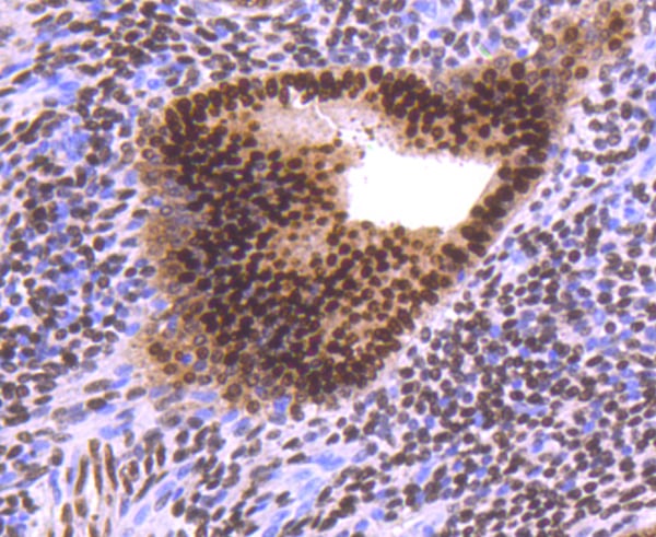
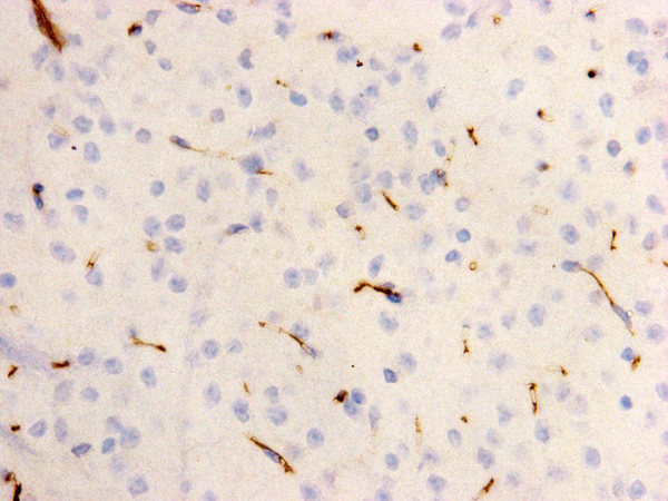
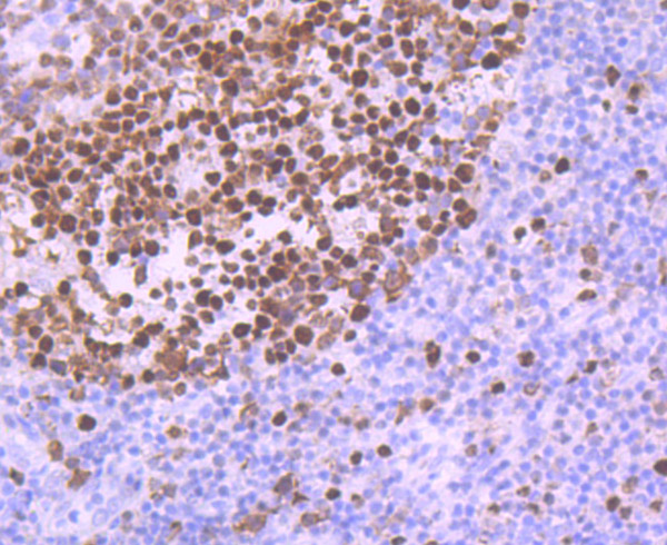
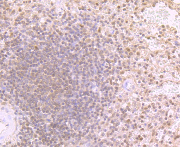
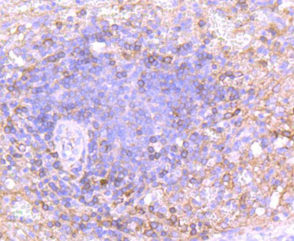
 IHC (Immunohistochemistry)
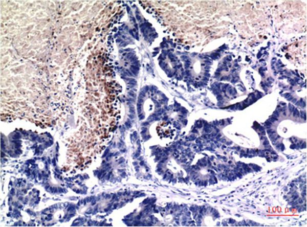
(Immunohistochemical analysis of paraffin-embedded human ovarian cancer tissue using anti- XRN2 antibody. Counter stained with hematoxylin.)
IHC (Immunohistochemistry)
(Immunohistochemical analysis of paraffin-embedded human ovarian cancer tissue using anti- XRN2 antibody. Counter stained with hematoxylin.)
XRN2, Monoclonal Antibody (Cat# AAA312049)
 FCM/FACS (Flow Cytometry)
(Flow cytometric analysis of Hela cells with JARID2 antibody at 1/100 dilution (green) compared with an unlabelled control (cells without incubation with primary antibody; red).)
FCM/FACS (Flow Cytometry)
(Flow cytometric analysis of Hela cells with JARID2 antibody at 1/100 dilution (green) compared with an unlabelled control (cells without incubation with primary antibody; red).)
JARID2, Monoclonal Antibody (Cat# AAA312053)
 FCM/FACS (Flow Cytometry)
(Flow cytometric analysis of SK-N-SH cells with AP2 beta antibody at 1/100 dilution (green) compared with an unlabelled control (cells without incubation with primary antibody; red).)
FCM/FACS (Flow Cytometry)
(Flow cytometric analysis of SK-N-SH cells with AP2 beta antibody at 1/100 dilution (green) compared with an unlabelled control (cells without incubation with primary antibody; red).)
AP2 beta, Monoclonal Antibody (Cat# AAA312057)
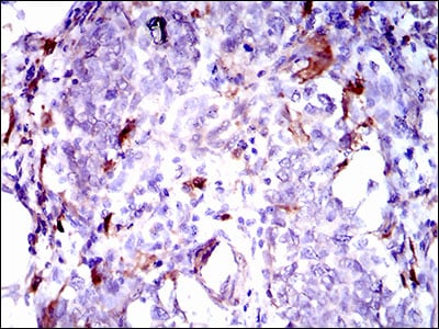
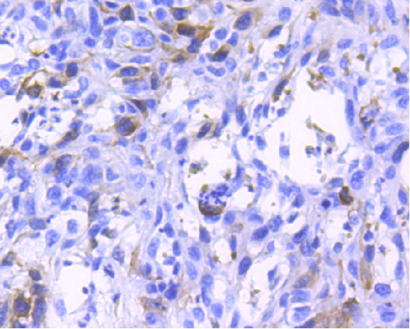
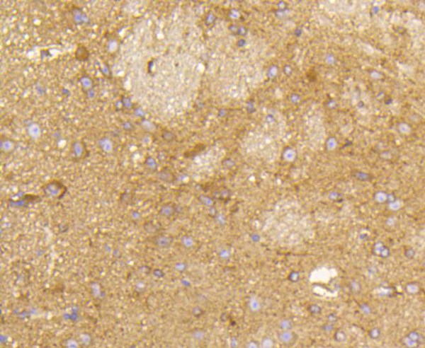
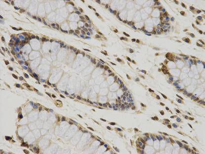
 IHC (Immunohistochemistry)
(Immunohistochemical analysis of paraffin-embedded human esophagus cancer tissue using anti-GPNMB antibody. Counter stained with hematoxylin.)
IHC (Immunohistochemistry)
(Immunohistochemical analysis of paraffin-embedded human esophagus cancer tissue using anti-GPNMB antibody. Counter stained with hematoxylin.)
GPNMB, Monoclonal Antibody (Cat# AAA312065)
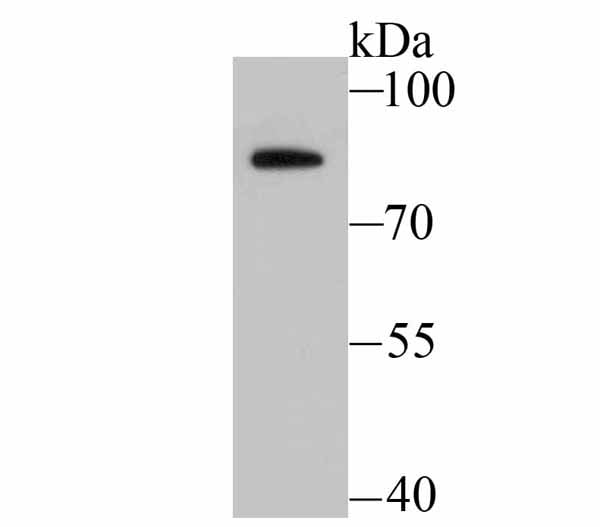
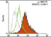 Application Data
(Intracellular FCM analysis of fixed and permeabilized control (green line histogram) and ErbB-4 transfected (solid orange histogram) NIH/3T3 cells. Dotted pink histogram represents the isotype control, normal mouse IgG2a.)
Application Data
(Intracellular FCM analysis of fixed and permeabilized control (green line histogram) and ErbB-4 transfected (solid orange histogram) NIH/3T3 cells. Dotted pink histogram represents the isotype control, normal mouse IgG2a.)
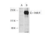
HER4/ErbB4, Monoclonal Antibody (Cat# AAA311987)
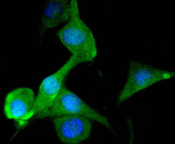
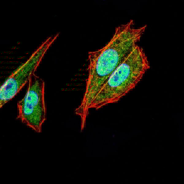
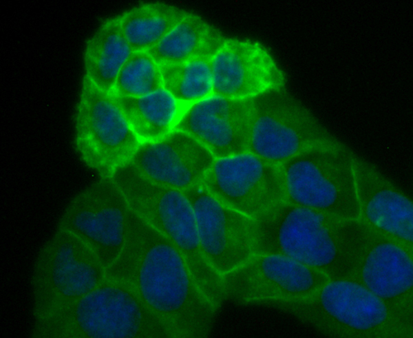
 IF (Immunofluorescence)
(Immunofluorescent staining of F9 cells using anti-Kidins220 Mouse mAb.)
IF (Immunofluorescence)
(Immunofluorescent staining of F9 cells using anti-Kidins220 Mouse mAb.)
Kinase D-Interacting Substrate 220, Monoclonal Antibody (Cat# AAA311878)
 ICC (Immunocytochemistry)
(ICC staining CCDC111 in HepG2 cells (red). Cells were fixed in paraformaldehyde, permeabilised with 0.25% Triton X100/PBS.)
ICC (Immunocytochemistry)
(ICC staining CCDC111 in HepG2 cells (red). Cells were fixed in paraformaldehyde, permeabilised with 0.25% Triton X100/PBS.)
CCDC111, Monoclonal Antibody (Cat# AAA311895)
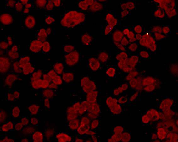
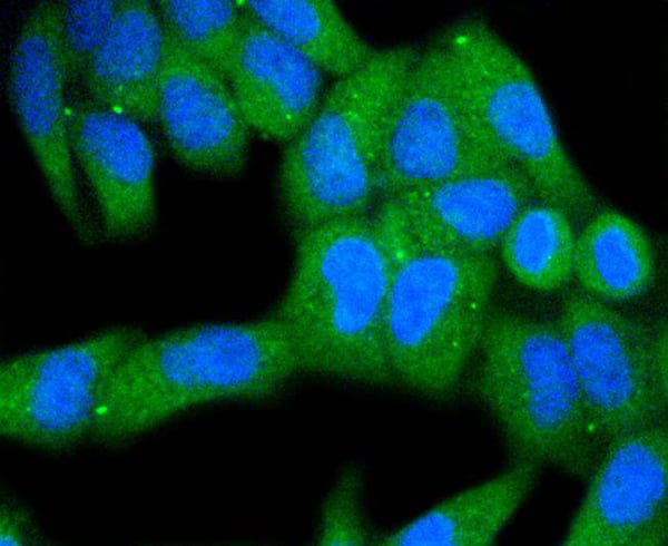
 ICC (Immunocytochemistry)
(ICC staining GRP78 in HepG2 cells (red). Cells were fixed in paraformaldehyde, permeabilised with 0.25% Triton X100/PBS.)
ICC (Immunocytochemistry)
(ICC staining GRP78 in HepG2 cells (red). Cells were fixed in paraformaldehyde, permeabilised with 0.25% Triton X100/PBS.)
GRP78 BiP, Monoclonal Antibody (Cat# AAA311907)
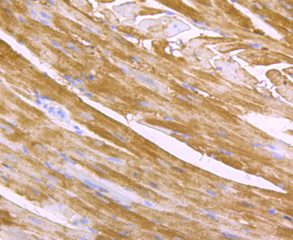
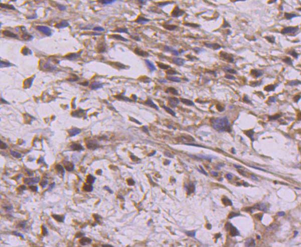
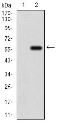
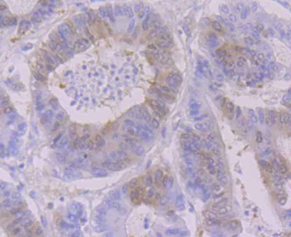
 IHC (Immunohistochemisry)
(Immunohistochemical analysis of paraffin-embedded mouse heart tissue using anti- Cardiac FABP3 antibody. Counter stained with hematoxylin.)
IHC (Immunohistochemisry)
(Immunohistochemical analysis of paraffin-embedded mouse heart tissue using anti- Cardiac FABP3 antibody. Counter stained with hematoxylin.)
FABP3, Monoclonal Antibody (Cat# AAA312581)
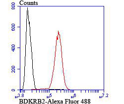 FCM/FACS (Flow Cytometry)
(Flow cytometric analysis of SH-SY5Y cells with BDKRB2 antibody at 1/100 dilution (red) compared with an unlabelled control (cells without incubation with primary antibody; black).)
FCM/FACS (Flow Cytometry)
(Flow cytometric analysis of SH-SY5Y cells with BDKRB2 antibody at 1/100 dilution (red) compared with an unlabelled control (cells without incubation with primary antibody; black).)
BDKRB2, Monoclonal Antibody (Cat# AAA312593)
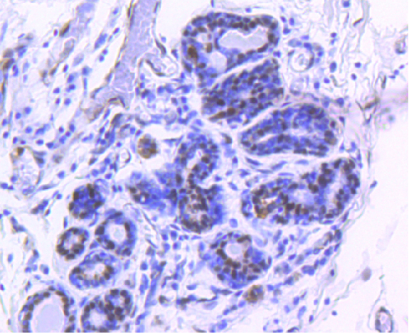
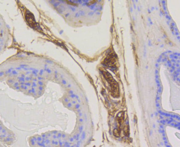
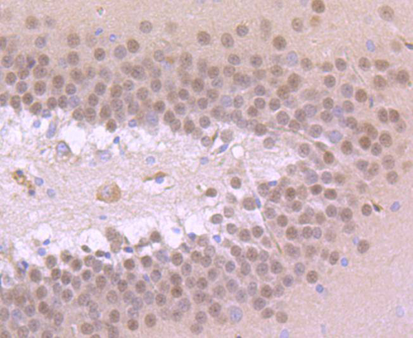
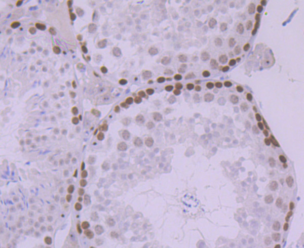 IHC (Immunohistochemistry)
(Immunohistochemical analysis of paraffin-embedded mouse testis tissue using anti-MCM5 antibody. Counter stained with hematoxylin.)
IHC (Immunohistochemistry)
(Immunohistochemical analysis of paraffin-embedded mouse testis tissue using anti-MCM5 antibody. Counter stained with hematoxylin.)
MCM5, Monoclonal Antibody (Cat# AAA312613)
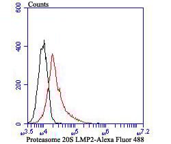 FCM/FACS (Flow Cytometry)
(Flow cytometric analysis of Daudi cells with Proteasome 20S LMP2 antibody at 1/100 dilution (red) compared with an unlabelled control (cells without incubation with primary antibody; black).)
FCM/FACS (Flow Cytometry)
(Flow cytometric analysis of Daudi cells with Proteasome 20S LMP2 antibody at 1/100 dilution (red) compared with an unlabelled control (cells without incubation with primary antibody; black).)
Proteasome 20S LMP2, Monoclonal Antibody (Cat# AAA312616)
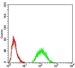
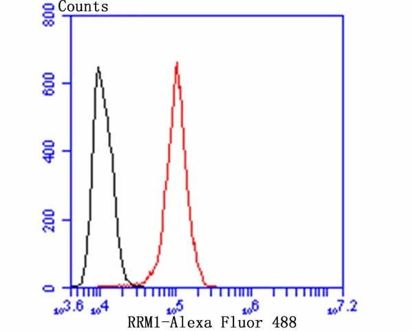 FCM/FACS (Flow Cytometry)
(Flow cytometric analysis of A431 cells with RRM1 antibody at 1/50 dilution (blue) compared with an unlabelled control (cells without incubation with primary antibody; red). Alexa Fluor 488-conjugated goat anti-rabbit IgG was used as the secondary antibody.)
FCM/FACS (Flow Cytometry)
(Flow cytometric analysis of A431 cells with RRM1 antibody at 1/50 dilution (blue) compared with an unlabelled control (cells without incubation with primary antibody; red). Alexa Fluor 488-conjugated goat anti-rabbit IgG was used as the secondary antibody.)
RRM1, Monoclonal Antibody (Cat# AAA312638)
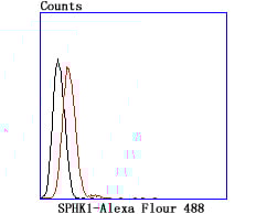 FCM/FACS (Flow Cytometry)
(Flow cytometric analysis of Raji cells with SPHK1 antibody at 1/100 dilution (red) compared with an unlabelled control (cells without incubation with primary antibody; black).)
FCM/FACS (Flow Cytometry)
(Flow cytometric analysis of Raji cells with SPHK1 antibody at 1/100 dilution (red) compared with an unlabelled control (cells without incubation with primary antibody; black).)
SPHK1, Monoclonal Antibody (Cat# AAA312514)
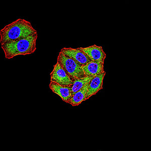
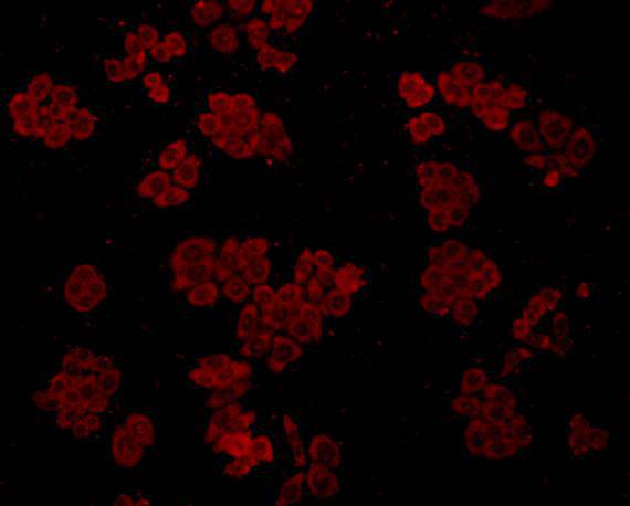
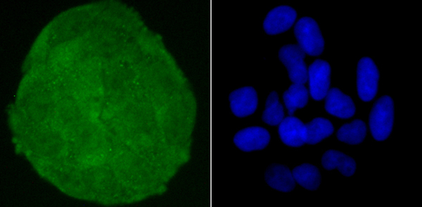
 ICC (Immunocytochemistry)
(ICC staining PLAP in HepG2 cells (green). The nuclear counter stain is DAPI (blue). Cells were fixed in paraformaldehyde, permeabilised with 0.25% Triton X100/PBS.)
ICC (Immunocytochemistry)
(ICC staining PLAP in HepG2 cells (green). The nuclear counter stain is DAPI (blue). Cells were fixed in paraformaldehyde, permeabilised with 0.25% Triton X100/PBS.)
Placental Alkaline Phosphatase, Monoclonal Antibody (Cat# AAA312527)

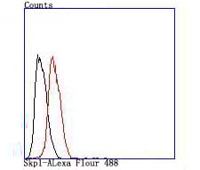 FCM/FACS (Flow Cytometry)
(Flow cytometric analysis of Hela cells with Skp1 antibody at 1/100 dilution (red) compared with an unlabelled control (cells without incubation with primary antibody; black).)
FCM/FACS (Flow Cytometry)
(Flow cytometric analysis of Hela cells with Skp1 antibody at 1/100 dilution (red) compared with an unlabelled control (cells without incubation with primary antibody; black).)
SKP1, Monoclonal Antibody (Cat# AAA312535)
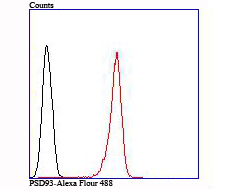 FCM/FACS (Flow Cytometry)
(Flow cytometric analysis of SH-SY5Y cells with PSD93 antibody at 1/100 dilution (red) compared with an unlabelled control (cells without incubation with primary antibody; black).)
FCM/FACS (Flow Cytometry)
(Flow cytometric analysis of SH-SY5Y cells with PSD93 antibody at 1/100 dilution (red) compared with an unlabelled control (cells without incubation with primary antibody; black).)
PSD93, Monoclonal Antibody (Cat# AAA312556)
 ICC (Immunocytochemistry)
(ICC staining GluR2 in SH-SY5Y cells (green). The nuclear counter stain is DAPI (blue). Cells were fixed in paraformaldehyde, permeabilised with 0.25% Triton X100/PBS.)
ICC (Immunocytochemistry)
(ICC staining GluR2 in SH-SY5Y cells (green). The nuclear counter stain is DAPI (blue). Cells were fixed in paraformaldehyde, permeabilised with 0.25% Triton X100/PBS.)
GluR2, Monoclonal Antibody (Cat# AAA312561)
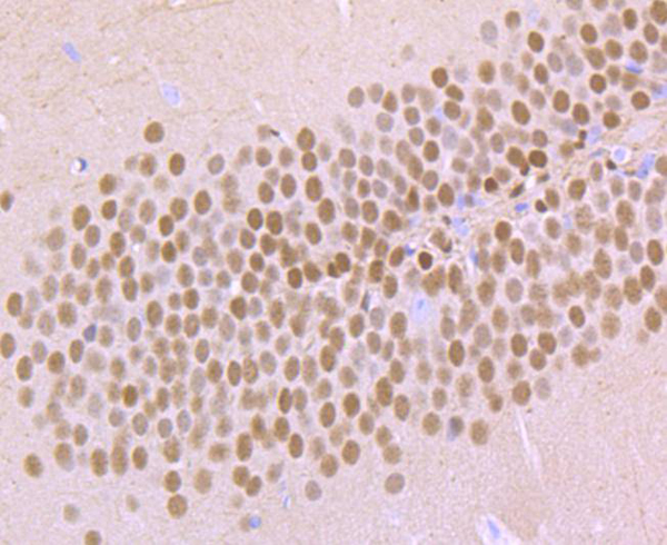
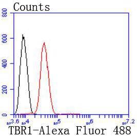 FCM/FACS (Flow Cytometry)
(Flow cytometric analysis of SH-SY-5Y cells with TBR1 antibody at 1/50 dilution (red) compared with an unlabelled control (cells without incubation with primary antibody; black). Alexa Fluor 488-conjugated goat anti rabbit IgG was used as the secondary antibody)
FCM/FACS (Flow Cytometry)
(Flow cytometric analysis of SH-SY-5Y cells with TBR1 antibody at 1/50 dilution (red) compared with an unlabelled control (cells without incubation with primary antibody; black). Alexa Fluor 488-conjugated goat anti rabbit IgG was used as the secondary antibody)
TBR1, Monoclonal Antibody (Cat# AAA312444)
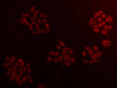
 ICC (Immunocytochemistry)
(ICC staining IgA in HepG2 cells (red). The nuclear counter stain is DAPI (blue). Cells were fixed in paraformaldehyde, permeabilised with 0.25% Triton X100/PBS.)
ICC (Immunocytochemistry)
(ICC staining IgA in HepG2 cells (red). The nuclear counter stain is DAPI (blue). Cells were fixed in paraformaldehyde, permeabilised with 0.25% Triton X100/PBS.)
IgA, Monoclonal Antibody (Cat# AAA312450)
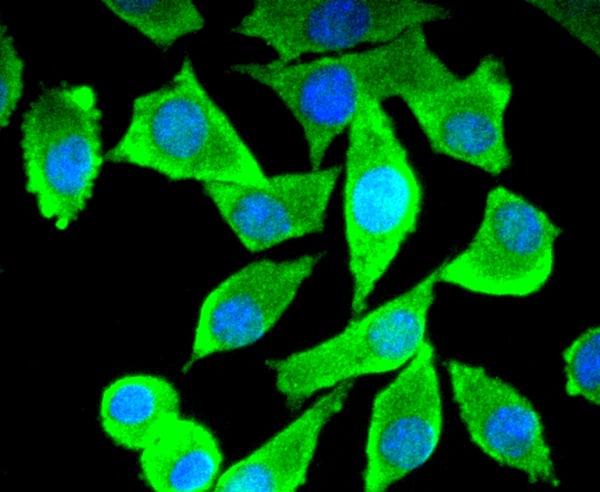
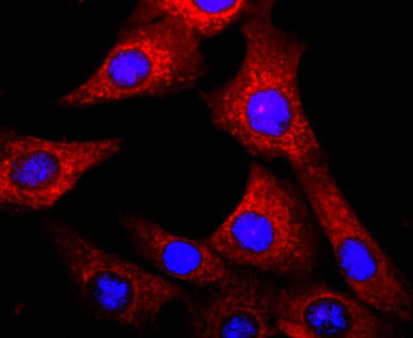 ICC (Immunocytochemistry)
(ICC staining AhR in NIH/3T3 cells (red). The nuclear counter stain is DAPI (blue). Cells were fixed in paraformaldehyde, permeabilised with 0.25% Triton X100/PBS.)
ICC (Immunocytochemistry)
(ICC staining AhR in NIH/3T3 cells (red). The nuclear counter stain is DAPI (blue). Cells were fixed in paraformaldehyde, permeabilised with 0.25% Triton X100/PBS.)
Aryl Hydrocarbon, Monoclonal Antibody (Cat# AAA312451)
What are Monoclonal Antibodies?
Monoclonal antibodies are specialized laboratory-produced proteins developed for binding to specific biological antigens or other molecular targets. Since they come from a single cell (or clone), they are especially consistent and accurate in the data they are involved in producing.
This type of antibody material has been shown to be a powerful tool in finding and subsequently destroying harmful cells in an organism, such as those found in cancers or various autoimmune diseases. This makes them excellent aids in medical testing and research, which is why they are so widely used.
AAA Biotech offers a comprehensive range of high-quality monoclonal antibodies that perform effectively in various laboratory tests, including (amongst others) ELISA, western blotting, immunohistochemistry, and flow cytometry. All of the products in our catalog are thoroughly quality tested to make sure that they are reliable and will consistently perform well in your research.
What Are The Uses of Monoclonal Antibodies
Monoclonal antibodies are used in many lab tests, including (amongst others) ELISA, western blotting, immunohistochemistry, and flow cytometry.
ELISA is a test that helps detect a specific substance/analyte in a sample. It uses antibodies (often monoclonal) bound to a solid surface (such as the well of a microplate) to “capture” the substance/analyte in the sample and immobilize it so that the detection antibody component can then bind to it and produce a signal, which can then be measured.
Western blotting identifies specific proteins in a sample. The sample is first separated on a gel, and then antibodies are applied that will typically bind to the target, which will all be localized to a single band in a lane.
Immunohistochemistry helps locate specific proteins in cells or tissue samples using antibodies.
Flow cytometry looks at and sorts cells. It uses antibodies that are conjugated to reporter molecules called “fluorophores”, which, under special lights, emit light themselves, which can then be measured by a detector instrument.
How Monoclonal Antibodies Are Used as Medicine?
Please note that all of the products listed in AAA Biotech’s also known as AAA Bio or AAABio catalog are strictly for research-use only (RUO).
Monoclonal antibodies can also be used as therapeutic/medical treatments, particularly in the context of cancers. They are designed to find and bind to specific cells or proteins, helping the immune system recognize and attack the cancer. These treatments work in different ways, such as:
- Radioimmunotherapy attaches a small amount of radioactive molecule to the antibody, so it delivers the radiation directly to the cancer cells that the antibody is specifically binding to.
- Antibody-directed enzyme prodrug therapy uses antibodies that are specifically bound to special enzymes. These enzymes activate a harmless drug in the body and turn it into a cancer-killing drug only near the cancer cells—this helps avoid harming healthy cells.
- Immunoliposomes are tiny “bubbles” filled with medicine/drug and coated with antibodies. They carry the drug straight to the cancer cells.
Why Buy Monoclonal Antibodies From Us?
At AAA Biotech, we provide high-performance monoclonal antibodies designed to support a wide range of research needs.
1. Validated for Versatile Applications
The antibodies in our catalog are extensively validated and compatible with multiple techniques, including (but not limited to) ELISA, flow cytometry (FC), immunocytochemistry (ICC), immunofluorescence (IF), immunohistochemistry (IHC), immunoprecipitation (IP), and western blotting (WB).
2. Wide Selection & Specialized Options
We offer antibodies for common and rare species, that are available in various conjugated forms, and also in recombinant formats. Essentially, there is almost anything one might need to meet their experimental model’s requirements.
3. High-Quality Proteins
Our proteins meet high purity standards—90% or more as confirmed by SDS-PAGE. Many are available with tags like His, Flag, GST, or MBP, and we also supply native and biologically active proteins for functional studies.
Frequently Asked Questions
1. Are your monoclonal antibodies validated for specific applications?
Yes, our antibodies are tested and validated for use in methods such as ELISA, western blot, IHC, flow cytometry, and more. Refer to specific product pages or datasheets for individual product information.
2. How do I choose the right monoclonal antibody for my application?
Review the product details directly for application validation, species reactivity, and target information. You may also contact our support team at any time for help.
3. How quickly can I receive my order?
Most orders are processed and shipped within 1–3 business days, depending on product availability and your shipping location.