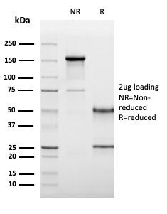
Filters
▼Clonality
▼Type
▼Reactivity
▼Gene Name
▼Isotype
▼Host
▼Application
▼Clone
▼Monoclonal Antibodies
Get accurate results in your research with our Monoclonal Antibodies, which are specially made to target exactly what you require for your research, and will produce consistent, reliable performance in lab tests.
Viewing 9250-9300 of 27597 product results
 IHC (Immunohistochemisry)
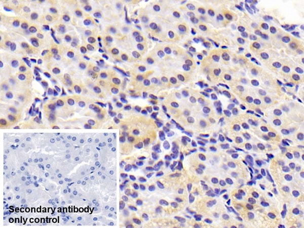
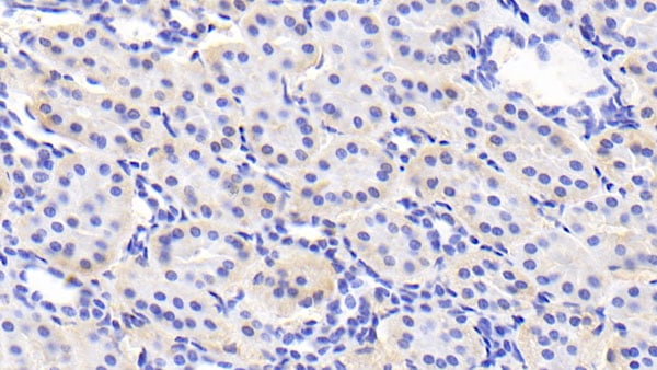
(Immunohistochemical analysis of paraffin-embedded human thyroid tissue using anti-L1CAM antibody. Counter stained with hematoxylin.)
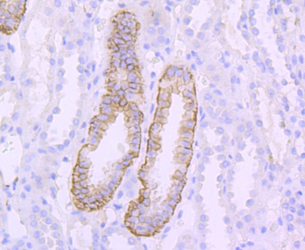
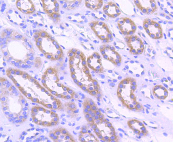
IHC (Immunohistochemisry)
(Immunohistochemical analysis of paraffin-embedded human thyroid tissue using anti-L1CAM antibody. Counter stained with hematoxylin.)
L1CAM, Monoclonal Antibody (Cat# AAA312466)
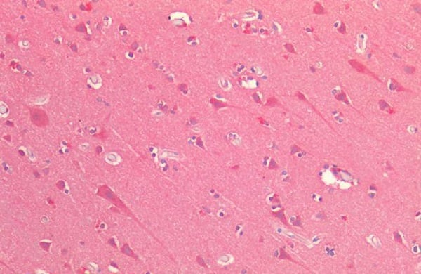
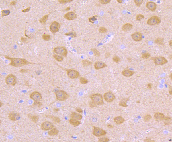
 IHC (Immunohistochemisry)
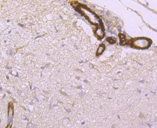
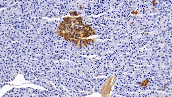
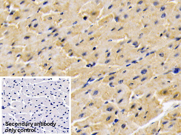
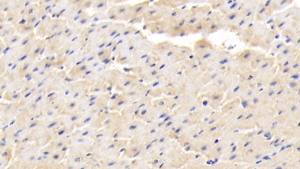
(Immunohistochemical analysis of paraffin-embedded rat brain tissue using anti- Hepcidin antibody. Counter stained with hematoxylin.)
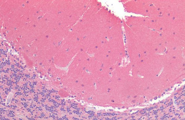
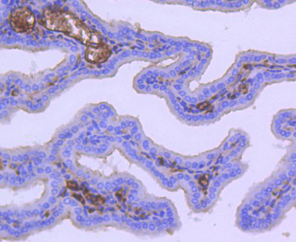
IHC (Immunohistochemisry)
(Immunohistochemical analysis of paraffin-embedded rat brain tissue using anti- Hepcidin antibody. Counter stained with hematoxylin.)
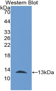
Hepcidin, Monoclonal Antibody (Cat# AAA312488)
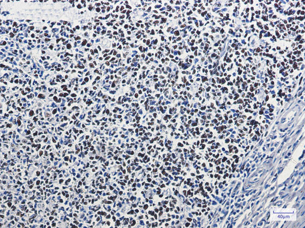
CD8a, Monoclonal Antibody (Cat# AAA129127)
CD79a, Monoclonal Antibody (Cat# AAA129134)
TCR alpha/beta, Monoclonal Antibody (Cat# AAA129175)
CD61, Monoclonal Antibody (Cat# AAA129037)
CD61, Monoclonal Antibody (Cat# AAA129040)
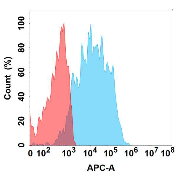
 FCM/FACS (Flow Cytometry)
(Figure 2. Flow cytometry analysis of antigen binding of anti-human TIM-1 mAb(AAA129465).)
FCM/FACS (Flow Cytometry)
(Figure 2. Flow cytometry analysis of antigen binding of anti-human TIM-1 mAb(AAA129465).)
TIM-1, Monoclonal Antibody (Cat# AAA129465)
SCIMP, Monoclonal Antibody (Cat# AAA128910)
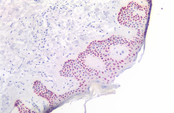
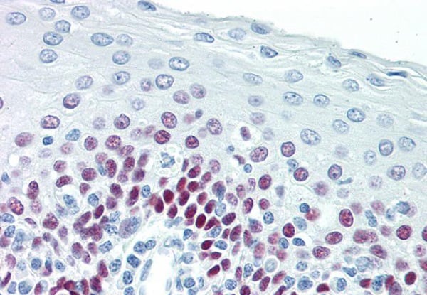 IHC (Immunohiostchemistry)
(TP63/p63 Antibody-Human Tonsil: Formalin-Fixed, Paraffin-Embedded (FFPE))
IHC (Immunohiostchemistry)
(TP63/p63 Antibody-Human Tonsil: Formalin-Fixed, Paraffin-Embedded (FFPE))
TP63/p63, Monoclonal Antibody (Cat# AAA163088)
 IHC (Immunohistochemisry)
(OSMR/IL-31R-Beta Antibody-Human Brain, Cerebellum: Formalin-Fixed, Paraffin-Embedded (FFPE))
IHC (Immunohistochemisry)
(OSMR/IL-31R-Beta Antibody-Human Brain, Cerebellum: Formalin-Fixed, Paraffin-Embedded (FFPE))
OSMR/IL-31R-Beta, Monoclonal Antibody (Cat# AAA163337)
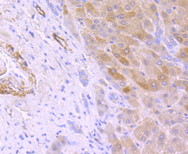
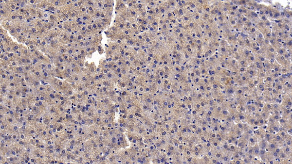
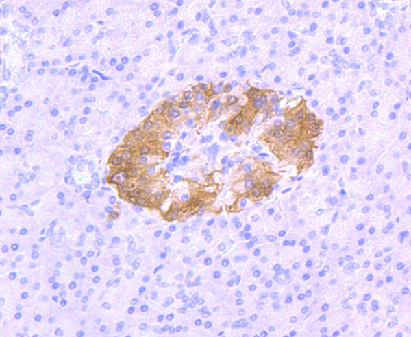
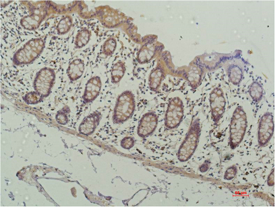
 IHC (Immunohiostchemistry)
(DAB staining on IHC-P;Sample: Rat Stomach Tissue; Primary Ab: 20ug/ml Mouse Anti-human FGF9 AntibodySecond Ab: 2ug/mL HRP-Linked Caprine Anti-Mouse IgG Polyclonal Antibody)
IHC (Immunohiostchemistry)
(DAB staining on IHC-P;Sample: Rat Stomach Tissue; Primary Ab: 20ug/ml Mouse Anti-human FGF9 AntibodySecond Ab: 2ug/mL HRP-Linked Caprine Anti-Mouse IgG Polyclonal Antibody)
Fibroblast Growth Factor 9 (FGF9), Monoclonal Antibody (Cat# AAA152563)
Casein Kappa (CSN3), Monoclonal Antibody (Cat# AAA162031)
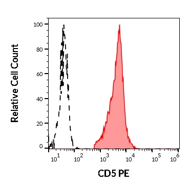
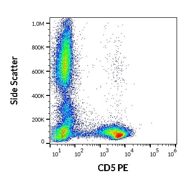 FCM/FACS (Flow Cytometry)
(Flow cytometry surface staining pattern of human peripheral whole blood stained using anti-human CD5 (L17F12) PE antibody (10 ul reagent / 100 ul of peripheral whole blood).)
FCM/FACS (Flow Cytometry)
(Flow cytometry surface staining pattern of human peripheral whole blood stained using anti-human CD5 (L17F12) PE antibody (10 ul reagent / 100 ul of peripheral whole blood).)
CD5, Monoclonal Antibody (Cat# AAA128653)
CD8a, Monoclonal Antibody (Cat# AAA128365)
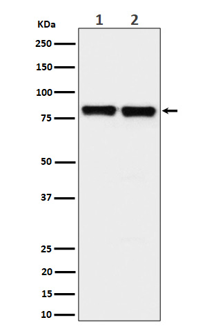
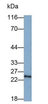 WB (Western Blot)
(Western Blot; Sample: PC3 cell lysate Primary Ab: 0.5ug/ml Mouse Anti-human PSMD10 Antibody Second Ab: 0.2ug/mL HRP-Linked Caprine Anti-Mouse IgG Polyclonal Antibody)
WB (Western Blot)
(Western Blot; Sample: PC3 cell lysate Primary Ab: 0.5ug/ml Mouse Anti-human PSMD10 Antibody Second Ab: 0.2ug/mL HRP-Linked Caprine Anti-Mouse IgG Polyclonal Antibody)
Proteasome 26S Subunit, Non ATPase 10 (PSMD10), Monoclonal Antibody (Cat# AAA152944)
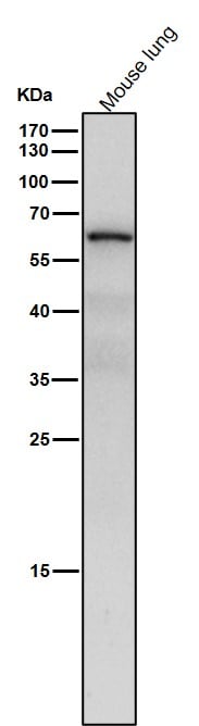
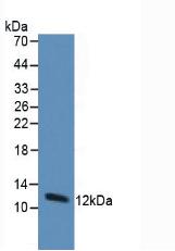
 WB (Western Blot)
(Western Blot; Sample: Porcine SeuM Primary Ab: 2ug/ml Mouse Anti-Porcine CLU Antibody Second Ab: 0.2ug/mL HRP-Linked Caprine Anti-Mouse IgG Polyclonal Antibody)
WB (Western Blot)
(Western Blot; Sample: Porcine SeuM Primary Ab: 2ug/ml Mouse Anti-Porcine CLU Antibody Second Ab: 0.2ug/mL HRP-Linked Caprine Anti-Mouse IgG Polyclonal Antibody)
Clusterin (CLU), Monoclonal Antibody (Cat# AAA152836)
 WB (Western Blot)
(All lanes use the Antibody at 1:1W dilution for 1 hour at room temperature.)
WB (Western Blot)
(All lanes use the Antibody at 1:1W dilution for 1 hour at room temperature.)
PKC delta + PKC theta, Monoclonal Antibody (Cat# AAA128127)
 WB (Western Blot)
(Western blot analysis of Phospho-Chk1 (S280) expression in 293T treated with Calyculin A cell lysate.)
WB (Western Blot)
(Western blot analysis of Phospho-Chk1 (S280) expression in 293T treated with Calyculin A cell lysate.)
Chk1, Monoclonal Antibody (Cat# AAA128129)


 WB (Western Blot)
(All lanes use the Antibody at 1:4K dilution for 1 hour at room temperature.)
WB (Western Blot)
(All lanes use the Antibody at 1:4K dilution for 1 hour at room temperature.)
NR2C2/TR4, Monoclonal Antibody (Cat# AAA128150)
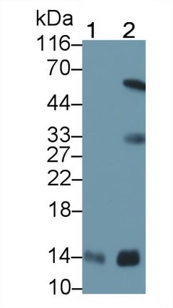
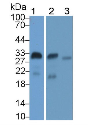
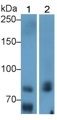
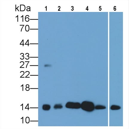 WB (Western Blot)
(Western Blot; Sample: Lane1: Gallus Heart lysate; Lane2: Canine Heart lysate; Lane3: Caprine Heart lysate; Lane4: Cavia Heart lysate; Lane5: Rabbit Heart lysate; Lane6: Equine Heart lysatePrimary Ab: 0.2ug/ml Mouse AntiHuman CYCS AntibodySecond Ab: 0.2ug/mL HRPLinked Caprine AntiMouse IgG Polyclonal Antibody(Catalog: SAA544Mu19))
WB (Western Blot)
(Western Blot; Sample: Lane1: Gallus Heart lysate; Lane2: Canine Heart lysate; Lane3: Caprine Heart lysate; Lane4: Cavia Heart lysate; Lane5: Rabbit Heart lysate; Lane6: Equine Heart lysatePrimary Ab: 0.2ug/ml Mouse AntiHuman CYCS AntibodySecond Ab: 0.2ug/mL HRPLinked Caprine AntiMouse IgG Polyclonal Antibody(Catalog: SAA544Mu19))
Cytochrome C (CYCS), Monoclonal Antibody (Cat# AAA151581)
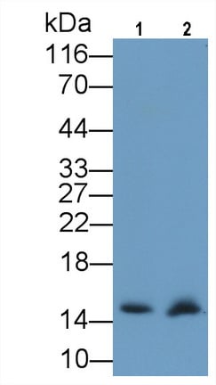
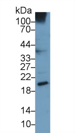
 WB (Western Blot)
(Western Blot; Sample: Porcine Skin lysate Primary Ab: 6ug/ml Mouse AntiMultispecies PIIINP Antibody Second Ab: 0.2ug/mL HRPLinked Caprine AntiMouse IgG Polyclonal Antibody (Catalog: SAA544Mu19))
WB (Western Blot)
(Western Blot; Sample: Porcine Skin lysate Primary Ab: 6ug/ml Mouse AntiMultispecies PIIINP Antibody Second Ab: 0.2ug/mL HRPLinked Caprine AntiMouse IgG Polyclonal Antibody (Catalog: SAA544Mu19))
Procollagen III NTerminal Propeptide (PIIINP), Monoclonal Antibody (Cat# AAA151577)
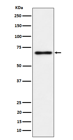
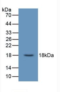
 WB (Western Blot)
(Western Blot Sample: Lane1: human SeuM; Lane2: Rat Heart lysate; Lane3: HepG2 cell lysate Primary Ab: 2 ug/ml Mouse Anti-human ADPN Antibody Second Ab: 0.2ug/mL HRP-Linked Caprine Anti-Mouse IgG Polyclonal Antibody)
WB (Western Blot)
(Western Blot Sample: Lane1: human SeuM; Lane2: Rat Heart lysate; Lane3: HepG2 cell lysate Primary Ab: 2 ug/ml Mouse Anti-human ADPN Antibody Second Ab: 0.2ug/mL HRP-Linked Caprine Anti-Mouse IgG Polyclonal Antibody)
Adiponectin (ADPN), Monoclonal Antibody (Cat# AAA152569)
 WB (Western Blot)
(Western Blot:Sample: Human A549 cell lysate;Primary Ab: 3ug/ml Mouse Anti-Human UCN3 AntibodySecond Ab: 0.2ug/mL HRP-Linked Caprine Anti-Mouse IgG Polyclonal Antibody)
WB (Western Blot)
(Western Blot:Sample: Human A549 cell lysate;Primary Ab: 3ug/ml Mouse Anti-Human UCN3 AntibodySecond Ab: 0.2ug/mL HRP-Linked Caprine Anti-Mouse IgG Polyclonal Antibody)
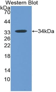
Urocortin 3 (UCN3), Monoclonal Antibody (Cat# AAA130610)
 WB (Western Blot)
(Western Blot: Sample: Recombinant Trx, Human.)
WB (Western Blot)
(Western Blot: Sample: Recombinant Trx, Human.)
Thioredoxin (Trx), Monoclonal Antibody (Cat# AAA130621)
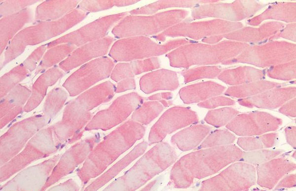
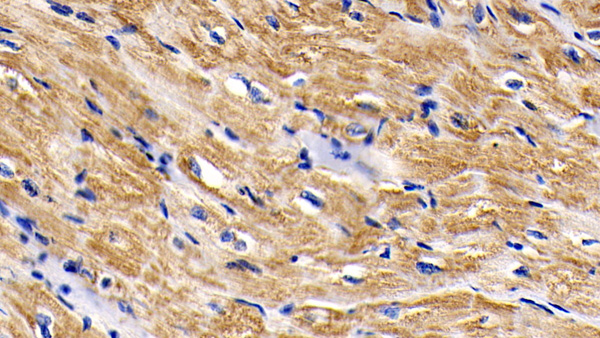
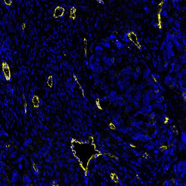
 IHC (Immunohistochemistry)
(DAB staining on IHC-P;Samples: Human Cardiac Muscle Tissue;Primary Ab: 20ug/ml Mouse Anti-Human MX1 AntibodySecond Ab: 2ug/mL HRPLinked Caprine Anti-Mouse IgG Polyclonal Antibody (Catalog: ))
IHC (Immunohistochemistry)
(DAB staining on IHC-P;Samples: Human Cardiac Muscle Tissue;Primary Ab: 20ug/ml Mouse Anti-Human MX1 AntibodySecond Ab: 2ug/mL HRPLinked Caprine Anti-Mouse IgG Polyclonal Antibody (Catalog: ))
Myxovirus Resistance 1 (MX1), Monoclonal Antibody (Cat# AAA130646)
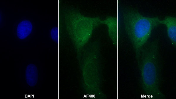
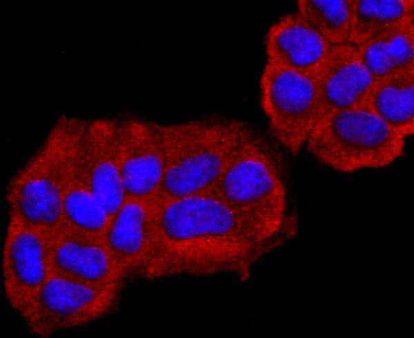
 ICC (Immunocytochemistry)
(ICC staining Cdx1 in HOVO cells (green). The nuclear counter stain is DAPI (blue). Cells were fixed in paraformaldehyde, permeabilised with 0.25% Triton X100/PBS.)
ICC (Immunocytochemistry)
(ICC staining Cdx1 in HOVO cells (green). The nuclear counter stain is DAPI (blue). Cells were fixed in paraformaldehyde, permeabilised with 0.25% Triton X100/PBS.)
CDX1, Monoclonal Antibody (Cat# AAA312677)
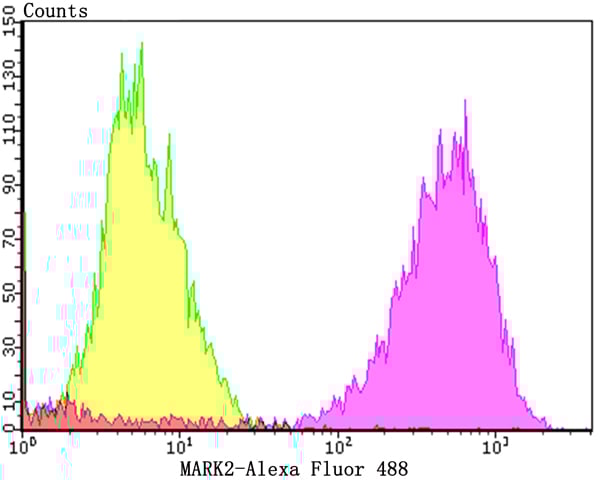 FCM/FACS (Flow Cytometry)
(Flow cytometric analysis of MCF-7 cells with MARK2 antibody at 1/100 dilution (purple) compared with an unlabelled control (cells without incubation with primary antibody; yellow). Alexa Fluor 488-conjugated goat anti-rabbit IgG was used as the secondary antibody.)
FCM/FACS (Flow Cytometry)
(Flow cytometric analysis of MCF-7 cells with MARK2 antibody at 1/100 dilution (purple) compared with an unlabelled control (cells without incubation with primary antibody; yellow). Alexa Fluor 488-conjugated goat anti-rabbit IgG was used as the secondary antibody.)
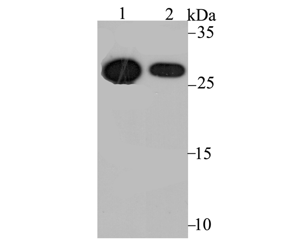
MARK2, Monoclonal Antibody (Cat# AAA312696)
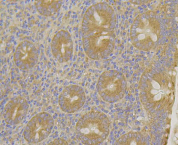
 IHC (Immunohiostchemistry)
(Immunohistochemical analysis of paraffin-embedded mouse placenta tissue using anti-Acetyl CoA Carboxylase 1 antibody. Counter stained with hematoxylin.)
IHC (Immunohiostchemistry)
(Immunohistochemical analysis of paraffin-embedded mouse placenta tissue using anti-Acetyl CoA Carboxylase 1 antibody. Counter stained with hematoxylin.)
Acetyl CoA Carboxylase 1 (ACC1), Monoclonal Antibody (Cat# AAA312708)
ANP, Monoclonal Antibody (Cat# AAA312823)
NR1D1, Monoclonal Antibody (Cat# AAA312825)
IgG, Monoclonal Secondary Antibody (Cat# AAA312920)
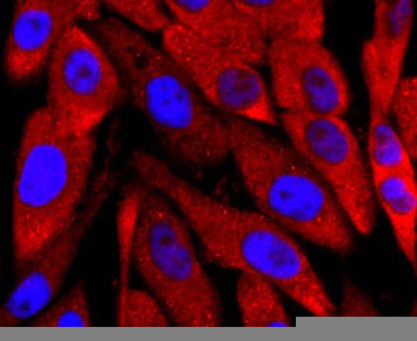
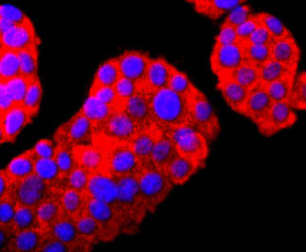 ICC (Immunocytochemistry)
(ICC staining PGP9.5 in PC-12 cells (red). The nuclear counter stain is DAPI (blue). Cells were fixed in paraformaldehyde, permeabilised with 0.25% Triton X100/PBS.)
ICC (Immunocytochemistry)
(ICC staining PGP9.5 in PC-12 cells (red). The nuclear counter stain is DAPI (blue). Cells were fixed in paraformaldehyde, permeabilised with 0.25% Triton X100/PBS.)
PGP9.5, Monoclonal Antibody (Cat# AAA312436)
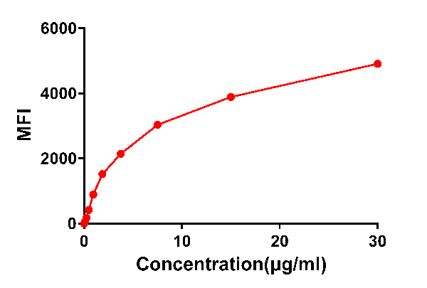
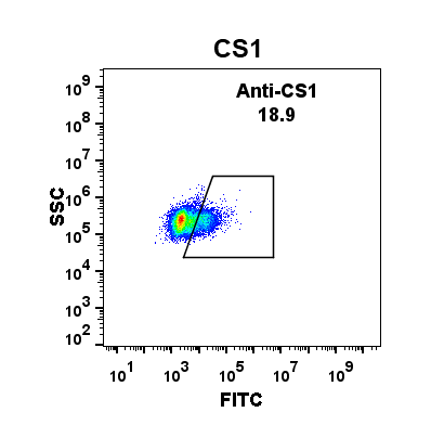
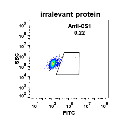
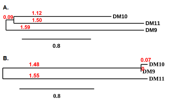 Application Data
(Figure 4. Phylogenetic analysis of amino acid sequence of different Rabbit Anti-CS1 mAb clones. A) Heavy chain and B) Light chain.)
Application Data
(Figure 4. Phylogenetic analysis of amino acid sequence of different Rabbit Anti-CS1 mAb clones. A) Heavy chain and B) Light chain.)
CS1, Monoclonal Antibody (Cat# AAA314236)
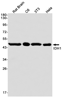
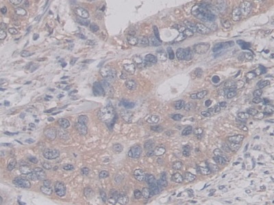
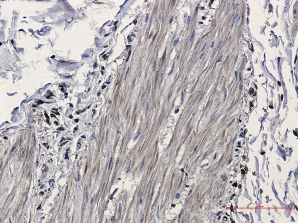 IHC (Immunohiostchemistry)
(Immunohistochemistry of Isocitrate dehydrogenase in paraffin-embedded Human Cholangiocarcinoma using Isocitrate dehydrogenase Rabbit mAb at dilution 1/20)
IHC (Immunohiostchemistry)
(Immunohistochemistry of Isocitrate dehydrogenase in paraffin-embedded Human Cholangiocarcinoma using Isocitrate dehydrogenase Rabbit mAb at dilution 1/20)
IDH1, Monoclonal Antibody (Cat# AAA314256)
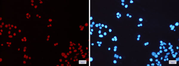
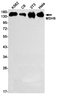
 IHC (Immunohistochemisry)
(Immunohistochemistry of MSH6 in paraffin-embedded Human tonsil using MSH6 Rabbit mAb at dilution 1/1)
IHC (Immunohistochemisry)
(Immunohistochemistry of MSH6 in paraffin-embedded Human tonsil using MSH6 Rabbit mAb at dilution 1/1)
MSH6, Monoclonal Antibody (Cat# AAA314276)

 Application Data
(Dilution: IF: 1:50-200 IHC 1:100-200)
Application Data
(Dilution: IF: 1:50-200 IHC 1:100-200)
HDAC1, Monoclonal Antibody (Cat# AAA293771)
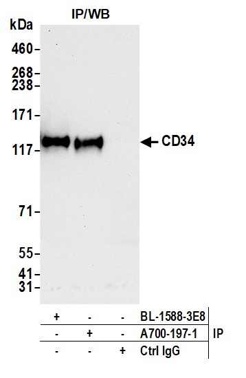
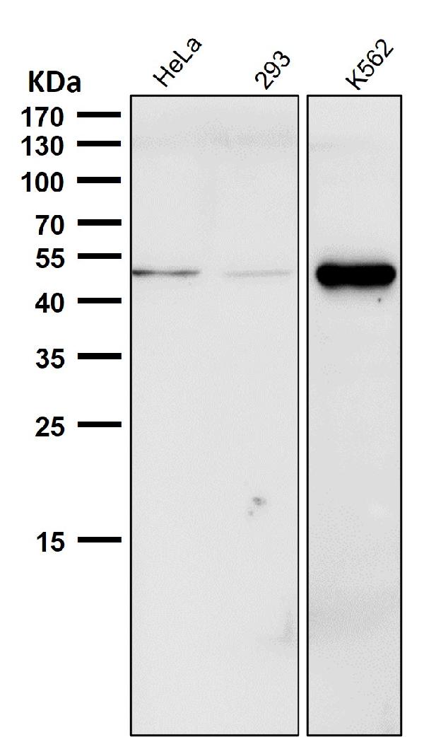
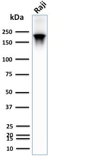
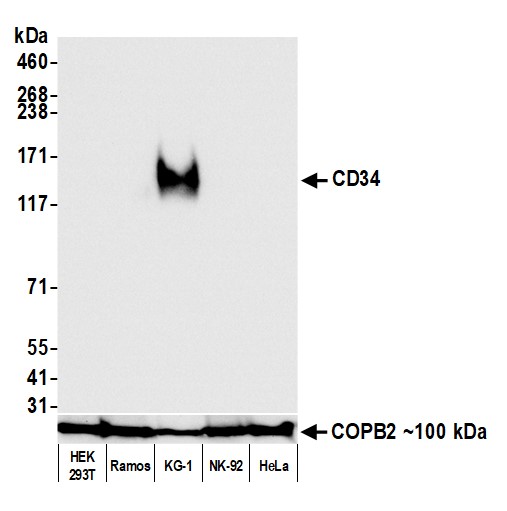 WB (Western Blot)
(Detection of human CD34 by western blot. Samples: Whole cell lysate (50 ug) from HEK293T, Ramos, KG-1, NK-92, and HeLa cells prepared using NETN lysis buffer. Antibody: Rabbit anti-CD34 recombinant monoclonal antibody (AAA213636 lot 1) used at 1:1000. Secondary: HRP-conjugated goat anti-rabbit IgG . Detection: Chemiluminescence with an exposure time of 1 second. Lower Panel: Rabbit anti-COPB2 antibody .)
WB (Western Blot)
(Detection of human CD34 by western blot. Samples: Whole cell lysate (50 ug) from HEK293T, Ramos, KG-1, NK-92, and HeLa cells prepared using NETN lysis buffer. Antibody: Rabbit anti-CD34 recombinant monoclonal antibody (AAA213636 lot 1) used at 1:1000. Secondary: HRP-conjugated goat anti-rabbit IgG . Detection: Chemiluminescence with an exposure time of 1 second. Lower Panel: Rabbit anti-COPB2 antibody .)
CD34, Monoclonal Recombinant Antibody (Cat# AAA213636)
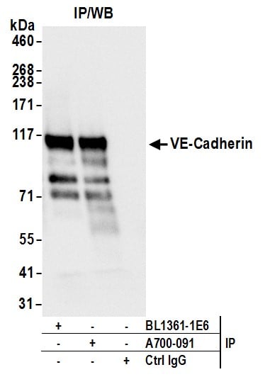
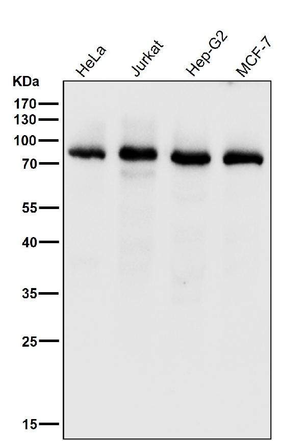
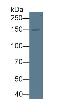
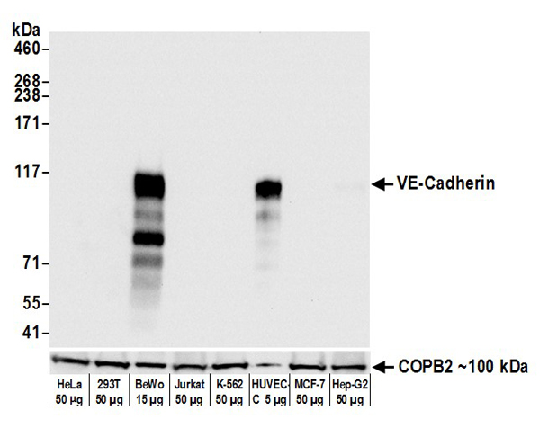 WB (Western Blot)
(Detection of human VE-Cadherin by western blot. Samples: Whole cell lysate from HeLa, HEK293T, BeWo, Jurkat, K-562, HUVEC-C, MCF-7, and Hep-G2 cells prepared using NETN lysis buffer. Antibody: Rabbit anti-VE-Cadherin recombinant monoclonal antibody (AAA213570 lot 1) used at 1:1000. Secondary: HRP-conjugated goat anti-rabbit IgG . Detection: Chemiluminescence with an exposure time of 3 seconds. Lower Panel: Rabbit anti-COPB2 .)
WB (Western Blot)
(Detection of human VE-Cadherin by western blot. Samples: Whole cell lysate from HeLa, HEK293T, BeWo, Jurkat, K-562, HUVEC-C, MCF-7, and Hep-G2 cells prepared using NETN lysis buffer. Antibody: Rabbit anti-VE-Cadherin recombinant monoclonal antibody (AAA213570 lot 1) used at 1:1000. Secondary: HRP-conjugated goat anti-rabbit IgG . Detection: Chemiluminescence with an exposure time of 3 seconds. Lower Panel: Rabbit anti-COPB2 .)
VE-Cadherin, Monoclonal Recombinant Antibody (Cat# AAA213570)
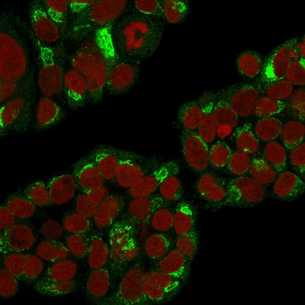 IF (Immunofluorescence)
(Immunofluorescence Analysis of Human MCF-7 cells labeling RCAS1 with RCAS1 Mouse Monoclonal Antibody (CPTC-EBAG9-1) followed by Goat anti-Mouse IgG-CF488 (Green). The nuclear counterstain is Reddot (Red).)
IF (Immunofluorescence)
(Immunofluorescence Analysis of Human MCF-7 cells labeling RCAS1 with RCAS1 Mouse Monoclonal Antibody (CPTC-EBAG9-1) followed by Goat anti-Mouse IgG-CF488 (Green). The nuclear counterstain is Reddot (Red).)
RCAS1/Estrogen Receptor Binding Site Associated, Antigen 9, Monoclonal Antibody (Cat# AAA215278)
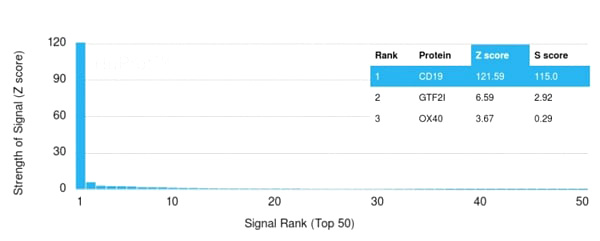 Application Data
(Analysis of Protein Array containing more than 19,000 full-length human proteins using CD19 Monospecific Mouse Monoclonal Antibody (CD19/3117). Z- and S- Score: The Z-score represents the strength of a signal that a monoclonal antibody (MAb) (in combination with a fluorescently-tagged anti-IgG secondary antibody) produces when binding to a particular protein on the HuProtTM array. Z-scores are described in units of standard deviations (SD’s) above the mean value of all signals generated on that array. If targets on HuProtTM are arranged in descending order of the Z-score, the S-score is the difference (also in units of SD’s) between the Z-score. S-score therefore represents the relative target specificity of a MAb to its intended target. A MAb is considered to specific to its intended target, if the MAb has an S-score of at least 2.5. For example, if a MAb binds to protein X with a Z-score of 43 and to protein Y with a Z-score of 14, then the S-score for the binding of that MAb to protein X is equal to 29.)
Application Data
(Analysis of Protein Array containing more than 19,000 full-length human proteins using CD19 Monospecific Mouse Monoclonal Antibody (CD19/3117). Z- and S- Score: The Z-score represents the strength of a signal that a monoclonal antibody (MAb) (in combination with a fluorescently-tagged anti-IgG secondary antibody) produces when binding to a particular protein on the HuProtTM array. Z-scores are described in units of standard deviations (SD’s) above the mean value of all signals generated on that array. If targets on HuProtTM are arranged in descending order of the Z-score, the S-score is the difference (also in units of SD’s) between the Z-score. S-score therefore represents the relative target specificity of a MAb to its intended target. A MAb is considered to specific to its intended target, if the MAb has an S-score of at least 2.5. For example, if a MAb binds to protein X with a Z-score of 43 and to protein Y with a Z-score of 14, then the S-score for the binding of that MAb to protein X is equal to 29.)
CD19, Monoclonal Antibody (Cat# AAA215293)
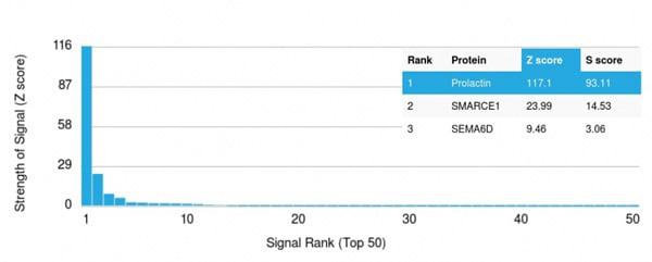 Application Data
(Analysis of Protein Array containing more than 19,000 full-length human proteins using Prolactin Mouse Monoclonal Antibody (PRL/2642). Z- and S- Score: The Z-score represents the strength of a signal that a monoclonal antibody (Monoclonal Antibody) (in combination with a fluorescently-tagged anti-IgG secondary antibody) produces when binding to a particular protein on the HuProtTM array. Z-scores are described in units of standard deviations (SD’s) above the mean value of all signals generated on that array. If targets on HuProtTM are arranged in descending order of the Z-score, the S-score is the difference (also in units of SD’s) between the Z-score. S-score therefore represents the relative target specificity of a Monoclonal Antibody to its intended target. A Monoclonal Antibody is considered to specific to its intended target, if the Monoclonal Antibody has an S-score of at least 2.5. For example, if a Monoclonal Antibody binds to protein X with a Z-score of 43 and to protein Y with a Z-score of 14, then the S-score for the binding of that Monoclonal Antibody to protein X is equal to 29.)
Application Data
(Analysis of Protein Array containing more than 19,000 full-length human proteins using Prolactin Mouse Monoclonal Antibody (PRL/2642). Z- and S- Score: The Z-score represents the strength of a signal that a monoclonal antibody (Monoclonal Antibody) (in combination with a fluorescently-tagged anti-IgG secondary antibody) produces when binding to a particular protein on the HuProtTM array. Z-scores are described in units of standard deviations (SD’s) above the mean value of all signals generated on that array. If targets on HuProtTM are arranged in descending order of the Z-score, the S-score is the difference (also in units of SD’s) between the Z-score. S-score therefore represents the relative target specificity of a Monoclonal Antibody to its intended target. A Monoclonal Antibody is considered to specific to its intended target, if the Monoclonal Antibody has an S-score of at least 2.5. For example, if a Monoclonal Antibody binds to protein X with a Z-score of 43 and to protein Y with a Z-score of 14, then the S-score for the binding of that Monoclonal Antibody to protein X is equal to 29.)
Prolactin, Monoclonal Antibody (Cat# AAA215161)

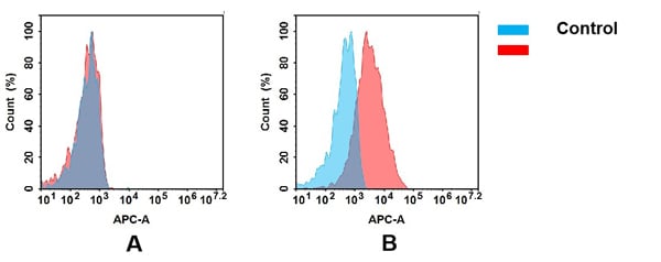
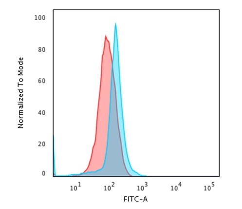 FCM/FACS (Flow Cytometry)
(Flow Cytometric Analysis of paraformaldehyde-fixed Jurkat cells using CD45 Mouse Recombinant Monoclonal Antibody (PTPRC/1666) followed by Goat anti-mouse- IgG-CF488 (Blue); Isotype Control (Red).)
FCM/FACS (Flow Cytometry)
(Flow Cytometric Analysis of paraformaldehyde-fixed Jurkat cells using CD45 Mouse Recombinant Monoclonal Antibody (PTPRC/1666) followed by Goat anti-mouse- IgG-CF488 (Blue); Isotype Control (Red).)
CD45/LCA, Monoclonal Antibody (Cat# AAA215369)
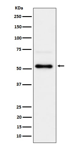
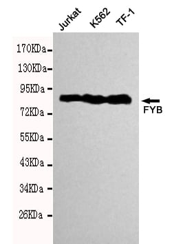
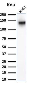 WB (Western Blot)
(Western Blot Analysis of K562 cell lysate using CD43 Rabbit Polyclonal Antibody.)
WB (Western Blot)
(Western Blot Analysis of K562 cell lysate using CD43 Rabbit Polyclonal Antibody.)
CD43, Monoclonal Antibody (Cat# AAA215400)
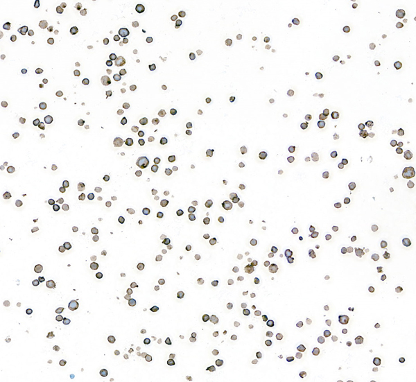
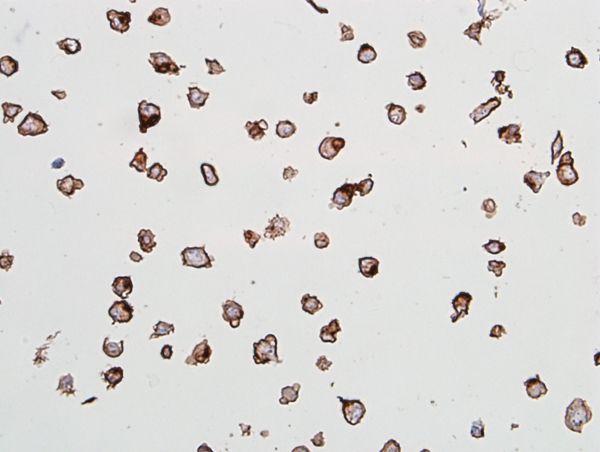
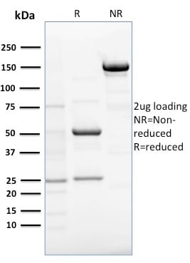
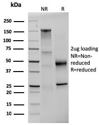
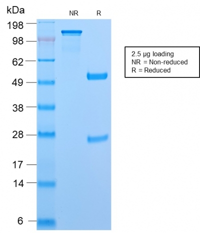
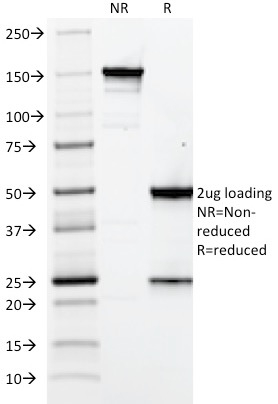 SDS_PAGE
(SDS-PAGE Analysis Purified Elastin Mouse Monoclonal Antibody (ELN/2069). Confirmation of Integrity and Purity of Antibody.)
SDS_PAGE
(SDS-PAGE Analysis Purified Elastin Mouse Monoclonal Antibody (ELN/2069). Confirmation of Integrity and Purity of Antibody.)
Elastin (ELN), Monoclonal Antibody (Cat# AAA215403)
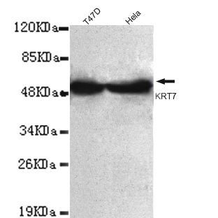
 Application Data
(HeLa cells using anti- KRT7 (N-terminus) antibody diluted 1:150)
Application Data
(HeLa cells using anti- KRT7 (N-terminus) antibody diluted 1:150)
Keratin 7, Monoclonal Antibody (Cat# AAA300807)
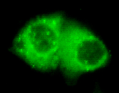
 ICC (Immunocytochemistry)
(Immunocytochemistry stain of Hela using FYB antibody (1:300).)
ICC (Immunocytochemistry)
(Immunocytochemistry stain of Hela using FYB antibody (1:300).)
FYB, Monoclonal Antibody (Cat# AAA300700)
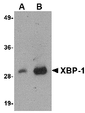
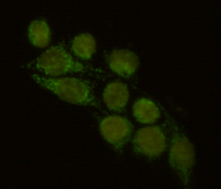
 ICC (Immunocytochemistry)
(Immunocytochemistry of XBP-1 in HepG2 cells with XBP-1 antibody at 2 ug/mL.)
ICC (Immunocytochemistry)
(Immunocytochemistry of XBP-1 in HepG2 cells with XBP-1 antibody at 2 ug/mL.)
XBP-1, Monoclonal Antibody (Cat# AAA300707)
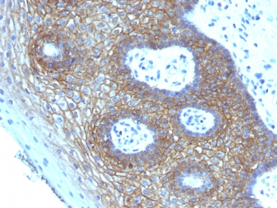
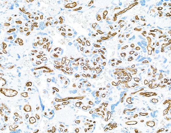
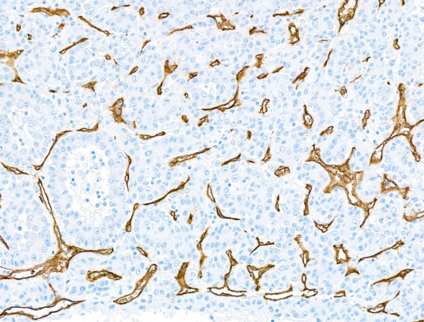
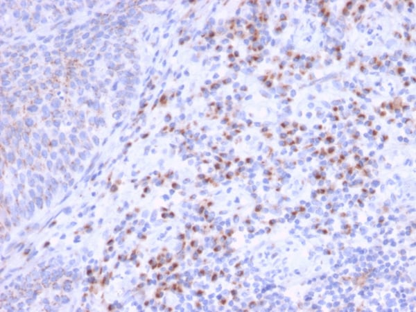
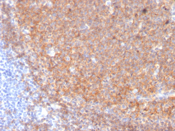
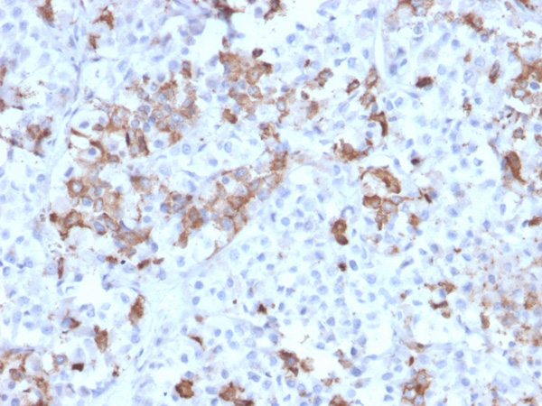
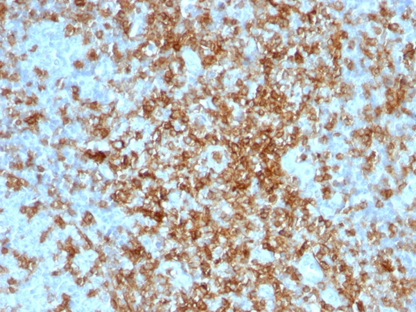
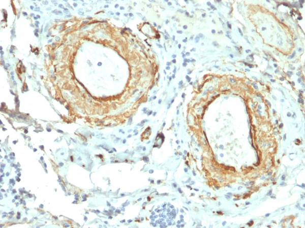
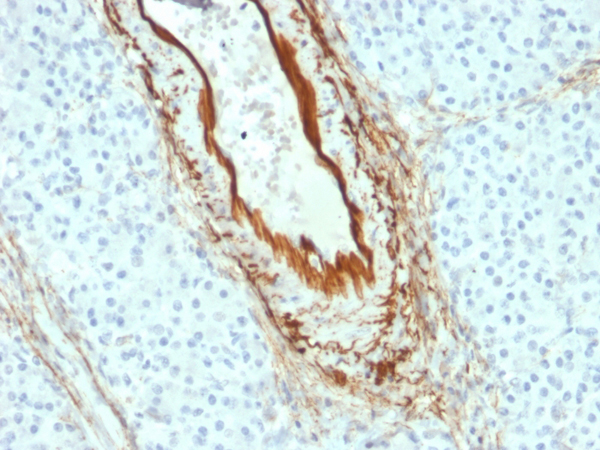
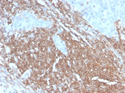
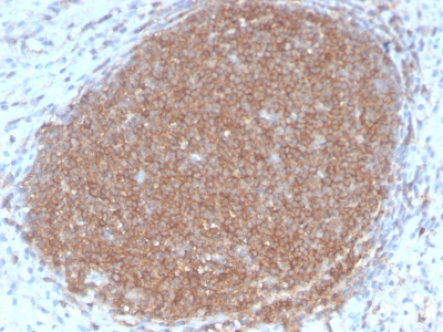
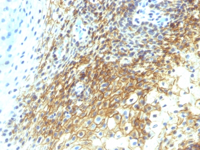 IHC (Immunohiostchemistry)
(Formalin-paraffin human Tongue Squamous Cell Carcinoma stained with CD44v4 Monoclonal Antibody (CD44v4/1219))
IHC (Immunohiostchemistry)
(Formalin-paraffin human Tongue Squamous Cell Carcinoma stained with CD44v4 Monoclonal Antibody (CD44v4/1219))
CD44v4, Monoclonal Antibody (Cat# AAA214606)
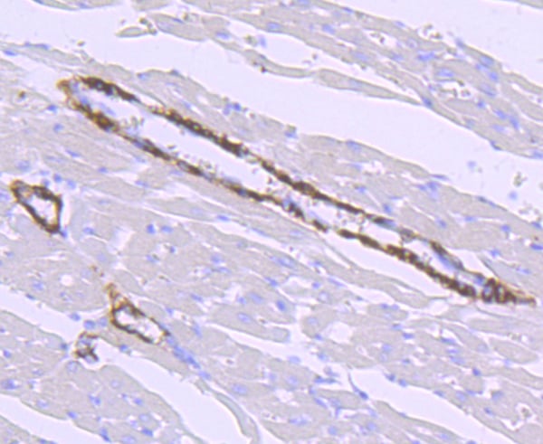
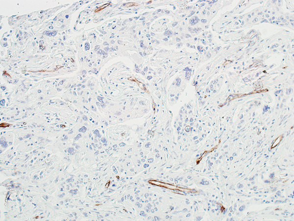
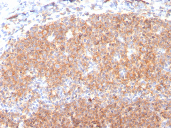
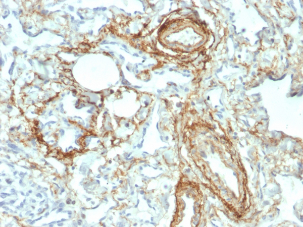
 IHC (Immunohistochemistry)
(Formalin-fixed, paraffin-embedded human Lymph Node stained with CD45RB Rabbit Recombinant Monoclonal Antibody (PTPRC/2877R).)
IHC (Immunohistochemistry)
(Formalin-fixed, paraffin-embedded human Lymph Node stained with CD45RB Rabbit Recombinant Monoclonal Antibody (PTPRC/2877R).)
CD45RB, Monoclonal Antibody (Cat# AAA214804)
Does not react with rat, rhesus monkey. Others not known.
What are Monoclonal Antibodies?
Monoclonal antibodies are specialized laboratory-produced proteins developed for binding to specific biological antigens or other molecular targets. Since they come from a single cell (or clone), they are especially consistent and accurate in the data they are involved in producing.
This type of antibody material has been shown to be a powerful tool in finding and subsequently destroying harmful cells in an organism, such as those found in cancers or various autoimmune diseases. This makes them excellent aids in medical testing and research, which is why they are so widely used.
AAA Biotech offers a comprehensive range of high-quality monoclonal antibodies that perform effectively in various laboratory tests, including (amongst others) ELISA, western blotting, immunohistochemistry, and flow cytometry. All of the products in our catalog are thoroughly quality tested to make sure that they are reliable and will consistently perform well in your research.
What Are The Uses of Monoclonal Antibodies
Monoclonal antibodies are used in many lab tests, including (amongst others) ELISA, western blotting, immunohistochemistry, and flow cytometry.
ELISA is a test that helps detect a specific substance/analyte in a sample. It uses antibodies (often monoclonal) bound to a solid surface (such as the well of a microplate) to “capture” the substance/analyte in the sample and immobilize it so that the detection antibody component can then bind to it and produce a signal, which can then be measured.
Western blotting identifies specific proteins in a sample. The sample is first separated on a gel, and then antibodies are applied that will typically bind to the target, which will all be localized to a single band in a lane.
Immunohistochemistry helps locate specific proteins in cells or tissue samples using antibodies.
Flow cytometry looks at and sorts cells. It uses antibodies that are conjugated to reporter molecules called “fluorophores”, which, under special lights, emit light themselves, which can then be measured by a detector instrument.
How Monoclonal Antibodies Are Used as Medicine?
Please note that all of the products listed in AAA Biotech’s also known as AAA Bio or AAABio catalog are strictly for research-use only (RUO).
Monoclonal antibodies can also be used as therapeutic/medical treatments, particularly in the context of cancers. They are designed to find and bind to specific cells or proteins, helping the immune system recognize and attack the cancer. These treatments work in different ways, such as:
- Radioimmunotherapy attaches a small amount of radioactive molecule to the antibody, so it delivers the radiation directly to the cancer cells that the antibody is specifically binding to.
- Antibody-directed enzyme prodrug therapy uses antibodies that are specifically bound to special enzymes. These enzymes activate a harmless drug in the body and turn it into a cancer-killing drug only near the cancer cells—this helps avoid harming healthy cells.
- Immunoliposomes are tiny “bubbles” filled with medicine/drug and coated with antibodies. They carry the drug straight to the cancer cells.
Why Buy Monoclonal Antibodies From Us?
At AAA Biotech, we provide high-performance monoclonal antibodies designed to support a wide range of research needs.
1. Validated for Versatile Applications
The antibodies in our catalog are extensively validated and compatible with multiple techniques, including (but not limited to) ELISA, flow cytometry (FC), immunocytochemistry (ICC), immunofluorescence (IF), immunohistochemistry (IHC), immunoprecipitation (IP), and western blotting (WB).
2. Wide Selection & Specialized Options
We offer antibodies for common and rare species, that are available in various conjugated forms, and also in recombinant formats. Essentially, there is almost anything one might need to meet their experimental model’s requirements.
3. High-Quality Proteins
Our proteins meet high purity standards—90% or more as confirmed by SDS-PAGE. Many are available with tags like His, Flag, GST, or MBP, and we also supply native and biologically active proteins for functional studies.
Frequently Asked Questions
1. Are your monoclonal antibodies validated for specific applications?
Yes, our antibodies are tested and validated for use in methods such as ELISA, western blot, IHC, flow cytometry, and more. Refer to specific product pages or datasheets for individual product information.
2. How do I choose the right monoclonal antibody for my application?
Review the product details directly for application validation, species reactivity, and target information. You may also contact our support team at any time for help.
3. How quickly can I receive my order?
Most orders are processed and shipped within 1–3 business days, depending on product availability and your shipping location.