Filters
▼Clonality
▼Type
▼Reactivity
▼Gene Name
▼Isotype
▼Host
▼Application
▼Clone
▼Monoclonal Antibodies
Get accurate results in your research with our Monoclonal Antibodies, which are specially made to target exactly what you require for your research, and will produce consistent, reliable performance in lab tests.
Viewing 9150-9200 of 27597 product results
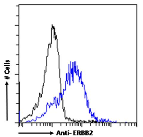 FCM/FACS (Flow Cytometry)
(Flow-cytometry using the anti-ERBB2 antibody SAIC-02A-7 (AAA72066). MCF7 cells were stained with anti-Fluorescein IgG antibody (4-4-20; isotype control, black line) or the rabbit IgG1 version of SAIC-02A-7 (, blue line) at a dilution of 1:100 for 1h at RT. After washing, bound antibody was detected using a goat anti-mouse IgG AlexaFluor 488 antibody at a dilution of 1:1000 and cells analyzed using a FACSCanto flow-cytometer.)
FCM/FACS (Flow Cytometry)
(Flow-cytometry using the anti-ERBB2 antibody SAIC-02A-7 (AAA72066). MCF7 cells were stained with anti-Fluorescein IgG antibody (4-4-20; isotype control, black line) or the rabbit IgG1 version of SAIC-02A-7 (, blue line) at a dilution of 1:100 for 1h at RT. After washing, bound antibody was detected using a goat anti-mouse IgG AlexaFluor 488 antibody at a dilution of 1:1000 and cells analyzed using a FACSCanto flow-cytometer.)
ERBB2, Monoclonal Antibody (Cat# AAA72066)
AChE, Monoclonal Antibody (Cat# AAA74347)
alpha 2 Macroglobulin, Monoclonal Antibody (Cat# AAA74349)
CA 19-9, Monoclonal Antibody (Cat# AAA74356)
Factor VIIIc, Monoclonal Antibody (Cat# AAA74357)
Factor VIII: 0%
Factor related antigens: 0%
NSE, Monoclonal Antibody (Cat# AAA74362)
HSV1 + HSV2, Monoclonal Antibody (Cat# AAA74373)
Synaptopodin, Monoclonal Antibody (Cat# AAA74375)
Methadone, Monoclonal Antibody (Cat# AAA74384)
Salmonella, Monoclonal Antibody (Cat# AAA74386)
MHC Class I, Monoclonal Antibody (Cat# AAA74392)
Factor VII, Monoclonal Antibody (Cat# AAA74395)
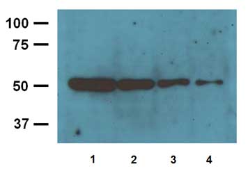 Application Data
(Fig. 02 1:4000 (0.25ug/mL) Ab dilution used in WB of 10ug/lane (1), 5ug/lane (2), 2.5ug/lane (3) and 1.25ug/lane (4) mouse brain tissue lysates)
Application Data
(Fig. 02 1:4000 (0.25ug/mL) Ab dilution used in WB of 10ug/lane (1), 5ug/lane (2), 2.5ug/lane (3) and 1.25ug/lane (4) mouse brain tissue lysates)
beta-Tubulin, Monoclonal Antibody (Cat# AAA74307)
Tau, Monoclonal Antibody (Cat# AAA74314)
Gram Negative Endotoxin, Monoclonal Antibody (Cat# AAA74324)
Progesterone, Monoclonal Antibody (Cat# AAA74327)
Cortisol: 0.1%
Testosterone: 0.2%
Pregnenolone: 1.5%
DHEA: 0.0%
DHEA-S: 0.0%
Estradiol: <0.1%
11-Deoxycortisol: <0.1%
Corticosterone: <0.01%
11-Deoxycorticosterone: <0.1%
Troponin T, Monoclonal Antibody (Cat# AAA74329)
 Application Data
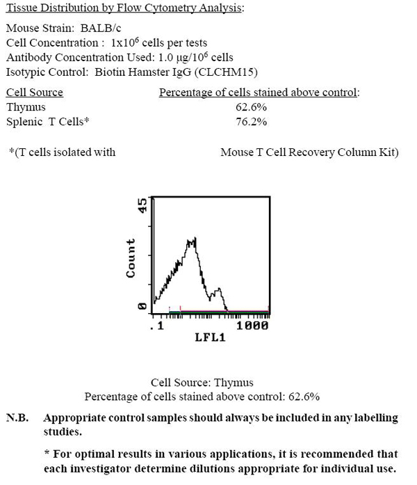
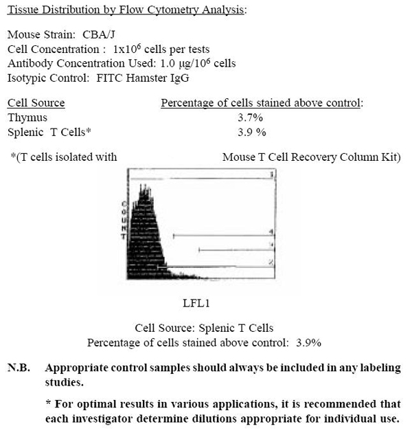
alpha/beta TCR, Monoclonal Antibody (Cat# AAA74128)
 Application Data
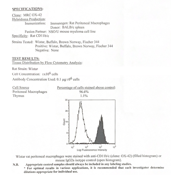
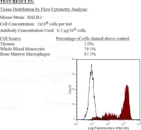
CD11b/c, Monoclonal Antibody (Cat# AAA74135)
 Application Data
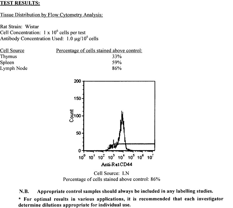
CD44, Monoclonal Antibody (Cat# AAA74147)
 Application Data
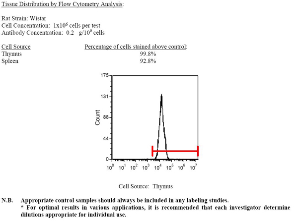
CD45, Monoclonal Antibody (Cat# AAA74152)
 Application Data
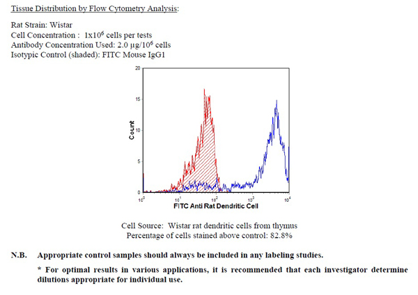
Dendritic cell, Monoclonal Antibody (Cat# AAA74170)
 Application Data
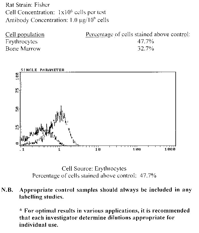
Erythrocytes, Monoclonal Antibody (Cat# AAA74172)
 Application Data
gamma/delta TCR, Monoclonal Antibody (Cat# AAA74176)
 Application Data
(Balb/c mouse bone marrow (high SSC population) was stained with anti-Granulocytes (Gr-1) (clone:RB6-8C5) (filled histogram) or rat IgG2b isotype control (open histogram).N.B. Appropriate control samples should always be included in any labeling studies.* For optimal results in various applications, it is recommended that each investigator determine dilutions appropriate for individual use.)
Application Data
(Balb/c mouse bone marrow (high SSC population) was stained with anti-Granulocytes (Gr-1) (clone:RB6-8C5) (filled histogram) or rat IgG2b isotype control (open histogram).N.B. Appropriate control samples should always be included in any labeling studies.* For optimal results in various applications, it is recommended that each investigator determine dilutions appropriate for individual use.)
Granulocytes (Gr-1), Monoclonal Antibody (Cat# AAA74178)
I-Ab and I-Ad, Monoclonal Antibody (Cat# AAA74187)
 Application Data
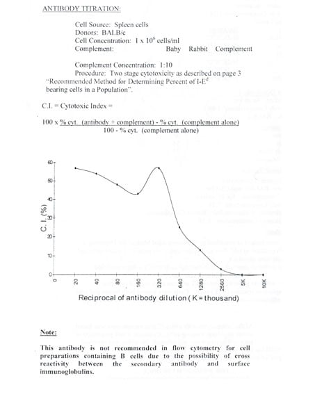
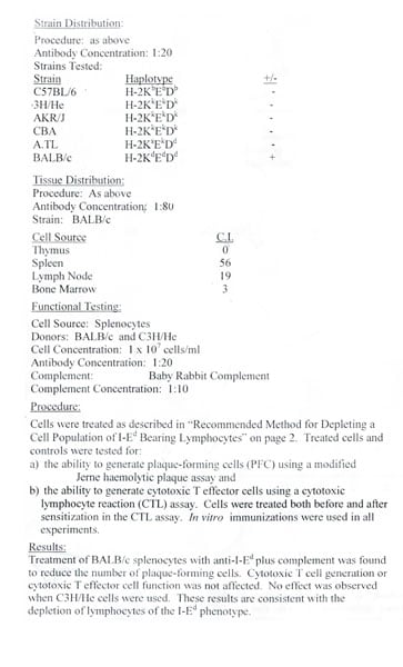
I-Ed (private; Ia.m23), Monoclonal Antibody (Cat# AAA74189)
 Application Data
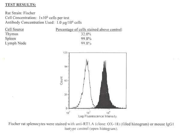
RT1.A, Monoclonal Antibody (Cat# AAA74190)
 Application Data
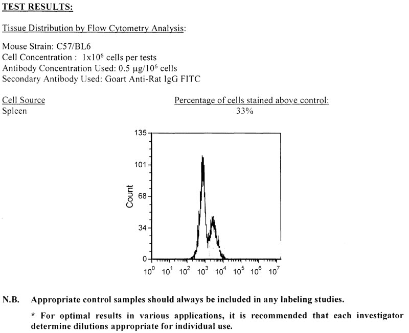
CD19, Monoclonal Antibody (Cat# AAA74202)
Prothrombin (Calcium dependant), Monoclonal Antibody (Cat# AAA74203)
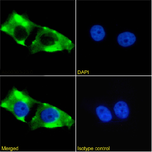
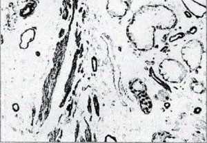 IHC (Immunohistochemistry)
(IHC: Human lung tissue FFPE slides stained with mouse anti-Laminin monoclonal antibody (AAA71320) at 1:200 for 10 min @ RT, Visualized by using DAB chromogen color system. Note: Staining of formalin-fixed tissue requires boiling tissue sections in 10 mM Citrate Buffer, pH 6.0 for 10 min followed by cooling at RT for 20 min.)
IHC (Immunohistochemistry)
(IHC: Human lung tissue FFPE slides stained with mouse anti-Laminin monoclonal antibody (AAA71320) at 1:200 for 10 min @ RT, Visualized by using DAB chromogen color system. Note: Staining of formalin-fixed tissue requires boiling tissue sections in 10 mM Citrate Buffer, pH 6.0 for 10 min followed by cooling at RT for 20 min.)
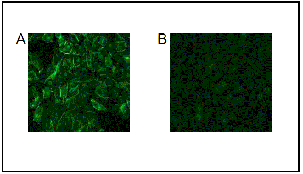
Laminin, Monoclonal Antibody (Cat# AAA71320)
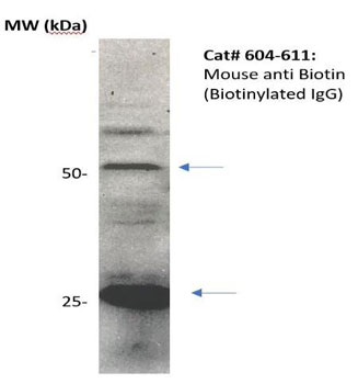 WB (Western Blot)
(Western Blot: The biotinylated IgG was resolved onto 12% SDS-PAGE, transferred onto NC membrane, and detected by Mouse anti-Biotin (Cat# 604-100) at 1:1000. Observed two major immunoreactive bands at molecular weight 50kDa and 25 kDa.)
WB (Western Blot)
(Western Blot: The biotinylated IgG was resolved onto 12% SDS-PAGE, transferred onto NC membrane, and detected by Mouse anti-Biotin (Cat# 604-100) at 1:1000. Observed two major immunoreactive bands at molecular weight 50kDa and 25 kDa.)
Biotin, Monoclonal Antibody (Cat# AAA71343)
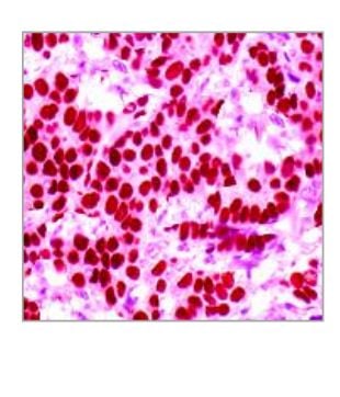 IHC (Immunohiostchemistry)
(IHC: Human breastcarcinoma tissue stainedwith Rat anti HSF1 antibody,at 1:100 for10 min @ RT. Staining offormalin-fixed tissuerequires boiling tissuesections in 10 mM CitrateBuffer, pH 6.0 for 10 minfollowed by cooling at RT for20 min.)
IHC (Immunohiostchemistry)
(IHC: Human breastcarcinoma tissue stainedwith Rat anti HSF1 antibody,at 1:100 for10 min @ RT. Staining offormalin-fixed tissuerequires boiling tissuesections in 10 mM CitrateBuffer, pH 6.0 for 10 minfollowed by cooling at RT for20 min.)
Heat Shock Factor 1, Monoclonal Antibody (Cat# AAA71351)
HBcAg, Monoclonal Antibody (Cat# AAA71355)
CD94, Monoclonal Antibody (Cat# AAA71366)
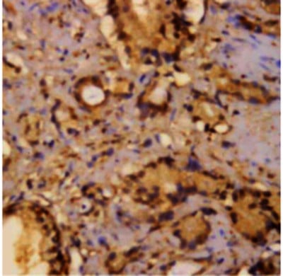 IHC (Immunohiostchemistry)
(Immunohistochemistry: Human breast carcinoma (FFPE) stained with Mouse anti-pSER (Cat# AAA71295) at 1:200 for 10 min @ RT. Staining of formalin-fixed tissue requires boiling tissue sections in 10 mM Citrate Buffer, pH 6.0 for 10 min followed by cooling at RT for 20 min.)
IHC (Immunohiostchemistry)
(Immunohistochemistry: Human breast carcinoma (FFPE) stained with Mouse anti-pSER (Cat# AAA71295) at 1:200 for 10 min @ RT. Staining of formalin-fixed tissue requires boiling tissue sections in 10 mM Citrate Buffer, pH 6.0 for 10 min followed by cooling at RT for 20 min.)
Phosphoserine, Monoclonal Antibody (Cat# AAA71295)
 Application Data
BUP mAb1, Monoclonal Antibody (Cat# AAA71738)
Purity: >95%
MET-Ab2, Monoclonal Antibody (Cat# AAA71740)
HSA, Monoclonal Antibody (Cat# AAA71759)
Protein A affinity chromatography
Plague-F1 mAbl, Monoclonal Antibody (Cat# AAA71761)
Protein A affinity purified
Plague-F1 mAb2, Monoclonal Antibody (Cat# AAA71766)
Affinity chromatography purified.
Lipoprotein a, Monoclonal Antibody (Cat# AAA71772)
Purification: Protein G
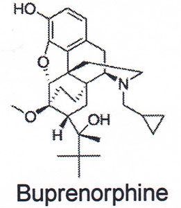
Fentanyl, Monoclonal Antibody (Cat# AAA71773)
Purification: Protein A affinit chromatography purified
 Application Data
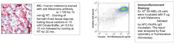
Melanoma/NG2, Monoclonal Antibody (Cat# AAA71435)
CD71, Monoclonal Antibody (Cat# AAA71444)
Apo-A1, Monoclonal Antibody (Cat# AAA71452)
 Application Data
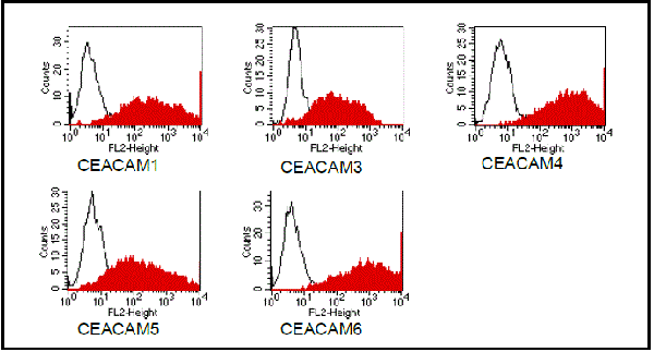
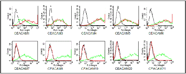
(Fig. 1: BOSC cells were transiently transfected with expression vectors containing either the cDNA of CEACAM1, CEACAM3- CEACAM8 or CEACAM19-21. Recognition of CEACAM4 was tested on CHO cells stably transfected with a CEACAM4 expression vector. Expression of the constructs was confirmed with monoclonal antibodies known to recognise the corresponding proteins (CEACAM1: 4/3/17, CEACAM3,4: D14HD11, CEACAM5: 26/3/13, CEACAM6: 9A6, CEACAM7: BAC2, CEACAM8: GM-2H6, CEACAM19-21: anti-myc, green curves). An irrelevant monoclonal antibody served as a negative control (black curves). For specificity testing, protein G purified D14HD11 was tested on all CEACAM transfectants. A positive signal was obtained with CEACAM1, CEACAM3, CEACAM4, CEACAM5 and CEACAM6 expressing cells (red curves).)
Application Data
(Fig. 1: BOSC cells were transiently transfected with expression vectors containing either the cDNA of CEACAM1, CEACAM3- CEACAM8 or CEACAM19-21. Recognition of CEACAM4 was tested on CHO cells stably transfected with a CEACAM4 expression vector. Expression of the constructs was confirmed with monoclonal antibodies known to recognise the corresponding proteins (CEACAM1: 4/3/17, CEACAM3,4: D14HD11, CEACAM5: 26/3/13, CEACAM6: 9A6, CEACAM7: BAC2, CEACAM8: GM-2H6, CEACAM19-21: anti-myc, green curves). An irrelevant monoclonal antibody served as a negative control (black curves). For specificity testing, protein G purified D14HD11 was tested on all CEACAM transfectants. A positive signal was obtained with CEACAM1, CEACAM3, CEACAM4, CEACAM5 and CEACAM6 expressing cells (red curves).)
CEACAM1,3,4,5,6, Monoclonal Antibody (Cat# AAA71461)
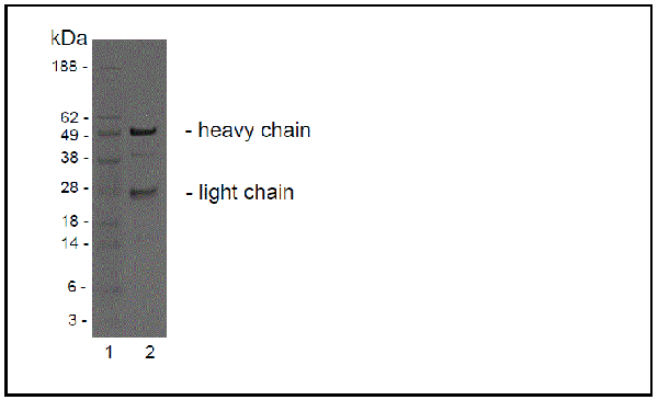 Application Data
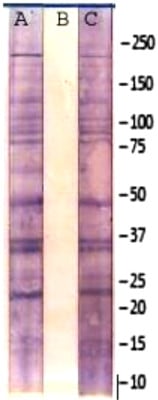
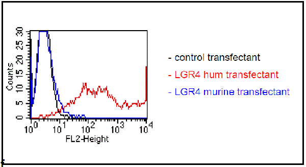
(SDS page analysis of BBX-1H1 The antibody was purified by protein G affinity chromatography from cell culture supernatants Fig.2: SDS-PAGE analysis of purified BBX-1H1 monoclonal antibody. Lane 1: molecular weight marker, Lane 2: 2 ?g of purified BBX-1H1 antibody. Proteins were separated by SDSPAGE and stained with RAPID StainTM Reagent.)
Application Data
(SDS page analysis of BBX-1H1 The antibody was purified by protein G affinity chromatography from cell culture supernatants Fig.2: SDS-PAGE analysis of purified BBX-1H1 monoclonal antibody. Lane 1: molecular weight marker, Lane 2: 2 ?g of purified BBX-1H1 antibody. Proteins were separated by SDSPAGE and stained with RAPID StainTM Reagent.)
LGR4, Monoclonal Antibody (Cat# AAA71462)
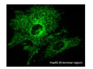
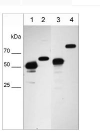 WB (Western Blot)
(Western blot image of cell structure markers in NCI-H1915 lung carcinoma cells. The blot was probed with anti-Vimentin intermediate filament protein VM4341 (lane 1), anti-Nucleoporin p62nM4361 (lane 2), anti-Hsp60 mitochondrial protein HM4381 (lane 3), and anti-Calnexin endoplasmic reticulum protein CM4371 (lane 4).)
WB (Western Blot)
(Western blot image of cell structure markers in NCI-H1915 lung carcinoma cells. The blot was probed with anti-Vimentin intermediate filament protein VM4341 (lane 1), anti-Nucleoporin p62nM4361 (lane 2), anti-Hsp60 mitochondrial protein HM4381 (lane 3), and anti-Calnexin endoplasmic reticulum protein CM4371 (lane 4).)
Hsp60, Monoclonal Antibody (Cat# AAA71646)
What are Monoclonal Antibodies?
Monoclonal antibodies are specialized laboratory-produced proteins developed for binding to specific biological antigens or other molecular targets. Since they come from a single cell (or clone), they are especially consistent and accurate in the data they are involved in producing.
This type of antibody material has been shown to be a powerful tool in finding and subsequently destroying harmful cells in an organism, such as those found in cancers or various autoimmune diseases. This makes them excellent aids in medical testing and research, which is why they are so widely used.
AAA Biotech offers a comprehensive range of high-quality monoclonal antibodies that perform effectively in various laboratory tests, including (amongst others) ELISA, western blotting, immunohistochemistry, and flow cytometry. All of the products in our catalog are thoroughly quality tested to make sure that they are reliable and will consistently perform well in your research.
What Are The Uses of Monoclonal Antibodies
Monoclonal antibodies are used in many lab tests, including (amongst others) ELISA, western blotting, immunohistochemistry, and flow cytometry.
ELISA is a test that helps detect a specific substance/analyte in a sample. It uses antibodies (often monoclonal) bound to a solid surface (such as the well of a microplate) to “capture” the substance/analyte in the sample and immobilize it so that the detection antibody component can then bind to it and produce a signal, which can then be measured.
Western blotting identifies specific proteins in a sample. The sample is first separated on a gel, and then antibodies are applied that will typically bind to the target, which will all be localized to a single band in a lane.
Immunohistochemistry helps locate specific proteins in cells or tissue samples using antibodies.
Flow cytometry looks at and sorts cells. It uses antibodies that are conjugated to reporter molecules called “fluorophores”, which, under special lights, emit light themselves, which can then be measured by a detector instrument.
How Monoclonal Antibodies Are Used as Medicine?
Please note that all of the products listed in AAA Biotech’s also known as AAA Bio or AAABio catalog are strictly for research-use only (RUO).
Monoclonal antibodies can also be used as therapeutic/medical treatments, particularly in the context of cancers. They are designed to find and bind to specific cells or proteins, helping the immune system recognize and attack the cancer. These treatments work in different ways, such as:
- Radioimmunotherapy attaches a small amount of radioactive molecule to the antibody, so it delivers the radiation directly to the cancer cells that the antibody is specifically binding to.
- Antibody-directed enzyme prodrug therapy uses antibodies that are specifically bound to special enzymes. These enzymes activate a harmless drug in the body and turn it into a cancer-killing drug only near the cancer cells—this helps avoid harming healthy cells.
- Immunoliposomes are tiny “bubbles” filled with medicine/drug and coated with antibodies. They carry the drug straight to the cancer cells.
Why Buy Monoclonal Antibodies From Us?
At AAA Biotech, we provide high-performance monoclonal antibodies designed to support a wide range of research needs.
1. Validated for Versatile Applications
The antibodies in our catalog are extensively validated and compatible with multiple techniques, including (but not limited to) ELISA, flow cytometry (FC), immunocytochemistry (ICC), immunofluorescence (IF), immunohistochemistry (IHC), immunoprecipitation (IP), and western blotting (WB).
2. Wide Selection & Specialized Options
We offer antibodies for common and rare species, that are available in various conjugated forms, and also in recombinant formats. Essentially, there is almost anything one might need to meet their experimental model’s requirements.
3. High-Quality Proteins
Our proteins meet high purity standards—90% or more as confirmed by SDS-PAGE. Many are available with tags like His, Flag, GST, or MBP, and we also supply native and biologically active proteins for functional studies.
Frequently Asked Questions
1. Are your monoclonal antibodies validated for specific applications?
Yes, our antibodies are tested and validated for use in methods such as ELISA, western blot, IHC, flow cytometry, and more. Refer to specific product pages or datasheets for individual product information.
2. How do I choose the right monoclonal antibody for my application?
Review the product details directly for application validation, species reactivity, and target information. You may also contact our support team at any time for help.
3. How quickly can I receive my order?
Most orders are processed and shipped within 1–3 business days, depending on product availability and your shipping location.