Filters
▼Clonality
▼Type
▼Reactivity
▼Gene Name
▼Isotype
▼Host
▼Application
▼Clone
▼Monoclonal Antibodies
Get accurate results in your research with our Monoclonal Antibodies, which are specially made to target exactly what you require for your research, and will produce consistent, reliable performance in lab tests.
Viewing 9000-9050 of 27597 product results
 Application Data
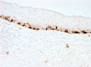
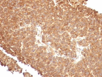
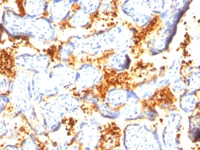
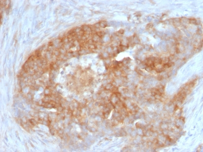
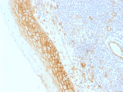
(Formalin-fixed, paraffin-embedded human Skin stained with MART-1 Monoclonal Antibody (M2-7C10).)
Application Data
(Formalin-fixed, paraffin-embedded human Skin stained with MART-1 Monoclonal Antibody (M2-7C10).)
MART-1 / Melan-A / MLANA, Monoclonal Antibody (Cat# AAA62664)
Does not react with Mouse and Rat.
Others not tested
 IHC (Immunohiostchemistry)
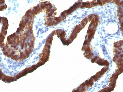
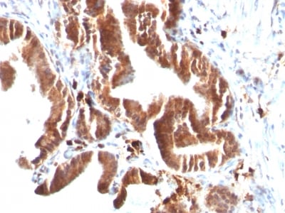
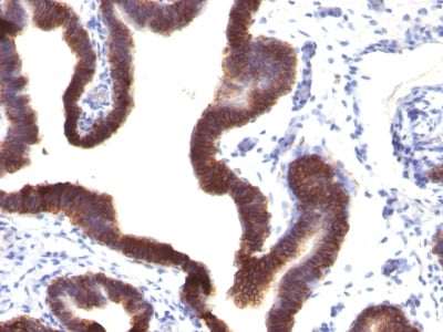
(Formalin-fixed, paraffin-embedded human Lung Carcinoma stained with Cytokeratin 7 Monoclonal Antibody (KRT7/760 + OV-TL12/30))
IHC (Immunohiostchemistry)
(Formalin-fixed, paraffin-embedded human Lung Carcinoma stained with Cytokeratin 7 Monoclonal Antibody (KRT7/760 + OV-TL12/30))
Cytokeratin 7 (KRT7), Monoclonal Antibody (Cat# AAA62667)
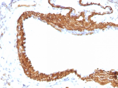
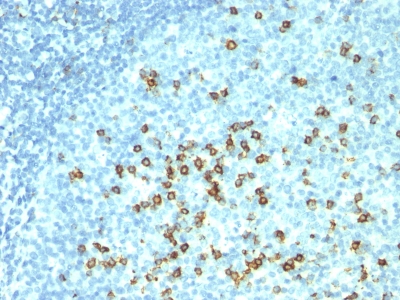 IHC (Immunohistochemistry)
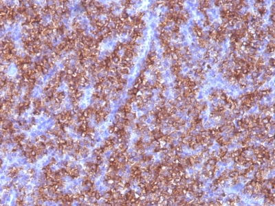
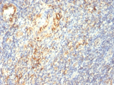
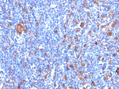
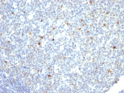
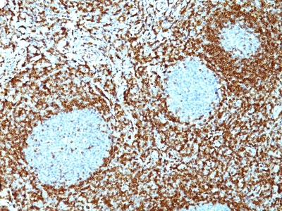
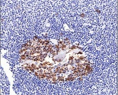
(Formalin-fixed, paraffin-embedded human Tonsil stained with CD57 Monoclonal Antibody (HNK-1 + NK-1).)
IHC (Immunohistochemistry)
(Formalin-fixed, paraffin-embedded human Tonsil stained with CD57 Monoclonal Antibody (HNK-1 + NK-1).)
CD57 / B3GAT1, Monoclonal Antibody (Cat# AAA62673)
Does not react with rat. Others not known.
 IHC (Immunohiostchemistry)
(Formalin-fixed, paraffin-embedded human Thyroid stained with TTF-1 Monoclonal Antibody (8G7G3/1 + NX2.1/690))
IHC (Immunohiostchemistry)
(Formalin-fixed, paraffin-embedded human Thyroid stained with TTF-1 Monoclonal Antibody (8G7G3/1 + NX2.1/690))
TTF-1 / NKX2.1, Monoclonal Antibody (Cat# AAA62683)
 IHC (Immunohistochemisry)
(Formalin-fixed, paraffin-embedded human Pancreas stained with Topo I, MT Monoclonal Antibody (TOP1MT/488).)
IHC (Immunohistochemisry)
(Formalin-fixed, paraffin-embedded human Pancreas stained with Topo I, MT Monoclonal Antibody (TOP1MT/488).)
Topoisomerase (DNA) I, Mitochondrial (TOP1MT), Monoclonal Antibody (Cat# AAA62691)
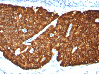
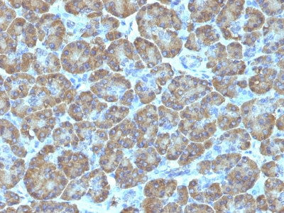
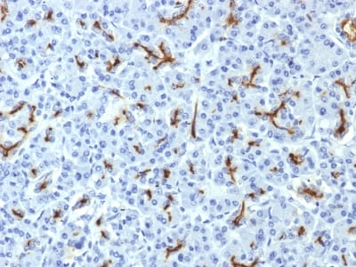
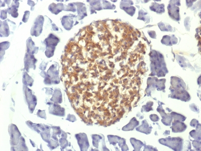
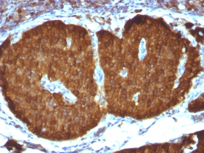
 IHC (Immunohistochemistry)
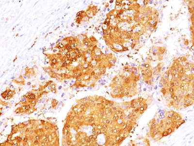
(Formalin-fixed, paraffin-embedded human Adrenal Gland stained with Chromogranin A Monoclonal Antibody (CGA/413))
IHC (Immunohistochemistry)
(Formalin-fixed, paraffin-embedded human Adrenal Gland stained with Chromogranin A Monoclonal Antibody (CGA/413))
Chromogranin A / CHGA, Monoclonal Antibody (Cat# AAA62696)
Others not known
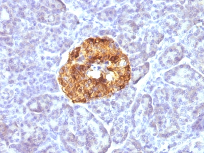
 IHC (Immunohiostchemistry)
(Formalin-fixed, paraffin-embedded human Pancreas stained with Cytochrome C Monoclonal Antibody (7H8.2C12 + CYCS/1010).)
IHC (Immunohiostchemistry)
(Formalin-fixed, paraffin-embedded human Pancreas stained with Cytochrome C Monoclonal Antibody (7H8.2C12 + CYCS/1010).)
Cytochrome C, Monoclonal Antibody (Cat# AAA62707)
Others not known

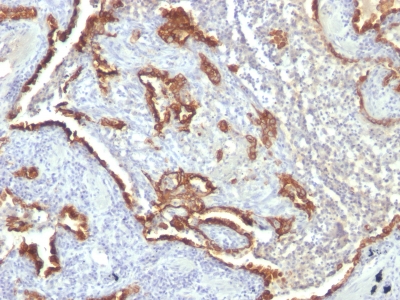
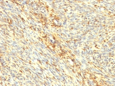
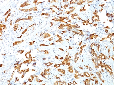
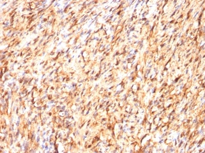
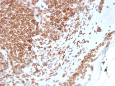
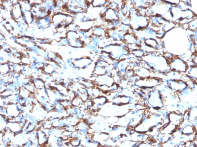 IHC (Immunohistochemistry)
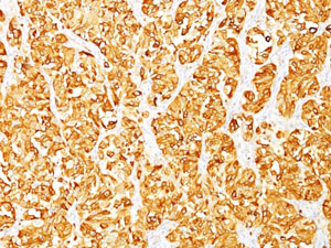
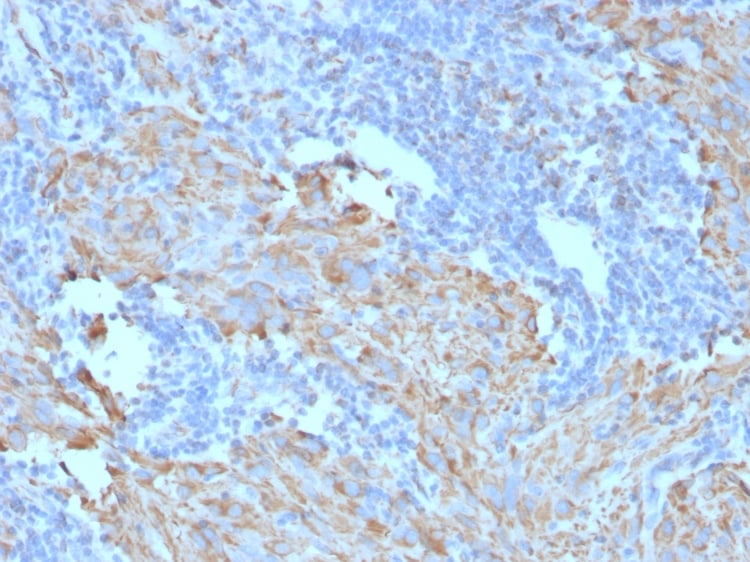
(Formalin-fixed, paraffin-embedded human Angiosarcoma stained with Smooth Muscle Actin Monoclonal Antibody (1A4 + ACTA2/791).)
IHC (Immunohistochemistry)
(Formalin-fixed, paraffin-embedded human Angiosarcoma stained with Smooth Muscle Actin Monoclonal Antibody (1A4 + ACTA2/791).)
Actin, Monoclonal Antibody (Cat# AAA62866)
Predicted to show a broad reactivity
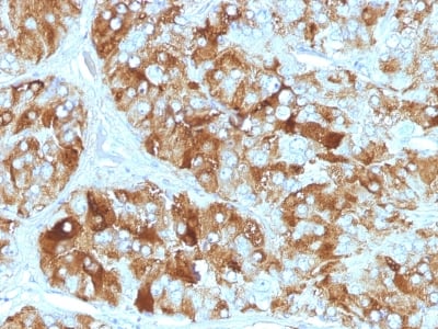
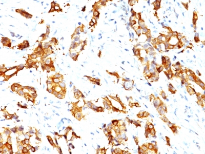
 IHC (Immunohistochemistry)
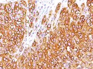
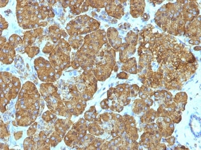
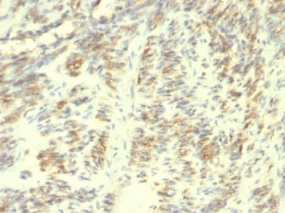
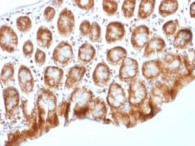
(Formalin-fixed, paraffin-embedded human Hepatocellular Carcinoma stained with ARG1 Monoclonal Antibody (ARG1/1125 + ARG1/1126).)
IHC (Immunohistochemistry)
(Formalin-fixed, paraffin-embedded human Hepatocellular Carcinoma stained with ARG1 Monoclonal Antibody (ARG1/1125 + ARG1/1126).)
Arginase 1, Monoclonal Antibody (Cat# AAA62732)
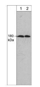
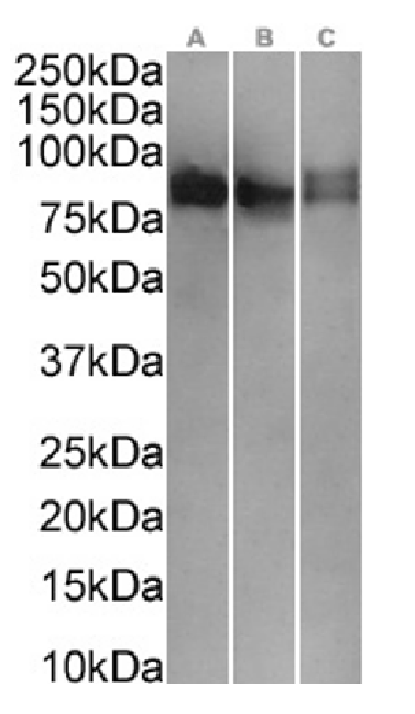
 WB (Western Blot)
(Western Blot Analysis of HCT116 Cell Lysate using EpCAM Monoclonal Antibody (EGP40/1120))
WB (Western Blot)
(Western Blot Analysis of HCT116 Cell Lysate using EpCAM Monoclonal Antibody (EGP40/1120))
Ep-CAM /CD326, Monoclonal Antibody (Cat# AAA62749)
Does not react with Mouse and Rat
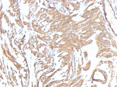
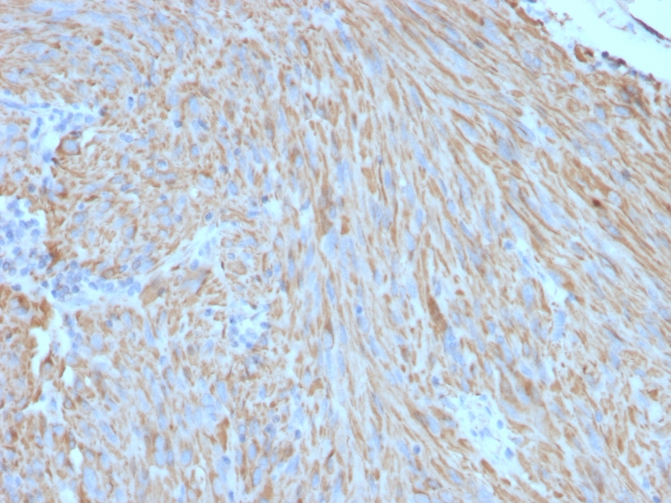
 IHC (Immunohistochemistry)
(Formalin-fixed, paraffin-embedded human Leiomyosarcoma stained with Transglutaminase II Monoclonal Antibody (TGM2/419))
IHC (Immunohistochemistry)
(Formalin-fixed, paraffin-embedded human Leiomyosarcoma stained with Transglutaminase II Monoclonal Antibody (TGM2/419))
Transglutaminase II (TGM2), Monoclonal Antibody (Cat# AAA62779)
 WB (Western Blot)
(Western Blot of HeLa Cell Lysate using Fascin-1 Monoclonal Antibody (FSN1/417))
WB (Western Blot)
(Western Blot of HeLa Cell Lysate using Fascin-1 Monoclonal Antibody (FSN1/417))
Fascin-1, Monoclonal Antibody (Cat# AAA62794)
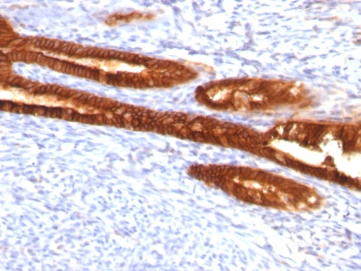 IHC (Immunohistochemisry)
(Formalin-fixed, paraffin-embedded human Endometrial Carcinoma stained with Cytokeratin 7 Monoclonal Antibody (KRT7/760 + KRT7/903))
IHC (Immunohistochemisry)
(Formalin-fixed, paraffin-embedded human Endometrial Carcinoma stained with Cytokeratin 7 Monoclonal Antibody (KRT7/760 + KRT7/903))
Cytokeratin 7 (KRT7), Monoclonal Antibody (Cat# AAA62799)
MUC5AC (Mucin 5AC / Gastric Mucin), Monoclonal Antibody (Cat# AAA62802)
Does not react with Pig and hedgehog
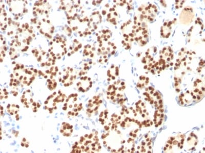
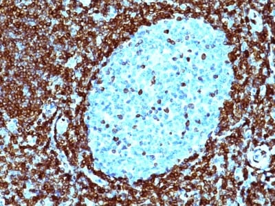
 IHC (Immunohiostchemistry)
(Formalin-fixed, paraffin-embedded human Tonsil stained with PCNA Monoclonal Antibody (PCNA/694))
IHC (Immunohiostchemistry)
(Formalin-fixed, paraffin-embedded human Tonsil stained with PCNA Monoclonal Antibody (PCNA/694))
PCNA (Proliferating Cell Nuclear Antigen), Monoclonal Antibody (Cat# AAA62806)
Shows broad species reactivity
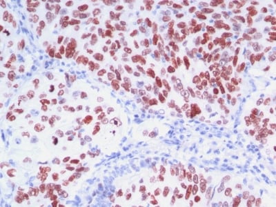
 IHC (Immunohiostchemistry)
(Formalin-fixed, paraffin-embedded human Breast Carcinoma stained with HSP27 Monoclonal Antibody (SPM252))
IHC (Immunohiostchemistry)
(Formalin-fixed, paraffin-embedded human Breast Carcinoma stained with HSP27 Monoclonal Antibody (SPM252))
HSP27 (Heat Shock Protein 27), Monoclonal Antibody (Cat# AAA62525)
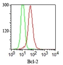
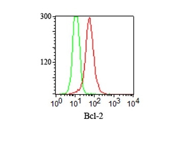
 WB (Western Blot)
(Western Blot of Bcl-2 in human Skin using Bcl-2 Monoclonal Antibody (SPM530).)
WB (Western Blot)
(Western Blot of Bcl-2 in human Skin using Bcl-2 Monoclonal Antibody (SPM530).)
Bcl-2, Monoclonal Antibody (Cat# AAA62532)
Does not react with Mouse and Rat
 WB (Western Blot)
(Western Blot Analysis of Raji Cell Lysate using IgM Monoclonal Antibody (IM260))
WB (Western Blot)
(Western Blot Analysis of Raji Cell Lysate using IgM Monoclonal Antibody (IM260))
IgM (Immunoglobulin Mu Heavy Chain), Monoclonal Antibody (Cat# AAA62544)
 FCM/FACS (Flow Cytometry)
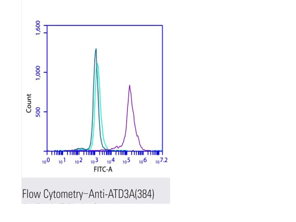
(Flow cytometric analysis of 2% paraformaldehyde-fixed Jurkat (Human T cell leukemia cells from peripheral blood) cells labeling ATD3A(384) with M25064 at 1/200 dilution (red) compared with a mouse monoclonal IgG isotype control (blue) and an unlabelled control (cells without incubation with primary antibody, green). Goat anti-mouse IgG (FITC) at 1/300 dilution was used as the secondary antibody.)
FCM/FACS (Flow Cytometry)
(Flow cytometric analysis of 2% paraformaldehyde-fixed Jurkat (Human T cell leukemia cells from peripheral blood) cells labeling ATD3A(384) with M25064 at 1/200 dilution (red) compared with a mouse monoclonal IgG isotype control (blue) and an unlabelled control (cells without incubation with primary antibody, green). Goat anti-mouse IgG (FITC) at 1/300 dilution was used as the secondary antibody.)
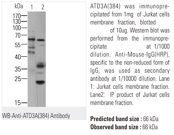
ATD3A, Monoclonal Antibody (Cat# AAA62317)

 FCM/FACS (Flow Cytometry)
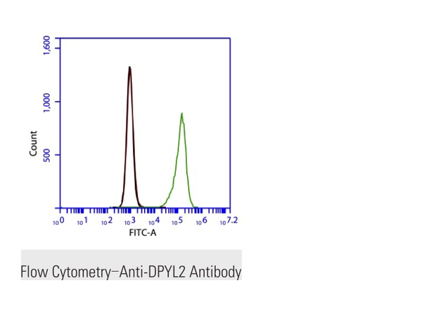
(Flow cytometric analysis of 2% paraformaldehyde-fixed Jurkat (Human T cell leukemia cells from peripheral blood) cells labeling DPYL2 with M25017 at 1/200 dilution (green) compared with a mouse monoclonal IgG isotype control (black) and an unlabelled control (cells without incubation with primary antibody, blue). Goat anti-mouse IgG (FITC) at 1/300 dilution was used as the secondary antibody.)
FCM/FACS (Flow Cytometry)
(Flow cytometric analysis of 2% paraformaldehyde-fixed Jurkat (Human T cell leukemia cells from peripheral blood) cells labeling DPYL2 with M25017 at 1/200 dilution (green) compared with a mouse monoclonal IgG isotype control (black) and an unlabelled control (cells without incubation with primary antibody, blue). Goat anti-mouse IgG (FITC) at 1/300 dilution was used as the secondary antibody.)
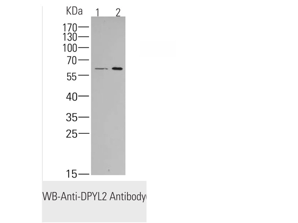
CRMP2, Monoclonal Antibody (Cat# AAA62332)
 FCM/FACS (Flow Cytometry)
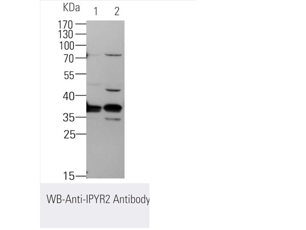
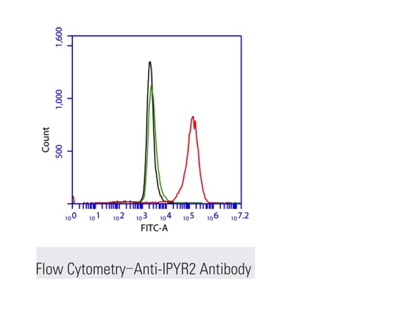
(Flow cytometric analysis of 2% paraformaldehyde-fixed THP1 (Human acute monocytic leukemia cell line) cells labeling IPYR2 with M25206 at 1/200 dilution (red) compared with a mouse monoclonal IgG isotype control (black) and an unlabelled control (cells without incubation with primary antibody, blue). Goat anti-mouse IgG (FITC) at 1/300 dilution was used as the secondary antibody.)
FCM/FACS (Flow Cytometry)
(Flow cytometric analysis of 2% paraformaldehyde-fixed THP1 (Human acute monocytic leukemia cell line) cells labeling IPYR2 with M25206 at 1/200 dilution (red) compared with a mouse monoclonal IgG isotype control (black) and an unlabelled control (cells without incubation with primary antibody, blue). Goat anti-mouse IgG (FITC) at 1/300 dilution was used as the secondary antibody.)
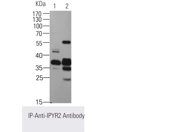
IPYR2/PPA2, Monoclonal Antibody (Cat# AAA62360)
 FCM/FACS (Flow Cytometry)
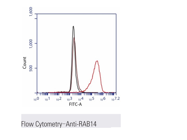
(Flow cytometric analysis of 2% paraformaldehyde-fixed THP1 (Human acute monocytic leukemia cell line) cells labeling RAB14 with M25201 at 1/200 dilution (red) compared with a mouse monoclonal IgG isotype control (black) and an unlabelled control (cells without incubation with primary antibody, purple). Goat anti-mouse IgG (FITC) at 1/300 dilution was used as the secondary antibody.)
FCM/FACS (Flow Cytometry)
(Flow cytometric analysis of 2% paraformaldehyde-fixed THP1 (Human acute monocytic leukemia cell line) cells labeling RAB14 with M25201 at 1/200 dilution (red) compared with a mouse monoclonal IgG isotype control (black) and an unlabelled control (cells without incubation with primary antibody, purple). Goat anti-mouse IgG (FITC) at 1/300 dilution was used as the secondary antibody.)
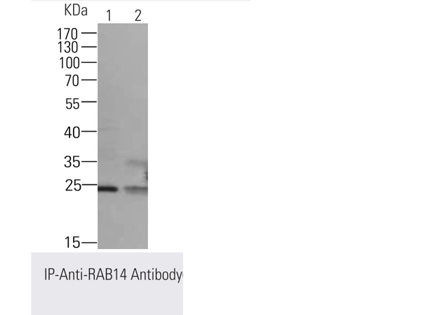
RAB14, Monoclonal Antibody (Cat# AAA62364)
 FCM/FACS (Flow Cytometry)
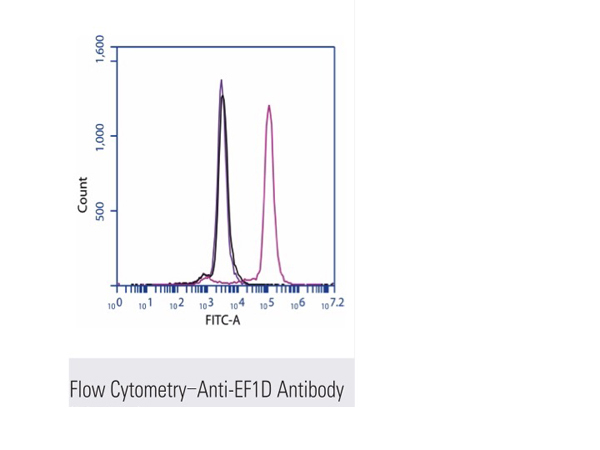
(Flow cytometric analysis of 2% paraformaldehyde-fixed Jurkat (Human T cell leukemia cells from peripheral blood) cells labeling EF1D with M25112 at 1/200 dilution (red) compared with a mouse monoclonal IgG isotype control (black) and an unlabelled control (cells without incubation with primary antibody, blue). Goat anti-mouse IgG (FITC) at 1/300 dilution was used as the secondary antibody.)
FCM/FACS (Flow Cytometry)
(Flow cytometric analysis of 2% paraformaldehyde-fixed Jurkat (Human T cell leukemia cells from peripheral blood) cells labeling EF1D with M25112 at 1/200 dilution (red) compared with a mouse monoclonal IgG isotype control (black) and an unlabelled control (cells without incubation with primary antibody, blue). Goat anti-mouse IgG (FITC) at 1/300 dilution was used as the secondary antibody.)
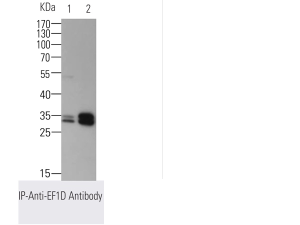
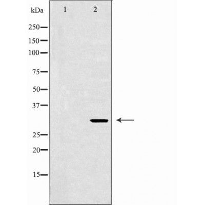
EEF1D, Monoclonal Antibody (Cat# AAA62369)
 FCM/FACS (Flow Cytometry)
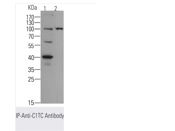
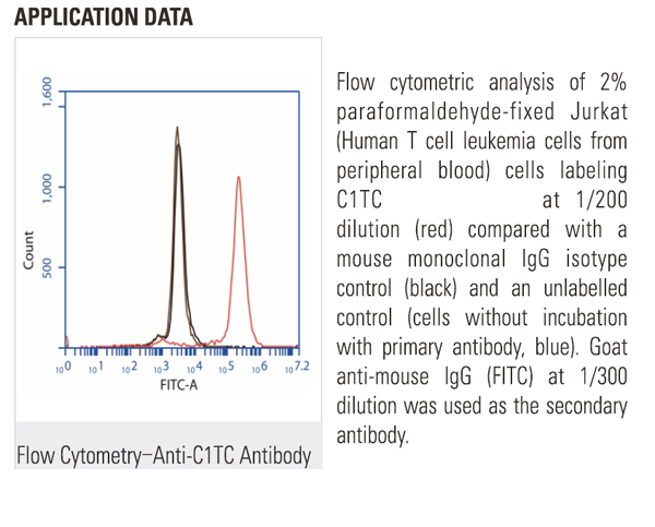
(Flow cytometric analysis of 2% paraformaldehyde-fixed Jurkat (Human T cell leukemia cells from peripheral blood) cells labeling C1TC with M25094 at 1/200 dilution (red) compared with a mouse monoclonal IgG isotype control (black) and an unlabelled control (cells without incubation with primary antibody, blue). Goat anti-mouse IgG (FITC) at 1/300 dilution was used as the secondary antibody.)
FCM/FACS (Flow Cytometry)
(Flow cytometric analysis of 2% paraformaldehyde-fixed Jurkat (Human T cell leukemia cells from peripheral blood) cells labeling C1TC with M25094 at 1/200 dilution (red) compared with a mouse monoclonal IgG isotype control (black) and an unlabelled control (cells without incubation with primary antibody, blue). Goat anti-mouse IgG (FITC) at 1/300 dilution was used as the secondary antibody.)
MTHFD1, Monoclonal Antibody (Cat# AAA62387)
 FCM/FACS (Flow Cytometry)
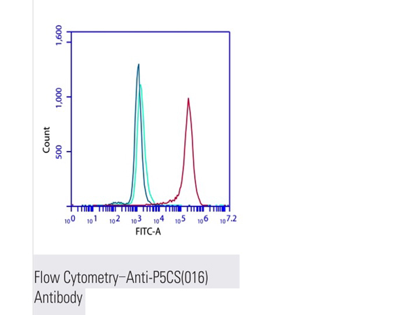
(Flow cytometric analysis of 2% paraformaldehyde-fixed Jurkat (Human T cell leukemia cells from peripheral blood) cells labeling P5CS(016) with M25084 at 1/200 dilution (red) compared with a mouse monoclonal IgG isotype control (blue) and an unlabelled control (cells without incubation with primary antibody, green). Goat anti-mouse IgG (FITC) at 1/300 dilution was used as the secondary antibody.)
FCM/FACS (Flow Cytometry)
(Flow cytometric analysis of 2% paraformaldehyde-fixed Jurkat (Human T cell leukemia cells from peripheral blood) cells labeling P5CS(016) with M25084 at 1/200 dilution (red) compared with a mouse monoclonal IgG isotype control (blue) and an unlabelled control (cells without incubation with primary antibody, green). Goat anti-mouse IgG (FITC) at 1/300 dilution was used as the secondary antibody.)
P5CS, Monoclonal Antibody (Cat# AAA62394)
Cartilage Link Protein, Monoclonal Antibody (Cat# AAA63027)
Hyaluronic Acid Binding Region, Monoclonal Antibody (Cat# AAA63032)
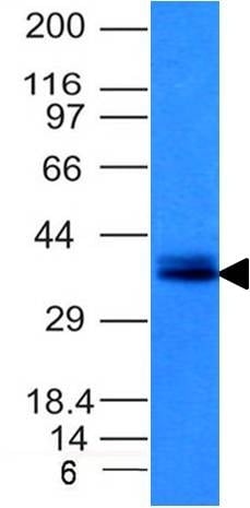
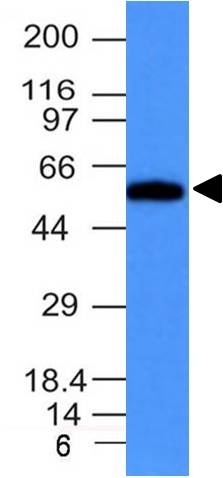
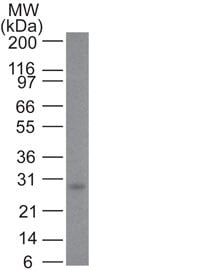
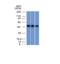
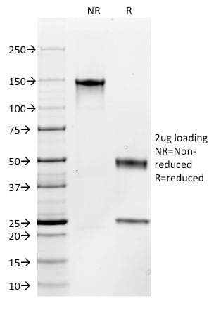
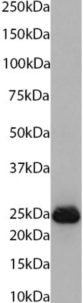
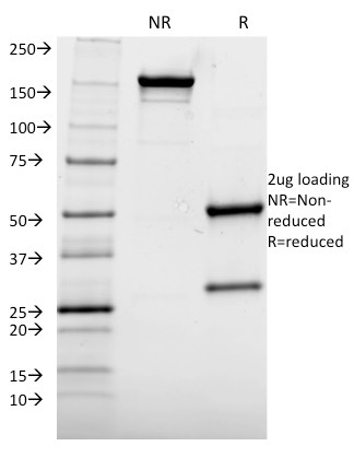 SDS-PAGE
(SDS-PAGE Analysis Purified AKT1 Mouse Monoclonal Antibody (AKT1/2552). Confirmation of Integrity and Purity of Antibody.)
SDS-PAGE
(SDS-PAGE Analysis Purified AKT1 Mouse Monoclonal Antibody (AKT1/2552). Confirmation of Integrity and Purity of Antibody.)
p21WAF1, Monoclonal Antibody (Cat# AAA62907)
Does not react with Mouse and Rat. Others not known.
 IHC (Immunohiostchemistry)
(Formalin-fixed, paraffin-embedded human Schwanoma stained with S100B Mouse Monoclonal Antibody (S100B/1012).)
IHC (Immunohiostchemistry)
(Formalin-fixed, paraffin-embedded human Schwanoma stained with S100B Mouse Monoclonal Antibody (S100B/1012).)
CFTR (Cystic Fibrosis Transmembrane Conductance Regulator), Monoclonal Antibody (Cat# AAA62918)

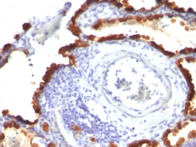
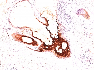
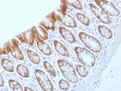 IHC (Immunohistochemistry)
(Formalin-fixed, paraffin-embedded Rat Colon stained with Beta-Catenin (p120) Monoclonal Antibody (CTNNB1/1509).)
IHC (Immunohistochemistry)
(Formalin-fixed, paraffin-embedded Rat Colon stained with Beta-Catenin (p120) Monoclonal Antibody (CTNNB1/1509).)
Catenin, beta, Monoclonal Antibody (Cat# AAA62949)
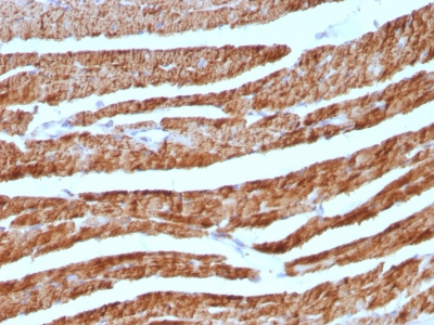
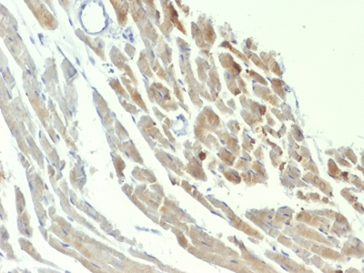 IHC (Immunohistochemistry)
(Formalin-fixed, paraffin-embedded Rat Heart stained with Formalin-fixed, paraffin-embedded Rat Heart stained with NSE gamma Monoclonal Antibody (ENO2/1375).)
IHC (Immunohistochemistry)
(Formalin-fixed, paraffin-embedded Rat Heart stained with Formalin-fixed, paraffin-embedded Rat Heart stained with NSE gamma Monoclonal Antibody (ENO2/1375).)
NSE gamma (Neuron Specific Enolase, gamma), Monoclonal Antibody (Cat# AAA62963)
 WB (Western Blot)
(Western Blot of Bcl-2 in human skin using Bcl-2 Monoclonal Antibody (100/D5 + 124).)
WB (Western Blot)
(Western Blot of Bcl-2 in human skin using Bcl-2 Monoclonal Antibody (100/D5 + 124).)
Bcl-2, Monoclonal Antibody (Cat# AAA62560)
Does not react with Mouse and Rat
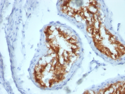
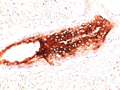
 IHC (Immunohistochemisry)
(Formalin-fixed, paraffin-embedded human GIST stained with CD117 Mouse Monoclonal Antibody (C117/370).)
IHC (Immunohistochemisry)
(Formalin-fixed, paraffin-embedded human GIST stained with CD117 Mouse Monoclonal Antibody (C117/370).)
CD117, Monoclonal Antibody (Cat# AAA62592)
 Application Data
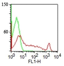
(Surface staining of human lymphocytes with CDw75 Monoclonal Antibody (LN-1) (red) and isotype control (green). PPI negative population analyzed.)
Application Data
(Surface staining of human lymphocytes with CDw75 Monoclonal Antibody (LN-1) (red) and isotype control (green). PPI negative population analyzed.)
CDw75, Monoclonal Antibody (Cat# AAA62593)
 WB (Western Blot)
(Western Blot of HeLa Lysate using CK17 Monoclonal Antibody (SPM560).)
WB (Western Blot)
(Western Blot of HeLa Lysate using CK17 Monoclonal Antibody (SPM560).)
Cytokeratin 17 (KRT17), Monoclonal Antibody (Cat# AAA62620)
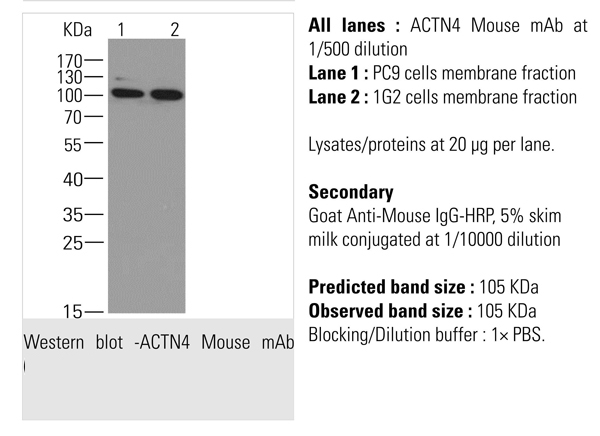
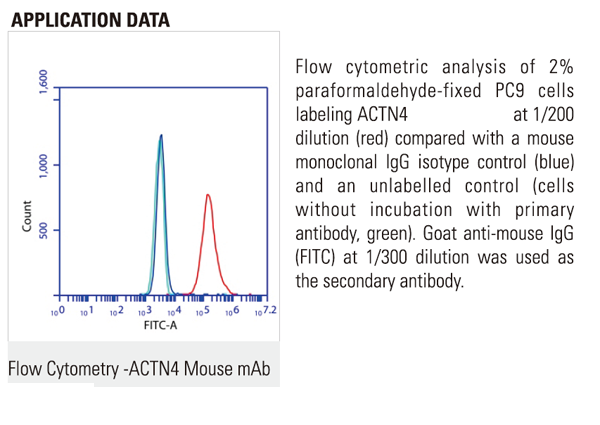 FCM/FACS (Flow Cytometry)
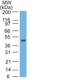
alpha Actinin 4 (ACTN4), Monoclonal Antibody (Cat# AAA62453)
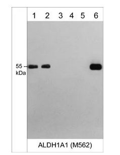
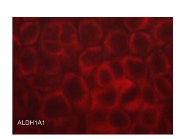 ICC (Immunocytochemistry)
(Immunocytochemical labeling of ALDH1A1 in aldehyde fixed and NP-40 permeabilized human A431 cells. The cells were labeled with mouse monoclonal anti-ALDH1A1 (AM5621). The antibody was detected using goat anti-mouse DyLight 594.)
ICC (Immunocytochemistry)
(Immunocytochemical labeling of ALDH1A1 in aldehyde fixed and NP-40 permeabilized human A431 cells. The cells were labeled with mouse monoclonal anti-ALDH1A1 (AM5621). The antibody was detected using goat anti-mouse DyLight 594.)
ALDH1A1, Monoclonal Antibody (Cat# AAA71563)
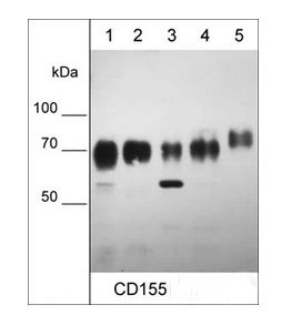
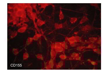 ICC (Immunocytochemistry)
(Immunocytochemical labeling of CD155 in aldehyde fixed and NP-40 permeabilized human NCI-H446 small cell lung carcinoma cells. The cells were labeled with mouse monoclonal anti-CD155 (CM0481). The antibody was detected using goat anti-mouse DyLight 594.)
ICC (Immunocytochemistry)
(Immunocytochemical labeling of CD155 in aldehyde fixed and NP-40 permeabilized human NCI-H446 small cell lung carcinoma cells. The cells were labeled with mouse monoclonal anti-CD155 (CM0481). The antibody was detected using goat anti-mouse DyLight 594.)
CD155/PVR, Monoclonal Antibody (Cat# AAA71588)
 ICC (Immunocytochemistry)
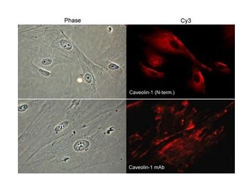
(Immunocytochemical labeling of caveolin-1 in paraformaldehyde-fixed and NP-40-permeabilized rabbit spleen fibroblasts. The cells were labeled with rabbit polyclonal Caveolin-1 (N-terminal region) and mouse monoclonal Caveolin-1 antibodies, and detected using appropriate secondary antibodies conjugated to Cy3. Phase contrast images (left) and immunofluorescent images (right).)
ICC (Immunocytochemistry)
(Immunocytochemical labeling of caveolin-1 in paraformaldehyde-fixed and NP-40-permeabilized rabbit spleen fibroblasts. The cells were labeled with rabbit polyclonal Caveolin-1 (N-terminal region) and mouse monoclonal Caveolin-1 antibodies, and detected using appropriate secondary antibodies conjugated to Cy3. Phase contrast images (left) and immunofluorescent images (right).)
Caveolin-1, Monoclonal Antibody (Cat# AAA71592)
 ICC (Immunocytochemistry)
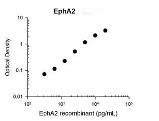
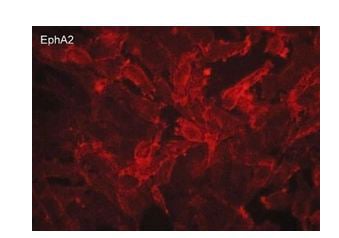
(Immunocytochemical labeling of EphA2 in aldehyde fixed human MDA-MB-231 breast carcinoma cells. The cells were labeled with mouse monoclonal anti-EphA2 (EM0881). The antibody was detected using goat anti-mouse DyLight 594.)
ICC (Immunocytochemistry)
(Immunocytochemical labeling of EphA2 in aldehyde fixed human MDA-MB-231 breast carcinoma cells. The cells were labeled with mouse monoclonal anti-EphA2 (EM0881). The antibody was detected using goat anti-mouse DyLight 594.)
EphA2, Monoclonal Antibody (Cat# AAA71623)
 ICC (Immunocytochemistry)
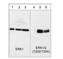
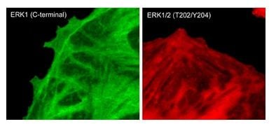
(Immunocytochemical labeling of phosphorylated ERK1 in paraformaldehyde-fixed and NP-40-permeabilized rat A7r5 cells treated with calyculin A. The fixed cells were labeled with mouse monoclonal antibodies to anti-ERK1 (EM2331) and anti-ERK1/2 (Thr-202/Tyr-204) (EM2061). The antibodies were detected using Goat anti-Mouse secondary antibodies conjugated to DyLight 488 (left) and DyLight 594 (right).)
ICC (Immunocytochemistry)
(Immunocytochemical labeling of phosphorylated ERK1 in paraformaldehyde-fixed and NP-40-permeabilized rat A7r5 cells treated with calyculin A. The fixed cells were labeled with mouse monoclonal antibodies to anti-ERK1 (EM2331) and anti-ERK1/2 (Thr-202/Tyr-204) (EM2061). The antibodies were detected using Goat anti-Mouse secondary antibodies conjugated to DyLight 488 (left) and DyLight 594 (right).)
ERK1, Monoclonal Antibody (Cat# AAA71624)
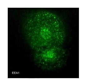 ICC (Immunocytochemistry)
(Immunocytochemical labeling of EEA1 in paraformaldehyde-fixed and NP40-permeabilized A7r5 cells. The cells were labeled with mouse monoclonal EEA1 (EM3471). The antibody was detected using goat anti-mouse DyLight 488.)
ICC (Immunocytochemistry)
(Immunocytochemical labeling of EEA1 in paraformaldehyde-fixed and NP40-permeabilized A7r5 cells. The cells were labeled with mouse monoclonal EEA1 (EM3471). The antibody was detected using goat anti-mouse DyLight 488.)
Early Endosome Antigen 1 (EEA1), Monoclonal Antibody (Cat# AAA71626)
alpha Synuclein, Monoclonal Antibody (Cat# AAA72837)
Reactivity assumed based on 100% sequence homology: most mammals
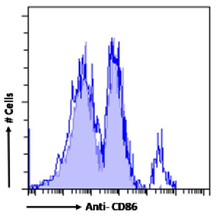 FCM/FACS (Flow Cytometry)
(Flow-cytometryusingtheanti-CD86antibodyOX-48(AAA72467). Ratsplenocyteswerestainedwithanti-FluoresceinIgGantibody(4-4-20;isotypecontrol,shadedline)ortherabbitIgG1versionofOX-48(AAA72467,blueline)atadilutionof1:100for1hatRT.Afterwashing,boundantibodywasdetectedusingagoatanti-mouseIgGAlexaFluor488antibodyatadilutionof1:1000andcellsanalyzedusingaFACSCantoflow-cytometer.)
FCM/FACS (Flow Cytometry)
(Flow-cytometryusingtheanti-CD86antibodyOX-48(AAA72467). Ratsplenocyteswerestainedwithanti-FluoresceinIgGantibody(4-4-20;isotypecontrol,shadedline)ortherabbitIgG1versionofOX-48(AAA72467,blueline)atadilutionof1:100for1hatRT.Afterwashing,boundantibodywasdetectedusingagoatanti-mouseIgGAlexaFluor488antibodyatadilutionof1:1000andcellsanalyzedusingaFACSCantoflow-cytometer.)
CD86, Monoclonal Recombinant Antibody (Cat# AAA72467)
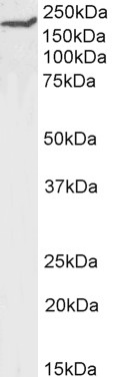 WB (Western Blot)
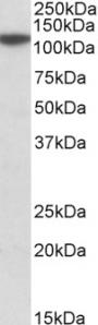
(Western Blotusinganti-CD45RAorA/B(Receptor-typetyrosine-proteinphosphataseC)antibodyOX-30(AAA72475) Ratthymuslysatesamples(35ugproteininRIPAbuffer)wereresolvedona10%SDSPAGEgelandblotsprobedwiththechimericrabbitversionofOX-30(AAA72475)at2ug/mlbeforedetectionusingananti-rabbitsecondaryantibody.Aprimaryincubationof1hwasusedandproteinwasdetectedbychemiluminescence.TheexpectedbandsizeforunmodifiedCD45RAis~143.3kDa,butthisproteinisalsohighlyglycosylated(UniProtId:P04157).AAA72475successfullydetectedCD45RAorA/Binratthymustissuelysate.)
WB (Western Blot)
(Western Blotusinganti-CD45RAorA/B(Receptor-typetyrosine-proteinphosphataseC)antibodyOX-30(AAA72475) Ratthymuslysatesamples(35ugproteininRIPAbuffer)wereresolvedona10%SDSPAGEgelandblotsprobedwiththechimericrabbitversionofOX-30(AAA72475)at2ug/mlbeforedetectionusingananti-rabbitsecondaryantibody.Aprimaryincubationof1hwasusedandproteinwasdetectedbychemiluminescence.TheexpectedbandsizeforunmodifiedCD45RAis~143.3kDa,butthisproteinisalsohighlyglycosylated(UniProtId:P04157).AAA72475successfullydetectedCD45RAorA/Binratthymustissuelysate.)
CD45RA or A/B, Monoclonal Recombinant Antibody (Cat# AAA72475)
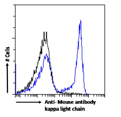

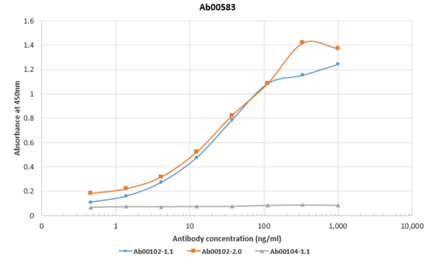 ELISA
(ELISAusingOX-20(AAA72483),Ab00102-1.1,Ab00102-2.0andAb00104-1.1. BindingcurvesoftherabbitIgGchimericversionoftheAnti-HAantibodyOX-20againstAb00102-1.1(blueline),Ab00102-2.0(orangeline)andAb00104-1.1(greyline)toanELISAplatecoatedwithAb00102-1.1,Ab00102-2.0andAb00104-1.1ataconcentrationof2.5ug/ml.A3-foldserialdilutionfrom3000to0.0169ng/mlwasperformedusingAAA72483antibody.Forsignaldetection,a1:4000dilutionofaHRP-conjugatedanti-rabbitIgGantibodywasused.)
ELISA
(ELISAusingOX-20(AAA72483),Ab00102-1.1,Ab00102-2.0andAb00104-1.1. BindingcurvesoftherabbitIgGchimericversionoftheAnti-HAantibodyOX-20againstAb00102-1.1(blueline),Ab00102-2.0(orangeline)andAb00104-1.1(greyline)toanELISAplatecoatedwithAb00102-1.1,Ab00102-2.0andAb00104-1.1ataconcentrationof2.5ug/ml.A3-foldserialdilutionfrom3000to0.0169ng/mlwasperformedusingAAA72483antibody.Forsignaldetection,a1:4000dilutionofaHRP-conjugatedanti-rabbitIgGantibodywasused.)
kappa light chain, Monoclonal Recombinant Antibody (Cat# AAA72483)
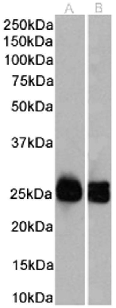
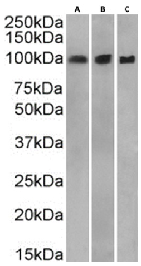
 WB (Western Blot)
(Western BlotusingAnti-CD43antibodyOX-56(AAA72489). Ratthymus(A),ratlymphnode(B)andratspleen(C)tissuelysates(35ugproteininRIPAbuffer)wereresolvedonaSDSPAGEgelandblotswereprobedwiththechimericrabbitversionofOX-56(AAA72489)at0.3ug/mlbeforedetectionusingananti-rabbitsecondaryantibody.Aprimaryincubationof1hwasusedandproteinwasdetectedbychemiluminescence.)
WB (Western Blot)
(Western BlotusingAnti-CD43antibodyOX-56(AAA72489). Ratthymus(A),ratlymphnode(B)andratspleen(C)tissuelysates(35ugproteininRIPAbuffer)wereresolvedonaSDSPAGEgelandblotswereprobedwiththechimericrabbitversionofOX-56(AAA72489)at0.3ug/mlbeforedetectionusingananti-rabbitsecondaryantibody.Aprimaryincubationof1hwasusedandproteinwasdetectedbychemiluminescence.)
CD43, Monoclonal Recombinant Antibody (Cat# AAA72489)
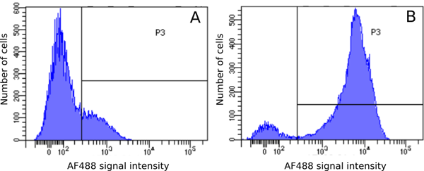
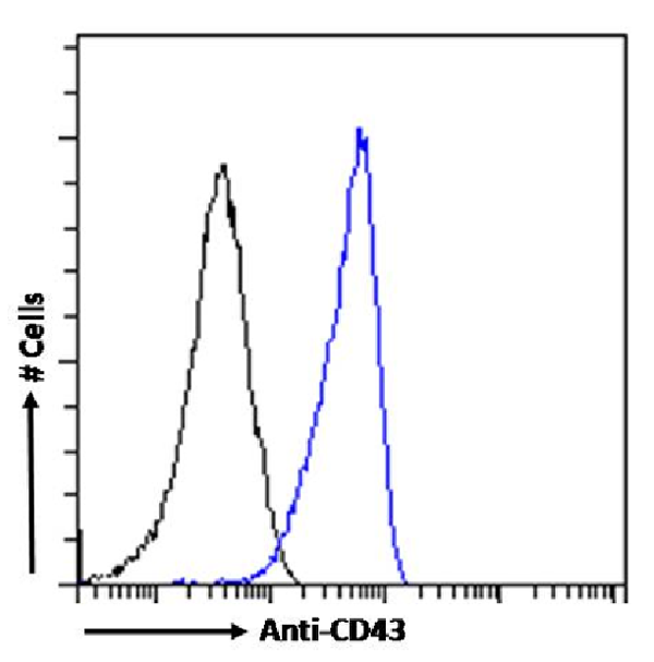
 FCM/FACS (Flow Cytometry)
(Flowcytometryusinganti-SiderocalinantibodyMAB1857(AAA72493). Paraformaldehyde-fixedRAW264.7cellspermeabilizedwith0.5%Tritonwerestainedwiththeanti-unknownspecificityantibodyortherabbitIgGversionofMAB1857(AAA72493,blueline)atadilutionof1:100for1hatRT.Afterwashing,theboundantibodywasdetectedusingagoatanti-rabbitIgGAlexaFluor488antibodyatadilutionof1:1000,andthecellswereanalyzedusingaFACSCantoflow-cytometer.)
FCM/FACS (Flow Cytometry)
(Flowcytometryusinganti-SiderocalinantibodyMAB1857(AAA72493). Paraformaldehyde-fixedRAW264.7cellspermeabilizedwith0.5%Tritonwerestainedwiththeanti-unknownspecificityantibodyortherabbitIgGversionofMAB1857(AAA72493,blueline)atadilutionof1:100for1hatRT.Afterwashing,theboundantibodywasdetectedusingagoatanti-rabbitIgGAlexaFluor488antibodyatadilutionof1:1000,andthecellswereanalyzedusingaFACSCantoflow-cytometer.)
Siderocalin, Monoclonal Recombinant Antibody (Cat# AAA72493)
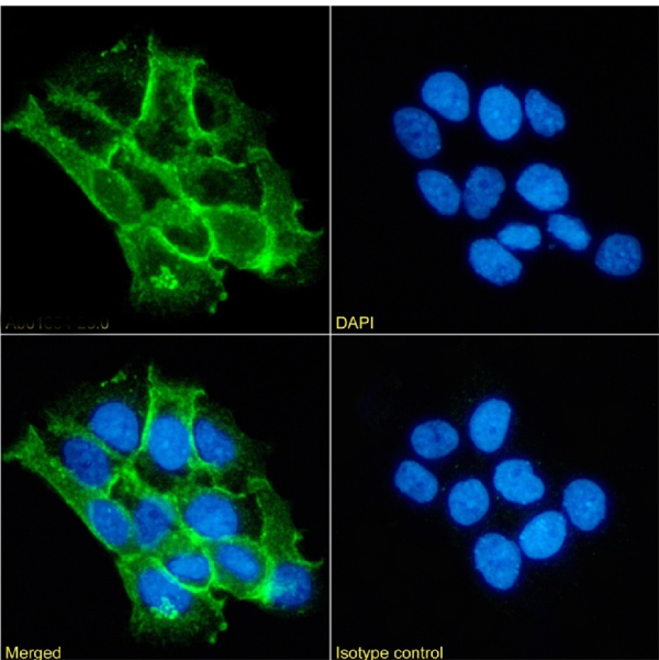
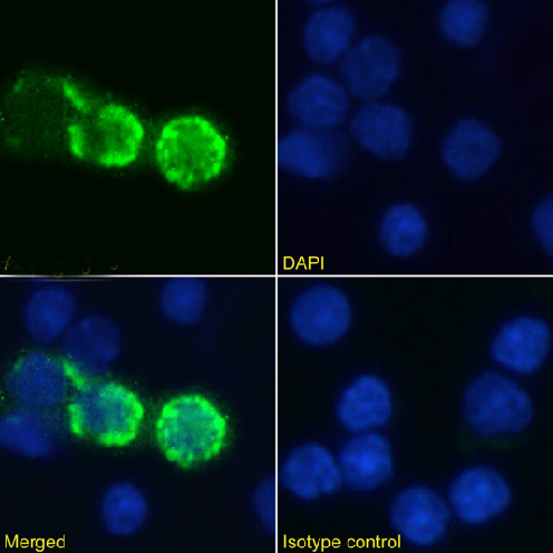
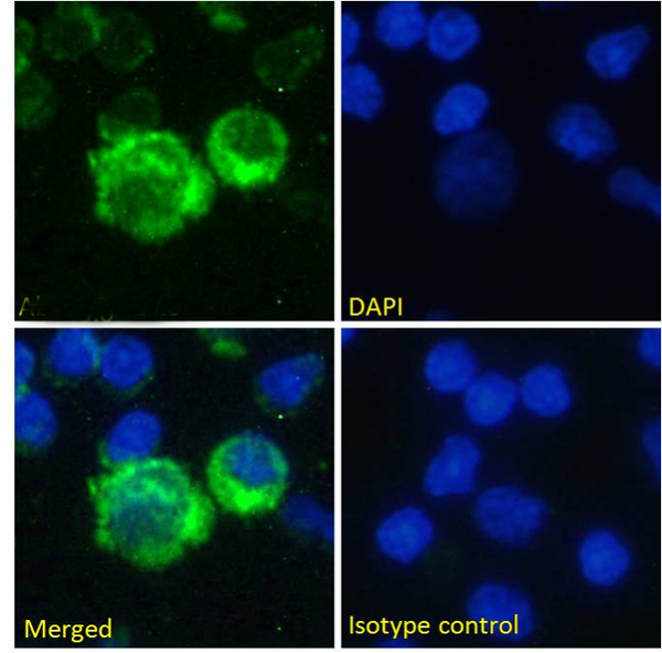
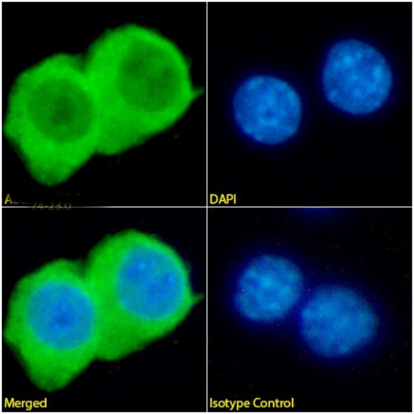
 IF (Immunofluorescence)
(ImmunofluorescencestainingoffixedA431cellswithanti-alphacateninantibody1G5(AAA72522) ImmunofluorescenceanalysisofparaformaldehydefixedA431cellsonShi-fixcoverslipsstainedwiththechimericrabbitIgGversionof1G5(AAA72522)at10ug/mlfor1hfollowedbyAlexaFluor488secondaryantibody(2ug/ml),showingmembranestaining.ThenuclearstainisDAPI(blue).Panelsshowfromleft-right,top-bottomAAA72522,DAPI,mergedchannelsandanisotypecontrol.TheisotypecontrolwasanunknownspecificityantibodyfollowedbystainingwithAlexaFluor488secondaryantibody.)
IF (Immunofluorescence)
(ImmunofluorescencestainingoffixedA431cellswithanti-alphacateninantibody1G5(AAA72522) ImmunofluorescenceanalysisofparaformaldehydefixedA431cellsonShi-fixcoverslipsstainedwiththechimericrabbitIgGversionof1G5(AAA72522)at10ug/mlfor1hfollowedbyAlexaFluor488secondaryantibody(2ug/ml),showingmembranestaining.ThenuclearstainisDAPI(blue).Panelsshowfromleft-right,top-bottomAAA72522,DAPI,mergedchannelsandanisotypecontrol.TheisotypecontrolwasanunknownspecificityantibodyfollowedbystainingwithAlexaFluor488secondaryantibody.)
alpha catenin, Monoclonal Recombinant Antibody (Cat# AAA72522)


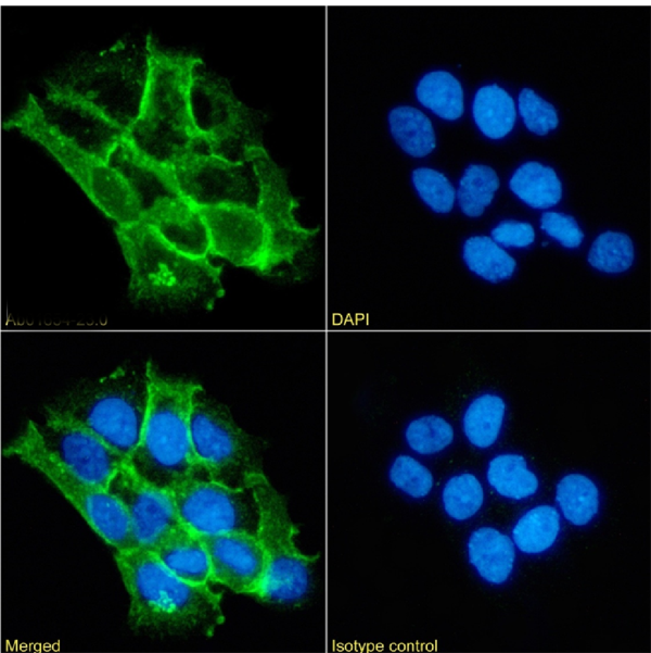
 IF (Immunofluorescence)
(ImmunofluorescencestainingoffixedA431cellswithanti-alphacateninantibody1G5(AAA72523) ImmunofluorescenceanalysisofparaformaldehydefixedA431cellsonShi-fixcoverslipsstainedwiththechimericrabbitIgGversionof1G5(AAA72523)at10ug/mlfor1hfollowedbyAlexaFluor488secondaryantibody(2ug/ml),showingmembranestaining.ThenuclearstainisDAPI(blue).Panelsshowfromleft-right,top-bottomAAA72523,DAPI,mergedchannelsandanisotypecontrol.TheisotypecontrolwasanunknownspecificityantibodyfollowedbystainingwithAlexaFluor488secondaryantibody.)
IF (Immunofluorescence)
(ImmunofluorescencestainingoffixedA431cellswithanti-alphacateninantibody1G5(AAA72523) ImmunofluorescenceanalysisofparaformaldehydefixedA431cellsonShi-fixcoverslipsstainedwiththechimericrabbitIgGversionof1G5(AAA72523)at10ug/mlfor1hfollowedbyAlexaFluor488secondaryantibody(2ug/ml),showingmembranestaining.ThenuclearstainisDAPI(blue).Panelsshowfromleft-right,top-bottomAAA72523,DAPI,mergedchannelsandanisotypecontrol.TheisotypecontrolwasanunknownspecificityantibodyfollowedbystainingwithAlexaFluor488secondaryantibody.)
alpha catenin, Monoclonal Recombinant Antibody (Cat# AAA72523)
What are Monoclonal Antibodies?
Monoclonal antibodies are specialized laboratory-produced proteins developed for binding to specific biological antigens or other molecular targets. Since they come from a single cell (or clone), they are especially consistent and accurate in the data they are involved in producing.
This type of antibody material has been shown to be a powerful tool in finding and subsequently destroying harmful cells in an organism, such as those found in cancers or various autoimmune diseases. This makes them excellent aids in medical testing and research, which is why they are so widely used.
AAA Biotech offers a comprehensive range of high-quality monoclonal antibodies that perform effectively in various laboratory tests, including (amongst others) ELISA, western blotting, immunohistochemistry, and flow cytometry. All of the products in our catalog are thoroughly quality tested to make sure that they are reliable and will consistently perform well in your research.
What Are The Uses of Monoclonal Antibodies
Monoclonal antibodies are used in many lab tests, including (amongst others) ELISA, western blotting, immunohistochemistry, and flow cytometry.
ELISA is a test that helps detect a specific substance/analyte in a sample. It uses antibodies (often monoclonal) bound to a solid surface (such as the well of a microplate) to “capture” the substance/analyte in the sample and immobilize it so that the detection antibody component can then bind to it and produce a signal, which can then be measured.
Western blotting identifies specific proteins in a sample. The sample is first separated on a gel, and then antibodies are applied that will typically bind to the target, which will all be localized to a single band in a lane.
Immunohistochemistry helps locate specific proteins in cells or tissue samples using antibodies.
Flow cytometry looks at and sorts cells. It uses antibodies that are conjugated to reporter molecules called “fluorophores”, which, under special lights, emit light themselves, which can then be measured by a detector instrument.
How Monoclonal Antibodies Are Used as Medicine?
Please note that all of the products listed in AAA Biotech’s also known as AAA Bio or AAABio catalog are strictly for research-use only (RUO).
Monoclonal antibodies can also be used as therapeutic/medical treatments, particularly in the context of cancers. They are designed to find and bind to specific cells or proteins, helping the immune system recognize and attack the cancer. These treatments work in different ways, such as:
- Radioimmunotherapy attaches a small amount of radioactive molecule to the antibody, so it delivers the radiation directly to the cancer cells that the antibody is specifically binding to.
- Antibody-directed enzyme prodrug therapy uses antibodies that are specifically bound to special enzymes. These enzymes activate a harmless drug in the body and turn it into a cancer-killing drug only near the cancer cells—this helps avoid harming healthy cells.
- Immunoliposomes are tiny “bubbles” filled with medicine/drug and coated with antibodies. They carry the drug straight to the cancer cells.
Why Buy Monoclonal Antibodies From Us?
At AAA Biotech, we provide high-performance monoclonal antibodies designed to support a wide range of research needs.
1. Validated for Versatile Applications
The antibodies in our catalog are extensively validated and compatible with multiple techniques, including (but not limited to) ELISA, flow cytometry (FC), immunocytochemistry (ICC), immunofluorescence (IF), immunohistochemistry (IHC), immunoprecipitation (IP), and western blotting (WB).
2. Wide Selection & Specialized Options
We offer antibodies for common and rare species, that are available in various conjugated forms, and also in recombinant formats. Essentially, there is almost anything one might need to meet their experimental model’s requirements.
3. High-Quality Proteins
Our proteins meet high purity standards—90% or more as confirmed by SDS-PAGE. Many are available with tags like His, Flag, GST, or MBP, and we also supply native and biologically active proteins for functional studies.
Frequently Asked Questions
1. Are your monoclonal antibodies validated for specific applications?
Yes, our antibodies are tested and validated for use in methods such as ELISA, western blot, IHC, flow cytometry, and more. Refer to specific product pages or datasheets for individual product information.
2. How do I choose the right monoclonal antibody for my application?
Review the product details directly for application validation, species reactivity, and target information. You may also contact our support team at any time for help.
3. How quickly can I receive my order?
Most orders are processed and shipped within 1–3 business days, depending on product availability and your shipping location.