Filters
▼Clonality
▼Type
▼Reactivity
▼Gene Name
▼Isotype
▼Host
▼Application
▼Clone
▼Monoclonal Antibodies
Get accurate results in your research with our Monoclonal Antibodies, which are specially made to target exactly what you require for your research, and will produce consistent, reliable performance in lab tests.
Viewing 8900-8950 of 27597 product results
 IHC (Immunohistochemistry)
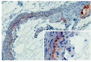
(MBL-A (8G6) deposition in developing murine atherosclerotic lesions. Staining of frozen tissue sections with antibody 8G6 (Cat. # AAA77763). Anti-mouse MBL-A at 2ug/ml (2h, RT). MBL-A was detected on the intima to media border as well as throughout the media (insert). Furthermore, extensive MBL-A deposition was seen at sites of necrosis (upper right corner).)
IHC (Immunohistochemistry)
(MBL-A (8G6) deposition in developing murine atherosclerotic lesions. Staining of frozen tissue sections with antibody 8G6 (Cat. # AAA77763). Anti-mouse MBL-A at 2ug/ml (2h, RT). MBL-A was detected on the intima to media border as well as throughout the media (insert). Furthermore, extensive MBL-A deposition was seen at sites of necrosis (upper right corner).)
MBL-A, Monoclonal Antibody (Cat# AAA77763)
 IA (Immunoassay)
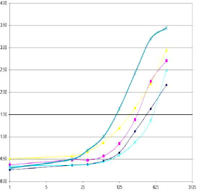
(Immuno Assay experiment with (AAA77770) as capture antibody used in different concentrations.)
IA (Immunoassay)
(Immuno Assay experiment with (AAA77770) as capture antibody used in different concentrations.)
TNF-RII, Monoclonal Antibody (Cat# AAA77770)
Rift Valley Fever (RVFV-NP), Monoclonal Antibody (Cat# AAA77427)
Ion Exchange Purified
Hepatitis B Surface (HBsAg), Monoclonal Antibody (Cat# AAA77435)
6-Monoacetylmorphine (6-MAM), Monoclonal Antibody (Cat# AAA77299)
Tramadol, Monoclonal Antibody (Cat# AAA77239)
Chorionic Gonadotropin beta (HCGbeta), Monoclonal Antibody (Cat# AAA77254)
Helicobacter pylori, Monoclonal Antibody (Cat# AAA77257)
Ferritin, Monoclonal Antibody (Cat# AAA77261)
Benzodiazepine, Monoclonal Antibody (Cat# AAA77274)
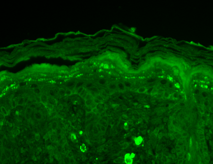
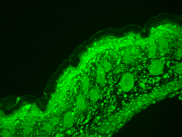
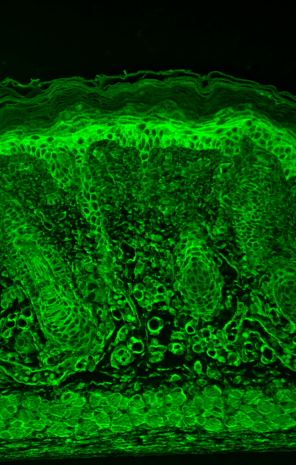
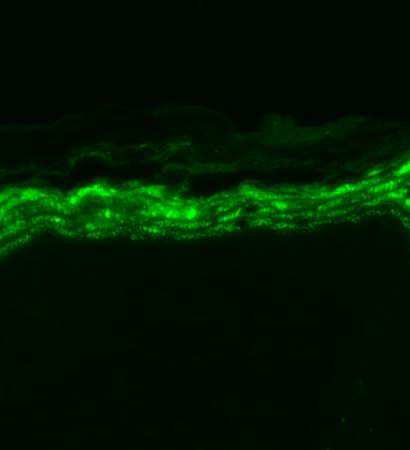 IHC (Immunohistochemistry)
(Immunohistochemistry analysis using Mouse Anti-Nav1.7 Sodium Channel Monoclonal Antibody, Clone S68-6. Tissue: backskin. Species: Mouse. Fixation: Bouin's Fixative and paraffin-embedded. Primary Antibody: Mouse Anti-Nav1.7 Sodium Channel Monoclonal Antibody at 1:100 for 1 hour at RT. Secondary Antibody: FITC Goat Anti-Mouse (green) at 1:50 for 1 hour at RT.)
IHC (Immunohistochemistry)
(Immunohistochemistry analysis using Mouse Anti-Nav1.7 Sodium Channel Monoclonal Antibody, Clone S68-6. Tissue: backskin. Species: Mouse. Fixation: Bouin's Fixative and paraffin-embedded. Primary Antibody: Mouse Anti-Nav1.7 Sodium Channel Monoclonal Antibody at 1:100 for 1 hour at RT. Secondary Antibody: FITC Goat Anti-Mouse (green) at 1:50 for 1 hour at RT.)
Nav1.7, Monoclonal Antibody (Cat# AAA103249)
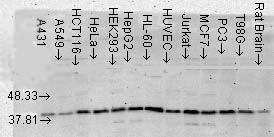 WB (Western Blot)
(Western Blot analysis of Human Cell lysates showing detection of p38 MAPK protein using Mouse Anti-p38 MAPK Monoclonal Antibody, Clone 9F12. Load: 15 ug. Block: 1.5% BSA for 30 minutes at RT. Primary Antibody: Mouse Anti-p38 MAPK Monoclonal Antibody at 1:1000 for 2 hours at RT. Secondary Antibody: Sheep Anti-Mouse IgG: HRP for 1 hour at RT.)
WB (Western Blot)
(Western Blot analysis of Human Cell lysates showing detection of p38 MAPK protein using Mouse Anti-p38 MAPK Monoclonal Antibody, Clone 9F12. Load: 15 ug. Block: 1.5% BSA for 30 minutes at RT. Primary Antibody: Mouse Anti-p38 MAPK Monoclonal Antibody at 1:1000 for 2 hours at RT. Secondary Antibody: Sheep Anti-Mouse IgG: HRP for 1 hour at RT.)
p38 alpha, Monoclonal Antibody (Cat# AAA103260)
 WB (Western Blot)
(Western Blot analysis of Rat brain membrane lysate showing detection of SHANK3 protein using Mouse Anti-SHANK3 Monoclonal Antibody, Clone S69-46. Load: 15 ug. Block: 1.5% BSA for 30 minutes at RT. Primary Antibody: Mouse Anti-SHANK3 Monoclonal Antibody at 1:1000 for 2 hours at RT. Secondary Antibody: Sheep Anti-Mouse IgG: HRP for 1 hour at RT.)
WB (Western Blot)
(Western Blot analysis of Rat brain membrane lysate showing detection of SHANK3 protein using Mouse Anti-SHANK3 Monoclonal Antibody, Clone S69-46. Load: 15 ug. Block: 1.5% BSA for 30 minutes at RT. Primary Antibody: Mouse Anti-SHANK3 Monoclonal Antibody at 1:1000 for 2 hours at RT. Secondary Antibody: Sheep Anti-Mouse IgG: HRP for 1 hour at RT.)
SHANK3, Monoclonal Antibody (Cat# AAA103269)


 WB (Western Blot)
(Western Blot analysis of Human Cell lysates showing detection of p38 MAPK protein using Mouse Anti-p38 MAPK Monoclonal Antibody, Clone 9F12. Load: 15 ug. Block: 1.5% BSA for 30 minutes at RT. Primary Antibody: Mouse Anti-p38 MAPK Monoclonal Antibody at 1:1000 for 2 hours at RT. Secondary Antibody: Sheep Anti-Mouse IgG: HRP for 1 hour at RT.)
WB (Western Blot)
(Western Blot analysis of Human Cell lysates showing detection of p38 MAPK protein using Mouse Anti-p38 MAPK Monoclonal Antibody, Clone 9F12. Load: 15 ug. Block: 1.5% BSA for 30 minutes at RT. Primary Antibody: Mouse Anti-p38 MAPK Monoclonal Antibody at 1:1000 for 2 hours at RT. Secondary Antibody: Sheep Anti-Mouse IgG: HRP for 1 hour at RT.)
p38 alpha, Monoclonal Antibody (Cat# AAA103279)
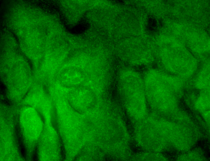
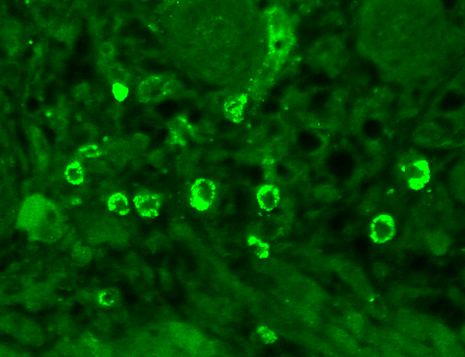
 IHC (Immunohiostchemistry)
(Immunohistochemistry analysis using Mouse Anti-CaV1.3 Calcium Channel Monoclonal Antibody, Clone S48A-9. Tissue: hippocampus. Species: Human. Fixation: Bouin's Fixative and paraffin-embedded. Primary Antibody: Mouse Anti-CaV1.3 Calcium Channel Monoclonal Antibody at 1:1000 for 1 hour at RT. Secondary Antibody: FITC Goat Anti-Mouse (green) at 1:50 for 1 hour at RT.)
IHC (Immunohiostchemistry)
(Immunohistochemistry analysis using Mouse Anti-CaV1.3 Calcium Channel Monoclonal Antibody, Clone S48A-9. Tissue: hippocampus. Species: Human. Fixation: Bouin's Fixative and paraffin-embedded. Primary Antibody: Mouse Anti-CaV1.3 Calcium Channel Monoclonal Antibody at 1:1000 for 1 hour at RT. Secondary Antibody: FITC Goat Anti-Mouse (green) at 1:50 for 1 hour at RT.)
Cav1.3, Monoclonal Antibody (Cat# AAA103295)
 IHC (Immunohistochemisry)
(Immunohistochemistry analysis using Mouse Anti-KCNQ4 Monoclonal Antibody, Clone S43-6. Tissue: hippocampus. Species: Human. Fixation: Bouin's Fixative and paraffin-embedded. Primary Antibody: Mouse Anti-KCNQ4 Monoclonal Antibody at 1:1000 for 1 hour at RT. Secondary Antibody: FITC Goat Anti-Mouse (green) at 1:50 for 1 hour at RT.)
IHC (Immunohistochemisry)
(Immunohistochemistry analysis using Mouse Anti-KCNQ4 Monoclonal Antibody, Clone S43-6. Tissue: hippocampus. Species: Human. Fixation: Bouin's Fixative and paraffin-embedded. Primary Antibody: Mouse Anti-KCNQ4 Monoclonal Antibody at 1:1000 for 1 hour at RT. Secondary Antibody: FITC Goat Anti-Mouse (green) at 1:50 for 1 hour at RT.)
KCNQ4, Monoclonal Antibody (Cat# AAA103296)
 WB (Western Blot)
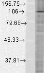
(Western Blot analysis of Human Cervical cancer cell line (HeLa) lysate showing detection of HIF1 alpha protein using Mouse Anti-HIF1 alpha Monoclonal Antibody, Clone ESEE122. Load: 15 ug. Block: 1.5% BSA for 30 minutes at RT. Primary Antibody: Mouse Anti-HIF1 alpha Monoclonal Antibody at 1:500 for 2 hours at RT. Secondary Antibody: Sheep Anti-Mouse IgG: HRP for 1 hour at RT.)
WB (Western Blot)
(Western Blot analysis of Human Cervical cancer cell line (HeLa) lysate showing detection of HIF1 alpha protein using Mouse Anti-HIF1 alpha Monoclonal Antibody, Clone ESEE122. Load: 15 ug. Block: 1.5% BSA for 30 minutes at RT. Primary Antibody: Mouse Anti-HIF1 alpha Monoclonal Antibody at 1:500 for 2 hours at RT. Secondary Antibody: Sheep Anti-Mouse IgG: HRP for 1 hour at RT.)
HIF1 alpha, Monoclonal Antibody (Cat# AAA103300)
 WB (Western Blot)
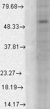
(Western Blot analysis of Human Cell line lysates showing detection of GABA A Receptor protein using Mouse Anti-GABA A Receptor Monoclonal Antibody, Clone S95-35. Load: 15 ug. Block: 1.5% BSA for 30 minutes at RT. Primary Antibody: Mouse Anti-GABA A Receptor Monoclonal Antibody at 1:1000 for 2 hours at RT. Secondary Antibody: Sheep Anti-Mouse IgG: HRP for 1 hour at RT.)
WB (Western Blot)
(Western Blot analysis of Human Cell line lysates showing detection of GABA A Receptor protein using Mouse Anti-GABA A Receptor Monoclonal Antibody, Clone S95-35. Load: 15 ug. Block: 1.5% BSA for 30 minutes at RT. Primary Antibody: Mouse Anti-GABA A Receptor Monoclonal Antibody at 1:1000 for 2 hours at RT. Secondary Antibody: Sheep Anti-Mouse IgG: HRP for 1 hour at RT.)
GABA(A) Receptor Alpha1, Monoclonal Antibody (Cat# AAA103303)
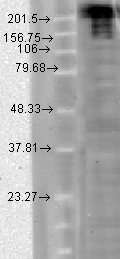
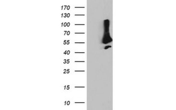
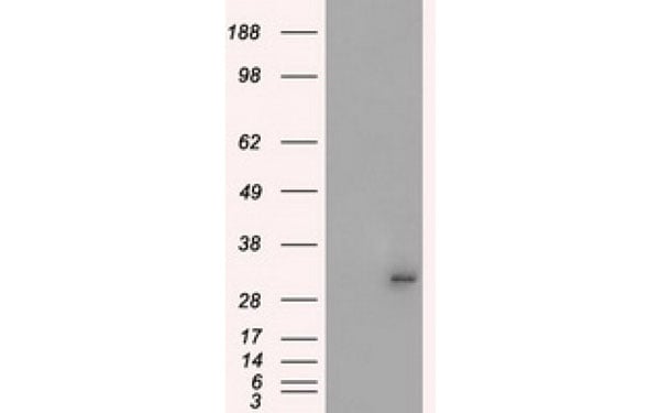
 WB (Western Blot)
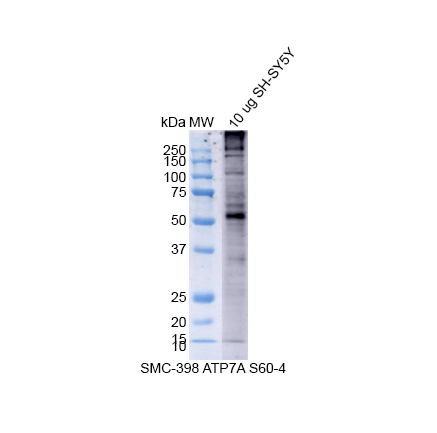
(Western Blot analysis of Human SH-SY5Y showing detection of Copper Transporting ATPase 1 protein using Mouse Anti-Copper Transporting ATPase 1 Monoclonal Antibody, Clone S60-4 . Lane 1: MW Ladder. Lane 2: 10 ug SH-SY5Y. Load: 10 ug. Block: 5% Skim Milk powder in TBST. Primary Antibody: Mouse Anti-Copper Transporting ATPase 1 Monoclonal Antibody at 1:500 for 2 hours at RT with shaking. Secondary Antibody: Goat anti-mouse IgG:HRP at 1:4000 for 1 hour at RT with shaking. Color Development: Chemiluminescent for HRP (Moss) for 5 min in RT.)
WB (Western Blot)
(Western Blot analysis of Human SH-SY5Y showing detection of Copper Transporting ATPase 1 protein using Mouse Anti-Copper Transporting ATPase 1 Monoclonal Antibody, Clone S60-4 . Lane 1: MW Ladder. Lane 2: 10 ug SH-SY5Y. Load: 10 ug. Block: 5% Skim Milk powder in TBST. Primary Antibody: Mouse Anti-Copper Transporting ATPase 1 Monoclonal Antibody at 1:500 for 2 hours at RT with shaking. Secondary Antibody: Goat anti-mouse IgG:HRP at 1:4000 for 1 hour at RT with shaking. Color Development: Chemiluminescent for HRP (Moss) for 5 min in RT.)
Copper-Transporting ATPase1, Monoclonal Antibody (Cat# AAA103305)

 WB (Western Blot)
(Western Blot analysis of Human Cervical cancer cell line (HeLa) lysate showing detection of HIF1 alpha protein using Mouse Anti-HIF1 alpha Monoclonal Antibody, Clone ESEE122. Load: 15 ug. Block: 1.5% BSA for 30 minutes at RT. Primary Antibody: Mouse Anti-HIF1 alpha Monoclonal Antibody at 1:500 for 2 hours at RT. Secondary Antibody: Sheep Anti-Mouse IgG: HRP for 1 hour at RT.)
WB (Western Blot)
(Western Blot analysis of Human Cervical cancer cell line (HeLa) lysate showing detection of HIF1 alpha protein using Mouse Anti-HIF1 alpha Monoclonal Antibody, Clone ESEE122. Load: 15 ug. Block: 1.5% BSA for 30 minutes at RT. Primary Antibody: Mouse Anti-HIF1 alpha Monoclonal Antibody at 1:500 for 2 hours at RT. Secondary Antibody: Sheep Anti-Mouse IgG: HRP for 1 hour at RT.)
HIF1 alpha, Monoclonal Antibody (Cat# AAA103314)
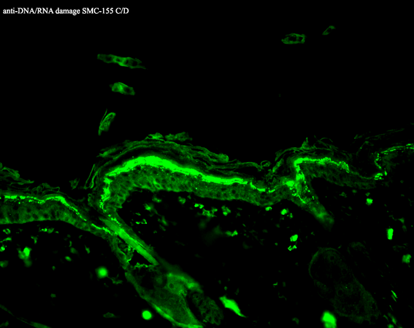
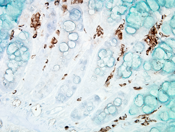
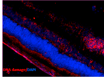
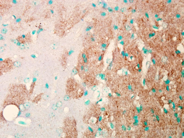
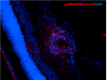
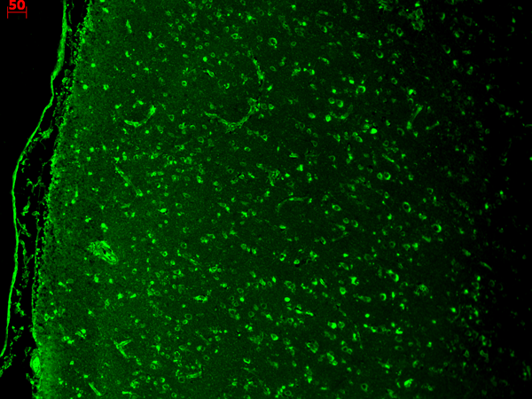
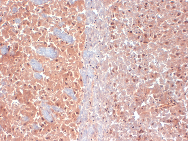
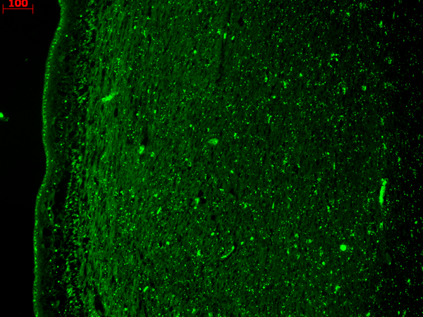
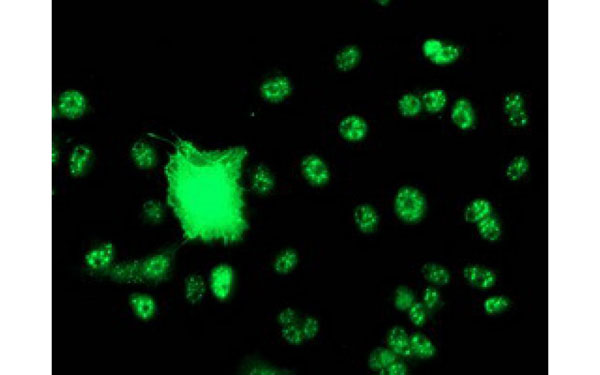
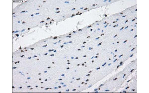
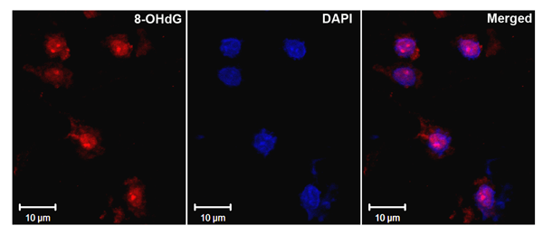 IHC (Immunohistochemistry)
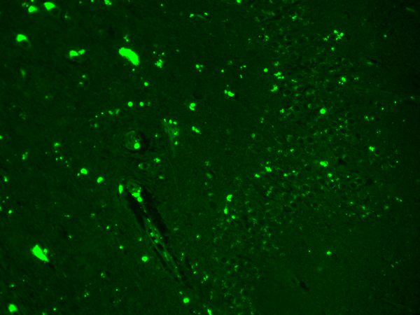
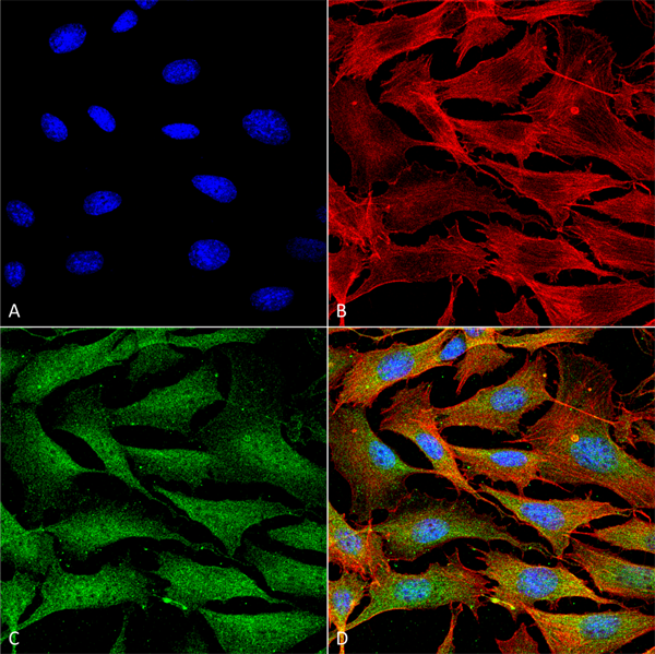
(Immunohistochemistry analysis using Mouse Anti-DNA Damage Monoclonal Antibody, Clone 15A3. Tissue: Ischemic fresh brain tissue. Species: Rat. Primary Antibody: Mouse Anti-DNA Damage Monoclonal Antibody at 1:1000 for 16 hours at RT. Secondary Antibody: Alexa Fluor 546 Goat Anti-mouse (Red) at 1:500 for 1 hour at RT. Localization: Cerebral Cortex. Courtesy of: Dr. Yi Yang, U. New Mexico.)
IHC (Immunohistochemistry)
(Immunohistochemistry analysis using Mouse Anti-DNA Damage Monoclonal Antibody, Clone 15A3. Tissue: Ischemic fresh brain tissue. Species: Rat. Primary Antibody: Mouse Anti-DNA Damage Monoclonal Antibody at 1:1000 for 16 hours at RT. Secondary Antibody: Alexa Fluor 546 Goat Anti-mouse (Red) at 1:500 for 1 hour at RT. Localization: Cerebral Cortex. Courtesy of: Dr. Yi Yang, U. New Mexico.)
DNA/RNA Damage, Monoclonal Antibody (Cat# AAA103315)
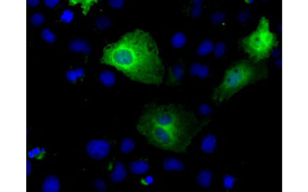
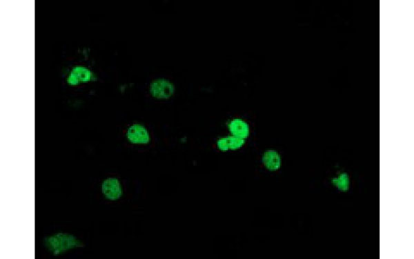
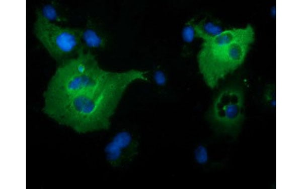
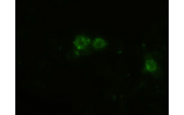
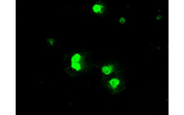
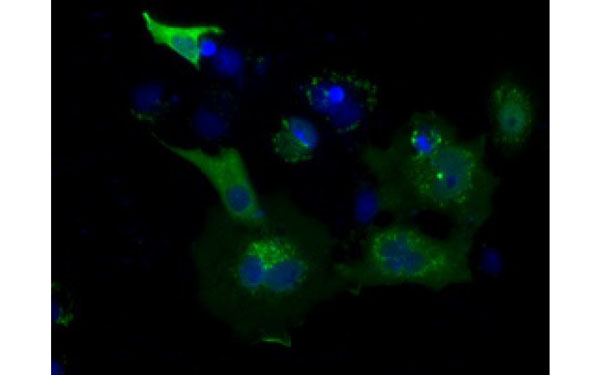
 IF (Immunofluorescence)
(Immunofluorescent staining of COS7 cells transiently transfected with recombinant SLC7A8 protein using SLC7A8 antibody)
IF (Immunofluorescence)
(Immunofluorescent staining of COS7 cells transiently transfected with recombinant SLC7A8 protein using SLC7A8 antibody)
SLC7A8, Monoclonal Antibody (Cat# AAA107242)
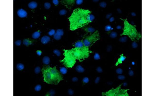 IF (Immunofluorescence)
(Immunofluorescent staining of COS7 cells transiently transfected with recombinant EPN2 protein using EPN2 antibody)
IF (Immunofluorescence)
(Immunofluorescent staining of COS7 cells transiently transfected with recombinant EPN2 protein using EPN2 antibody)
EPN2, Monoclonal Antibody (Cat# AAA107243)
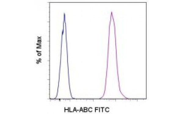 FCM/FACS (Flow Cytometry)
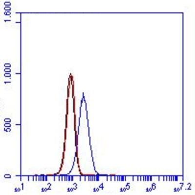
(Staining of normal human peripheral blood cells with Mouse IgG2a K Isotype Control (FITC) (blue histogram) or HLA-ABC antibody (FITC) (purple histogram). Cells in the lymphocyte gate were used for analysis.)
FCM/FACS (Flow Cytometry)
(Staining of normal human peripheral blood cells with Mouse IgG2a K Isotype Control (FITC) (blue histogram) or HLA-ABC antibody (FITC) (purple histogram). Cells in the lymphocyte gate were used for analysis.)
HLAABC, Monoclonal Antibody (Cat# AAA107258)

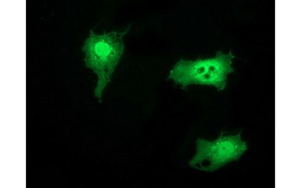
 IF (Immunofluorescence)
(Immunofluorescent staining of COS7 cells transiently transfected with recombinant TTLL12 protein using TTLL12 antibody)
IF (Immunofluorescence)
(Immunofluorescent staining of COS7 cells transiently transfected with recombinant TTLL12 protein using TTLL12 antibody)
TTLL12, Monoclonal Antibody (Cat# AAA107265)
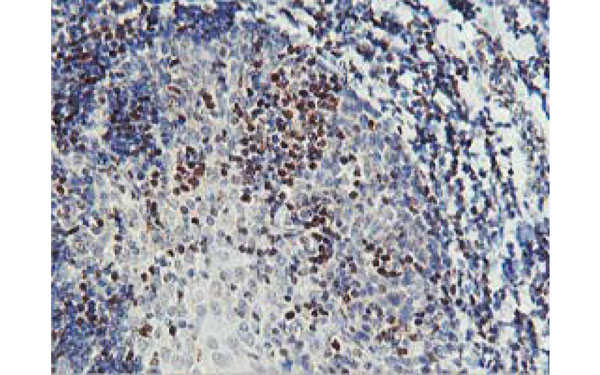
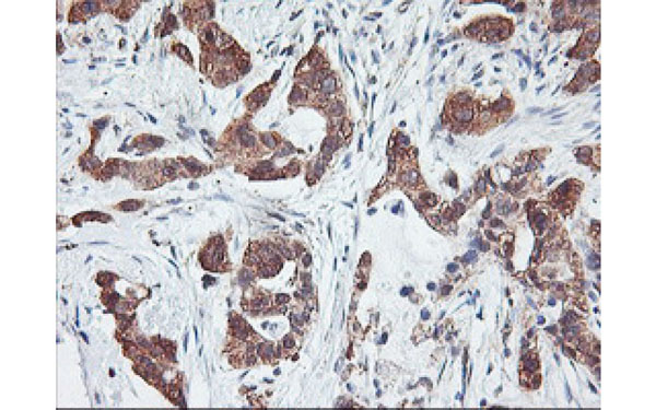 IHC (Immunohistochemisry)
(Immunohistochemical analysis of PFDN6 protein in paraffin embedded Carcinoma of Human lung tissue using PFDN6 antibody)
IHC (Immunohistochemisry)
(Immunohistochemical analysis of PFDN6 protein in paraffin embedded Carcinoma of Human lung tissue using PFDN6 antibody)
PFDN6, Monoclonal Antibody (Cat# AAA107271)
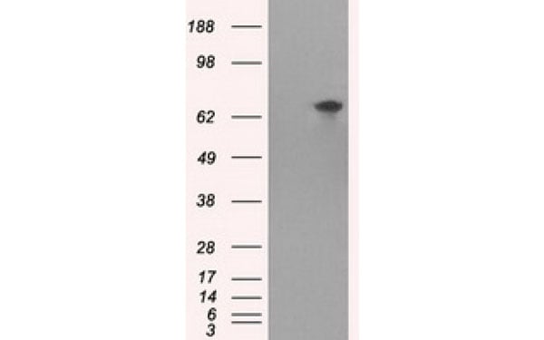
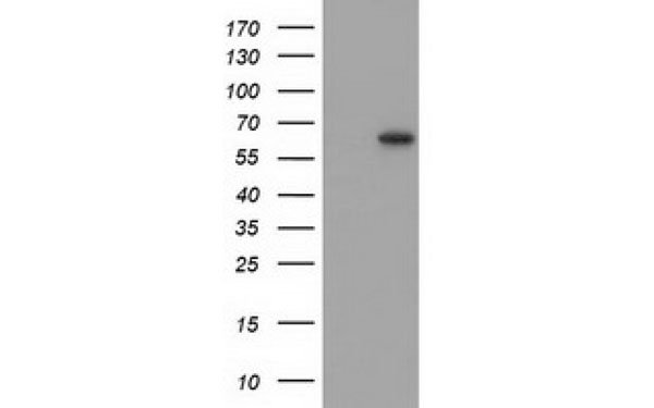 WB (Western Blot)
(Western Blot analysis of HEK293T cell lysates (5 ug) transfected with either recombinant SNTG1 protein (Right) or empty vector (Left) detected with SNTG1 antibody)
WB (Western Blot)
(Western Blot analysis of HEK293T cell lysates (5 ug) transfected with either recombinant SNTG1 protein (Right) or empty vector (Left) detected with SNTG1 antibody)
SNTG1, Monoclonal Antibody (Cat# AAA107275)
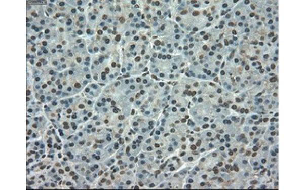
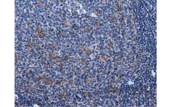
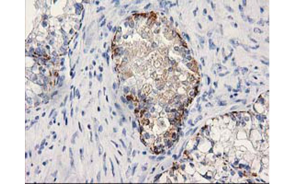 IHC (Immunohiostchemistry)
(Immunohistochemical analysis of MGLL protein in paraffin embedded Adenocarcinoma of Human ovary tissue using MGLL antibody)
IHC (Immunohiostchemistry)
(Immunohistochemical analysis of MGLL protein in paraffin embedded Adenocarcinoma of Human ovary tissue using MGLL antibody)
MGLL, Monoclonal Antibody (Cat# AAA107277)
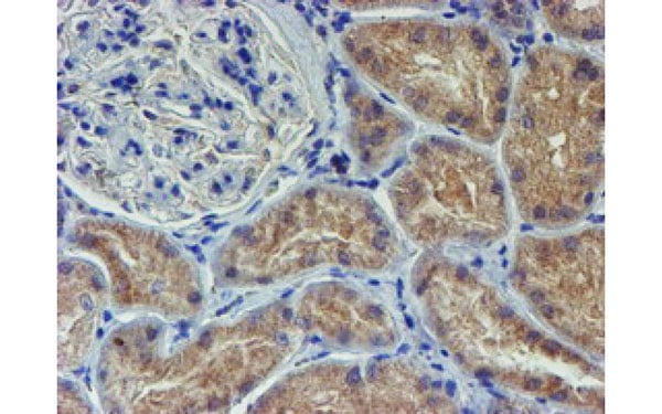
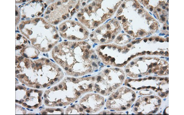
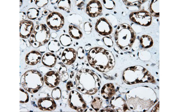 IHC (Immunohistochemisry)
(Immunohistochemical analysis of TTLL12 protein in paraffin embedded Human kidney tissue using TTLL12 antibody)
IHC (Immunohistochemisry)
(Immunohistochemical analysis of TTLL12 protein in paraffin embedded Human kidney tissue using TTLL12 antibody)
TTLL12, Monoclonal Antibody (Cat# AAA107280)

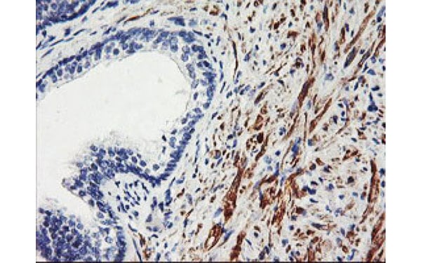
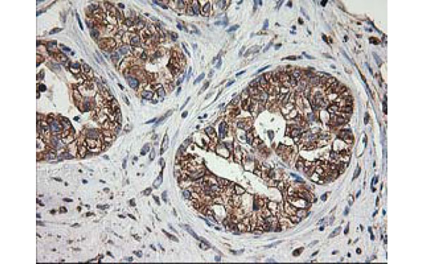
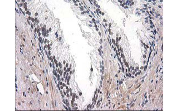 IHC (Immunohistochemisry)
(Immunohistochemical analysis of UBA2 protein in paraffin embedded Human prostate tissue using UBA2 antibody)
IHC (Immunohistochemisry)
(Immunohistochemical analysis of UBA2 protein in paraffin embedded Human prostate tissue using UBA2 antibody)
UBA2, Monoclonal Antibody (Cat# AAA107285)
 IHC (Immunohistochemisry)
(Immunohistochemical analysis of SPR protein in paraffin embedded Carcinoma of Human lung tissue using SPR antibody)
IHC (Immunohistochemisry)
(Immunohistochemical analysis of SPR protein in paraffin embedded Carcinoma of Human lung tissue using SPR antibody)
SPR, Monoclonal Antibody (Cat# AAA107292)
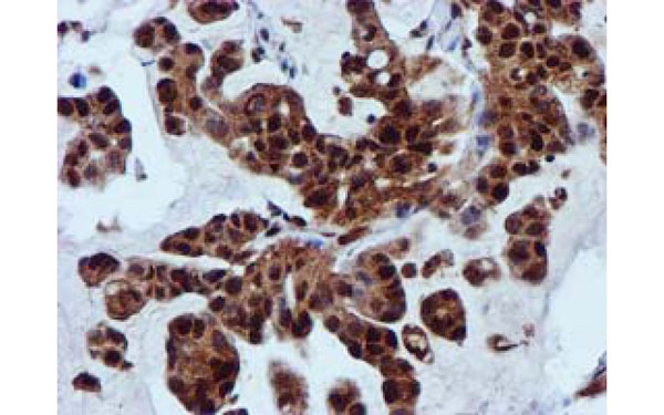
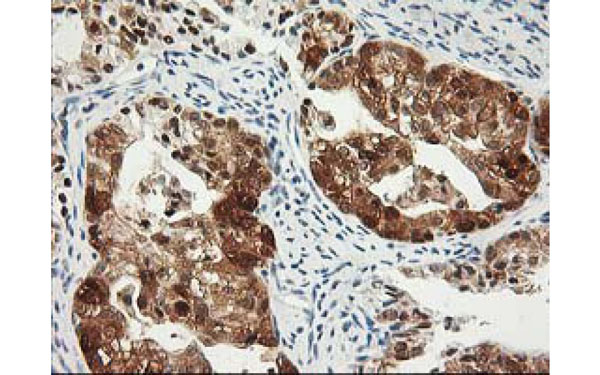 IHC (Immunohistochemisry)
(Immunohistochemical analysis of PDXK protein in paraffin embedded Adenocarcinoma of Human ovary tissue using PDXK antibody)
IHC (Immunohistochemisry)
(Immunohistochemical analysis of PDXK protein in paraffin embedded Adenocarcinoma of Human ovary tissue using PDXK antibody)
PDXK, Monoclonal Antibody (Cat# AAA107303)
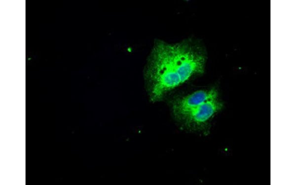
 IF (Immunofluorescence)
(Immunofluorescent staining of COS7 cells transiently transfected with recombinant PDE4A protein using PDE4A antibody)
IF (Immunofluorescence)
(Immunofluorescent staining of COS7 cells transiently transfected with recombinant PDE4A protein using PDE4A antibody)
PDE4A, Monoclonal Antibody (Cat# AAA107311)
 IF (Immunofluorescence)
(Immunofluorescent staining of COS7 cells transiently transfected with recombinant ZFAND2B protein using ZFAND2B antibody)
IF (Immunofluorescence)
(Immunofluorescent staining of COS7 cells transiently transfected with recombinant ZFAND2B protein using ZFAND2B antibody)
ZFAND2B, Monoclonal Antibody (Cat# AAA107474)
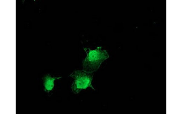
 IF (Immunofluorescence)
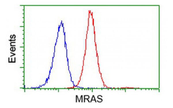
(Immunofluorescent staining of COS7 cells transiently transfected with recombinant MRAS protein using MRAS antibody)
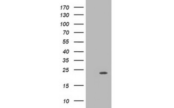
IF (Immunofluorescence)
(Immunofluorescent staining of COS7 cells transiently transfected with recombinant MRAS protein using MRAS antibody)
MRAS, Monoclonal Antibody (Cat# AAA107485)
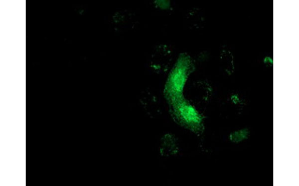
 IF (Immunofluorescence)
(Immunofluorescent staining of COS7 cells transiently transfected with recombinant MICAL1 protein using MICAL1 antibody)
IF (Immunofluorescence)
(Immunofluorescent staining of COS7 cells transiently transfected with recombinant MICAL1 protein using MICAL1 antibody)
MICAL1, Monoclonal Antibody (Cat# AAA107491)
 IF (Immunofluorescence)
(Immunofluorescent staining of COS7 cells transiently transfected with recombinant LOX protein using LOX antibody)
IF (Immunofluorescence)
(Immunofluorescent staining of COS7 cells transiently transfected with recombinant LOX protein using LOX antibody)
LOX, Monoclonal Antibody (Cat# AAA107492)
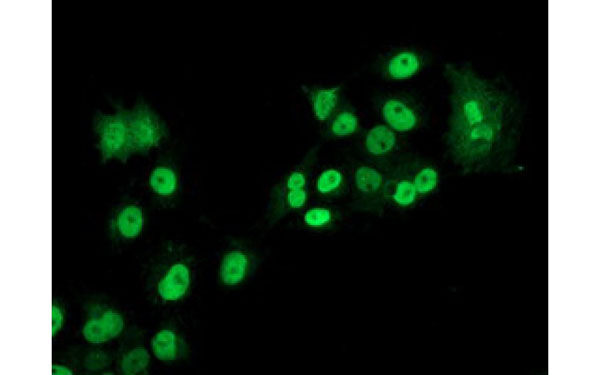
 IF (Immunofluorescence)
(Immunofluorescent staining of COS7 cells transiently transfected with recombinant PIK3AP1 protein using PIK3AP1 antibody)
IF (Immunofluorescence)
(Immunofluorescent staining of COS7 cells transiently transfected with recombinant PIK3AP1 protein using PIK3AP1 antibody)
PIK3AP1, Monoclonal Antibody (Cat# AAA107504)
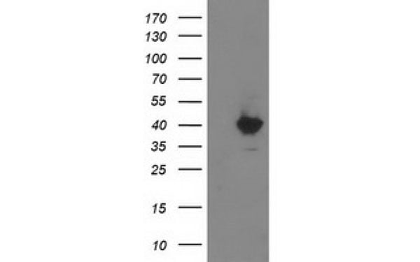
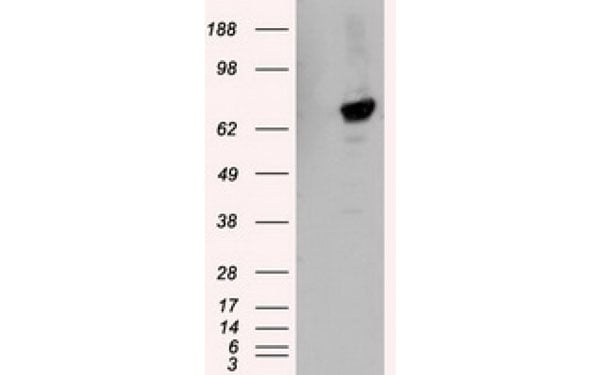
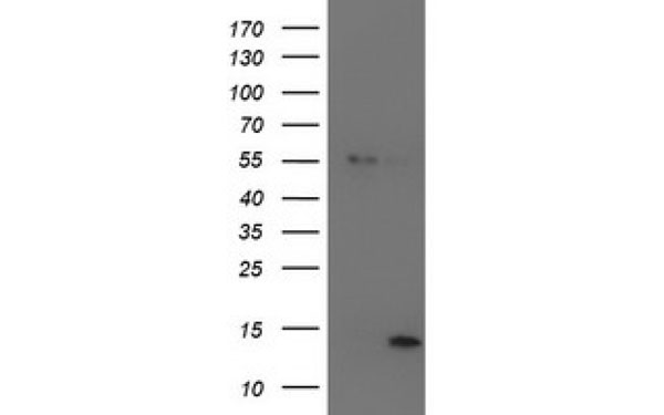 WB (Western Blot)
(Western Blot analysis of HEK293T cell lysates (5 ug) transfected with either recombinant LECT2 protein (Right) or empty vector (Left) detected with LECT2 antibody)
WB (Western Blot)
(Western Blot analysis of HEK293T cell lysates (5 ug) transfected with either recombinant LECT2 protein (Right) or empty vector (Left) detected with LECT2 antibody)
LECT2, Monoclonal Antibody (Cat# AAA107506)
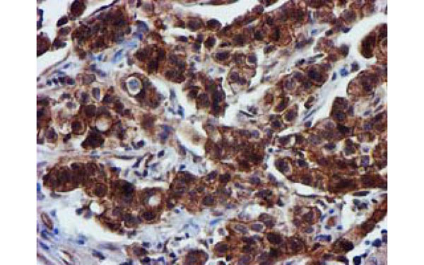
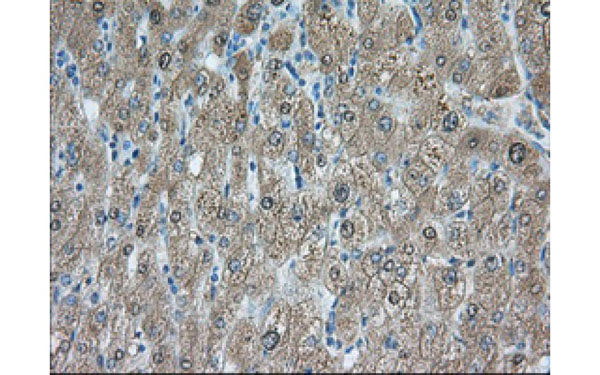
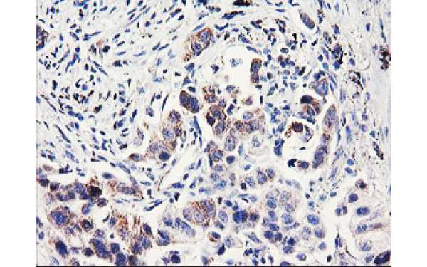 IHC (Immunohistochemisry)
(Immunohistochemical analysis of PDSS2 protein in paraffin embedded Carcinoma of Human lung tissue using PDSS2 antibody)
IHC (Immunohistochemisry)
(Immunohistochemical analysis of PDSS2 protein in paraffin embedded Carcinoma of Human lung tissue using PDSS2 antibody)
PDSS2, Monoclonal Antibody (Cat# AAA107519)
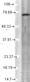
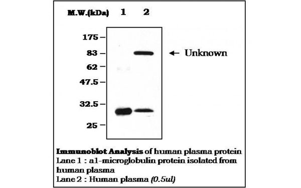 WB (Western Blot)
alpha 1 Microglobulin, Monoclonal Antibody (Cat# AAA107532)
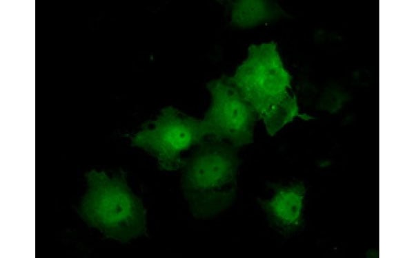
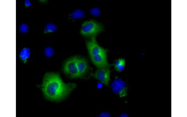
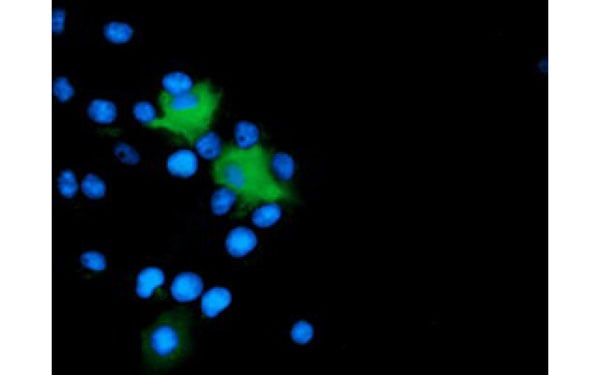
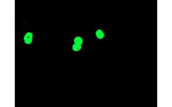
 IF (Immunofluorescence)
(Immunofluorescent staining of COS7 cells transiently transfected with recombinant SNX8 protein using SNX8 antibody)
IF (Immunofluorescence)
(Immunofluorescent staining of COS7 cells transiently transfected with recombinant SNX8 protein using SNX8 antibody)
SNX8, Monoclonal Antibody (Cat# AAA107538)
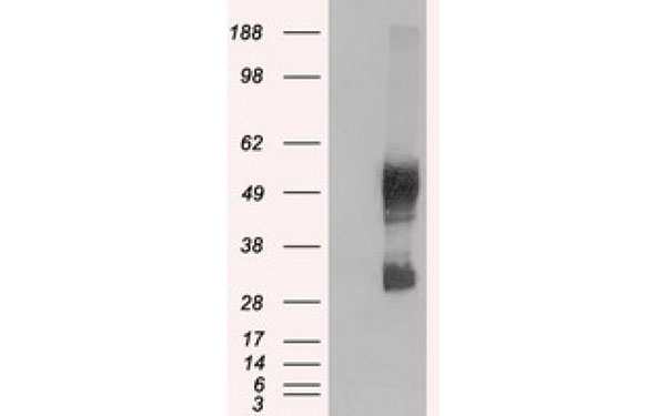
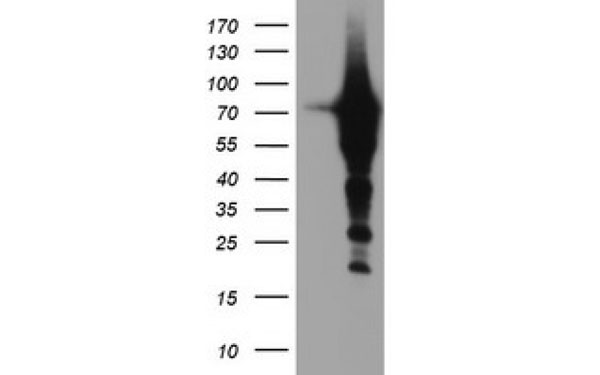
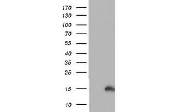
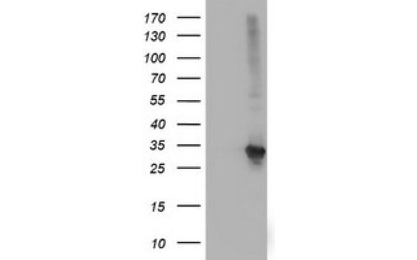
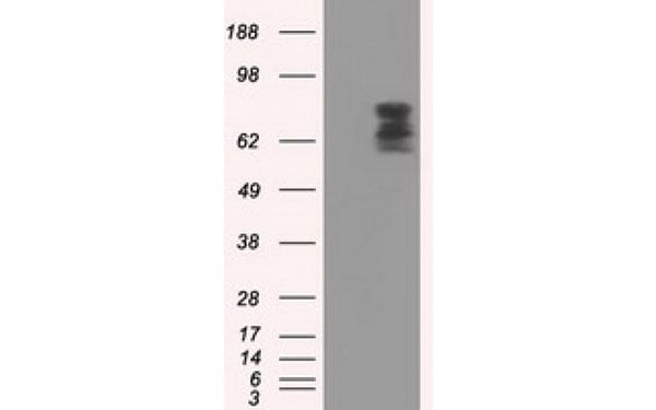
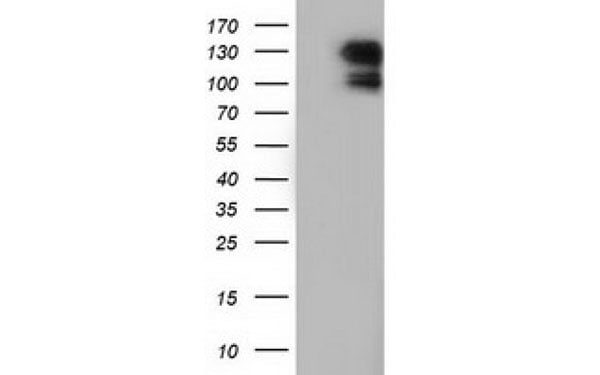
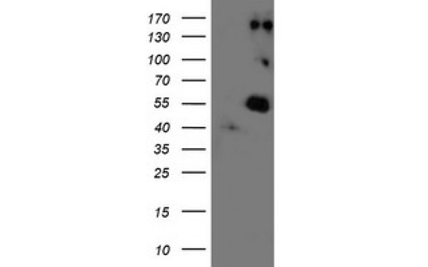
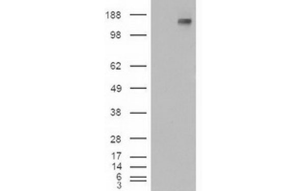
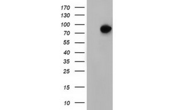
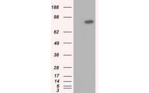
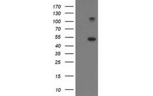
 WB (Western Blot)
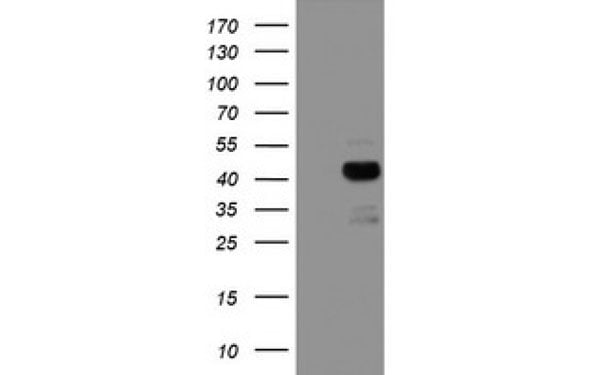
(Western Blot analysis of HEK293T cell lysates (5 ug) transfected with either recombinant PDSS2 protein (Right) or empty vector (Left) detected with PDSS2 antibody)
WB (Western Blot)
(Western Blot analysis of HEK293T cell lysates (5 ug) transfected with either recombinant PDSS2 protein (Right) or empty vector (Left) detected with PDSS2 antibody)
PDSS2, Monoclonal Antibody (Cat# AAA107336)
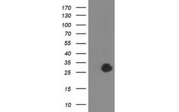
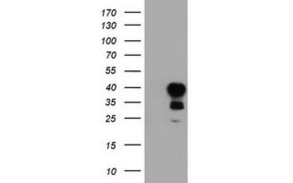
 WB (Western Blot)
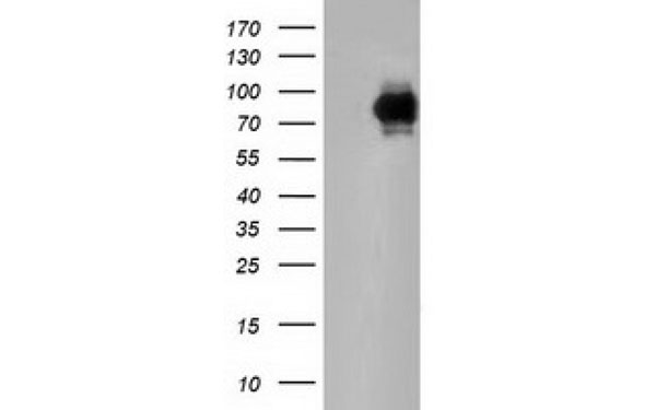
(Western Blot analysis of HEK293T cell lysates (5 ug) transfected with either recombinant PTCH1 protein (Right) or empty vector (Left) detected with PTCH1 antibody)
WB (Western Blot)
(Western Blot analysis of HEK293T cell lysates (5 ug) transfected with either recombinant PTCH1 protein (Right) or empty vector (Left) detected with PTCH1 antibody)
PTCH1, Monoclonal Antibody (Cat# AAA107340)
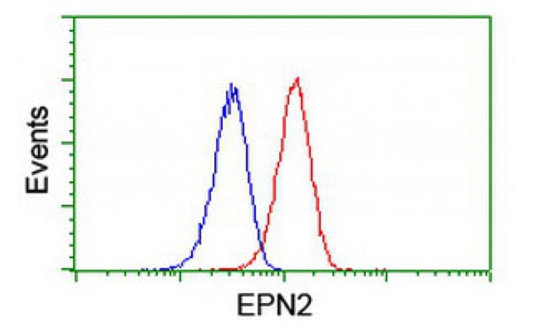
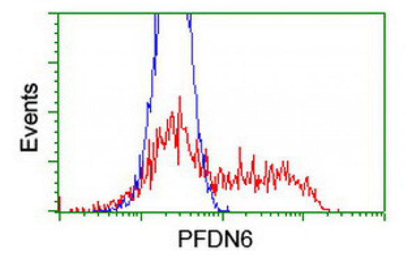
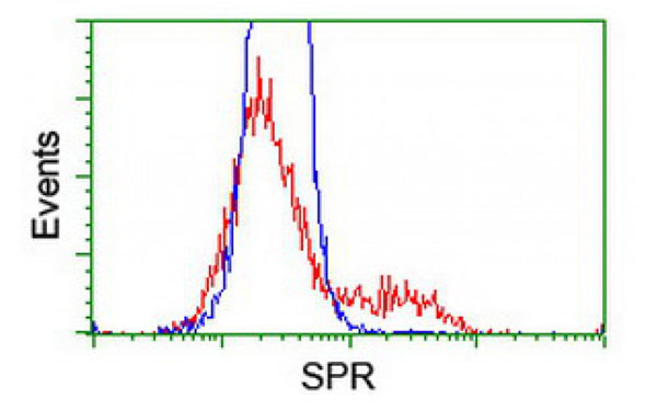
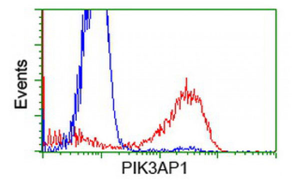
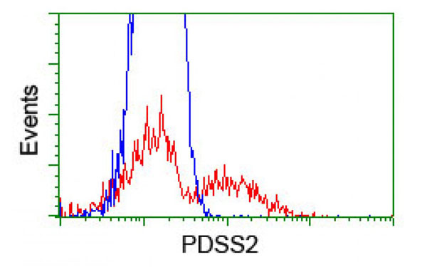
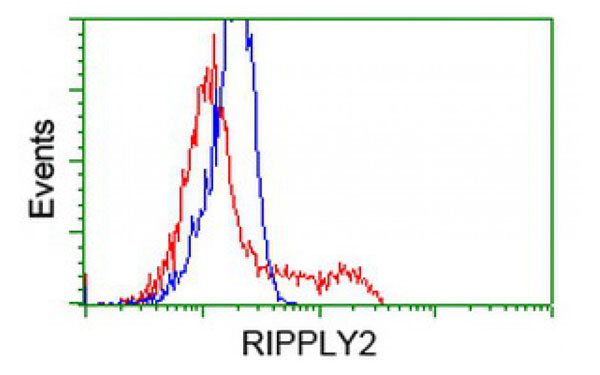
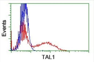 FCM/FACS (Flow Cytometry)
(Flow Cytometric analysis of HEK293T cells transfected with either recombinant TAL1 protein (red) or empty vector (blue) stained using TAL1 antibody.)
FCM/FACS (Flow Cytometry)
(Flow Cytometric analysis of HEK293T cells transfected with either recombinant TAL1 protein (red) or empty vector (blue) stained using TAL1 antibody.)
TAL1, Monoclonal Antibody (Cat# AAA107364)
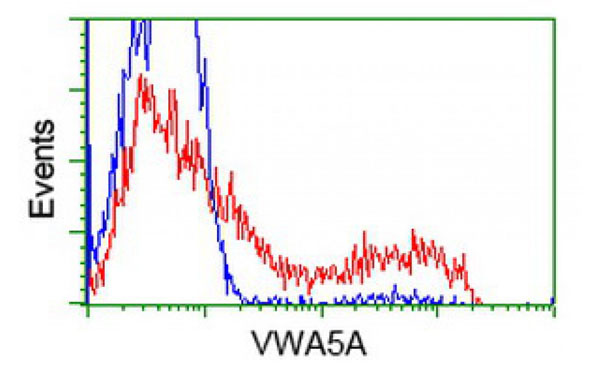
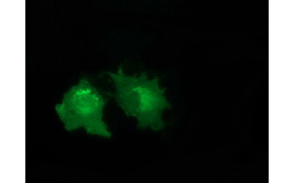 IF (Immunofluorescence)
(Immunofluorescent staining of COS7 cells transiently transfected with recombinant VWA5A protein using VWA5A antibody)
IF (Immunofluorescence)
(Immunofluorescent staining of COS7 cells transiently transfected with recombinant VWA5A protein using VWA5A antibody)
VWA5A, Monoclonal Antibody (Cat# AAA107368)
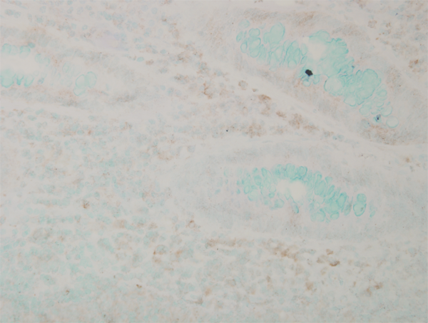
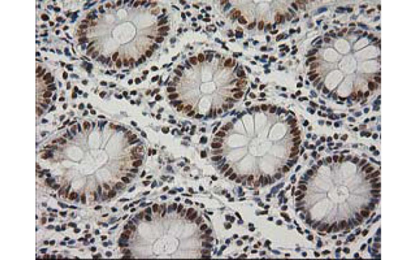
 IHC (Immunohistochemisry)
(Immunohistochemical analysis of PRKY protein in paraffin embedded Human colon tissue using PRKY antibody)
IHC (Immunohistochemisry)
(Immunohistochemical analysis of PRKY protein in paraffin embedded Human colon tissue using PRKY antibody)
PRKY, Monoclonal Antibody (Cat# AAA107391)
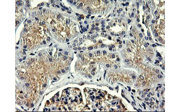
 IHC (Immunohistochemisry)
(Immunohistochemical analysis of USP5 protein in paraffin embedded Human Kidney tissue using USP5 antibody)
IHC (Immunohistochemisry)
(Immunohistochemical analysis of USP5 protein in paraffin embedded Human Kidney tissue using USP5 antibody)
USP5, Monoclonal Antibody (Cat# AAA107398)
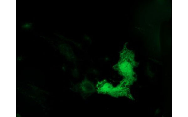 IF (Immunofluorescence)
(Immunofluorescent staining of COS7 cells transiently transfected with recombinant TUBB4 protein using TUBB4 antibody)
IF (Immunofluorescence)
(Immunofluorescent staining of COS7 cells transiently transfected with recombinant TUBB4 protein using TUBB4 antibody)
TUBB4, Monoclonal Antibody (Cat# AAA107434)
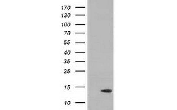 WB (Western Blot)
(Western Blot analysis of HEK293T cell lysates (5 ug) transfected with either recombinant RIPPLY2 protein (Right) or empty vector (Left) detected with RIPPLY2 antibody)
WB (Western Blot)
(Western Blot analysis of HEK293T cell lysates (5 ug) transfected with either recombinant RIPPLY2 protein (Right) or empty vector (Left) detected with RIPPLY2 antibody)
RIPPLY2, Monoclonal Antibody (Cat# AAA107439)
What are Monoclonal Antibodies?
Monoclonal antibodies are specialized laboratory-produced proteins developed for binding to specific biological antigens or other molecular targets. Since they come from a single cell (or clone), they are especially consistent and accurate in the data they are involved in producing.
This type of antibody material has been shown to be a powerful tool in finding and subsequently destroying harmful cells in an organism, such as those found in cancers or various autoimmune diseases. This makes them excellent aids in medical testing and research, which is why they are so widely used.
AAA Biotech offers a comprehensive range of high-quality monoclonal antibodies that perform effectively in various laboratory tests, including (amongst others) ELISA, western blotting, immunohistochemistry, and flow cytometry. All of the products in our catalog are thoroughly quality tested to make sure that they are reliable and will consistently perform well in your research.
What Are The Uses of Monoclonal Antibodies
Monoclonal antibodies are used in many lab tests, including (amongst others) ELISA, western blotting, immunohistochemistry, and flow cytometry.
ELISA is a test that helps detect a specific substance/analyte in a sample. It uses antibodies (often monoclonal) bound to a solid surface (such as the well of a microplate) to “capture” the substance/analyte in the sample and immobilize it so that the detection antibody component can then bind to it and produce a signal, which can then be measured.
Western blotting identifies specific proteins in a sample. The sample is first separated on a gel, and then antibodies are applied that will typically bind to the target, which will all be localized to a single band in a lane.
Immunohistochemistry helps locate specific proteins in cells or tissue samples using antibodies.
Flow cytometry looks at and sorts cells. It uses antibodies that are conjugated to reporter molecules called “fluorophores”, which, under special lights, emit light themselves, which can then be measured by a detector instrument.
How Monoclonal Antibodies Are Used as Medicine?
Please note that all of the products listed in AAA Biotech’s also known as AAA Bio or AAABio catalog are strictly for research-use only (RUO).
Monoclonal antibodies can also be used as therapeutic/medical treatments, particularly in the context of cancers. They are designed to find and bind to specific cells or proteins, helping the immune system recognize and attack the cancer. These treatments work in different ways, such as:
- Radioimmunotherapy attaches a small amount of radioactive molecule to the antibody, so it delivers the radiation directly to the cancer cells that the antibody is specifically binding to.
- Antibody-directed enzyme prodrug therapy uses antibodies that are specifically bound to special enzymes. These enzymes activate a harmless drug in the body and turn it into a cancer-killing drug only near the cancer cells—this helps avoid harming healthy cells.
- Immunoliposomes are tiny “bubbles” filled with medicine/drug and coated with antibodies. They carry the drug straight to the cancer cells.
Why Buy Monoclonal Antibodies From Us?
At AAA Biotech, we provide high-performance monoclonal antibodies designed to support a wide range of research needs.
1. Validated for Versatile Applications
The antibodies in our catalog are extensively validated and compatible with multiple techniques, including (but not limited to) ELISA, flow cytometry (FC), immunocytochemistry (ICC), immunofluorescence (IF), immunohistochemistry (IHC), immunoprecipitation (IP), and western blotting (WB).
2. Wide Selection & Specialized Options
We offer antibodies for common and rare species, that are available in various conjugated forms, and also in recombinant formats. Essentially, there is almost anything one might need to meet their experimental model’s requirements.
3. High-Quality Proteins
Our proteins meet high purity standards—90% or more as confirmed by SDS-PAGE. Many are available with tags like His, Flag, GST, or MBP, and we also supply native and biologically active proteins for functional studies.
Frequently Asked Questions
1. Are your monoclonal antibodies validated for specific applications?
Yes, our antibodies are tested and validated for use in methods such as ELISA, western blot, IHC, flow cytometry, and more. Refer to specific product pages or datasheets for individual product information.
2. How do I choose the right monoclonal antibody for my application?
Review the product details directly for application validation, species reactivity, and target information. You may also contact our support team at any time for help.
3. How quickly can I receive my order?
Most orders are processed and shipped within 1–3 business days, depending on product availability and your shipping location.