Filters
▼Clonality
▼Type
▼Reactivity
▼Gene Name
▼Isotype
▼Host
▼Application
▼Clone
▼Monoclonal Antibodies
Get accurate results in your research with our Monoclonal Antibodies, which are specially made to target exactly what you require for your research, and will produce consistent, reliable performance in lab tests.
Viewing 8800-8850 of 27597 product results

 IP (Immunoprecipitation)
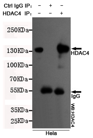
(Immunoprecipitation analysis of Hela cell lysates using HDAC4 mouse mAb.)
IP (Immunoprecipitation)
(Immunoprecipitation analysis of Hela cell lysates using HDAC4 mouse mAb.)
HDAC4, Monoclonal Antibody (Cat# AAA290426)
 IP (Immunoprecipitation)
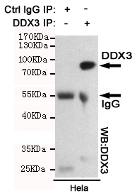
(Immunoprecipitation analysis of Hela cell lysates using DDX3 mouse mAb.)
IP (Immunoprecipitation)
(Immunoprecipitation analysis of Hela cell lysates using DDX3 mouse mAb.)
DDX3, Monoclonal Antibody (Cat# AAA290429)
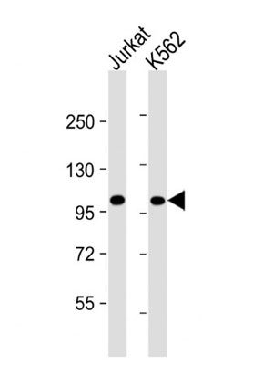 WB (Western Blot)
(All lanes : Anti-PRDM16 Antibody at 1:4000 dilutionLane 1: Jurkat whole cell lysateLane 2: K562 whole cell lysateLysates/proteins at 20 ug per lane.SecondaryGoat Anti-mouse IgG, (H+L), Peroxidase conjugated at 1/10000 dilution.Predicted band size : 140 kDaBlocking/Dilution buffer: 5% NFDM/TBST.)
WB (Western Blot)
(All lanes : Anti-PRDM16 Antibody at 1:4000 dilutionLane 1: Jurkat whole cell lysateLane 2: K562 whole cell lysateLysates/proteins at 20 ug per lane.SecondaryGoat Anti-mouse IgG, (H+L), Peroxidase conjugated at 1/10000 dilution.Predicted band size : 140 kDaBlocking/Dilution buffer: 5% NFDM/TBST.)
PRDM16, Monoclonal Antibody (Cat# AAA290741)
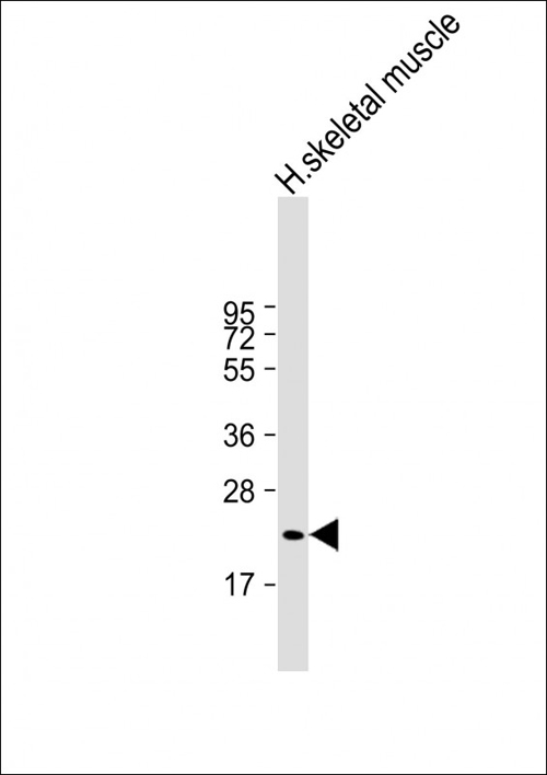 WB (Western Blot)
(Anti-FNDC5 Antibody at 1:2000 dilution + human skeletal muscle lysateLysates/proteins at 20 ug per lane.SecondaryGoat Anti-mouse IgG, (H+L), Peroxidase conjugated at 1/10000 dilution.Predicted band size : 23 kDaBlocking/Dilution buffer: 5% NFDM/TBST.)
WB (Western Blot)
(Anti-FNDC5 Antibody at 1:2000 dilution + human skeletal muscle lysateLysates/proteins at 20 ug per lane.SecondaryGoat Anti-mouse IgG, (H+L), Peroxidase conjugated at 1/10000 dilution.Predicted band size : 23 kDaBlocking/Dilution buffer: 5% NFDM/TBST.)
FNDC5, Monoclonal Antibody (Cat# AAA290742)
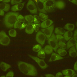
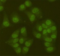
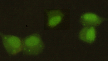
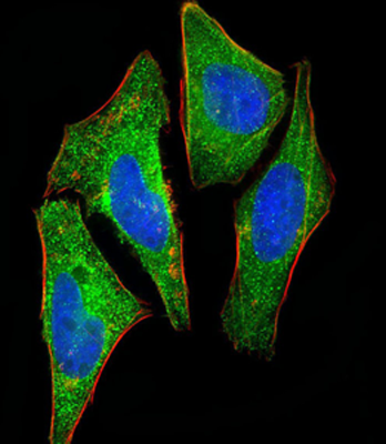 IF (Immunofluorescence)
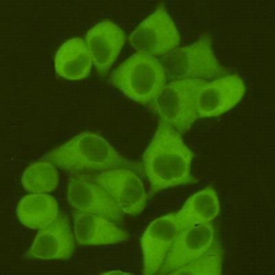
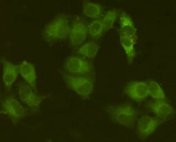
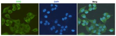
(Immunofluorescent analysis of 4% paraformaldehyde-fixed, 0.1% Triton X-100 permeabilized HeLa (human cervical epithelial adenocarcinoma cell line) cells labeling S100A2 with AAA290614 at 1/25 dilution, followed by Dylight® 488-conjugated goat anti-mouse IgG secondary antibody at 1/200 dilution (green). Immunofluorescence image showing cytoplasm and nucleus staining on HeLa cell line. Cytoplasmic actin is detected with Dylight® 554 Phalloidin at 1/100 dilution (red).The nuclear counter stain is DAPI (blue).)
IF (Immunofluorescence)
(Immunofluorescent analysis of 4% paraformaldehyde-fixed, 0.1% Triton X-100 permeabilized HeLa (human cervical epithelial adenocarcinoma cell line) cells labeling S100A2 with AAA290614 at 1/25 dilution, followed by Dylight® 488-conjugated goat anti-mouse IgG secondary antibody at 1/200 dilution (green). Immunofluorescence image showing cytoplasm and nucleus staining on HeLa cell line. Cytoplasmic actin is detected with Dylight® 554 Phalloidin at 1/100 dilution (red).The nuclear counter stain is DAPI (blue).)
S100A2, Monoclonal Antibody (Cat# AAA290614)
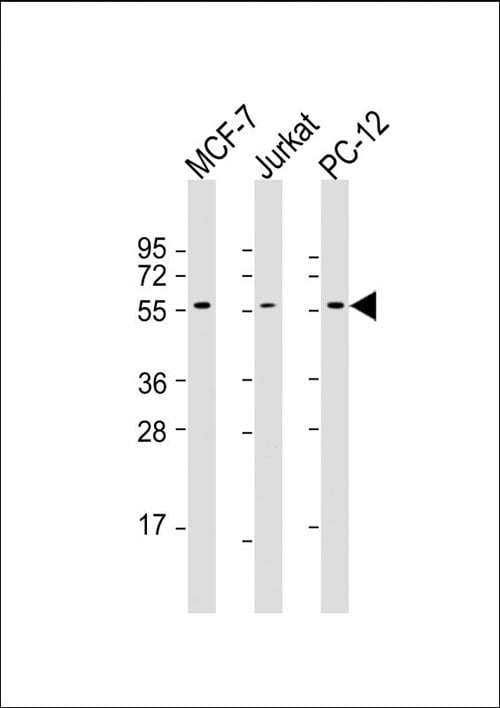
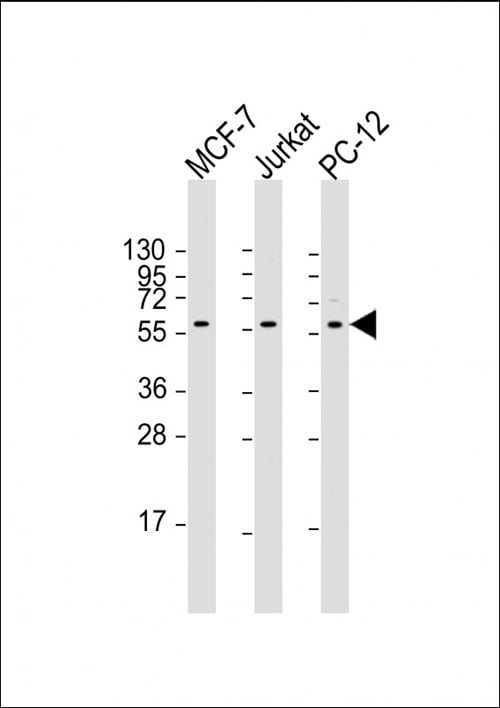 WB (Western Blot)
(All lanes : Anti-AKT2 Antibody at 1:2000 dilutionLane 1: MCF-7 whole cell lysateLane 2: Jurkat whole cell lysateLane 3: PC-12 whole cell lysateLysates/proteins at 20 ug per lane.SecondaryGoat Anti-mouse IgG, (H+L), Peroxidase conjugated at 1/10000 dilution.Predicted band size : 56 kDaBlocking/Dilution buffer: 5% NFDM/TBST.)
WB (Western Blot)
(All lanes : Anti-AKT2 Antibody at 1:2000 dilutionLane 1: MCF-7 whole cell lysateLane 2: Jurkat whole cell lysateLane 3: PC-12 whole cell lysateLysates/proteins at 20 ug per lane.SecondaryGoat Anti-mouse IgG, (H+L), Peroxidase conjugated at 1/10000 dilution.Predicted band size : 56 kDaBlocking/Dilution buffer: 5% NFDM/TBST.)
AKT2, Monoclonal Antibody (Cat# AAA290615)
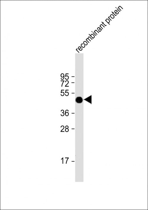 WB (Western Blot)
(Anti-FAT1 Antibody at 1:2000 dilution + recombinant proteinLysates/proteins at 20 ug per lane.SecondaryGoat Anti-mouse IgG, (H+L), Peroxidase conjugated at 1/10000 dilution.Predicted band size : 506 kDaBlocking/Dilution buffer: 5% NFDM/TBST.)
WB (Western Blot)
(Anti-FAT1 Antibody at 1:2000 dilution + recombinant proteinLysates/proteins at 20 ug per lane.SecondaryGoat Anti-mouse IgG, (H+L), Peroxidase conjugated at 1/10000 dilution.Predicted band size : 506 kDaBlocking/Dilution buffer: 5% NFDM/TBST.)
FAT1, Monoclonal Antibody (Cat# AAA290617)
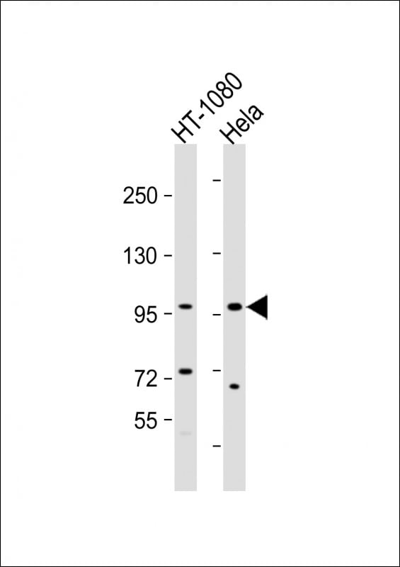
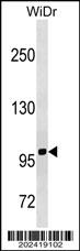 WB (Western Blot)
(TFRC Antibody western blot analysis in WiDr cell line lysates (35mug/lane).This demonstrates the TFRC antibody detected the TFRC protein (arrow).)
WB (Western Blot)
(TFRC Antibody western blot analysis in WiDr cell line lysates (35mug/lane).This demonstrates the TFRC antibody detected the TFRC protein (arrow).)
TFRC, Monoclonal Antibody (Cat# AAA290644)
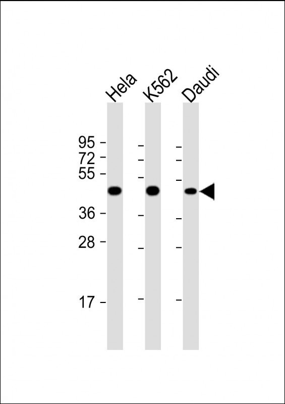

 WB (Western Blot)
(All lanes : Anti-PLIN3 Antibody at 1:2000 dilutionLane 1: Hela whole cell lysateLane 2: K562 whole cell lysateLane 3: Daudi whole cell lysateLysates/proteins at 20 ug per lane.SecondaryGoat Anti-mouse IgG, (H+L), Peroxidase conjugated at 1/10000 dilution.Predicted band size : 47 kDaBlocking/Dilution buffer: 5% NFDM/TBST.)
WB (Western Blot)
(All lanes : Anti-PLIN3 Antibody at 1:2000 dilutionLane 1: Hela whole cell lysateLane 2: K562 whole cell lysateLane 3: Daudi whole cell lysateLysates/proteins at 20 ug per lane.SecondaryGoat Anti-mouse IgG, (H+L), Peroxidase conjugated at 1/10000 dilution.Predicted band size : 47 kDaBlocking/Dilution buffer: 5% NFDM/TBST.)
PLIN3, Monoclonal Antibody (Cat# AAA290646)
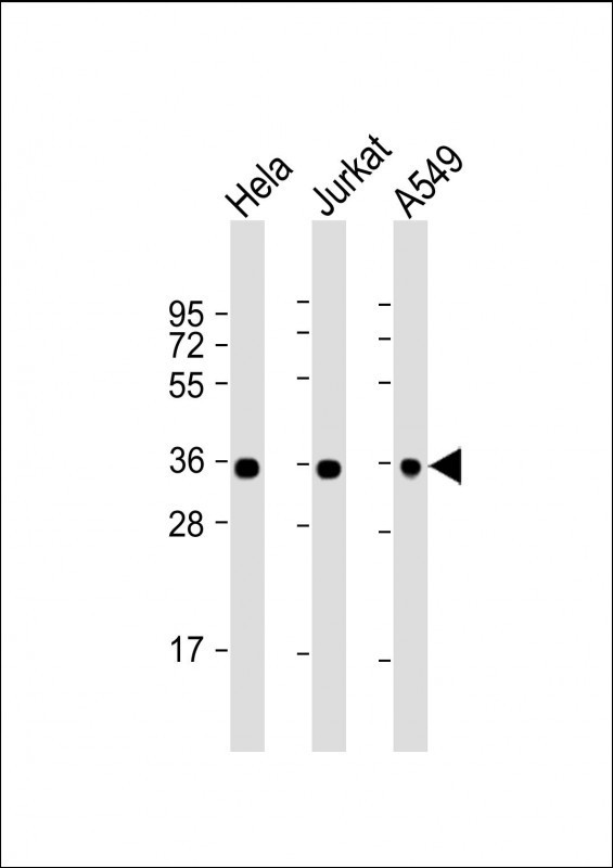
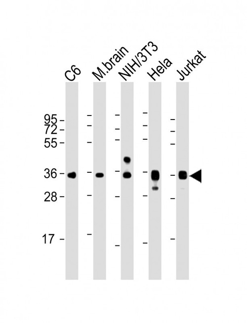
 WB (Western Blot)
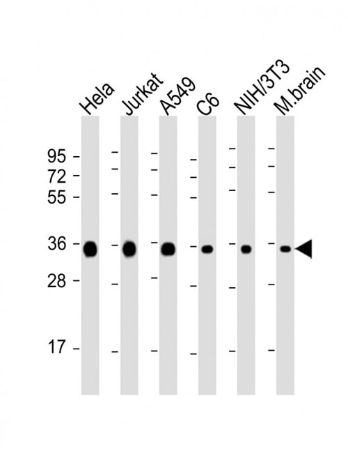
(All lanes : Anti-GAPDH Antibody at 1:8000 dilutionLane 1: Hela whole cell lysateLane 2: Jurkat whole cell lysateLane 3: A549 whole cell lysateLane 4: C6 whole cell lysateLane 5: NIH/3T3 whole cell lysateLane 6: mouse brain lysateLysates/proteins at 20 ug per lane.SecondaryGoat Anti-mouse IgG, (H+L), Peroxidase conjugated at 1/10000 dilution.Predicted band size : 36 kDaBlocking/Dilution buffer: 5% NFDM/TBST.)
WB (Western Blot)
(All lanes : Anti-GAPDH Antibody at 1:8000 dilutionLane 1: Hela whole cell lysateLane 2: Jurkat whole cell lysateLane 3: A549 whole cell lysateLane 4: C6 whole cell lysateLane 5: NIH/3T3 whole cell lysateLane 6: mouse brain lysateLysates/proteins at 20 ug per lane.SecondaryGoat Anti-mouse IgG, (H+L), Peroxidase conjugated at 1/10000 dilution.Predicted band size : 36 kDaBlocking/Dilution buffer: 5% NFDM/TBST.)
GAPDH, Monoclonal Antibody (Cat# AAA290647)
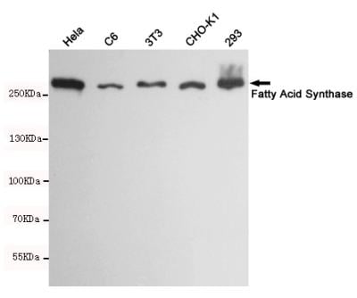
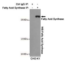 IP (Immunoprecipitation)
(Immunoprecipitation analysis of CHO-K1 cell lysates using Fatty Acid Synthase mouse mAb.)
IP (Immunoprecipitation)
(Immunoprecipitation analysis of CHO-K1 cell lysates using Fatty Acid Synthase mouse mAb.)
Fatty Acid Synthase, Monoclonal Antibody (Cat# AAA290857)
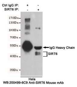 IP (Immunoprecipitation)
(Immunoprecipitation analysis of Hela cell lysates using SIRT6 mouse mAb.)
IP (Immunoprecipitation)
(Immunoprecipitation analysis of Hela cell lysates using SIRT6 mouse mAb.)
SIRT6, Monoclonal Antibody (Cat# AAA290859)
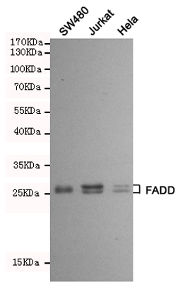
 IP (Immunoprecipitation)
(Immunoprecipitation analysis of Hela cell lysates using FADD mouse mAb.)
IP (Immunoprecipitation)
(Immunoprecipitation analysis of Hela cell lysates using FADD mouse mAb.)
FADD, Monoclonal Antibody (Cat# AAA290860)
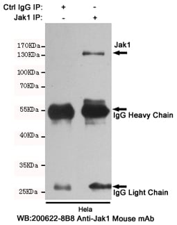 IP (Immunoprecipitation)
(Immunoprecipitation analysis of Hela cell lysates using Jak1 mouse mAb.)
IP (Immunoprecipitation)
(Immunoprecipitation analysis of Hela cell lysates using Jak1 mouse mAb.)
Anti-Jak1, Monoclonal Antibody (Cat# AAA290863)
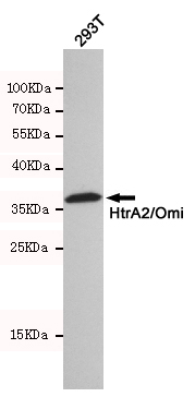
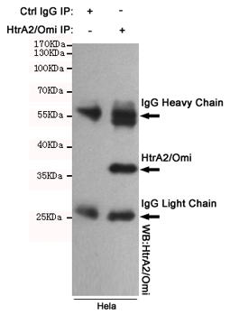 IP (Immunoprecipitation)
(Immunoprecipitation analysis of Hela cell lysates using HtrA2/Omi mouse mAb.)
IP (Immunoprecipitation)
(Immunoprecipitation analysis of Hela cell lysates using HtrA2/Omi mouse mAb.)
HTRA2, Monoclonal Antibody (Cat# AAA290869)
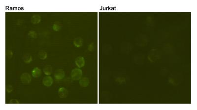
 FCM/FACS (Flow Cytometry)
(Flow cytometric analysis of Jurkat T cells (black) and Ramos B cells (red), using anti-CD19 mouse mAb.)
FCM/FACS (Flow Cytometry)
(Flow cytometric analysis of Jurkat T cells (black) and Ramos B cells (red), using anti-CD19 mouse mAb.)
CD19, Monoclonal Antibody (Cat# AAA290877)
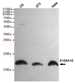
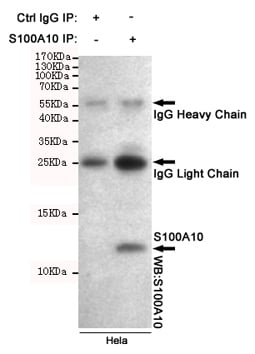 IP (Immunoprecipitation)
(Immunoprecipitation analysis of Hela cell lysates using S100A10 mouse mAb.)
IP (Immunoprecipitation)
(Immunoprecipitation analysis of Hela cell lysates using S100A10 mouse mAb.)
S100A10, Monoclonal Antibody (Cat# AAA290881)

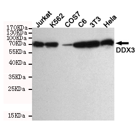
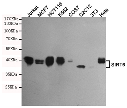
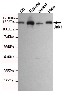
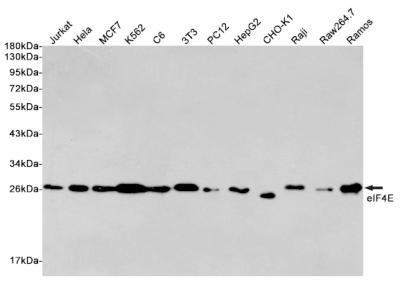 WB (Western Blot)
(Western blot detection of eIF4E in Jurkat, Hela, MCF7, K562, C6, 3T3, PC12, HepG2, CHO-K1, Raji, Raw264.7 and Ramos cell lysates using eIF4E mouse mAb (1:2000 diluted). Predicted band size: 25KDa. Observed band size:25KDa.)
WB (Western Blot)
(Western blot detection of eIF4E in Jurkat, Hela, MCF7, K562, C6, 3T3, PC12, HepG2, CHO-K1, Raji, Raw264.7 and Ramos cell lysates using eIF4E mouse mAb (1:2000 diluted). Predicted band size: 25KDa. Observed band size:25KDa.)
eIF4E, Monoclonal Antibody (Cat# AAA290883)
 ICC (Immunocytochemistry)
(Immunocytochemistry of HeLa cells fixed by Paraformaldehyde and using DR5 mouse mAb diluted 1:100.)
ICC (Immunocytochemistry)
(Immunocytochemistry of HeLa cells fixed by Paraformaldehyde and using DR5 mouse mAb diluted 1:100.)
DR5, Monoclonal Antibody (Cat# AAA290432)
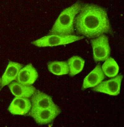
 ICC (Immunocytochemistry)
(Immunocytochemistry staining of HeLa cells fixed with 4% paraformaldehyde and using anti-Tubulin beta mouse mAb (dilution 1:500).)
ICC (Immunocytochemistry)
(Immunocytochemistry staining of HeLa cells fixed with 4% paraformaldehyde and using anti-Tubulin beta mouse mAb (dilution 1:500).)
beta Tubulin, Monoclonal Antibody (Cat# AAA290443)
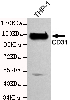
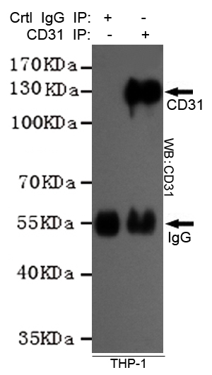 IP (Immunoprecipitation)
(Immunoprecipitation analysis of THP-1 cell lysates using CD31 mouse mAb.)
IP (Immunoprecipitation)
(Immunoprecipitation analysis of THP-1 cell lysates using CD31 mouse mAb.)
CD31, Monoclonal Antibody (Cat# AAA290445)
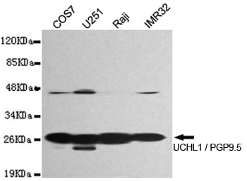
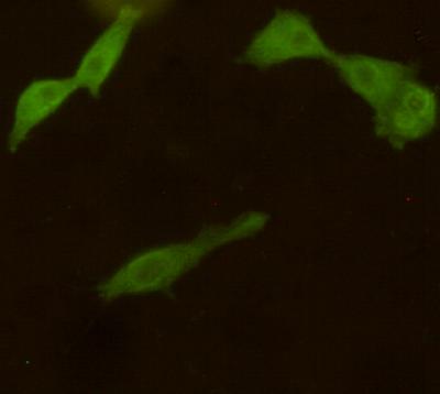 ICC (Immunocytochemistry)
(Immunocytochemistry stain of COS7 using UCHL1 / PGP9.5 mouse mAb (1:300).)
ICC (Immunocytochemistry)
(Immunocytochemistry stain of COS7 using UCHL1 / PGP9.5 mouse mAb (1:300).)
UCHL1 / PGP9.5, Monoclonal Antibody (Cat# AAA290446)
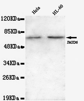
 ICC (Immunocytochemistry)
(Immunocytochemistry of HeLa cells using anti-JMJD6(N-terminus) mouse mAb diluted 1:200.)
ICC (Immunocytochemistry)
(Immunocytochemistry of HeLa cells using anti-JMJD6(N-terminus) mouse mAb diluted 1:200.)
JMJD6, Monoclonal Antibody (Cat# AAA290447)
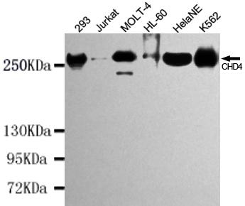
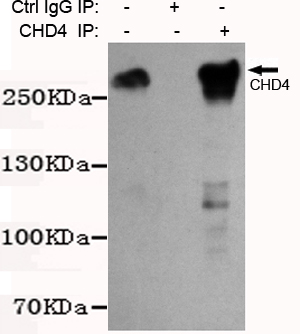 IP (Immunoprecipitation)
(Immunoprecipitation analysis of K562 cell lysates using CHD4 mouse mAb.)
IP (Immunoprecipitation)
(Immunoprecipitation analysis of K562 cell lysates using CHD4 mouse mAb.)
CHD4, Monoclonal Antibody (Cat# AAA290449)
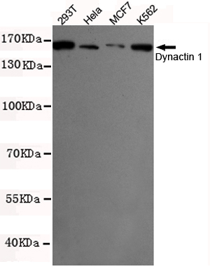
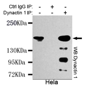 IP (Immunoprecipitation)
(Immunoprecipitation analysis of Hela cell lysates using Dynactin 1 mouse mAb.)
IP (Immunoprecipitation)
(Immunoprecipitation analysis of Hela cell lysates using Dynactin 1 mouse mAb.)
Dynactin 1, Monoclonal Antibody (Cat# AAA290452)
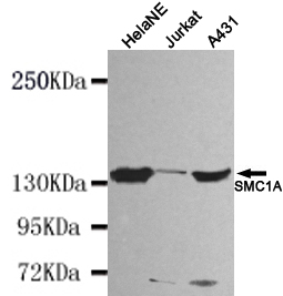
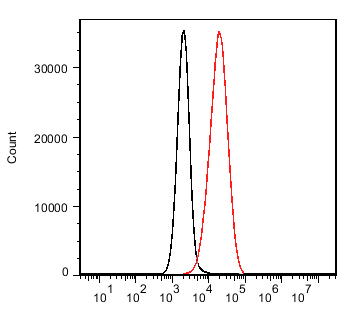
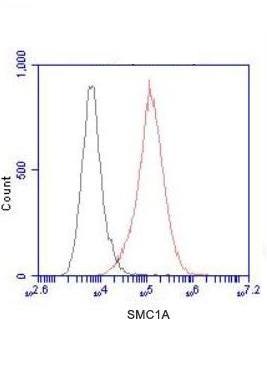 FCM/FACS (Flow Cytometry)
(Flow Cytometry analysis of HeLa cells stained with SMC1A (N-terminus) (red, 1/100 dilution), followed by FITC-conjugated goat anti-mouse IgG. Black line histogram represents the isotype control, normal mouse IgG.)
FCM/FACS (Flow Cytometry)
(Flow Cytometry analysis of HeLa cells stained with SMC1A (N-terminus) (red, 1/100 dilution), followed by FITC-conjugated goat anti-mouse IgG. Black line histogram represents the isotype control, normal mouse IgG.)
SMC1A, Monoclonal Antibody (Cat# AAA290454)
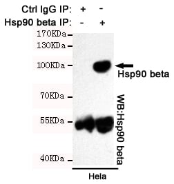 IP (Immunoprecipitation)
(Immunoprecipitation analysis of Hela cell lysates using Hsp90 beta mouse mAb.)
IP (Immunoprecipitation)
(Immunoprecipitation analysis of Hela cell lysates using Hsp90 beta mouse mAb.)
Hsp90 beta, Monoclonal Antibody (Cat# AAA290457)
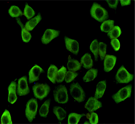
 ICC (Immunocytochemistry)
(Immunocytochemistry staining of HeLa cells fixed with 4% Paraformaldehyde and using anti-NFIC mouse mAb (dilution 1:200).)
ICC (Immunocytochemistry)
(Immunocytochemistry staining of HeLa cells fixed with 4% Paraformaldehyde and using anti-NFIC mouse mAb (dilution 1:200).)
NFIC, Monoclonal Antibody (Cat# AAA290458)
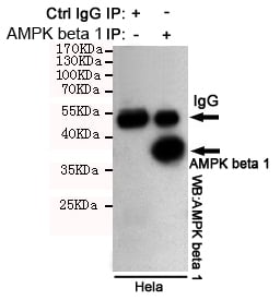
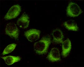
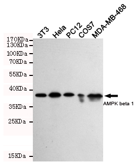
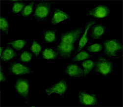
 IHC (Immunohistochemistry)
(Immunohistochemical analysis of paraffin-embedded Breast cancer using AMPK beta 1 mouse mAb (1/200 dilution).Antigen retrieval was performed by pressure cooking in citrate buffer (pH 6.0).)
IHC (Immunohistochemistry)
(Immunohistochemical analysis of paraffin-embedded Breast cancer using AMPK beta 1 mouse mAb (1/200 dilution).Antigen retrieval was performed by pressure cooking in citrate buffer (pH 6.0).)
AMPK beta 1, Monoclonal Antibody (Cat# AAA290459)
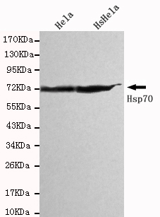
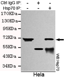 IP (Immunoprecipitation)
(Immunoprecipitation analysis of Hela cell lysates using Hsp70 (C-terminus) mouse mAb.)
IP (Immunoprecipitation)
(Immunoprecipitation analysis of Hela cell lysates using Hsp70 (C-terminus) mouse mAb.)
Hsp70, Monoclonal Antibody (Cat# AAA290466)
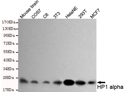
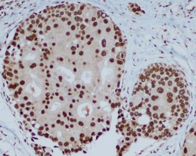 IHC (Immunohistochemisry)
(Immunohistochemical analysis of paraffin-embedded human breast carcinoma with HP1 alpha mouse mAb (3G2-H10-A6, 1:400 diluted),showing nuclear localization.A high pressure mediated antigen retrieval step was performed in citrat buffer(pH6.0).)
IHC (Immunohistochemisry)
(Immunohistochemical analysis of paraffin-embedded human breast carcinoma with HP1 alpha mouse mAb (3G2-H10-A6, 1:400 diluted),showing nuclear localization.A high pressure mediated antigen retrieval step was performed in citrat buffer(pH6.0).)
HP1 alpha, Monoclonal Antibody (Cat# AAA290471)
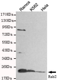
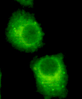 ICC (Immunocytochemistry)
(Immunocytochemistry of HeLa cells using anti-Rab2 mouse mAb diluted 1:200)
ICC (Immunocytochemistry)
(Immunocytochemistry of HeLa cells using anti-Rab2 mouse mAb diluted 1:200)
Rab2, Monoclonal Antibody (Cat# AAA290475)
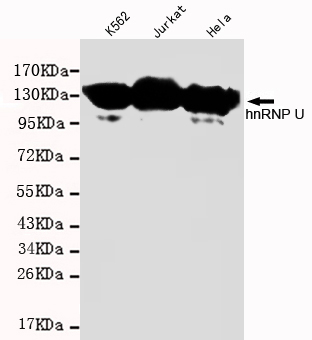 WB (Western Blot)
(Western blot detection of hnRNP U in K562,Jurkat and Hela cell lysates and using hnRNP U mouse mAb(1:1000 diluted).Predicted band size: 110KDa.Observed band size: 110KDa.)
WB (Western Blot)
(Western blot detection of hnRNP U in K562,Jurkat and Hela cell lysates and using hnRNP U mouse mAb(1:1000 diluted).Predicted band size: 110KDa.Observed band size: 110KDa.)
hnRNP U, Monoclonal Antibody (Cat# AAA290476)
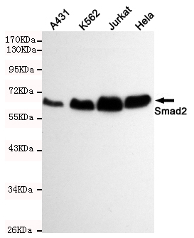
 ICC (Immunocytochemistry)
(Immunocytochemistry staining of HeLa cells fixed with 1% Paraformaldehyde and using Smad2 mouse mAb (dilution 1:100).)
ICC (Immunocytochemistry)
(Immunocytochemistry staining of HeLa cells fixed with 1% Paraformaldehyde and using Smad2 mouse mAb (dilution 1:100).)
Smad2, Monoclonal Antibody (Cat# AAA290477)
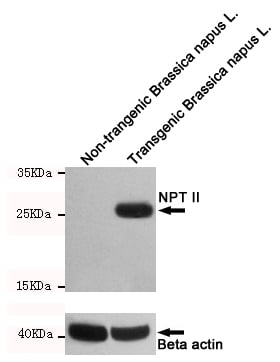 WB (Western Blot)
(Western blot detection of NPTII in non-trangenic Brassica napus L. and transgenic Brassica napus L. cell lysates using NPTII mouse mAb (1:1000 diluted).Predicted band size:29KDa.Observed band size:29KDa.)
WB (Western Blot)
(Western blot detection of NPTII in non-trangenic Brassica napus L. and transgenic Brassica napus L. cell lysates using NPTII mouse mAb (1:1000 diluted).Predicted band size:29KDa.Observed band size:29KDa.)
NPTII, Monoclonal Antibody (Cat# AAA290483)
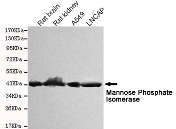
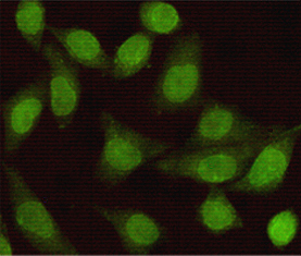
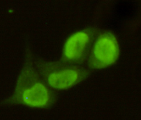
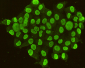
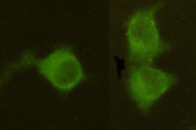 ICC (Immunocytochemistry)
(Immunocytochemistry stain of Hela using Mannose Phosphate Isomerase mouse mAb (1:300).)
ICC (Immunocytochemistry)
(Immunocytochemistry stain of Hela using Mannose Phosphate Isomerase mouse mAb (1:300).)
Mannose Phosphate Isomerase, Monoclonal Antibody (Cat# AAA290486)
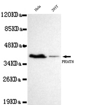
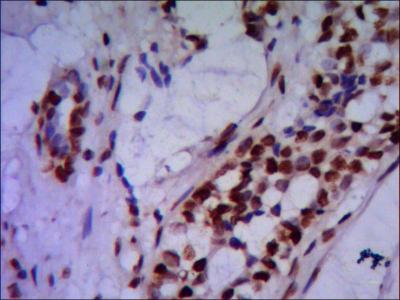 IHC (Immunohistochemisry)
(Immunohistochemistry stain of paraffin-embedded human breast cancer using PRMT6 mouse mAb (1:200).)
IHC (Immunohistochemisry)
(Immunohistochemistry stain of paraffin-embedded human breast cancer using PRMT6 mouse mAb (1:200).)
PRMT6, Monoclonal Antibody (Cat# AAA290492)
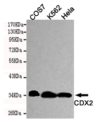
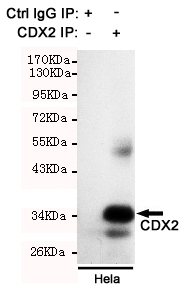 IP (Immunoprecipitation)
(Immunoprecipitation analysis of Hela cell lysate using CDX2 mouse mAb.)
IP (Immunoprecipitation)
(Immunoprecipitation analysis of Hela cell lysate using CDX2 mouse mAb.)
CDX2, Monoclonal Antibody (Cat# AAA290495)
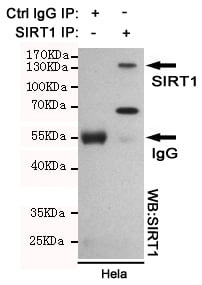 IP (Immunoprecipitation)
(Immunoprecipitation analysis of Hela cell lysates using SIRT1 mouse mAb.)
IP (Immunoprecipitation)
(Immunoprecipitation analysis of Hela cell lysates using SIRT1 mouse mAb.)
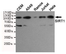
SIRT1, Monoclonal Antibody (Cat# AAA290497)
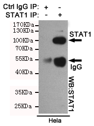 IP (Immunoprecipitation)
(Immunoprecipitation analysis of Hela cell lysates using STAT1 mouse mAb.)
IP (Immunoprecipitation)
(Immunoprecipitation analysis of Hela cell lysates using STAT1 mouse mAb.)
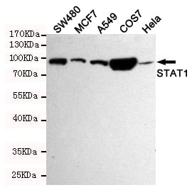
STAT1, Monoclonal Antibody (Cat# AAA290499)
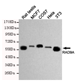
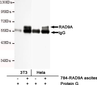 IP (Immunoprecipitation)
(Immunoprecipitation analysis of Hela and 3T3 cell lysates using RAD9A mouse mAb.)
IP (Immunoprecipitation)
(Immunoprecipitation analysis of Hela and 3T3 cell lysates using RAD9A mouse mAb.)
RAD9A, Monoclonal Antibody (Cat# AAA290502)
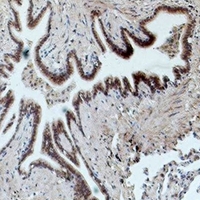
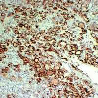
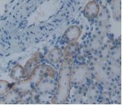
 IHC (Immunohiostchemistry)
(Immunohistochemical analysis of paraffin-embedded H. tonsil section using ITGB2 Antibody AAA290944. It was diluted at 1:25 dilution. A undiluted biotinylated goat polyvalent antibody was used as the secondary, followed by DAB staining.)
IHC (Immunohiostchemistry)
(Immunohistochemical analysis of paraffin-embedded H. tonsil section using ITGB2 Antibody AAA290944. It was diluted at 1:25 dilution. A undiluted biotinylated goat polyvalent antibody was used as the secondary, followed by DAB staining.)
ITGB2, Monoclonal Antibody (Cat# AAA290944)
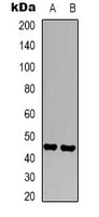
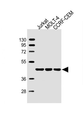 WB (Western Blot)
(All lanes : Anti-ADA Antibody (C-term) at 1:4000 dilutionLane 1: Jurkat whole cell lysateLane 2: MOLT-4 whole cell lysateLane 3: CCRF-CEM whole cell lysateLysates/proteins at 20 ug per lane.SecondaryGoat Anti-mouse IgG, (H+L), Peroxidase conjugated at 1/10000 dilution.Predicted band size : 41 kDaBlocking/Dilution buffer: 5% NFDM/TBST.)
WB (Western Blot)
(All lanes : Anti-ADA Antibody (C-term) at 1:4000 dilutionLane 1: Jurkat whole cell lysateLane 2: MOLT-4 whole cell lysateLane 3: CCRF-CEM whole cell lysateLysates/proteins at 20 ug per lane.SecondaryGoat Anti-mouse IgG, (H+L), Peroxidase conjugated at 1/10000 dilution.Predicted band size : 41 kDaBlocking/Dilution buffer: 5% NFDM/TBST.)
ADA, Monoclonal Antibody (Cat# AAA290954)
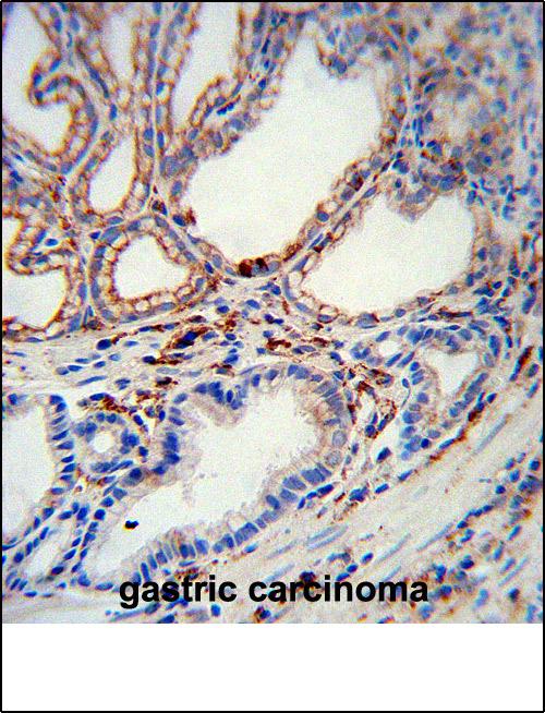
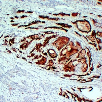
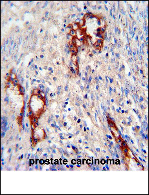 IHC (Immunohistochemistry)
(MMP2 Antibody immunohistochemistry analysis in formalin fixed and paraffin embedded human prostate carcinoma followed by peroxidase conjugation of the secondary antibody and DAB staining.This data demonstrates the use of MMP2 Antibody for immunohistochemistry. Clinical relevance has not been evaluated.)
IHC (Immunohistochemistry)
(MMP2 Antibody immunohistochemistry analysis in formalin fixed and paraffin embedded human prostate carcinoma followed by peroxidase conjugation of the secondary antibody and DAB staining.This data demonstrates the use of MMP2 Antibody for immunohistochemistry. Clinical relevance has not been evaluated.)
MMP2, Monoclonal Antibody (Cat# AAA290959)
 IHC (Immunohiostchemistry)
(Dilution: IH 1/50 - 1/200Immunohistochemical analysis of CD16 staining in human lung cancer formalin fixed paraffin embedded tissue section. The section was pre-treated using heat mediated antigen retrieval with sodium citrate buffer (pH 6.0). The section was then incubated with the antibody at room temperature and detected using an HRP conjugated compact polymer system. DAB was used as the chromogen. The section was then counterstained with haematoxylin and mounted with DPX.)
IHC (Immunohiostchemistry)
(Dilution: IH 1/50 - 1/200Immunohistochemical analysis of CD16 staining in human lung cancer formalin fixed paraffin embedded tissue section. The section was pre-treated using heat mediated antigen retrieval with sodium citrate buffer (pH 6.0). The section was then incubated with the antibody at room temperature and detected using an HRP conjugated compact polymer system. DAB was used as the chromogen. The section was then counterstained with haematoxylin and mounted with DPX.)
CD16, Monoclonal Antibody (Cat# AAA291251)
 IHC (Immunohiostchemistry)
(Dilution: IH 1/200 - 1/500Immunohistochemical analysis of Histone H2B staining in human breast cancer formalin fixed paraffin embedded tissue section. The section was pre-treated using heat mediated antigen retrieval with sodium citrate buffer (pH 6.0). The section was then incubated with the antibody at room temperature and detected using an HRP conjugated compact polymer system. DAB was used as the chromogen. The section was then counterstained with haematoxylin and mounted with DPX.)
IHC (Immunohiostchemistry)
(Dilution: IH 1/200 - 1/500Immunohistochemical analysis of Histone H2B staining in human breast cancer formalin fixed paraffin embedded tissue section. The section was pre-treated using heat mediated antigen retrieval with sodium citrate buffer (pH 6.0). The section was then incubated with the antibody at room temperature and detected using an HRP conjugated compact polymer system. DAB was used as the chromogen. The section was then counterstained with haematoxylin and mounted with DPX.)
Histone H2B, Monoclonal Antibody (Cat# AAA291252)

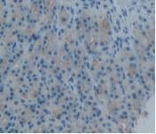
 IHC (Immunohiostchemistry)
(Dilution: IH 1/50 - 1/200Immunohistochemical analysis of 42278 staining in human tonsil formalin fixed paraffin embedded tissue section. The section was pre-treated using heat mediated antigen retrieval with sodium citrate buffer (pH 6.0). The section was then incubated with the antibody at room temperature and detected using an HRP conjugated compact polymer system. DAB was used as the chromogen. The section was then counterstained with haematoxylin and mounted with DPX.)
IHC (Immunohiostchemistry)
(Dilution: IH 1/50 - 1/200Immunohistochemical analysis of 42278 staining in human tonsil formalin fixed paraffin embedded tissue section. The section was pre-treated using heat mediated antigen retrieval with sodium citrate buffer (pH 6.0). The section was then incubated with the antibody at room temperature and detected using an HRP conjugated compact polymer system. DAB was used as the chromogen. The section was then counterstained with haematoxylin and mounted with DPX.)
OCT1, Monoclonal Antibody (Cat# AAA291253)
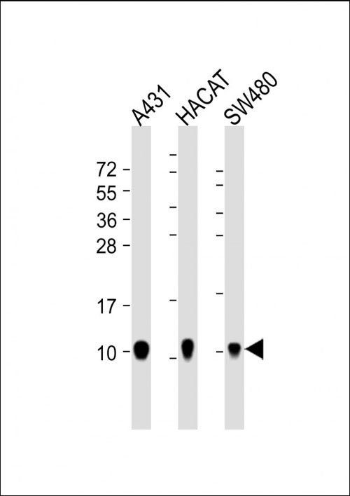
 IF (Immunofluorescence)
(Immunohistochemical analysis of Thyroid Peroxidase staining in human papillary thyroid carcinoma formalin fixed paraffin embedded tissue section. The section was pre-treated using heat mediated antigen retrieval with sodium citrate buffer (pH 6.0). The section was then incubated with the antibody at room temperature and detected using an HRP conjugated compact polymer system. DAB was used as the chromogen. The section was then counterstained with haematoxylin and mounted with DPX.)
IF (Immunofluorescence)
(Immunohistochemical analysis of Thyroid Peroxidase staining in human papillary thyroid carcinoma formalin fixed paraffin embedded tissue section. The section was pre-treated using heat mediated antigen retrieval with sodium citrate buffer (pH 6.0). The section was then incubated with the antibody at room temperature and detected using an HRP conjugated compact polymer system. DAB was used as the chromogen. The section was then counterstained with haematoxylin and mounted with DPX.)
Thyroid Peroxidase, Monoclonal Antibody (Cat# AAA291255)
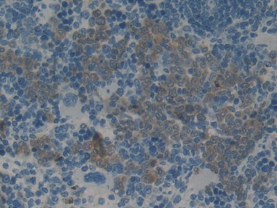

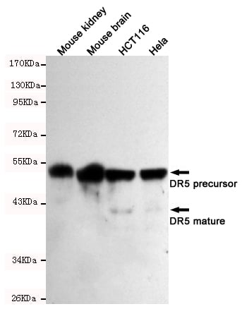
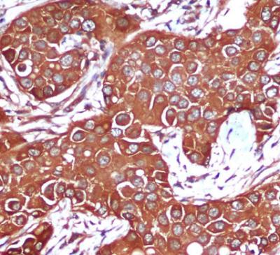
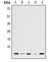
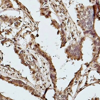
 IHC (Immunohistochemistry)
(DAB staining on IHC-P; Samples: Rat Kidney Tissue.)
IHC (Immunohistochemistry)
(DAB staining on IHC-P; Samples: Rat Kidney Tissue.)
Plasminogen Activator Inhibitor 2, Monoclonal Antibody (Cat# AAA141261)
Bradykinin, Monoclonal Antibody (Cat# AAA141305)
What are Monoclonal Antibodies?
Monoclonal antibodies are specialized laboratory-produced proteins developed for binding to specific biological antigens or other molecular targets. Since they come from a single cell (or clone), they are especially consistent and accurate in the data they are involved in producing.
This type of antibody material has been shown to be a powerful tool in finding and subsequently destroying harmful cells in an organism, such as those found in cancers or various autoimmune diseases. This makes them excellent aids in medical testing and research, which is why they are so widely used.
AAA Biotech offers a comprehensive range of high-quality monoclonal antibodies that perform effectively in various laboratory tests, including (amongst others) ELISA, western blotting, immunohistochemistry, and flow cytometry. All of the products in our catalog are thoroughly quality tested to make sure that they are reliable and will consistently perform well in your research.
What Are The Uses of Monoclonal Antibodies
Monoclonal antibodies are used in many lab tests, including (amongst others) ELISA, western blotting, immunohistochemistry, and flow cytometry.
ELISA is a test that helps detect a specific substance/analyte in a sample. It uses antibodies (often monoclonal) bound to a solid surface (such as the well of a microplate) to “capture” the substance/analyte in the sample and immobilize it so that the detection antibody component can then bind to it and produce a signal, which can then be measured.
Western blotting identifies specific proteins in a sample. The sample is first separated on a gel, and then antibodies are applied that will typically bind to the target, which will all be localized to a single band in a lane.
Immunohistochemistry helps locate specific proteins in cells or tissue samples using antibodies.
Flow cytometry looks at and sorts cells. It uses antibodies that are conjugated to reporter molecules called “fluorophores”, which, under special lights, emit light themselves, which can then be measured by a detector instrument.
How Monoclonal Antibodies Are Used as Medicine?
Please note that all of the products listed in AAA Biotech’s also known as AAA Bio or AAABio catalog are strictly for research-use only (RUO).
Monoclonal antibodies can also be used as therapeutic/medical treatments, particularly in the context of cancers. They are designed to find and bind to specific cells or proteins, helping the immune system recognize and attack the cancer. These treatments work in different ways, such as:
- Radioimmunotherapy attaches a small amount of radioactive molecule to the antibody, so it delivers the radiation directly to the cancer cells that the antibody is specifically binding to.
- Antibody-directed enzyme prodrug therapy uses antibodies that are specifically bound to special enzymes. These enzymes activate a harmless drug in the body and turn it into a cancer-killing drug only near the cancer cells—this helps avoid harming healthy cells.
- Immunoliposomes are tiny “bubbles” filled with medicine/drug and coated with antibodies. They carry the drug straight to the cancer cells.
Why Buy Monoclonal Antibodies From Us?
At AAA Biotech, we provide high-performance monoclonal antibodies designed to support a wide range of research needs.
1. Validated for Versatile Applications
The antibodies in our catalog are extensively validated and compatible with multiple techniques, including (but not limited to) ELISA, flow cytometry (FC), immunocytochemistry (ICC), immunofluorescence (IF), immunohistochemistry (IHC), immunoprecipitation (IP), and western blotting (WB).
2. Wide Selection & Specialized Options
We offer antibodies for common and rare species, that are available in various conjugated forms, and also in recombinant formats. Essentially, there is almost anything one might need to meet their experimental model’s requirements.
3. High-Quality Proteins
Our proteins meet high purity standards—90% or more as confirmed by SDS-PAGE. Many are available with tags like His, Flag, GST, or MBP, and we also supply native and biologically active proteins for functional studies.
Frequently Asked Questions
1. Are your monoclonal antibodies validated for specific applications?
Yes, our antibodies are tested and validated for use in methods such as ELISA, western blot, IHC, flow cytometry, and more. Refer to specific product pages or datasheets for individual product information.
2. How do I choose the right monoclonal antibody for my application?
Review the product details directly for application validation, species reactivity, and target information. You may also contact our support team at any time for help.
3. How quickly can I receive my order?
Most orders are processed and shipped within 1–3 business days, depending on product availability and your shipping location.