Filters
▼Clonality
▼Type
▼Reactivity
▼Gene Name
▼Isotype
▼Host
▼Application
▼Clone
▼Monoclonal Antibodies
Get accurate results in your research with our Monoclonal Antibodies, which are specially made to target exactly what you require for your research, and will produce consistent, reliable performance in lab tests.
Viewing 9050-9100 of 27597 product results
 Application Data
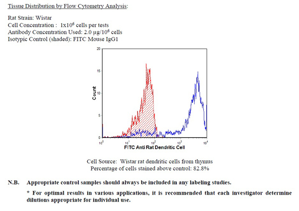
Dendritic cell, Monoclonal Antibody (Cat# AAA74170)
 Application Data
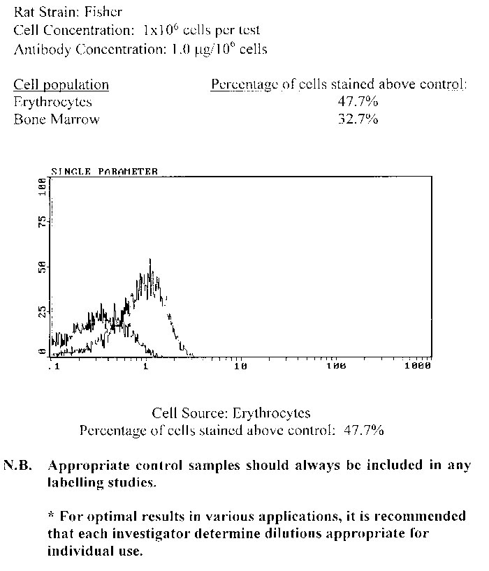
Erythrocytes, Monoclonal Antibody (Cat# AAA74172)
 Application Data
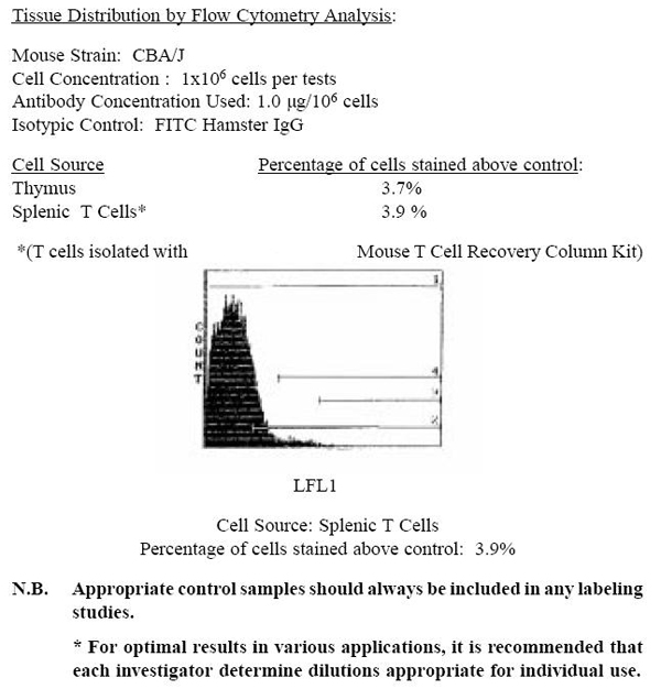
gamma/delta TCR, Monoclonal Antibody (Cat# AAA74176)
 Application Data
(Balb/c mouse bone marrow (high SSC population) was stained with anti-Granulocytes (Gr-1) (clone:RB6-8C5) (filled histogram) or rat IgG2b isotype control (open histogram).N.B. Appropriate control samples should always be included in any labeling studies.* For optimal results in various applications, it is recommended that each investigator determine dilutions appropriate for individual use.)
Application Data
(Balb/c mouse bone marrow (high SSC population) was stained with anti-Granulocytes (Gr-1) (clone:RB6-8C5) (filled histogram) or rat IgG2b isotype control (open histogram).N.B. Appropriate control samples should always be included in any labeling studies.* For optimal results in various applications, it is recommended that each investigator determine dilutions appropriate for individual use.)
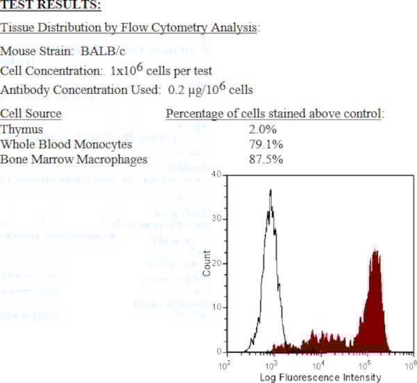
Granulocytes (Gr-1), Monoclonal Antibody (Cat# AAA74178)
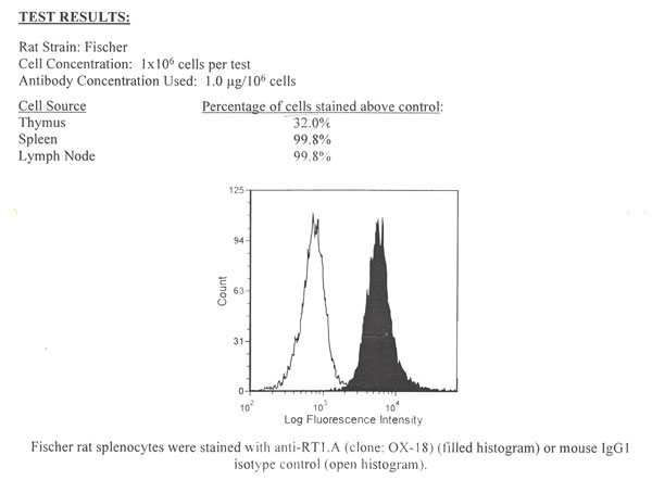
I-Ab and I-Ad, Monoclonal Antibody (Cat# AAA74187)
 Application Data
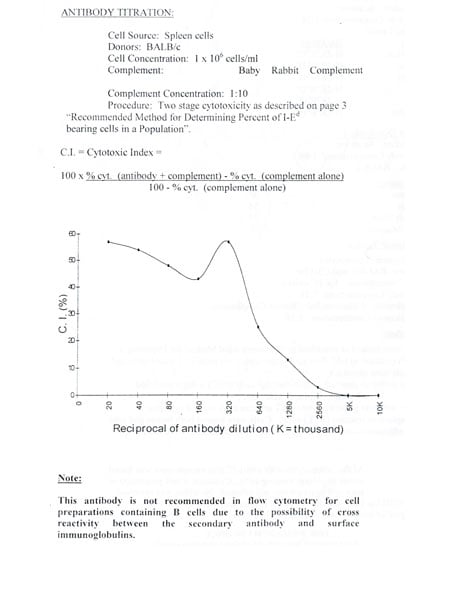
I-Ed (private; Ia.m23), Monoclonal Antibody (Cat# AAA74189)
 Application Data
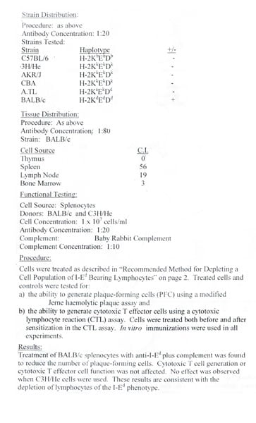
RT1.A, Monoclonal Antibody (Cat# AAA74190)
 Application Data
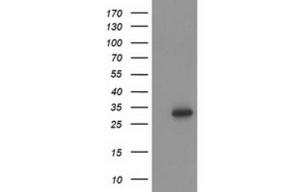
CD19, Monoclonal Antibody (Cat# AAA74202)
Prothrombin (Calcium dependant), Monoclonal Antibody (Cat# AAA74203)
 IF (Immunofluorescence)
(Immunofluorescent staining of COS7 cells transiently transfected with recombinant BTK protein using BTK antibody)
IF (Immunofluorescence)
(Immunofluorescent staining of COS7 cells transiently transfected with recombinant BTK protein using BTK antibody)
BTK, Monoclonal Antibody (Cat# AAA74749)
 IHC (Immunohiostchemistry)
(Immunohistochemical analysis of ID2 protein in paraffin embedded Carcinoma of Human liver tissue using ID2 antibody)
IHC (Immunohiostchemistry)
(Immunohistochemical analysis of ID2 protein in paraffin embedded Carcinoma of Human liver tissue using ID2 antibody)
ID2, Monoclonal Antibody (Cat# AAA74750)
 IHC (Immunohiostchemistry)
(Immunohistochemical analysis of paraffin-embedded human Uterus tissues using MUM1 antibody)
IHC (Immunohiostchemistry)
(Immunohistochemical analysis of paraffin-embedded human Uterus tissues using MUM1 antibody)
MUM1, Monoclonal Antibody (Cat# AAA74753)
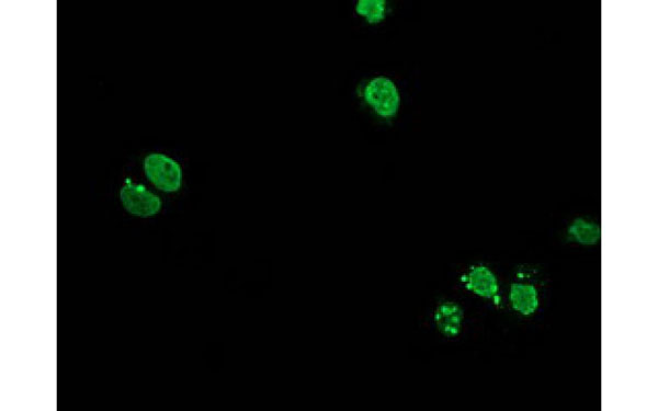
 IF (Immunofluorescence)
(Immunofluorescent staining of COS7 cells transiently transfected with recombinant FAM84B protein using FAM84B antibody)
IF (Immunofluorescence)
(Immunofluorescent staining of COS7 cells transiently transfected with recombinant FAM84B protein using FAM84B antibody)
FAM84B, Monoclonal Antibody (Cat# AAA74755)
 IHC (Immunohiostchemistry)
(Immunohistochemical analysis of paraffin-embedded human breast cancer,Lung breast tissues using EGF antibody)
IHC (Immunohiostchemistry)
(Immunohistochemical analysis of paraffin-embedded human breast cancer,Lung breast tissues using EGF antibody)
EGF, Monoclonal Antibody (Cat# AAA74759)
 IF (Immunofluorescence)
(Immunofluorescent staining of COS7 cells transiently transfected with recombinant CYP2J2 protein using CYP2J2 antibody)
IF (Immunofluorescence)
(Immunofluorescent staining of COS7 cells transiently transfected with recombinant CYP2J2 protein using CYP2J2 antibody)
CYP2J2, Monoclonal Antibody (Cat# AAA74774)
 IHC (Immunohiostchemistry)
(Immunohistochemical analysis of CENPH protein in paraffin embedded Carcinoma of Human bladder tissue using CENPH antibody)
IHC (Immunohiostchemistry)
(Immunohistochemical analysis of CENPH protein in paraffin embedded Carcinoma of Human bladder tissue using CENPH antibody)
CENPH, Monoclonal Antibody (Cat# AAA74778)
 IF (Immunofluorescence)
(Immunofluorescent staining of COS7 cells transiently transfected with recombinant AIPL1 protein using AIPL1 antibody)
IF (Immunofluorescence)
(Immunofluorescent staining of COS7 cells transiently transfected with recombinant AIPL1 protein using AIPL1 antibody)
AIPL1, Monoclonal Antibody (Cat# AAA74788)
 IF (Immunofluorescence)
(Immunofluorescent staining of COS7 cells transiently transfected with recombinant ERCC4 protein using ERCC4 antibody)
IF (Immunofluorescence)
(Immunofluorescent staining of COS7 cells transiently transfected with recombinant ERCC4 protein using ERCC4 antibody)
ERCC4, Monoclonal Antibody (Cat# AAA74791)
 IF (Immunofluorescence)
(Immunofluorescent staining of COS7 cells transiently transfected with recombinant HOXC11 protein using HOXC11 antibody)
IF (Immunofluorescence)
(Immunofluorescent staining of COS7 cells transiently transfected with recombinant HOXC11 protein using HOXC11 antibody)
HOXC11, Monoclonal Antibody (Cat# AAA74793)
GFAP, Monoclonal Antibody (Cat# AAA74800)
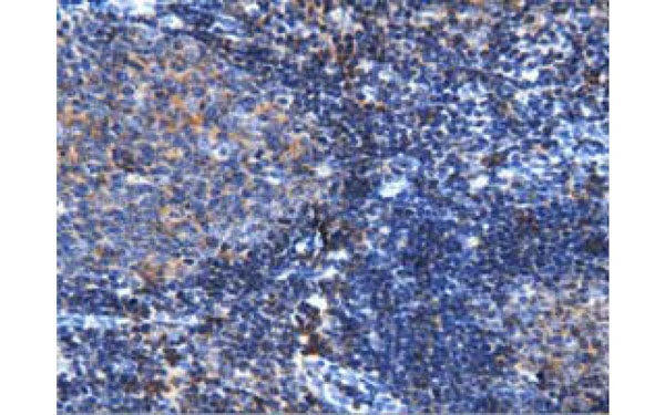 IHC (Immunohiostchemistry)
(Immunohistochemical analysis of ARHGAP25 protein in paraffin embedded Human lymph node tissue using ARHGAP25 antibody)
IHC (Immunohiostchemistry)
(Immunohistochemical analysis of ARHGAP25 protein in paraffin embedded Human lymph node tissue using ARHGAP25 antibody)
ARHGAP25, Monoclonal Antibody (Cat# AAA74810)
 IHC (Immunohiostchemistry)
(Immunohistochemical analysis of ACLY protein in paraffin embedded Human colon tissue using ACLY antibody)
IHC (Immunohiostchemistry)
(Immunohistochemical analysis of ACLY protein in paraffin embedded Human colon tissue using ACLY antibody)
ACLY, Monoclonal Antibody (Cat# AAA74827)
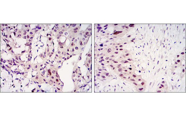 IHC (Immunohiostchemistry)
(Immunohistochemical analysis of paraffin-embedded mammary cancer tissues (left) and lung cancer tissues (right) using STAT3 antibody with DAB staining.)
IHC (Immunohiostchemistry)
(Immunohistochemical analysis of paraffin-embedded mammary cancer tissues (left) and lung cancer tissues (right) using STAT3 antibody with DAB staining.)
STAT3, Monoclonal Antibody (Cat# AAA74828)
 WB (Western Blot)
(Western Blotusinganti-CD45RAorA/B(Receptor-typetyrosine-proteinphosphataseC)antibodyOX-30(AAA72475) Ratthymuslysatesamples(35ugproteininRIPAbuffer)wereresolvedona10%SDSPAGEgelandblotsprobedwiththechimericrabbitversionofOX-30(AAA72475)at2ug/mlbeforedetectionusingananti-rabbitsecondaryantibody.Aprimaryincubationof1hwasusedandproteinwasdetectedbychemiluminescence.TheexpectedbandsizeforunmodifiedCD45RAis~143.3kDa,butthisproteinisalsohighlyglycosylated(UniProtId:P04157).AAA72475successfullydetectedCD45RAorA/Binratthymustissuelysate.)
WB (Western Blot)
(Western Blotusinganti-CD45RAorA/B(Receptor-typetyrosine-proteinphosphataseC)antibodyOX-30(AAA72475) Ratthymuslysatesamples(35ugproteininRIPAbuffer)wereresolvedona10%SDSPAGEgelandblotsprobedwiththechimericrabbitversionofOX-30(AAA72475)at2ug/mlbeforedetectionusingananti-rabbitsecondaryantibody.Aprimaryincubationof1hwasusedandproteinwasdetectedbychemiluminescence.TheexpectedbandsizeforunmodifiedCD45RAis~143.3kDa,butthisproteinisalsohighlyglycosylated(UniProtId:P04157).AAA72475successfullydetectedCD45RAorA/Binratthymustissuelysate.)
CD45RA or A/B, Monoclonal Recombinant Antibody (Cat# AAA72475)
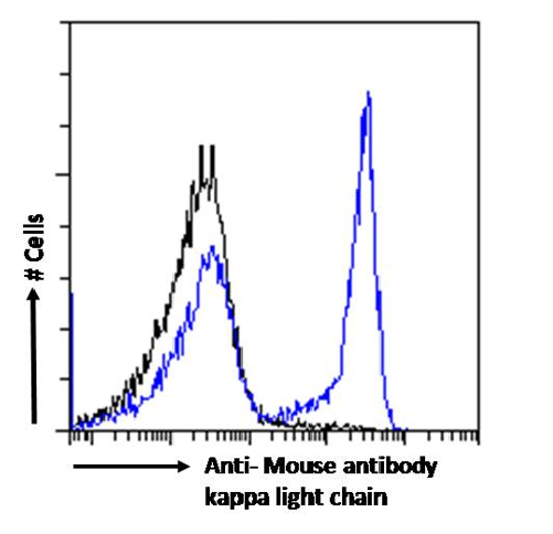

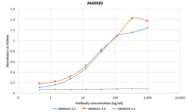 ELISA
(ELISAusingOX-20(AAA72483),Ab00102-1.1,Ab00102-2.0andAb00104-1.1. BindingcurvesoftherabbitIgGchimericversionoftheAnti-HAantibodyOX-20againstAb00102-1.1(blueline),Ab00102-2.0(orangeline)andAb00104-1.1(greyline)toanELISAplatecoatedwithAb00102-1.1,Ab00102-2.0andAb00104-1.1ataconcentrationof2.5ug/ml.A3-foldserialdilutionfrom3000to0.0169ng/mlwasperformedusingAAA72483antibody.Forsignaldetection,a1:4000dilutionofaHRP-conjugatedanti-rabbitIgGantibodywasused.)
ELISA
(ELISAusingOX-20(AAA72483),Ab00102-1.1,Ab00102-2.0andAb00104-1.1. BindingcurvesoftherabbitIgGchimericversionoftheAnti-HAantibodyOX-20againstAb00102-1.1(blueline),Ab00102-2.0(orangeline)andAb00104-1.1(greyline)toanELISAplatecoatedwithAb00102-1.1,Ab00102-2.0andAb00104-1.1ataconcentrationof2.5ug/ml.A3-foldserialdilutionfrom3000to0.0169ng/mlwasperformedusingAAA72483antibody.Forsignaldetection,a1:4000dilutionofaHRP-conjugatedanti-rabbitIgGantibodywasused.)
kappa light chain, Monoclonal Recombinant Antibody (Cat# AAA72483)
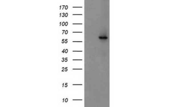
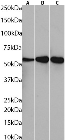
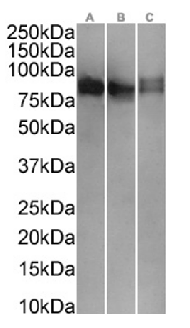 WB (Western Blot)
(Western BlotusingAnti-CD43antibodyOX-56(AAA72489). Ratthymus(A),ratlymphnode(B)andratspleen(C)tissuelysates(35ugproteininRIPAbuffer)wereresolvedonaSDSPAGEgelandblotswereprobedwiththechimericrabbitversionofOX-56(AAA72489)at0.3ug/mlbeforedetectionusingananti-rabbitsecondaryantibody.Aprimaryincubationof1hwasusedandproteinwasdetectedbychemiluminescence.)
WB (Western Blot)
(Western BlotusingAnti-CD43antibodyOX-56(AAA72489). Ratthymus(A),ratlymphnode(B)andratspleen(C)tissuelysates(35ugproteininRIPAbuffer)wereresolvedonaSDSPAGEgelandblotswereprobedwiththechimericrabbitversionofOX-56(AAA72489)at0.3ug/mlbeforedetectionusingananti-rabbitsecondaryantibody.Aprimaryincubationof1hwasusedandproteinwasdetectedbychemiluminescence.)
CD43, Monoclonal Recombinant Antibody (Cat# AAA72489)
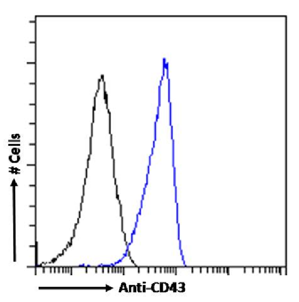
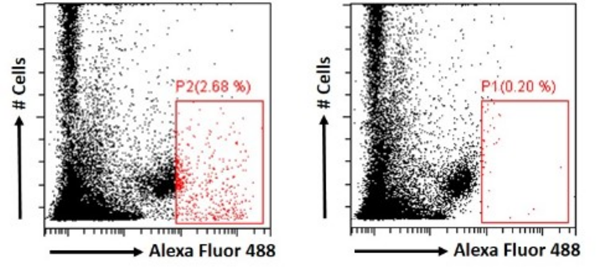
 FCM/FACS (Flow Cytometry)
(Flowcytometryusinganti-SiderocalinantibodyMAB1857(AAA72493). Paraformaldehyde-fixedRAW264.7cellspermeabilizedwith0.5%Tritonwerestainedwiththeanti-unknownspecificityantibodyortherabbitIgGversionofMAB1857(AAA72493,blueline)atadilutionof1:100for1hatRT.Afterwashing,theboundantibodywasdetectedusingagoatanti-rabbitIgGAlexaFluor488antibodyatadilutionof1:1000,andthecellswereanalyzedusingaFACSCantoflow-cytometer.)
FCM/FACS (Flow Cytometry)
(Flowcytometryusinganti-SiderocalinantibodyMAB1857(AAA72493). Paraformaldehyde-fixedRAW264.7cellspermeabilizedwith0.5%Tritonwerestainedwiththeanti-unknownspecificityantibodyortherabbitIgGversionofMAB1857(AAA72493,blueline)atadilutionof1:100for1hatRT.Afterwashing,theboundantibodywasdetectedusingagoatanti-rabbitIgGAlexaFluor488antibodyatadilutionof1:1000,andthecellswereanalyzedusingaFACSCantoflow-cytometer.)
Siderocalin, Monoclonal Recombinant Antibody (Cat# AAA72493)
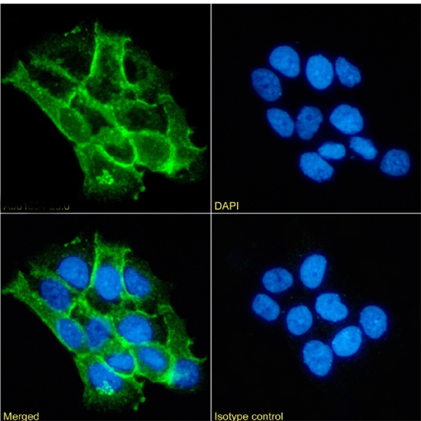
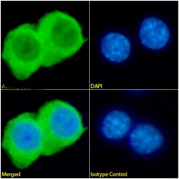
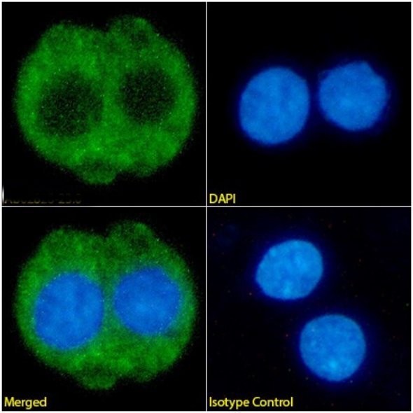
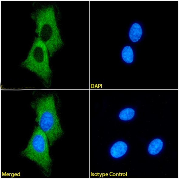
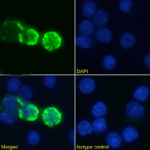
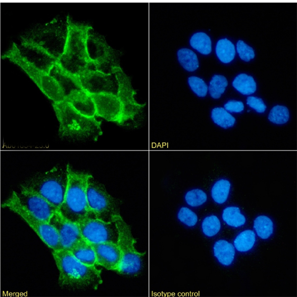 IF (Immunofluorescence)
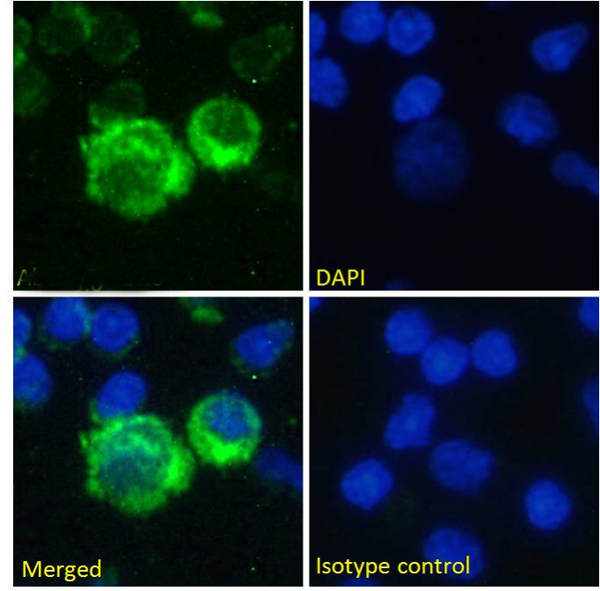
(ImmunofluorescencestainingoffixedA431cellswithanti-alphacateninantibody1G5(AAA72522) ImmunofluorescenceanalysisofparaformaldehydefixedA431cellsonShi-fixcoverslipsstainedwiththechimericrabbitIgGversionof1G5(AAA72522)at10ug/mlfor1hfollowedbyAlexaFluor488secondaryantibody(2ug/ml),showingmembranestaining.ThenuclearstainisDAPI(blue).Panelsshowfromleft-right,top-bottomAAA72522,DAPI,mergedchannelsandanisotypecontrol.TheisotypecontrolwasanunknownspecificityantibodyfollowedbystainingwithAlexaFluor488secondaryantibody.)
IF (Immunofluorescence)
(ImmunofluorescencestainingoffixedA431cellswithanti-alphacateninantibody1G5(AAA72522) ImmunofluorescenceanalysisofparaformaldehydefixedA431cellsonShi-fixcoverslipsstainedwiththechimericrabbitIgGversionof1G5(AAA72522)at10ug/mlfor1hfollowedbyAlexaFluor488secondaryantibody(2ug/ml),showingmembranestaining.ThenuclearstainisDAPI(blue).Panelsshowfromleft-right,top-bottomAAA72522,DAPI,mergedchannelsandanisotypecontrol.TheisotypecontrolwasanunknownspecificityantibodyfollowedbystainingwithAlexaFluor488secondaryantibody.)
alpha catenin, Monoclonal Recombinant Antibody (Cat# AAA72522)


 IF (Immunofluorescence)
(ImmunofluorescencestainingoffixedA431cellswithanti-alphacateninantibody1G5(AAA72523) ImmunofluorescenceanalysisofparaformaldehydefixedA431cellsonShi-fixcoverslipsstainedwiththechimericrabbitIgGversionof1G5(AAA72523)at10ug/mlfor1hfollowedbyAlexaFluor488secondaryantibody(2ug/ml),showingmembranestaining.ThenuclearstainisDAPI(blue).Panelsshowfromleft-right,top-bottomAAA72523,DAPI,mergedchannelsandanisotypecontrol.TheisotypecontrolwasanunknownspecificityantibodyfollowedbystainingwithAlexaFluor488secondaryantibody.)
IF (Immunofluorescence)
(ImmunofluorescencestainingoffixedA431cellswithanti-alphacateninantibody1G5(AAA72523) ImmunofluorescenceanalysisofparaformaldehydefixedA431cellsonShi-fixcoverslipsstainedwiththechimericrabbitIgGversionof1G5(AAA72523)at10ug/mlfor1hfollowedbyAlexaFluor488secondaryantibody(2ug/ml),showingmembranestaining.ThenuclearstainisDAPI(blue).Panelsshowfromleft-right,top-bottomAAA72523,DAPI,mergedchannelsandanisotypecontrol.TheisotypecontrolwasanunknownspecificityantibodyfollowedbystainingwithAlexaFluor488secondaryantibody.)
alpha catenin, Monoclonal Recombinant Antibody (Cat# AAA72523)
 FCM/FACS (Flow Cytometry)
(Flowcytometryusinganti-IL-4antibodySB240683(Pascolizumab,humanized3B9)(AAA72531). Humanbloodleukocytesrestedfor4hoursat37°C,thenfixedwith2%PFAandpermeabilizedwith0.5%Triton,werestainedwiththeanti-unknownspecificityantibodyortherabbitIgGversionofSB240683(AAA72531,left)atadilutionof1:100overnightat4°C.Afterwashing,theboundantibodywasdetectedusingagoatanti-rabbitIgGAlexaFluor488antibodyatadilutionof1:1000,andthecellswereanalyzedusingaFACSCantoflow-cytometer.)
FCM/FACS (Flow Cytometry)
(Flowcytometryusinganti-IL-4antibodySB240683(Pascolizumab,humanized3B9)(AAA72531). Humanbloodleukocytesrestedfor4hoursat37°C,thenfixedwith2%PFAandpermeabilizedwith0.5%Triton,werestainedwiththeanti-unknownspecificityantibodyortherabbitIgGversionofSB240683(AAA72531,left)atadilutionof1:100overnightat4°C.Afterwashing,theboundantibodywasdetectedusingagoatanti-rabbitIgGAlexaFluor488antibodyatadilutionof1:1000,andthecellswereanalyzedusingaFACSCantoflow-cytometer.)
IL-4, Monoclonal Recombinant Antibody (Cat# AAA72531)
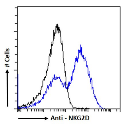
 FCM/FACS (Flow Cytometry)
(FlowcytometryusingtheAnti-NKG2DantibodyCX-5(AAA72535). Paraformaldehydefixedmousesplenocyteswerestainedwithanti-unknownspecificityantibodyortherabbitIgGversionofCX-5(AAA72535,blueline)atadilutionof1:100for1hatRT.Afterwashing,theboundantibodywasdetectedusingagoatanti-rabbitIgGAlexaFluor488antibodyatadilutionof1:1000andcellsanalyzedusingaFACSCantoflow-cytometer.)
FCM/FACS (Flow Cytometry)
(FlowcytometryusingtheAnti-NKG2DantibodyCX-5(AAA72535). Paraformaldehydefixedmousesplenocyteswerestainedwithanti-unknownspecificityantibodyortherabbitIgGversionofCX-5(AAA72535,blueline)atadilutionof1:100for1hatRT.Afterwashing,theboundantibodywasdetectedusingagoatanti-rabbitIgGAlexaFluor488antibodyatadilutionof1:1000andcellsanalyzedusingaFACSCantoflow-cytometer.)
NKG2D, Monoclonal Recombinant Antibody (Cat# AAA72535)
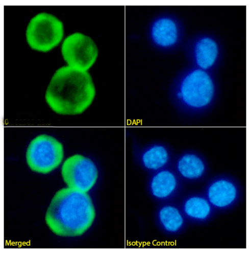
 IF (Immunofluorescence)
(ImmunofluorescencestainingofHepG2cellswithanti-Fibrinbeta-chainantibody59D8(AAA72540). ImmunofluorescenceanalysisofparaformaldehydefixedHepG2cellsonShi-fixcoverslipsstainedwiththechimericrabbitIgGversionof59D8(AAA72540)(1:100dilution)for1hfollowedbyAlexaFluor488secondaryantibody(1:1000dilution),showingcytoplasmicstaining.ThenuclearstainisDAPI(blue).Panelsshow,fromleft-right,top-bottom,AAA72540,DAPI,mergedchannelsandanisotypecontrol.TheisotypecontrolwasanunknownspecificityantibodyfollowedbystainingwithAlexaFluor488secondaryantibody.)
IF (Immunofluorescence)
(ImmunofluorescencestainingofHepG2cellswithanti-Fibrinbeta-chainantibody59D8(AAA72540). ImmunofluorescenceanalysisofparaformaldehydefixedHepG2cellsonShi-fixcoverslipsstainedwiththechimericrabbitIgGversionof59D8(AAA72540)(1:100dilution)for1hfollowedbyAlexaFluor488secondaryantibody(1:1000dilution),showingcytoplasmicstaining.ThenuclearstainisDAPI(blue).Panelsshow,fromleft-right,top-bottom,AAA72540,DAPI,mergedchannelsandanisotypecontrol.TheisotypecontrolwasanunknownspecificityantibodyfollowedbystainingwithAlexaFluor488secondaryantibody.)
Fibrin beta-chain, Monoclonal Recombinant Antibody (Cat# AAA72540)
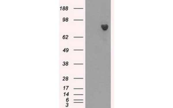
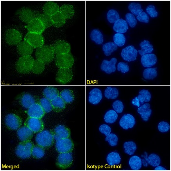
 IF (Immunofluorescence)
(ImmunofluorescencestainingofKellycellswithanti-MyelinbasicproteinantibodyF28C4(AAA72544). Immunofluorescenceanalysisofparaformaldehyde-fixedKellycellsonShi-fixcoverslipsstainedwiththechimericrabbitIgGversionofF28C4(AAA72544)(1:100dilution)for1hfollowedbyAlexaFluor488secondaryantibody(1:1000dilution),showingcytoplasmicstaining.ThenuclearstainisDAPI(blue).Panelsshow,fromleft-right,top-bottom,AAA72544,DAPI,mergedchannels,andanisotypecontrol.TheisotypecontrolwasanunknownspecificityantibodyfollowedbystainingwithAlexaFluor488secondaryantibody.)
IF (Immunofluorescence)
(ImmunofluorescencestainingofKellycellswithanti-MyelinbasicproteinantibodyF28C4(AAA72544). Immunofluorescenceanalysisofparaformaldehyde-fixedKellycellsonShi-fixcoverslipsstainedwiththechimericrabbitIgGversionofF28C4(AAA72544)(1:100dilution)for1hfollowedbyAlexaFluor488secondaryantibody(1:1000dilution),showingcytoplasmicstaining.ThenuclearstainisDAPI(blue).Panelsshow,fromleft-right,top-bottom,AAA72544,DAPI,mergedchannels,andanisotypecontrol.TheisotypecontrolwasanunknownspecificityantibodyfollowedbystainingwithAlexaFluor488secondaryantibody.)
Myelin basic protein, Monoclonal Recombinant Antibody (Cat# AAA72544)
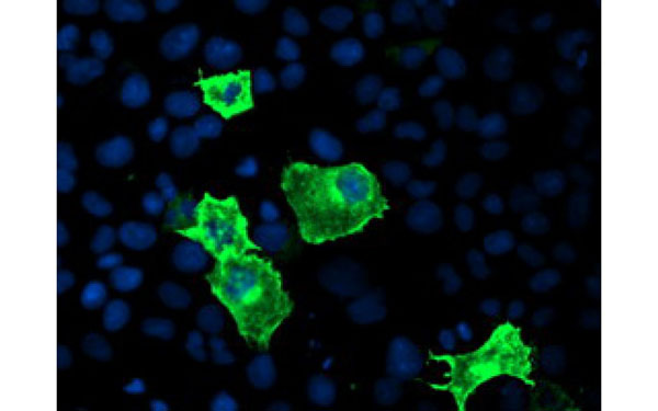
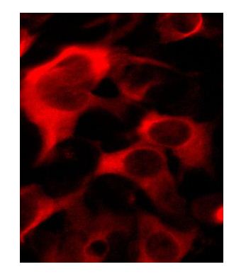
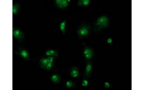
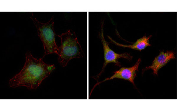 IF (Immunofluorescence)
(Immunofluorescence analysis of A549 (left) and SK-BR-3 (right) cells using CTNNB1 antibody (green). Red: Actin filaments have been labeled with DY-554 phalloidin. Blue: DRAQ5 fluorescent DNA dye.)
IF (Immunofluorescence)
(Immunofluorescence analysis of A549 (left) and SK-BR-3 (right) cells using CTNNB1 antibody (green). Red: Actin filaments have been labeled with DY-554 phalloidin. Blue: DRAQ5 fluorescent DNA dye.)
CTNNB1, Monoclonal Antibody (Cat# AAA74903)
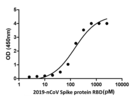
 Application Data
(Binding of Anti 2019-nCoV Spike Protein Llama heavy-chain to 2019-nCoV RBD determined by ELISA.)
Application Data
(Binding of Anti 2019-nCoV Spike Protein Llama heavy-chain to 2019-nCoV RBD determined by ELISA.)
COVID 19 Spike Protein Llama heavy-chain Coronavirus, Monoclonal Antibody (Cat# AAA73172)
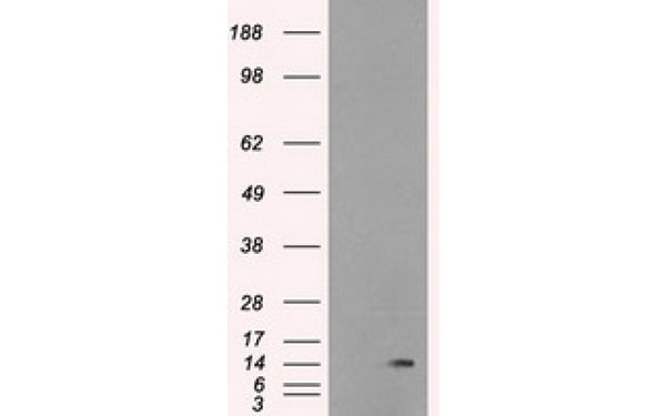
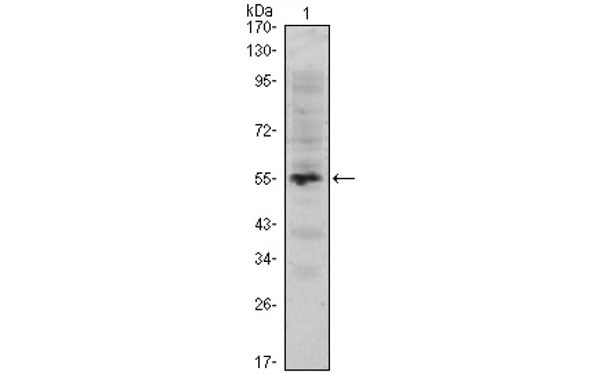
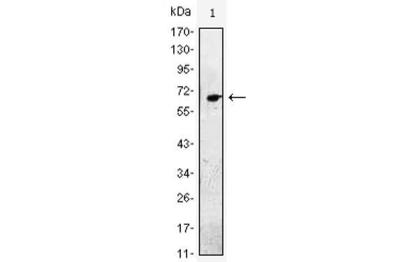
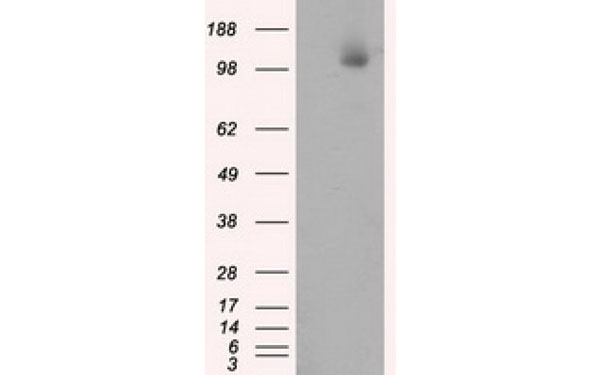
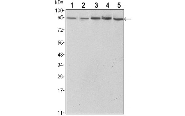
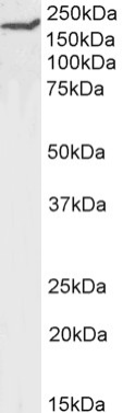
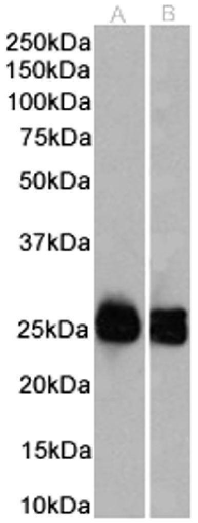
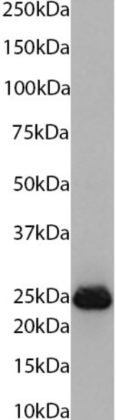
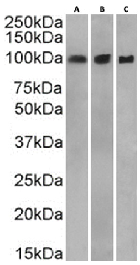
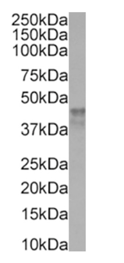
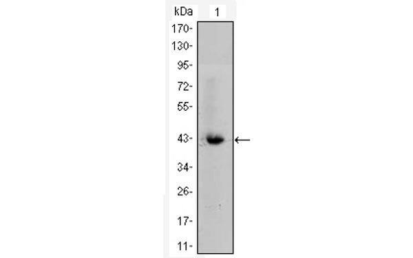
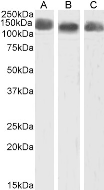 WB (Western Blot)
(Western Blotusinganti-CD43(Leukosialin)antibodyW3/13(AAA72434) Ratspleen(A),thymus(B)andlymphnode(C)lysatesamples(35ugproteininRIPAbuffer)wereresolvedona10%SDSPAGEgelandblotsprobedwiththechimericrabbitversionofW3/13(AAA72434)at0.1ug/mlbeforedetectionusingananti-rabbitsecondaryantibody.Aprimaryincubationof1hwasusedandproteinwasdetectedbychemiluminescence.TheexpectedrunningsizeforCD43is100-120kDa.Ab00successfullydetectedCD43inratspleen,thymusandlymphnodesamples.)
WB (Western Blot)
(Western Blotusinganti-CD43(Leukosialin)antibodyW3/13(AAA72434) Ratspleen(A),thymus(B)andlymphnode(C)lysatesamples(35ugproteininRIPAbuffer)wereresolvedona10%SDSPAGEgelandblotsprobedwiththechimericrabbitversionofW3/13(AAA72434)at0.1ug/mlbeforedetectionusingananti-rabbitsecondaryantibody.Aprimaryincubationof1hwasusedandproteinwasdetectedbychemiluminescence.TheexpectedrunningsizeforCD43is100-120kDa.Ab00successfullydetectedCD43inratspleen,thymusandlymphnodesamples.)
CD43, Monoclonal Recombinant Antibody (Cat# AAA72434)
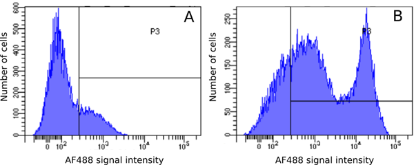
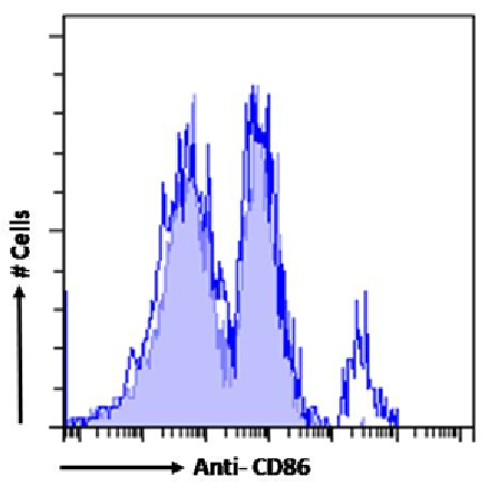 FCM/FACS (Flow Cytometry)
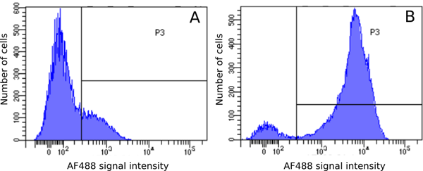
(Flow-cytometryusingtheanti-CD86antibodyOX-48(AAA72467). Ratsplenocyteswerestainedwithanti-FluoresceinIgGantibody(4-4-20;isotypecontrol,shadedline)ortherabbitIgG1versionofOX-48(AAA72467,blueline)atadilutionof1:100for1hatRT.Afterwashing,boundantibodywasdetectedusingagoatanti-mouseIgGAlexaFluor488antibodyatadilutionof1:1000andcellsanalyzedusingaFACSCantoflow-cytometer.)
FCM/FACS (Flow Cytometry)
(Flow-cytometryusingtheanti-CD86antibodyOX-48(AAA72467). Ratsplenocyteswerestainedwithanti-FluoresceinIgGantibody(4-4-20;isotypecontrol,shadedline)ortherabbitIgG1versionofOX-48(AAA72467,blueline)atadilutionof1:100for1hatRT.Afterwashing,boundantibodywasdetectedusingagoatanti-mouseIgGAlexaFluor488antibodyatadilutionof1:1000andcellsanalyzedusingaFACSCantoflow-cytometer.)
CD86, Monoclonal Recombinant Antibody (Cat# AAA72467)
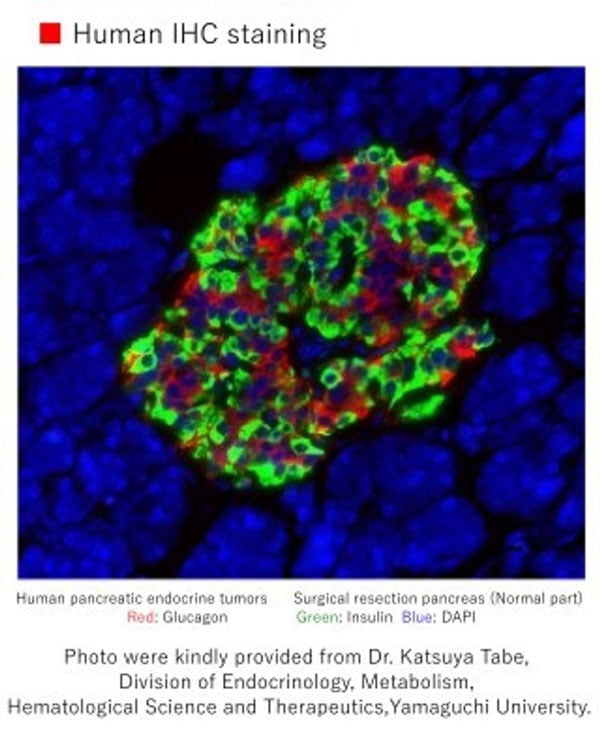
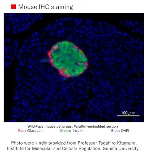 IHC (Immunohiostchemistry)
Glucagon (52A1A) IgG MoAb, Monoclonal Antibody (Cat# AAA72733)
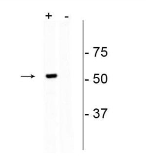
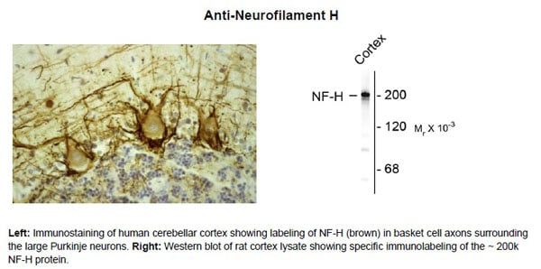 Application Data
Neurofilament NF-H ms, Monoclonal Antibody (Cat# AAA72746)
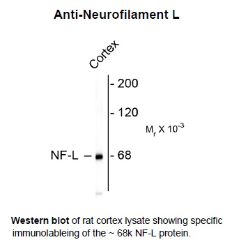 Application Data
Neurofilament NF-L ms, Monoclonal Antibody (Cat# AAA72747)
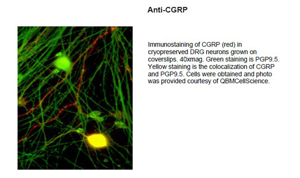 Application Data
Calcitonin Gene Related Peptide (CGRP), Monoclonal Antibody (Cat# AAA72751)
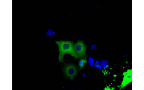
 IF (Immunofluorescence)
(Immunofluorescence of HEK293 cells transfected with Cas9 and labeled with Cas9 antibody.)
IF (Immunofluorescence)
(Immunofluorescence of HEK293 cells transfected with Cas9 and labeled with Cas9 antibody.)
CRISPR Cas9, Monoclonal Antibody (Cat# AAA72759)
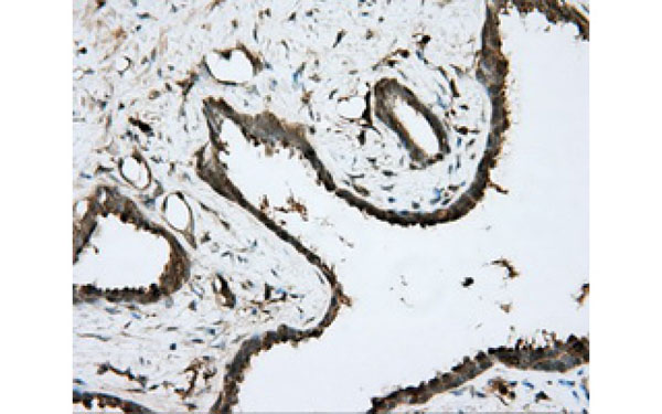
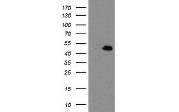
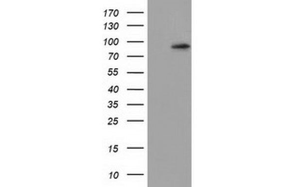
CD8, Monoclonal Antibody (Cat# AAA74674)
 IHC (Immunohiostchemistry)
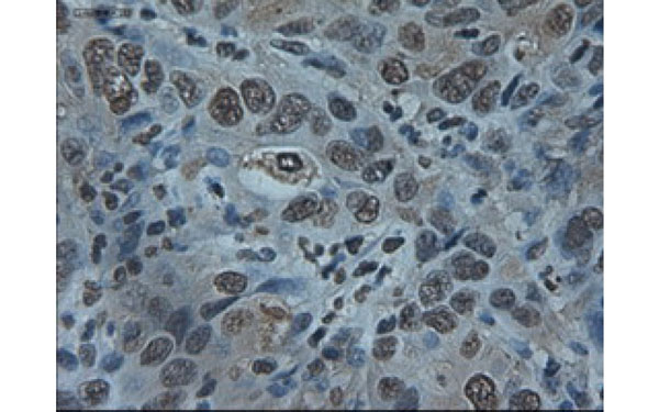
(Immunohistochemical analysis of ARNTL protein in paraffin embedded Adenocarcinoma of Human colon tissue using ARNTL antibody)
IHC (Immunohiostchemistry)
(Immunohistochemical analysis of ARNTL protein in paraffin embedded Adenocarcinoma of Human colon tissue using ARNTL antibody)
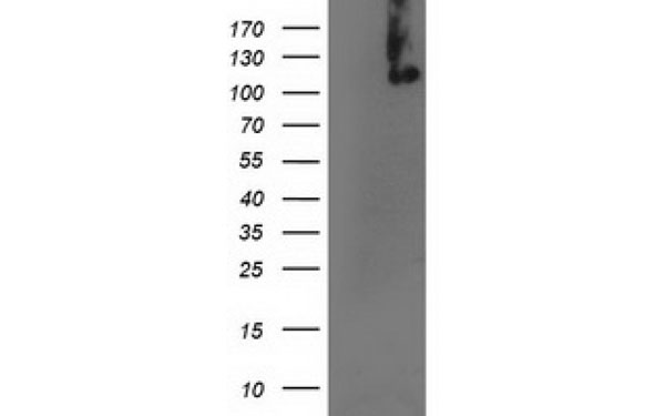
ARNTL, Monoclonal Antibody (Cat# AAA74676)
Adenovirus, Monoclonal Antibody (Cat# AAA74679)
Adenovirus antibody was purified by Ion exchange chromatography.
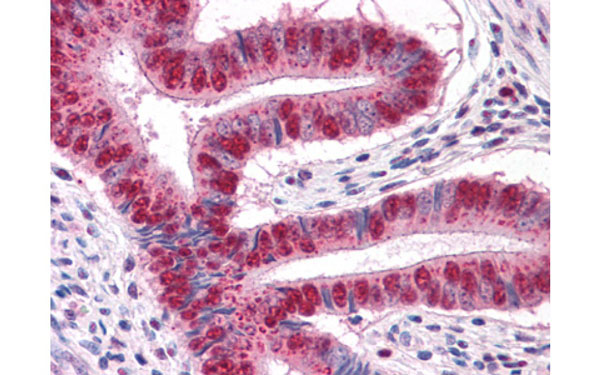
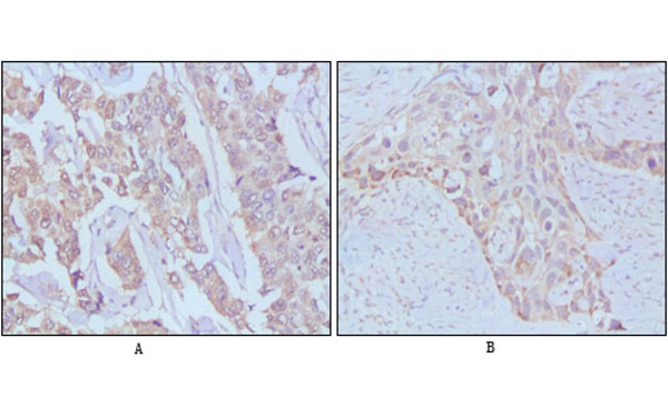
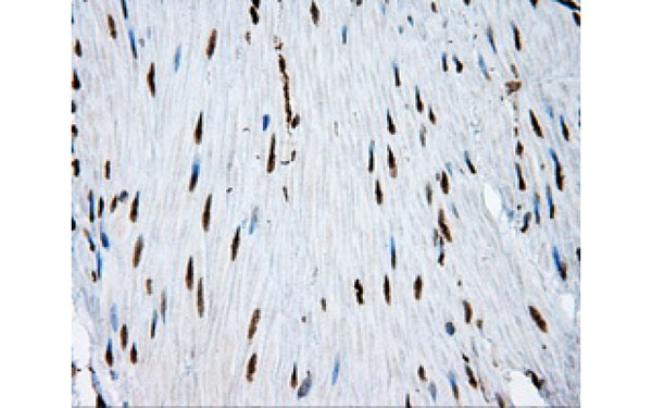
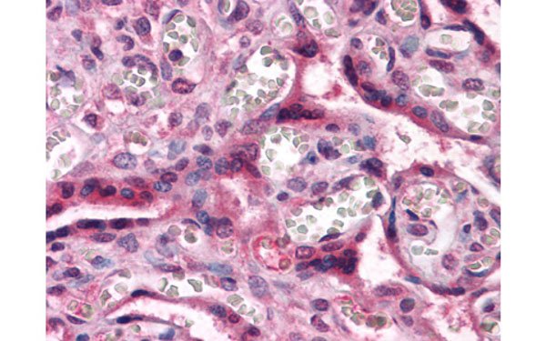
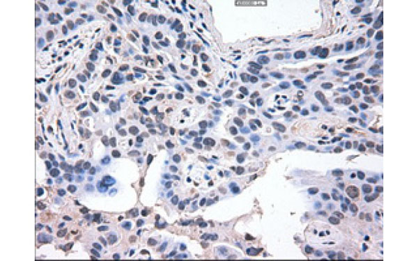
 IHC (Immunohiostchemistry)
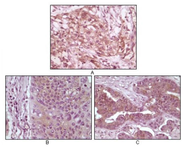
(Immunohistochemical analysis of paraffin-embedded human pancreas carcinoma(A), esophagus carcinoma tissue(B) and ovary tumor tissue(C), showing cytoplasmic and membrane localization using 4E-BP1 antibody with DAB staining.)
IHC (Immunohiostchemistry)
(Immunohistochemical analysis of paraffin-embedded human pancreas carcinoma(A), esophagus carcinoma tissue(B) and ovary tumor tissue(C), showing cytoplasmic and membrane localization using 4E-BP1 antibody with DAB staining.)
4EBP1, Monoclonal Antibody (Cat# AAA74680)
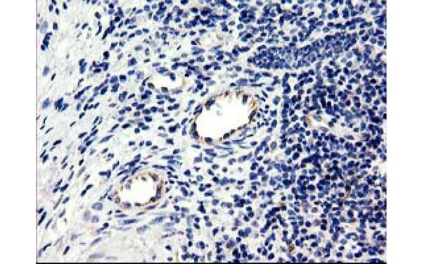
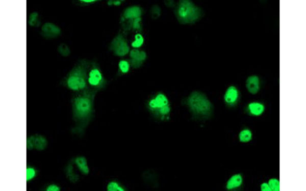
 IHC (Immunohiostchemistry)
(Immunohistochemical analysis of FBXO21 protein in paraffin embedded Human ovary tissue using FBXO21 antibody)
IHC (Immunohiostchemistry)
(Immunohistochemical analysis of FBXO21 protein in paraffin embedded Human ovary tissue using FBXO21 antibody)
FBXO21, Monoclonal Antibody (Cat# AAA74689)
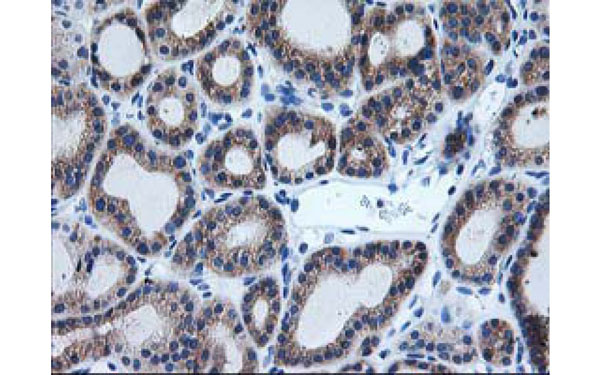
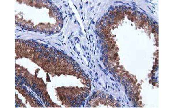 IHC (Immunohiostchemistry)
(Immunohistochemical analysis of ADI1 protein in paraffin embedded Human prostate tissue using ADI1 antibody)
IHC (Immunohiostchemistry)
(Immunohistochemical analysis of ADI1 protein in paraffin embedded Human prostate tissue using ADI1 antibody)
ADI1, Monoclonal Antibody (Cat# AAA74710)
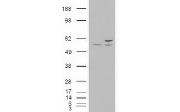
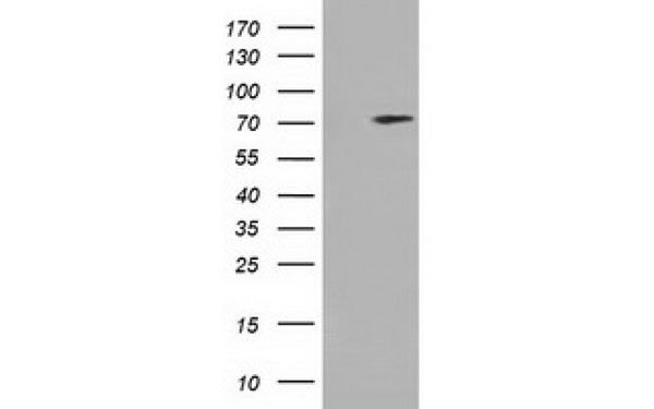
 IHC (Immunohiostchemistry)
(Immunohistochemical analysis of AKT3 protein in paraffin embedded Carcinoma of Human lung tissue using AKT3 antibody)
IHC (Immunohiostchemistry)
(Immunohistochemical analysis of AKT3 protein in paraffin embedded Carcinoma of Human lung tissue using AKT3 antibody)
AKT3, Monoclonal Antibody (Cat# AAA74729)
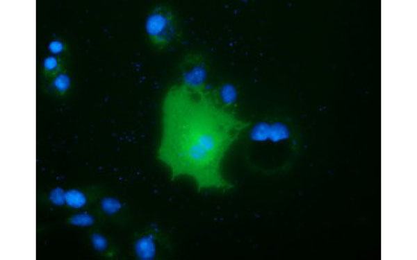
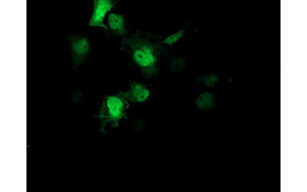
 IF (Immunofluorescence)
(Immunofluorescent staining of COS7 cells transiently transfected with recombinant CDCP1 protein using CDCP1 antibody)
IF (Immunofluorescence)
(Immunofluorescent staining of COS7 cells transiently transfected with recombinant CDCP1 protein using CDCP1 antibody)
CDCP1, Monoclonal Antibody (Cat# AAA74739)
What are Monoclonal Antibodies?
Monoclonal antibodies are specialized laboratory-produced proteins developed for binding to specific biological antigens or other molecular targets. Since they come from a single cell (or clone), they are especially consistent and accurate in the data they are involved in producing.
This type of antibody material has been shown to be a powerful tool in finding and subsequently destroying harmful cells in an organism, such as those found in cancers or various autoimmune diseases. This makes them excellent aids in medical testing and research, which is why they are so widely used.
AAA Biotech offers a comprehensive range of high-quality monoclonal antibodies that perform effectively in various laboratory tests, including (amongst others) ELISA, western blotting, immunohistochemistry, and flow cytometry. All of the products in our catalog are thoroughly quality tested to make sure that they are reliable and will consistently perform well in your research.
What Are The Uses of Monoclonal Antibodies
Monoclonal antibodies are used in many lab tests, including (amongst others) ELISA, western blotting, immunohistochemistry, and flow cytometry.
ELISA is a test that helps detect a specific substance/analyte in a sample. It uses antibodies (often monoclonal) bound to a solid surface (such as the well of a microplate) to “capture” the substance/analyte in the sample and immobilize it so that the detection antibody component can then bind to it and produce a signal, which can then be measured.
Western blotting identifies specific proteins in a sample. The sample is first separated on a gel, and then antibodies are applied that will typically bind to the target, which will all be localized to a single band in a lane.
Immunohistochemistry helps locate specific proteins in cells or tissue samples using antibodies.
Flow cytometry looks at and sorts cells. It uses antibodies that are conjugated to reporter molecules called “fluorophores”, which, under special lights, emit light themselves, which can then be measured by a detector instrument.
How Monoclonal Antibodies Are Used as Medicine?
Please note that all of the products listed in AAA Biotech’s also known as AAA Bio or AAABio catalog are strictly for research-use only (RUO).
Monoclonal antibodies can also be used as therapeutic/medical treatments, particularly in the context of cancers. They are designed to find and bind to specific cells or proteins, helping the immune system recognize and attack the cancer. These treatments work in different ways, such as:
- Radioimmunotherapy attaches a small amount of radioactive molecule to the antibody, so it delivers the radiation directly to the cancer cells that the antibody is specifically binding to.
- Antibody-directed enzyme prodrug therapy uses antibodies that are specifically bound to special enzymes. These enzymes activate a harmless drug in the body and turn it into a cancer-killing drug only near the cancer cells—this helps avoid harming healthy cells.
- Immunoliposomes are tiny “bubbles” filled with medicine/drug and coated with antibodies. They carry the drug straight to the cancer cells.
Why Buy Monoclonal Antibodies From Us?
At AAA Biotech, we provide high-performance monoclonal antibodies designed to support a wide range of research needs.
1. Validated for Versatile Applications
The antibodies in our catalog are extensively validated and compatible with multiple techniques, including (but not limited to) ELISA, flow cytometry (FC), immunocytochemistry (ICC), immunofluorescence (IF), immunohistochemistry (IHC), immunoprecipitation (IP), and western blotting (WB).
2. Wide Selection & Specialized Options
We offer antibodies for common and rare species, that are available in various conjugated forms, and also in recombinant formats. Essentially, there is almost anything one might need to meet their experimental model’s requirements.
3. High-Quality Proteins
Our proteins meet high purity standards—90% or more as confirmed by SDS-PAGE. Many are available with tags like His, Flag, GST, or MBP, and we also supply native and biologically active proteins for functional studies.
Frequently Asked Questions
1. Are your monoclonal antibodies validated for specific applications?
Yes, our antibodies are tested and validated for use in methods such as ELISA, western blot, IHC, flow cytometry, and more. Refer to specific product pages or datasheets for individual product information.
2. How do I choose the right monoclonal antibody for my application?
Review the product details directly for application validation, species reactivity, and target information. You may also contact our support team at any time for help.
3. How quickly can I receive my order?
Most orders are processed and shipped within 1–3 business days, depending on product availability and your shipping location.