Filters
▼Clonality
▼Type
▼Reactivity
▼Gene Name
▼Isotype
▼Host
▼Application
▼Clone
▼Monoclonal Antibodies
Get accurate results in your research with our Monoclonal Antibodies, which are specially made to target exactly what you require for your research, and will produce consistent, reliable performance in lab tests.
Viewing 9450-9500 of 27597 product results
Herpes Virus Type 6 (HHV-6) 37kDa EA, Monoclonal Antibody (Cat# AAA57633)
Human Papilloma Virus Type 16 (HPV) E7 Protein, Monoclonal Antibody (Cat# AAA57705)
Canine Distemper Virus (CDV), Surface envelope antigen, Monoclonal Antibody (Cat# AAA57706)
 DB (Dot Blot)
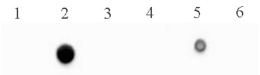
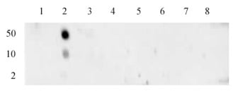
(5-Methylcytosine (5-mC) antibody (mAb) tested by dot blot analysis. DNA from the Methylated DNA Standard Kit were spotted (5 ng per spot) on to a positively charged nylon membrane and blotted with 5-Methylcytosine antibody (2 ug/ml dilution). Lane 1: single-stranded unmethylated DNA. Lane 2: single-stranded DNA containing 5-methylcytosine. Lane 3: single-stranded DNA containing 5-hydroxymethylcytosine. Lane 4: double-stranded unmethylated DNA. Lane 5: double-stranded DNA containing 5-methylcytosine. Lane 6: double-stranded DNA 5-hydroxymethylcytosine.)
DB (Dot Blot)
(5-Methylcytosine (5-mC) antibody (mAb) tested by dot blot analysis. DNA from the Methylated DNA Standard Kit were spotted (5 ng per spot) on to a positively charged nylon membrane and blotted with 5-Methylcytosine antibody (2 ug/ml dilution). Lane 1: single-stranded unmethylated DNA. Lane 2: single-stranded DNA containing 5-methylcytosine. Lane 3: single-stranded DNA containing 5-hydroxymethylcytosine. Lane 4: double-stranded unmethylated DNA. Lane 5: double-stranded DNA containing 5-methylcytosine. Lane 6: double-stranded DNA 5-hydroxymethylcytosine.)
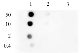
5-Methylcytosine, Monoclonal Antibody (Cat# AAA60030)
 DB (Dot Blot)
(Histone H3 monomethyl Lys4 antibody (mAb) tested by Dot Blot Dot Blot analysis was used to confirm the specificity of Histone H3 monomethyl Lys4 antibody (mAb) for monomethyl Lys4 histone H3. Methylated peptides corresponding to the immunogen or related peptides were spotted onto PVDF and probed with antibody at 1 ug/ml. Lane 1: unmodified peptide, Lane 2: Monomethyl Lys4, Lanes 3-8: various negative control peptides.)
DB (Dot Blot)
(Histone H3 monomethyl Lys4 antibody (mAb) tested by Dot Blot Dot Blot analysis was used to confirm the specificity of Histone H3 monomethyl Lys4 antibody (mAb) for monomethyl Lys4 histone H3. Methylated peptides corresponding to the immunogen or related peptides were spotted onto PVDF and probed with antibody at 1 ug/ml. Lane 1: unmodified peptide, Lane 2: Monomethyl Lys4, Lanes 3-8: various negative control peptides.)
Histone H3K4me1, Monoclonal Antibody (Cat# AAA59901)
 DB (Dot Blot)
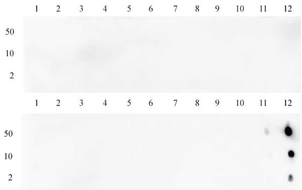
(Histone H4K20me3 antibody (mAb) tested by dot blot analysis. Dot blot analysis was used to confirm the specificity of Histone H4K20me3 antibody (mAb) for trimethyl-Lys20 of histone H4. Peptides were spotted onto PVDF and probed with antibody at a dilution of 0.2 ug/ml. The amount of peptide (in picomoles) spotted is indicated (2, 10, and 50). Column 1: H4K20me0. Column 2: H4K20me1. Column 3: H4K20me2. Column 4: H4K20me3.)
DB (Dot Blot)
(Histone H4K20me3 antibody (mAb) tested by dot blot analysis. Dot blot analysis was used to confirm the specificity of Histone H4K20me3 antibody (mAb) for trimethyl-Lys20 of histone H4. Peptides were spotted onto PVDF and probed with antibody at a dilution of 0.2 ug/ml. The amount of peptide (in picomoles) spotted is indicated (2, 10, and 50). Column 1: H4K20me0. Column 2: H4K20me1. Column 3: H4K20me2. Column 4: H4K20me3.)
Histone H4K20me3, Monoclonal Antibody (Cat# AAA59912)
 Application Data
(Histone H3 monomethyl Lys9 antibody specificity tested by peptide array analysis. Peptide array analysis was used to confirm the specificity of this antibody for its intended modification. Histone H3 monomethyl Lys9 antibody was applied at a dilution of 1:2,000 to MODified Histone Peptide Array spotted is indicated next to each row. Lane 1: monomethyl lysine 9 protein. Lane 2: dimethyl lysine 9 protein. Lane 3: trimethyl lysine 9 protein. No detection of proteins (unmodified, mono-, di-, or tri-methylated) corresponding to lysine 27 of histone H3 was observed with Histone H3 monomethyl Lys9 antibody.)
Application Data
(Histone H3 monomethyl Lys9 antibody specificity tested by peptide array analysis. Peptide array analysis was used to confirm the specificity of this antibody for its intended modification. Histone H3 monomethyl Lys9 antibody was applied at a dilution of 1:2,000 to MODified Histone Peptide Array spotted is indicated next to each row. Lane 1: monomethyl lysine 9 protein. Lane 2: dimethyl lysine 9 protein. Lane 3: trimethyl lysine 9 protein. No detection of proteins (unmodified, mono-, di-, or tri-methylated) corresponding to lysine 27 of histone H3 was observed with Histone H3 monomethyl Lys9 antibody.)
Histone H3K9me1, Monoclonal Antibody (Cat# AAA59914)
 DB (Dot Blot)
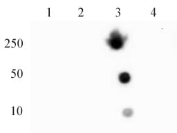
(Histone H3K36me3 antibody (mAb) tested by dot blot. Dot blot analysis was used to confirm the specificity of Histone H3K36me3 antibody for trimethyl Lys36 of histone H3. Recombinant methylated proteins corresponding to the immunogen and related sequences were spotted onto PVDF and probed with Histone H3K36me3 at 2 ug/ml. The amount of protein (picomoles) spotted is indicated next to each row. Top panel - Lane 1: unmodified H3 Lys4. Lane 2: H3K4me1. Lane 3: H3K4me2. Lane 4: H3K4me3. Lane 5: unmodified H3 Lys9. Lane 6: H3K9me1. Lane 7: H3K9me2. Lane 8: H3K9me3. Lane 9: unmodified H3 Lys79. Lane 10: H3K79me1. Lane 11: H3K79me2. Lane 12: H3K79me3. Bottom panel - Lane 1: unmodified H3 Lys23. Lane 2: H3K23me1. Lane 3: H3K23me2. Lane 4: H3K23me3. Lane 5: unmodified H3 Lys27. Lane 6: H3K27me1. Lane 7: H3K27me2. Lane 8: H3K27me3. Lane 9: unmodified H3 Lys36. Lane 10: H3K36me1. Lane 11: H3K36me2. Lane 12: H3K36me3.)
DB (Dot Blot)
(Histone H3K36me3 antibody (mAb) tested by dot blot. Dot blot analysis was used to confirm the specificity of Histone H3K36me3 antibody for trimethyl Lys36 of histone H3. Recombinant methylated proteins corresponding to the immunogen and related sequences were spotted onto PVDF and probed with Histone H3K36me3 at 2 ug/ml. The amount of protein (picomoles) spotted is indicated next to each row. Top panel - Lane 1: unmodified H3 Lys4. Lane 2: H3K4me1. Lane 3: H3K4me2. Lane 4: H3K4me3. Lane 5: unmodified H3 Lys9. Lane 6: H3K9me1. Lane 7: H3K9me2. Lane 8: H3K9me3. Lane 9: unmodified H3 Lys79. Lane 10: H3K79me1. Lane 11: H3K79me2. Lane 12: H3K79me3. Bottom panel - Lane 1: unmodified H3 Lys23. Lane 2: H3K23me1. Lane 3: H3K23me2. Lane 4: H3K23me3. Lane 5: unmodified H3 Lys27. Lane 6: H3K27me1. Lane 7: H3K27me2. Lane 8: H3K27me3. Lane 9: unmodified H3 Lys36. Lane 10: H3K36me1. Lane 11: H3K36me2. Lane 12: H3K36me3.)
Histone H3K36me3, Monoclonal Antibody (Cat# AAA59968)
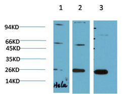
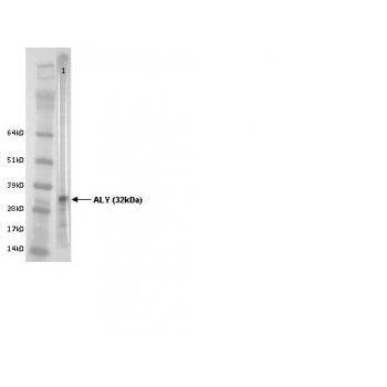 WB (Western Blot)
(Western blot using ALY Antibody (IQ221) and Hela cell lysate)
WB (Western Blot)
(Western blot using ALY Antibody (IQ221) and Hela cell lysate)
ALY/Ref, Monoclonal Antibody (Cat# AAA59541)
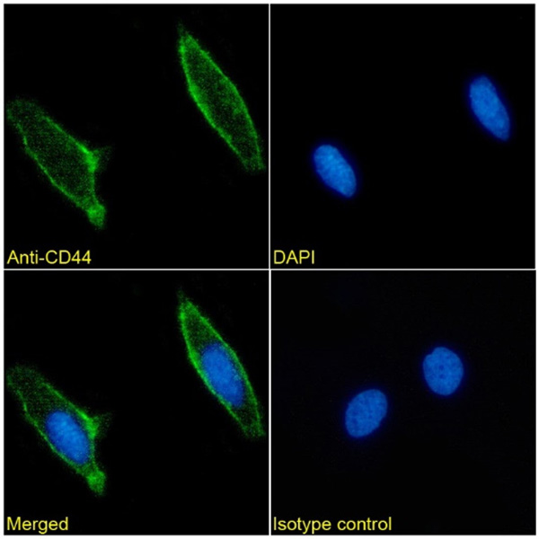
 FCM/FACS (Flow Cytometry)
(Flow cytometry-Anti CD44 Antibody [156-3C11])
FCM/FACS (Flow Cytometry)
(Flow cytometry-Anti CD44 Antibody [156-3C11])
CD44, Monoclonal Antibody (Cat# AAA59549)
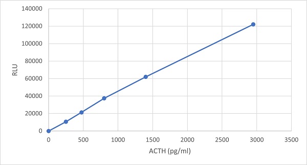
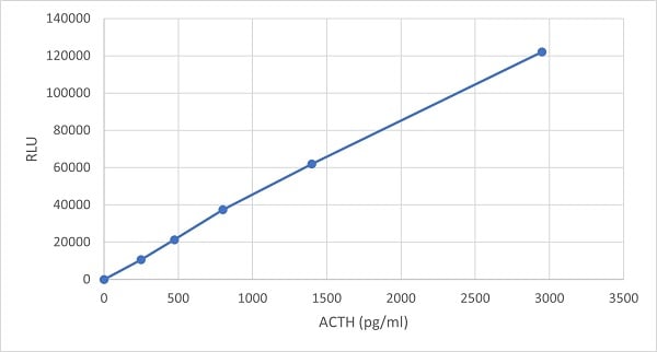 Application Data
(Chemiluminescent immunoassay measuring ACTH levels with Immuquest ACTH specific antibody Clone 6Y8 [57] used as capture antibody)
Application Data
(Chemiluminescent immunoassay measuring ACTH levels with Immuquest ACTH specific antibody Clone 6Y8 [57] used as capture antibody)
ACTH N-Terminal, Monoclonal Antibody (Cat# AAA59585)
CD49d, Monoclonal Antibody (Cat# AAA59596)
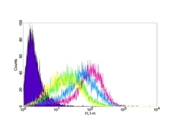
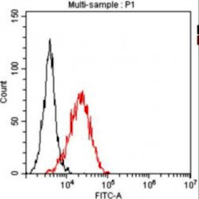 FCM/FACS (Flow Cytometry)
(A typical straining pattern obtained with the clone B-S6 (red line) on T-lymphoma cell line HUT78 compared to the isotype control (black line).)
FCM/FACS (Flow Cytometry)
(A typical straining pattern obtained with the clone B-S6 (red line) on T-lymphoma cell line HUT78 compared to the isotype control (black line).)
CD106, Monoclonal Antibody (Cat# AAA58573)
IL-12/p70, Monoclonal Antibody (Cat# AAA58583)
Vitamin D (25 OH Vitamin D), Monoclonal Antibody (Cat# AAA58288)
COVID 19 (SARS-CoV/SARS-CoV1 & SARS-CoV-2) Nucleocapsid (NP) Coronavirus, Monoclonal Antibody (Cat# AAA58437)
Purification: Protein A chromatography
Trichomonas vaginalis, Monoclonal Antibody (Cat# AAA58456)
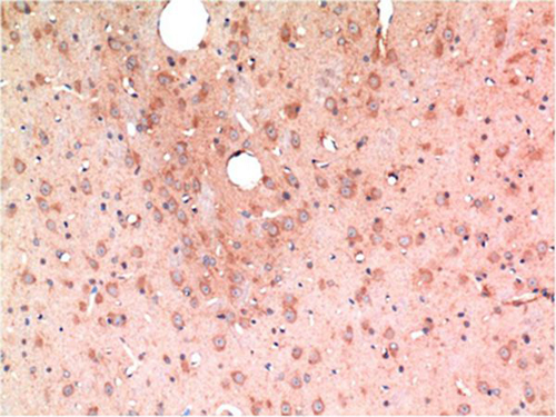
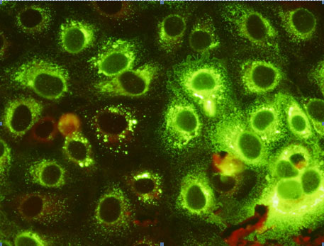 IF (Immunofluorescence)
(Legend: Indirect fluorescent antibody (IFA) image of JEV-infected cells stained with a JEV monoclonal antibody, Cat no. AAA58463 (these antibodies all show the same staining pattern). Cells were fixed with acetone. The JEV monoclonal antibody was used at a concentration of 20 ug/ml PBS and incubated for 30 minutes at room temperature, followed by a secondary FITC-conjugated antibody plus Evan’s Blue Counterstain for 30 minutes at room temperature. Magnification: 400X.)
IF (Immunofluorescence)
(Legend: Indirect fluorescent antibody (IFA) image of JEV-infected cells stained with a JEV monoclonal antibody, Cat no. AAA58463 (these antibodies all show the same staining pattern). Cells were fixed with acetone. The JEV monoclonal antibody was used at a concentration of 20 ug/ml PBS and incubated for 30 minutes at room temperature, followed by a secondary FITC-conjugated antibody plus Evan’s Blue Counterstain for 30 minutes at room temperature. Magnification: 400X.)
Japanese Encephalitis virus - NS1, Monoclonal Antibody (Cat# AAA58463)
MERS Coronavirus, Monoclonal Antibody (Cat# AAA58469)
Purification: Purified from ascites fluid or culture medium by protein A chromatography or sequential differential precipitations.
Gardnerella vaginalis, Monoclonal Antibody (Cat# AAA58491)
Acinetobacter species, Monoclonal Antibody (Cat# AAA58493)
 Application Data
H1 (A/NewCaledonia/20/99)(H1N1), Monoclonal Antibody (Cat# AAA62026)
Tetanus Toxin, Monoclonal Antibody (Cat# AAA62038)
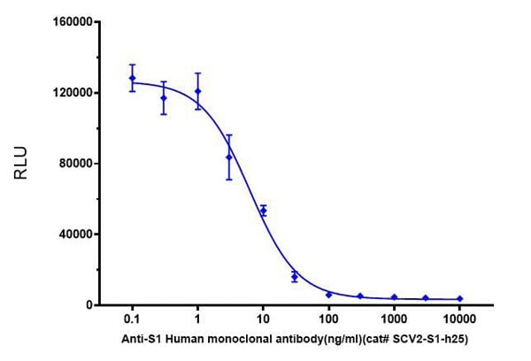 Application Data
(Figure 2. Pseudoviral particle (PP) infection assay challenged by neutralizing antibody (AAA62093). HEK293-ACE2 cells infected with SARS-CoV-2 pseudoviral particles under various amount of neutralizing antibodies)
Application Data
(Figure 2. Pseudoviral particle (PP) infection assay challenged by neutralizing antibody (AAA62093). HEK293-ACE2 cells infected with SARS-CoV-2 pseudoviral particles under various amount of neutralizing antibodies)
COVID 19 Spike S1 IgM Coronavirus, Monoclonal Antibody (Cat# AAA62093)
Kanamycin, Monoclonal Antibody (Cat# AAA81755)

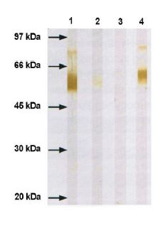
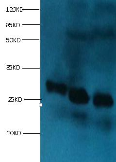
 SDS-PAGE
(PC-3 cells were subjected to SDS PAGE followed by western blot with AAA249063(EGFR Antibody) at dilution of 1:5000)
SDS-PAGE
(PC-3 cells were subjected to SDS PAGE followed by western blot with AAA249063(EGFR Antibody) at dilution of 1:5000)
EGFR, Monoclonal Antibody (Cat# AAA249063)
Protein A+G purified

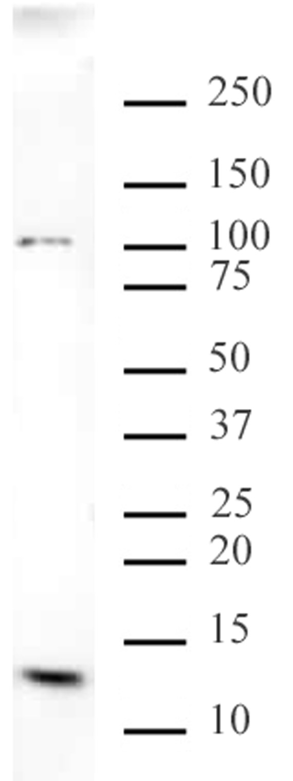
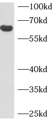
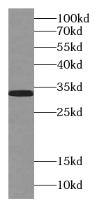
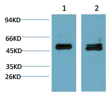
 WB (Western Blot)
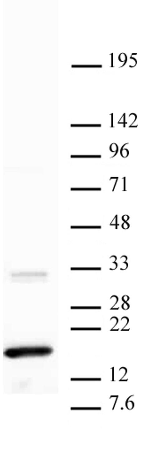
(human brain tissue were subjected to SDS PAGE followed by western blot with AAA248014 (NF-L antibody) at dilution of 1:1000)
WB (Western Blot)
(human brain tissue were subjected to SDS PAGE followed by western blot with AAA248014 (NF-L antibody) at dilution of 1:1000)
NF-L, Monoclonal Antibody (Cat# AAA248014)
Protein A+G purification
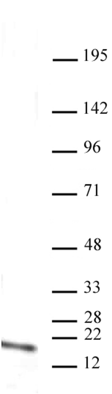
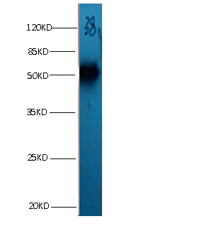
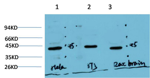
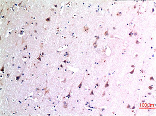
 WB (Western Blot)
(human brain tissue were subjected to SDS PAGE followed by western blot with AAA247941 (PTPRN antibody) at dilution of 1:500)
WB (Western Blot)
(human brain tissue were subjected to SDS PAGE followed by western blot with AAA247941 (PTPRN antibody) at dilution of 1:500)
IA-2/PTPRN, Monoclonal Antibody (Cat# AAA247941)
Protein A+G purification
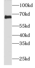
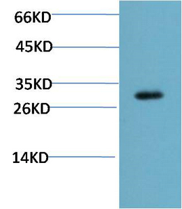
 WB (Western Blot)
(human brain tissue were subjected to SDS PAGE followed by western blot with AAA248099 (SULT4A1 antibody) at dilution of 1:1000)
WB (Western Blot)
(human brain tissue were subjected to SDS PAGE followed by western blot with AAA248099 (SULT4A1 antibody) at dilution of 1:1000)
SULT4A1, Monoclonal Antibody (Cat# AAA248099)
Protein A+G purification
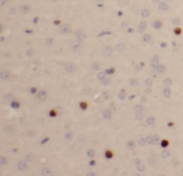
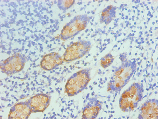
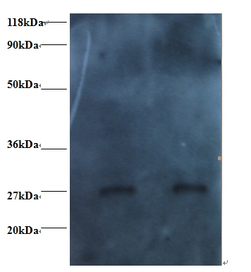
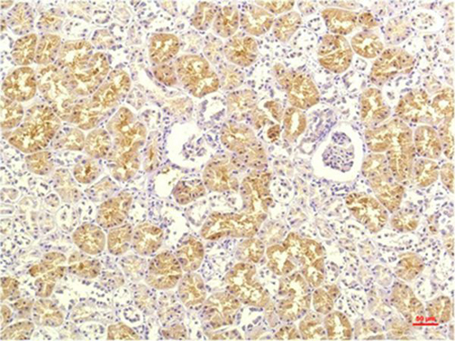
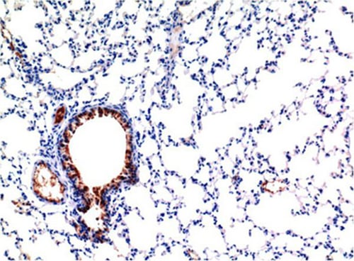
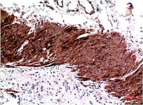
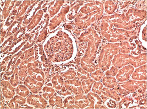
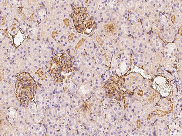 IHC (Immunohistochemisry)
(Immunochemical staining of rat ICAM1 in rat kidney with mouse monoclonal antibody at 1:30 dilution, formalin-fixed paraffin embedded sections.)
IHC (Immunohistochemisry)
(Immunochemical staining of rat ICAM1 in rat kidney with mouse monoclonal antibody at 1:30 dilution, formalin-fixed paraffin embedded sections.)
ICAM-1, Monoclonal Antibody (Cat# AAA257289)
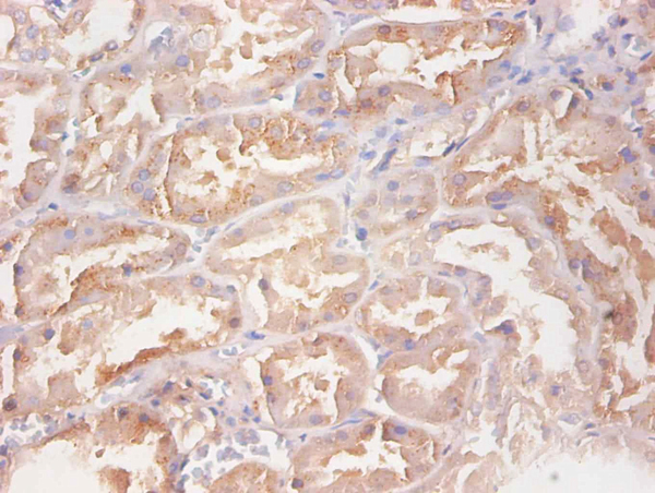
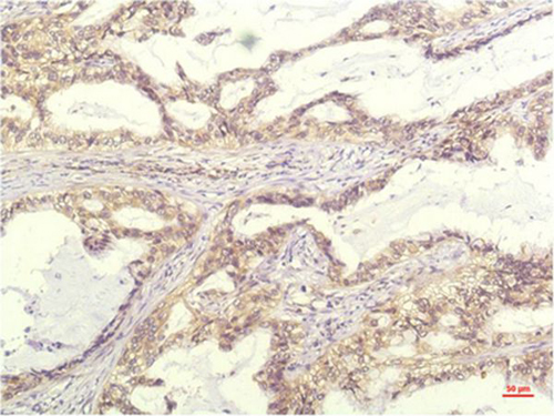
 IHC (Immunohiostchemistry)
(Immunohistochemical analysis of paraffin-embedded human kidney tissue using at dilution of 1:200.)
IHC (Immunohiostchemistry)
(Immunohistochemical analysis of paraffin-embedded human kidney tissue using at dilution of 1:200.)
Cystatin C, Monoclonal Antibody (Cat# AAA308394)

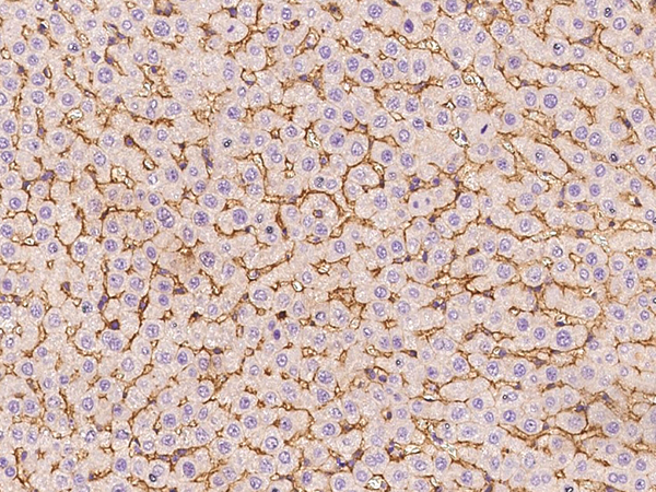
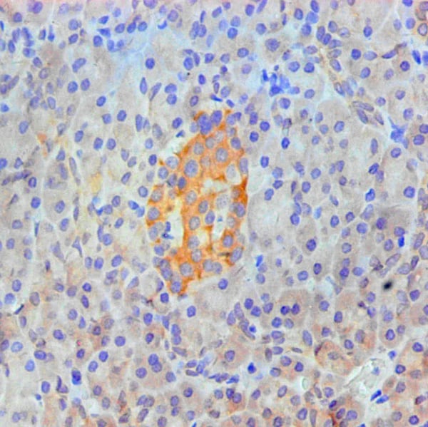
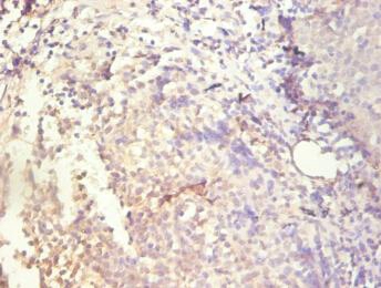
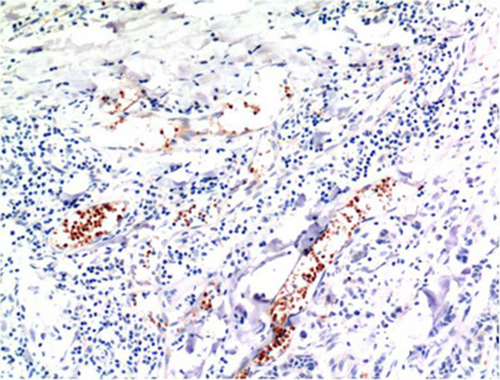
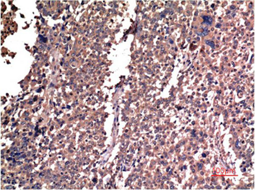
 IHC (Immunohiostchemistry)
(Immunohistochemical analysis of paraffin-embedded human spleen using at dilution of 1:200.)
IHC (Immunohiostchemistry)
(Immunohistochemical analysis of paraffin-embedded human spleen using at dilution of 1:200.)
S100A8, Monoclonal Antibody (Cat# AAA308397)

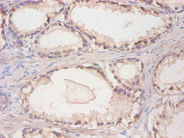
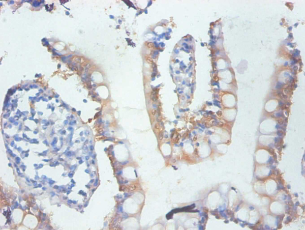
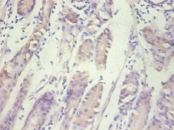
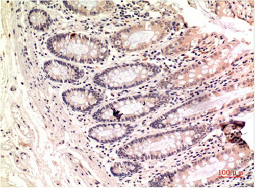
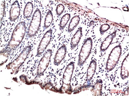
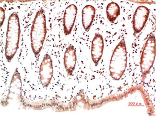
 IHC (Immunohiostchemistry)
(Immunohistochemical analysis of paraffin-embedded human small intestine using at dilution of 1:200.)
IHC (Immunohiostchemistry)
(Immunohistochemical analysis of paraffin-embedded human small intestine using at dilution of 1:200.)
TFF3, Monoclonal Antibody (Cat# AAA308399)
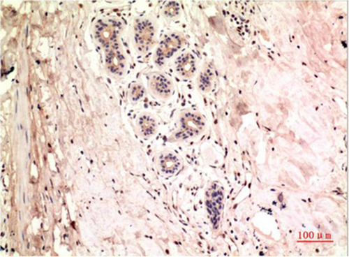
 IHC (Immunohiostchemistry)
(Immunohistochemical analysis of paraffin-embedded human small Intestinal tissue using at dilution of 1:200.)
IHC (Immunohiostchemistry)
(Immunohistochemical analysis of paraffin-embedded human small Intestinal tissue using at dilution of 1:200.)
GAL-3, Monoclonal Antibody (Cat# AAA308404)
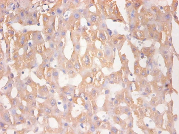
 IHC (Immunohistochemisry)
(Immunohistochemical analysis of paraffin-embedded Human pancreas tissue using at dilution of 1:200.)
IHC (Immunohistochemisry)
(Immunohistochemical analysis of paraffin-embedded Human pancreas tissue using at dilution of 1:200.)
ANXA2, Monoclonal Antibody (Cat# AAA308408)
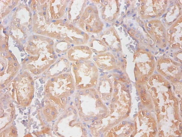
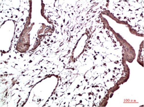
 IHC (Immunohistochemistry)
(Immunohistochemical analysis of paraffin-embedded human nephridial tissue using at dilution of 1:200.)
IHC (Immunohistochemistry)
(Immunohistochemical analysis of paraffin-embedded human nephridial tissue using at dilution of 1:200.)
RBP4, Monoclonal Antibody (Cat# AAA308409)
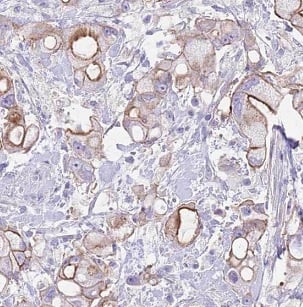
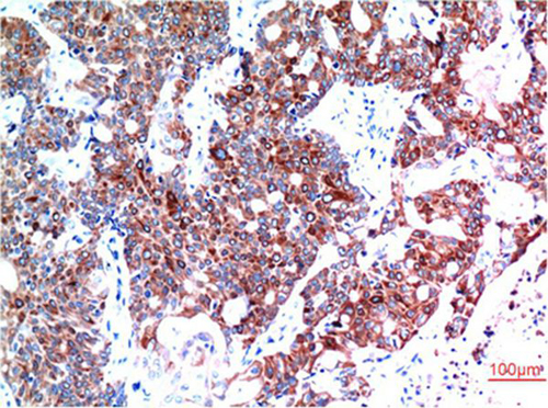
 IHC (Immunohiostchemistry)
(Immunohistochemical analysis of paraffin-embedded human gastric cancer tissue using at dilution of 1:200.)
IHC (Immunohiostchemistry)
(Immunohistochemical analysis of paraffin-embedded human gastric cancer tissue using at dilution of 1:200.)
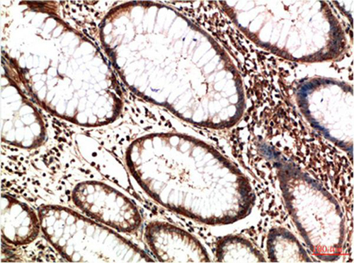
IL8, Monoclonal Antibody (Cat# AAA308410)
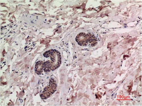
 IHC (Immunohiostchemistry)
(Immunohistochemical analysis of paraffin-embedded Human Kidney Tissue using IL-8 Mouse mAb diluted at 1:200.)
IHC (Immunohiostchemistry)
(Immunohistochemical analysis of paraffin-embedded Human Kidney Tissue using IL-8 Mouse mAb diluted at 1:200.)
IL-8, Monoclonal Antibody (Cat# AAA309476)
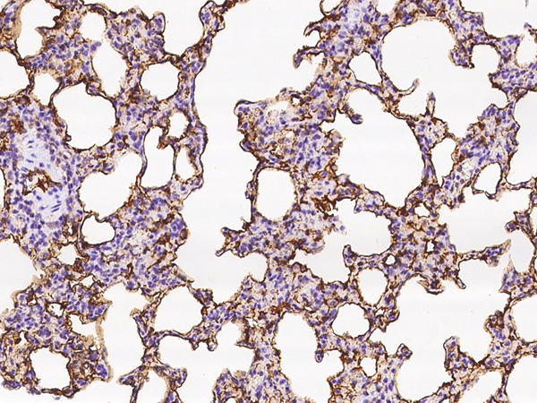
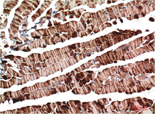
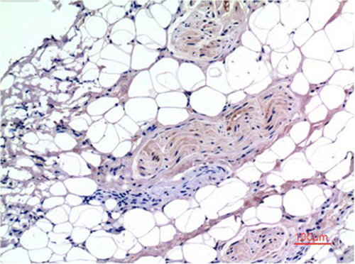
 IHC (Immunohistochemisry)
(Immunohistochemical analysis of paraffin-embedded Mouse Lung Tissue using TGFb1 Mouse mAb diluted at 1:200.)
IHC (Immunohistochemisry)
(Immunohistochemical analysis of paraffin-embedded Mouse Lung Tissue using TGFb1 Mouse mAb diluted at 1:200.)
TGFbeta1, Monoclonal Antibody (Cat# AAA309488)
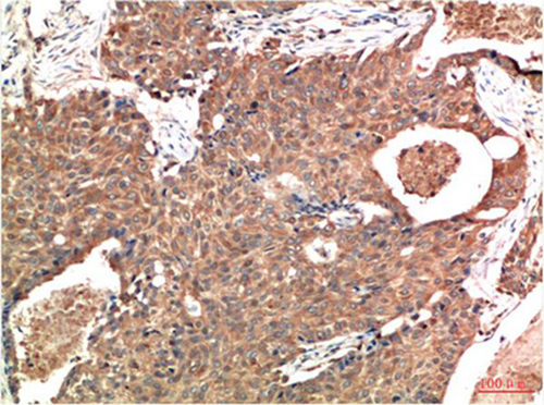
 IHC (Immunohiostchemistry)
(Immunohistochemical analysis of paraffin-embedded Human Breast Carcinoma Tissue using gamma Tutulin Mouse mAb diluted at 1:200.)
IHC (Immunohiostchemistry)
(Immunohistochemical analysis of paraffin-embedded Human Breast Carcinoma Tissue using gamma Tutulin Mouse mAb diluted at 1:200.)
Gamma Tubulin, Monoclonal Antibody (Cat# AAA309496)
 IHC (Immunohistochemisry)
(Immunohistochemical analysis of paraffin-embedded Human Colon Carcinoma Tissue using Muscle Actin Mouse mAb diluted at 1:200.)
IHC (Immunohistochemisry)
(Immunohistochemical analysis of paraffin-embedded Human Colon Carcinoma Tissue using Muscle Actin Mouse mAb diluted at 1:200.)
Muscle actin, Monoclonal Antibody (Cat# AAA309502)
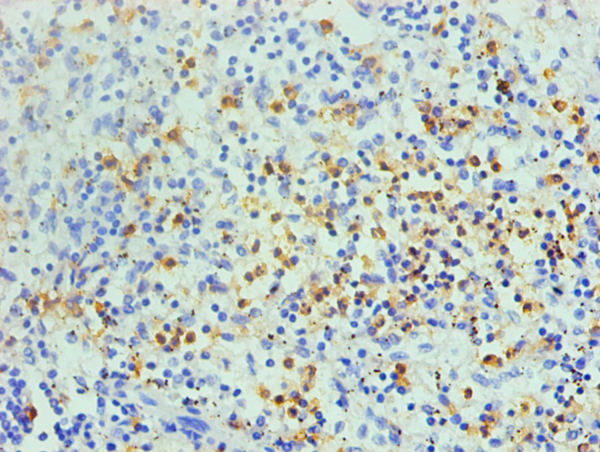
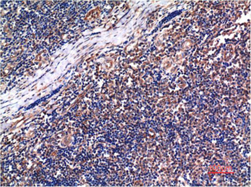 IHC (Immunohiostchemistry)
(Immunohistochemical analysis of paraffin-embedded Human Tonsil Tissue using Cyclin B1 Mouse mAb diluted at 1:200.)
IHC (Immunohiostchemistry)
(Immunohistochemical analysis of paraffin-embedded Human Tonsil Tissue using Cyclin B1 Mouse mAb diluted at 1:200.)
Cyclin B1, Monoclonal Antibody (Cat# AAA309508)
 IHC (Immunohiostchemistry)
(Immunohistochemical analysis of paraffin-embedded Human Breast Carcinoma Tissue using Beclin-1 Mouse mAb diluted at 1:200.)
IHC (Immunohiostchemistry)
(Immunohistochemical analysis of paraffin-embedded Human Breast Carcinoma Tissue using Beclin-1 Mouse mAb diluted at 1:200.)
Beclin-1, Monoclonal Antibody (Cat# AAA309510)
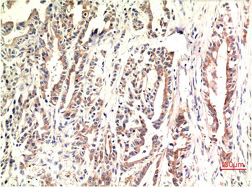
 IHC (Immunohistochemisry)
(Immunohistochemical analysis of paraffin-embedded Human Breast Carcinoma Tissue using Smad3 Mouse mAb diluted at 1:200.)
IHC (Immunohistochemisry)
(Immunohistochemical analysis of paraffin-embedded Human Breast Carcinoma Tissue using Smad3 Mouse mAb diluted at 1:200.)
Smad3, Monoclonal Antibody (Cat# AAA309513)
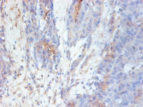
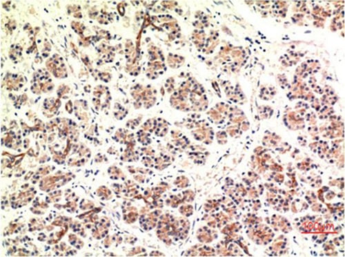 IHC (Immunohistochemisry)
(Immunohistochemical analysis of paraffin-embedded Human Pancreas Carcinoma Tissue using CHOP Mouse mAb diluted at 1:200.)
IHC (Immunohistochemisry)
(Immunohistochemical analysis of paraffin-embedded Human Pancreas Carcinoma Tissue using CHOP Mouse mAb diluted at 1:200.)
CHOP, Monoclonal Antibody (Cat# AAA309515)
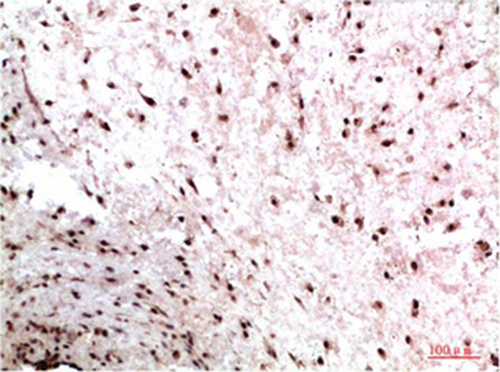
 IHC (Immunohiostchemistry)
(Immunohistochemical analysis of paraffin-embedded Human Breast Carcinoma Tissue using ATM Mouse mAb diluted at 1:200.)
IHC (Immunohiostchemistry)
(Immunohistochemical analysis of paraffin-embedded Human Breast Carcinoma Tissue using ATM Mouse mAb diluted at 1:200.)
ATM, Monoclonal Antibody (Cat# AAA309527)
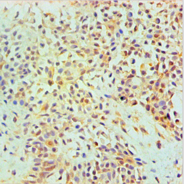
 IHC (Immunohistochemistry)
(Immunohistochemical analysis of paraffin-embedded Human Colon Carcinoma Tissue using HP-1g Mouse mAb diluted at 1:200)
IHC (Immunohistochemistry)
(Immunohistochemical analysis of paraffin-embedded Human Colon Carcinoma Tissue using HP-1g Mouse mAb diluted at 1:200)
HP-1gamma, Monoclonal Antibody (Cat# AAA309531)
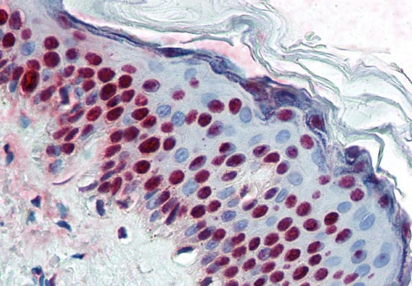
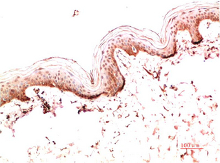
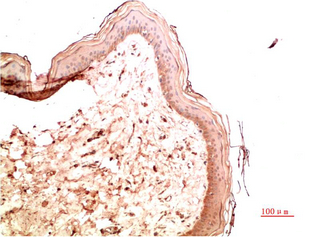 IHC (Immunohiostchemistry)
(Immunohistochemical analysis of paraffin-embedded Human Skin Tissue using Collagen IV Mouse mAb diluted at 1:200.)
IHC (Immunohiostchemistry)
(Immunohistochemical analysis of paraffin-embedded Human Skin Tissue using Collagen IV Mouse mAb diluted at 1:200.)
Collagen IV, Monoclonal Antibody (Cat# AAA309540)
 IHC (Immunohiostchemistry)
(Immunohistochemical analysis of paraffin-embedded Human Skin Tissue using Collagen I Mouse mAb diluted at 1:200.)
IHC (Immunohiostchemistry)
(Immunohistochemical analysis of paraffin-embedded Human Skin Tissue using Collagen I Mouse mAb diluted at 1:200.)
Collagen I, Monoclonal Antibody (Cat# AAA309543)
What are Monoclonal Antibodies?
Monoclonal antibodies are specialized laboratory-produced proteins developed for binding to specific biological antigens or other molecular targets. Since they come from a single cell (or clone), they are especially consistent and accurate in the data they are involved in producing.
This type of antibody material has been shown to be a powerful tool in finding and subsequently destroying harmful cells in an organism, such as those found in cancers or various autoimmune diseases. This makes them excellent aids in medical testing and research, which is why they are so widely used.
AAA Biotech offers a comprehensive range of high-quality monoclonal antibodies that perform effectively in various laboratory tests, including (amongst others) ELISA, western blotting, immunohistochemistry, and flow cytometry. All of the products in our catalog are thoroughly quality tested to make sure that they are reliable and will consistently perform well in your research.
What Are The Uses of Monoclonal Antibodies
Monoclonal antibodies are used in many lab tests, including (amongst others) ELISA, western blotting, immunohistochemistry, and flow cytometry.
ELISA is a test that helps detect a specific substance/analyte in a sample. It uses antibodies (often monoclonal) bound to a solid surface (such as the well of a microplate) to “capture” the substance/analyte in the sample and immobilize it so that the detection antibody component can then bind to it and produce a signal, which can then be measured.
Western blotting identifies specific proteins in a sample. The sample is first separated on a gel, and then antibodies are applied that will typically bind to the target, which will all be localized to a single band in a lane.
Immunohistochemistry helps locate specific proteins in cells or tissue samples using antibodies.
Flow cytometry looks at and sorts cells. It uses antibodies that are conjugated to reporter molecules called “fluorophores”, which, under special lights, emit light themselves, which can then be measured by a detector instrument.
How Monoclonal Antibodies Are Used as Medicine?
Please note that all of the products listed in AAA Biotech’s also known as AAA Bio or AAABio catalog are strictly for research-use only (RUO).
Monoclonal antibodies can also be used as therapeutic/medical treatments, particularly in the context of cancers. They are designed to find and bind to specific cells or proteins, helping the immune system recognize and attack the cancer. These treatments work in different ways, such as:
- Radioimmunotherapy attaches a small amount of radioactive molecule to the antibody, so it delivers the radiation directly to the cancer cells that the antibody is specifically binding to.
- Antibody-directed enzyme prodrug therapy uses antibodies that are specifically bound to special enzymes. These enzymes activate a harmless drug in the body and turn it into a cancer-killing drug only near the cancer cells—this helps avoid harming healthy cells.
- Immunoliposomes are tiny “bubbles” filled with medicine/drug and coated with antibodies. They carry the drug straight to the cancer cells.
Why Buy Monoclonal Antibodies From Us?
At AAA Biotech, we provide high-performance monoclonal antibodies designed to support a wide range of research needs.
1. Validated for Versatile Applications
The antibodies in our catalog are extensively validated and compatible with multiple techniques, including (but not limited to) ELISA, flow cytometry (FC), immunocytochemistry (ICC), immunofluorescence (IF), immunohistochemistry (IHC), immunoprecipitation (IP), and western blotting (WB).
2. Wide Selection & Specialized Options
We offer antibodies for common and rare species, that are available in various conjugated forms, and also in recombinant formats. Essentially, there is almost anything one might need to meet their experimental model’s requirements.
3. High-Quality Proteins
Our proteins meet high purity standards—90% or more as confirmed by SDS-PAGE. Many are available with tags like His, Flag, GST, or MBP, and we also supply native and biologically active proteins for functional studies.
Frequently Asked Questions
1. Are your monoclonal antibodies validated for specific applications?
Yes, our antibodies are tested and validated for use in methods such as ELISA, western blot, IHC, flow cytometry, and more. Refer to specific product pages or datasheets for individual product information.
2. How do I choose the right monoclonal antibody for my application?
Review the product details directly for application validation, species reactivity, and target information. You may also contact our support team at any time for help.
3. How quickly can I receive my order?
Most orders are processed and shipped within 1–3 business days, depending on product availability and your shipping location.