Filters
▼Clonality
▼Type
▼Reactivity
▼Gene Name
▼Isotype
▼Host
▼Application
▼Clone
▼Monoclonal Antibodies
Get accurate results in your research with our Monoclonal Antibodies, which are specially made to target exactly what you require for your research, and will produce consistent, reliable performance in lab tests.
Viewing 9900-9950 of 27597 product results
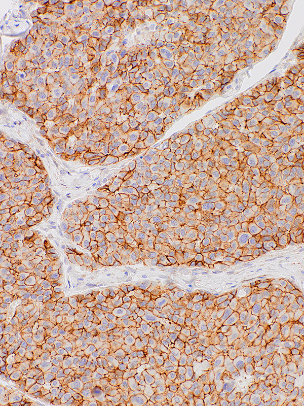
 IHC (Immunohiostchemistry)
Glycophorin A, Monoclonal Antibody (Cat# AAA59220)
 IHC (Immunohiostchemistry)
Myosin, Smooth Muscle, Monoclonal Antibody (Cat# AAA59238)
 IHC (Immunohiostchemistry)
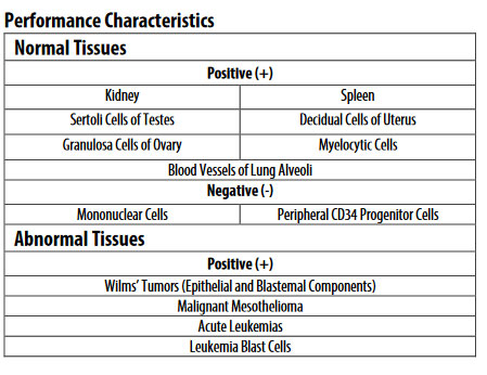 Application Data
(Performance Characteristics)
Application Data
(Performance Characteristics)
WT1, Monoclonal Antibody (Cat# AAA59246)
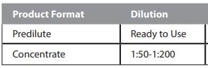 Dilution Info
fFN, Monoclonal Antibody (Cat# AAA59135)
Purification: Protein A or G purified
COVID 19 Spike S1 Coronavirus, Monoclonal Antibody (Cat# AAA58641)
Leptospira, Monoclonal Antibody (Cat# AAA58555)
IL-18Rb, Monoclonal Antibody (Cat# AAA58588)
IL-31, Monoclonal Antibody (Cat# AAA58590)
Canine Parvovirus, Monoclonal Antibody (Cat# AAA58446)
Purification: Ascites Fluid
Trichomonas vaginalis, Monoclonal Antibody (Cat# AAA58457)
West Nile Virus, Monoclonal Antibody (Cat# AAA58459)
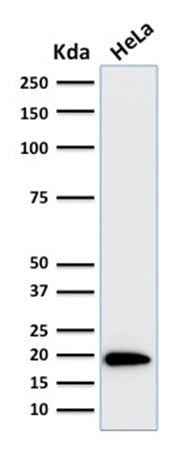
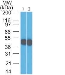 WB (Western Blot)
(Western Blot 1) HeLa and 2) A431 Lysate using Cytokeratin 18 Monoclonal Antibody (SPM265).)
WB (Western Blot)
(Western Blot 1) HeLa and 2) A431 Lysate using Cytokeratin 18 Monoclonal Antibody (SPM265).)
Cytokeratin 18 (KRT18), Monoclonal Antibody (Cat# AAA62608)
Does not react with Mouse, Rat, Sheep, Hamster, Cow, Dog and Pig
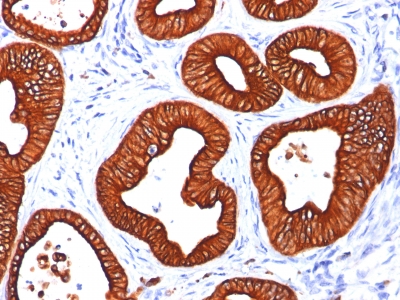
 IHC (Immunohistochemistry)
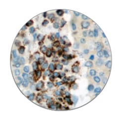
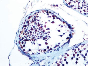
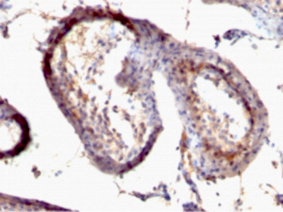
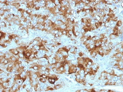
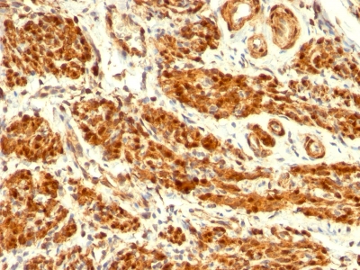
(Formalin-fixed, paraffin-embedded human Testicular Carcinoma stained with TGF alpha Monoclonal Antibody (SPM542))
IHC (Immunohistochemistry)
(Formalin-fixed, paraffin-embedded human Testicular Carcinoma stained with TGF alpha Monoclonal Antibody (SPM542))
TGF-alpha, Monoclonal Antibody (Cat# AAA62628)
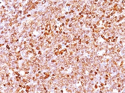
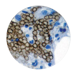
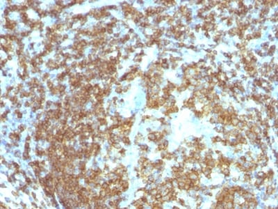
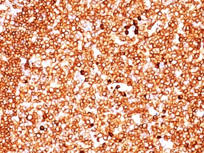 IHC (Immunohiostchemistry)
(Formalin-fixed, paraffin-embedded human Tonsil stained with CD79a Monoclonal Antibody (JCB117 + HM47/A9).)
IHC (Immunohiostchemistry)
(Formalin-fixed, paraffin-embedded human Tonsil stained with CD79a Monoclonal Antibody (JCB117 + HM47/A9).)
CD79a, Monoclonal Antibody (Cat# AAA62662)
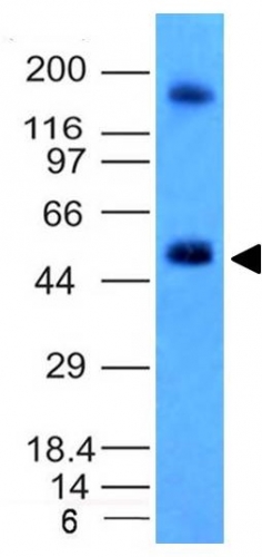 WB (Western Blot)
(Western Blot Analysis of HCT116 Cell Lysate using CAIX Monoclonal Antibody (CA9/781).)
WB (Western Blot)
(Western Blot Analysis of HCT116 Cell Lysate using CAIX Monoclonal Antibody (CA9/781).)
Carbonic Anhydrase IX, Monoclonal Antibody (Cat# AAA62798)
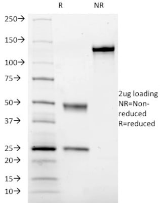 SDS-PAGE
(SDS-PAGE Analysis Purified CD41a Mouse Monoclonal Antibody (ITGA2B/1036). Confirmation of Integrity and Purity of Antibody.)
SDS-PAGE
(SDS-PAGE Analysis Purified CD41a Mouse Monoclonal Antibody (ITGA2B/1036). Confirmation of Integrity and Purity of Antibody.)
CD41a / Integrin alpha2b, Monoclonal Antibody (Cat# AAA62833)
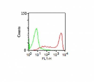 FCM/FACS (Flow Cytometry)
(FACS Analysis human of PBMC using CD45RA Monoclonal Antibody (158-4D3) (red) and isotype control (green).)
FCM/FACS (Flow Cytometry)
(FACS Analysis human of PBMC using CD45RA Monoclonal Antibody (158-4D3) (red) and isotype control (green).)
CD45RA, Monoclonal Antibody (Cat# AAA62550)
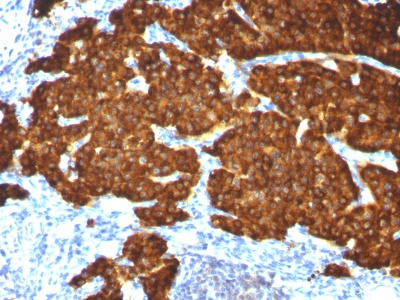
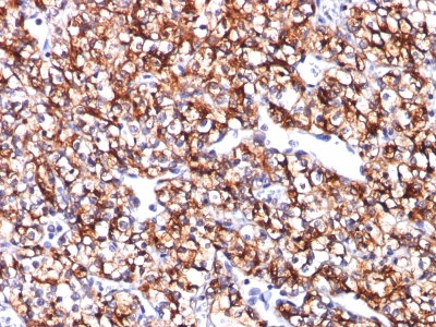
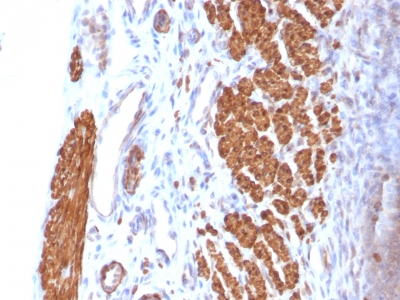
 IHC (Immunohistochemistry)
(Formalin-fixed, paraffin-embedded Adrenal Gland stained with Chromogranin A Monoclonal Antibody (CGA/413+ CHGA/777+ CHGA/798))
IHC (Immunohistochemistry)
(Formalin-fixed, paraffin-embedded Adrenal Gland stained with Chromogranin A Monoclonal Antibody (CGA/413+ CHGA/777+ CHGA/798))
Chromogranin A / CHGA, Monoclonal Antibody (Cat# AAA62853)
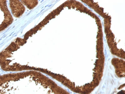
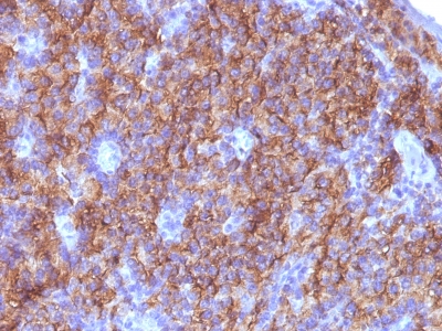
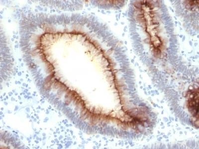
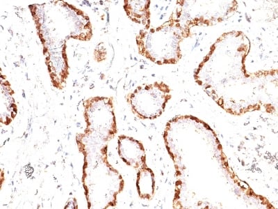
 IHC (Immunohiostchemistry)
(Formalin-fixed, paraffin-embedded human Prostate Carcinoma stained with IDH1 Monoclonal Antibody (IDH1/1152).)
IHC (Immunohiostchemistry)
(Formalin-fixed, paraffin-embedded human Prostate Carcinoma stained with IDH1 Monoclonal Antibody (IDH1/1152).)
IDH1 (Isocitrate Dehydrogenase), Monoclonal Antibody (Cat# AAA62882)
Others not known
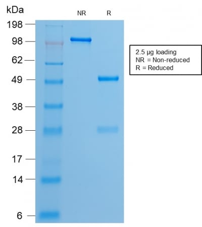
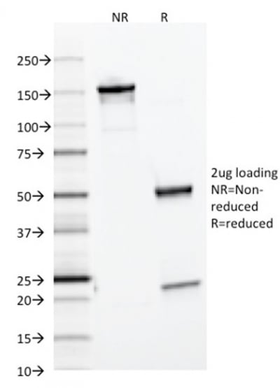
 SDS-PAGE
(SDS-PAGE Analysis of Purified CEA Mouse Recombinant Monoclonal Antibody (rC66/1009).)
SDS-PAGE
(SDS-PAGE Analysis of Purified CEA Mouse Recombinant Monoclonal Antibody (rC66/1009).)
Carcinoembryonic Antigen (CEA)/CD66, Monoclonal Antibody (Cat# AAA62913)
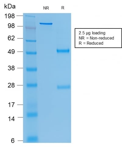
 SDS-PAGE
(SDS-PAGE Analysis of Purified Estrogen Receptor, alpha Rabbit Recombinant Monoclonal Antibody (ESR1/2299R).)
SDS-PAGE
(SDS-PAGE Analysis of Purified Estrogen Receptor, alpha Rabbit Recombinant Monoclonal Antibody (ESR1/2299R).)
Estrogen Receptor, alpha, Monoclonal Antibody (Cat# AAA62970)
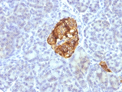
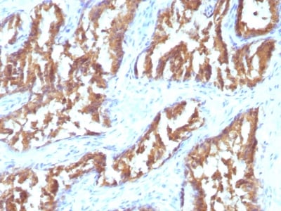
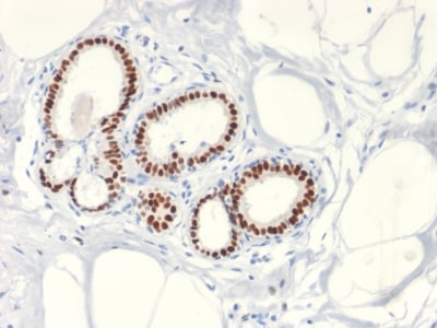
 IHC (Immunohistochemisry)
(Formalin-fixed, paraffin-embedded Human Pancreas stained with Ferritin, Light Chain Monoclonal Antibody (FTL/1386).)
IHC (Immunohistochemisry)
(Formalin-fixed, paraffin-embedded Human Pancreas stained with Ferritin, Light Chain Monoclonal Antibody (FTL/1386).)
Ferritin, Light Chain (FTL), Monoclonal Antibody (Cat# AAA62989)
Chondroitinase generated C-4-S & DS, Monoclonal Antibody (Cat# AAA63025)
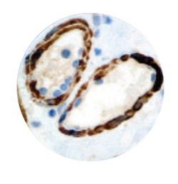
 IHC (Immunohistochemisry)
(Formalin-fixed, paraffin-embedded Rat Uterus stained with Calponin-1 Monoclonal Antibody (CNN1/832).)
IHC (Immunohistochemisry)
(Formalin-fixed, paraffin-embedded Rat Uterus stained with Calponin-1 Monoclonal Antibody (CNN1/832).)
Calponin-1, Monoclonal Antibody (Cat# AAA62692)
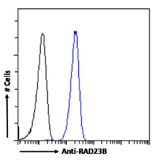
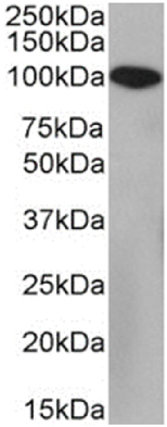
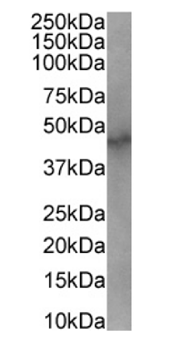 WB (Western Blot)
(Western Blot using anti-RAD23B antibody SAIC-28B-8 (AAA72076). Nuclear lysate of MCF7 cells (35ug protein in RIPA buffer) were resolved on a SDS PAGE gel and blots were probed with the chimeric rabbit version of SAIC-28B-8 () at 2ug/ml before detection using an anti-rabbit secondary antibody. A primary incubation of 1h was used and protein was detected by chemiluminescence.)
WB (Western Blot)
(Western Blot using anti-RAD23B antibody SAIC-28B-8 (AAA72076). Nuclear lysate of MCF7 cells (35ug protein in RIPA buffer) were resolved on a SDS PAGE gel and blots were probed with the chimeric rabbit version of SAIC-28B-8 () at 2ug/ml before detection using an anti-rabbit secondary antibody. A primary incubation of 1h was used and protein was detected by chemiluminescence.)
RAD23B, Monoclonal Antibody (Cat# AAA72076)
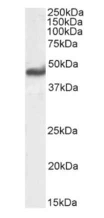
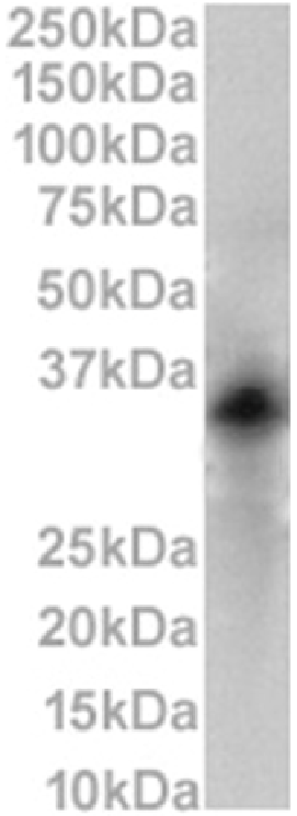
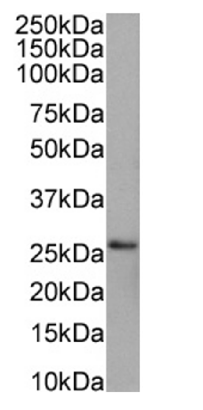 WB (Western Blot)
(Western Blot using anti-PRDX4 antibody SAIC-40C-8 (AAA72081). HEK293 cell lysates (35ug protein in RIPA buffer) were resolved on a SDS PAGE gel and blots were probed with the chimeric rabbit version of SAIC-40C-8 () at 0.001ug/ml before detection using an anti-rabbit secondary antibody. A primary incubation of 1h was used and protein was detected by chemiluminescence.)
WB (Western Blot)
(Western Blot using anti-PRDX4 antibody SAIC-40C-8 (AAA72081). HEK293 cell lysates (35ug protein in RIPA buffer) were resolved on a SDS PAGE gel and blots were probed with the chimeric rabbit version of SAIC-40C-8 () at 0.001ug/ml before detection using an anti-rabbit secondary antibody. A primary incubation of 1h was used and protein was detected by chemiluminescence.)
PRDX4, Monoclonal Antibody (Cat# AAA72081)
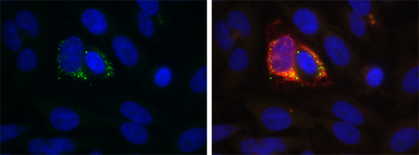 IF (Immunofluorescence)
(Immunofluorescence staining of Zika virus-infected A549 cells with anti-dsRNA antibody 1D3 Immunofluorescence analysis of Zika virus-infected A549 cells stained with the chimeric rabbit IgG version of 1D3 followed by a secondary antibody. The nuclear stain is DAPI (blue). Panels show from left-right: (green) with DAPI and merged channels , DAPI (blue), Zika's E protein (red)). Images are courtesy of Cecilia Alejandra Vazquez (IQUIBICEN (UBA-CONICET)).)
IF (Immunofluorescence)
(Immunofluorescence staining of Zika virus-infected A549 cells with anti-dsRNA antibody 1D3 Immunofluorescence analysis of Zika virus-infected A549 cells stained with the chimeric rabbit IgG version of 1D3 followed by a secondary antibody. The nuclear stain is DAPI (blue). Panels show from left-right: (green) with DAPI and merged channels , DAPI (blue), Zika's E protein (red)). Images are courtesy of Cecilia Alejandra Vazquez (IQUIBICEN (UBA-CONICET)).)
dsRNA, Monoclonal Antibody (Cat# AAA72083)
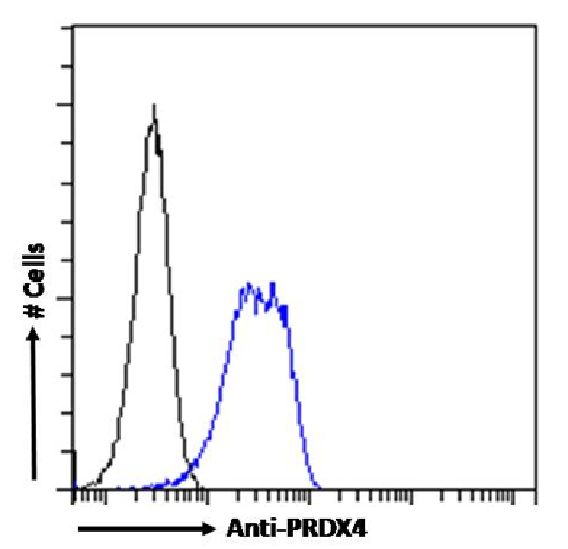
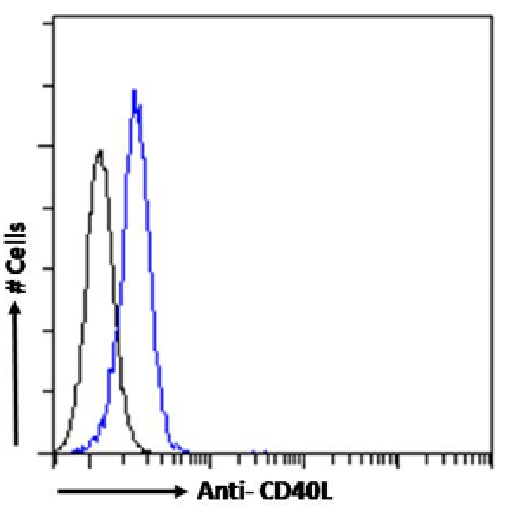 FCM/FACS (Flow Cytometry)
(Flow-cytometry using the anti-CD40L antibody AT161-10 (AAA72091). Human peripheral blood leukocytes were stained with anti-Fluorescein IgG antibody (4-4-20; isotype control, black line) or the rabbit IgG1 version of AT161-10 (, blue line) at a dilution of 1:100 for 1h at RT. After washing, bound antibody was detected using a goat anti-mouse IgG AlexaFluor 488 antibody at a dilution of 1:1000 and cells analyzed using a FACSCanto flow-cytometer.)
FCM/FACS (Flow Cytometry)
(Flow-cytometry using the anti-CD40L antibody AT161-10 (AAA72091). Human peripheral blood leukocytes were stained with anti-Fluorescein IgG antibody (4-4-20; isotype control, black line) or the rabbit IgG1 version of AT161-10 (, blue line) at a dilution of 1:100 for 1h at RT. After washing, bound antibody was detected using a goat anti-mouse IgG AlexaFluor 488 antibody at a dilution of 1:1000 and cells analyzed using a FACSCanto flow-cytometer.)
CD40L, Monoclonal Antibody (Cat# AAA72091)
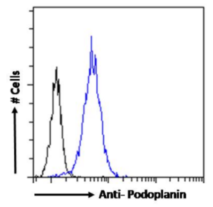 FCM/FACS (Flow Cytometry)
(Flow-cytometry using the anti-Podoplanin (MAP tag) antibody PMab-1 NIH3T3 cells were stained with anti-Fluorescein IgG antibody (4-4-20; isotype control, black line) or the rabbit IgG-chimeric version of PMab-1 at a dilution of 1:100 for 1h at RT. After washing, bound antibody was detected using a goat anti-rabbit IgG AlexaFluor 488 antibody at a dilution of 1:1000 and cells analyzed using a FACSCanto flow-cytometer.)
FCM/FACS (Flow Cytometry)
(Flow-cytometry using the anti-Podoplanin (MAP tag) antibody PMab-1 NIH3T3 cells were stained with anti-Fluorescein IgG antibody (4-4-20; isotype control, black line) or the rabbit IgG-chimeric version of PMab-1 at a dilution of 1:100 for 1h at RT. After washing, bound antibody was detected using a goat anti-rabbit IgG AlexaFluor 488 antibody at a dilution of 1:1000 and cells analyzed using a FACSCanto flow-cytometer.)
Podoplanin, Monoclonal Antibody (Cat# AAA72093)
 FCM/FACS (Flow Cytometry)
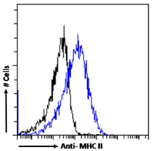
(Flow-cytometry using the anti-MHC II antibody P7/7 (AAA72094). Mouse splenocytes were stained with anti-Fluorescein IgG antibody (4-4-20; isotype control, black line) or the rabbit IgG1 version of P7/7 (, blue line) at a dilution of 1:100 for 1h at RT. After washing, bound antibody was detected using a goat anti-mouse IgG AlexaFluor 488 antibody at a dilution of 1:1000 and cells analyzed using a FACSCanto flow-cytometer.)
FCM/FACS (Flow Cytometry)
(Flow-cytometry using the anti-MHC II antibody P7/7 (AAA72094). Mouse splenocytes were stained with anti-Fluorescein IgG antibody (4-4-20; isotype control, black line) or the rabbit IgG1 version of P7/7 (, blue line) at a dilution of 1:100 for 1h at RT. After washing, bound antibody was detected using a goat anti-mouse IgG AlexaFluor 488 antibody at a dilution of 1:1000 and cells analyzed using a FACSCanto flow-cytometer.)
MHC II, Monoclonal Antibody (Cat# AAA72094)
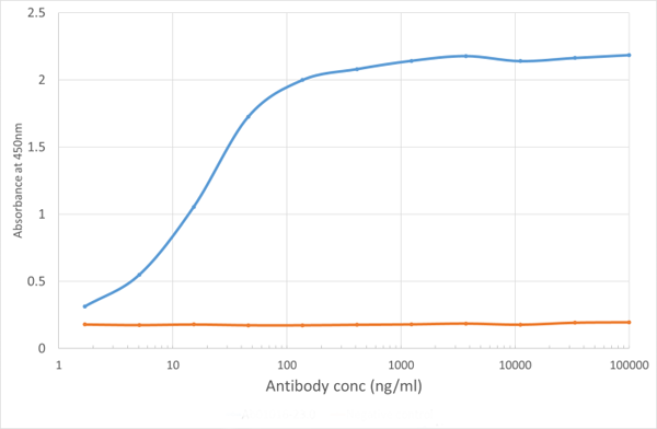 ELISA
(ELISA of anti-VISTA antibody on VISTA-Fc fusion protein. Binding curve of the rabbit chimeric version of the anti-VISTA antibody MH5A to an ELISA plate coated with mouse VISTA-Fc fusion protein (Pr00164-1.9) at a concentration of 5ug/ml. A 3-fold serial dilution from 10000 to 1.7 ng/ml was performed using For signal detection, a 1:4000 dilution of anti-rabbit IgG1 HRP (BioRad) antibody was used.)
ELISA
(ELISA of anti-VISTA antibody on VISTA-Fc fusion protein. Binding curve of the rabbit chimeric version of the anti-VISTA antibody MH5A to an ELISA plate coated with mouse VISTA-Fc fusion protein (Pr00164-1.9) at a concentration of 5ug/ml. A 3-fold serial dilution from 10000 to 1.7 ng/ml was performed using For signal detection, a 1:4000 dilution of anti-rabbit IgG1 HRP (BioRad) antibody was used.)
PD-1H, Monoclonal Antibody (Cat# AAA72098)
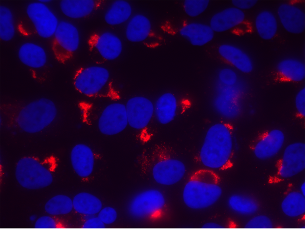
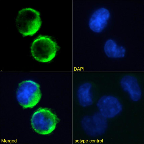
 IF (Immunofluorescence)
(Immunofluorescence staining of fixed RAW264.7 cells with anti-HVEM antibody HMHV-1B18 Immunofluorescence analysis of paraformaldehyde fixed RAW264.7 cells on Shi-fix coverslips stained with the chimeric rabbit IgG version of HMHV-1B18 at 10ug/ml for 1h followed by Alexa Fluor 488 secondary antibody (2ug/ml), showing membrane staining. The nuclear stain is DAPI (blue). Panels show from left-right, top-bottom DAPI, merged channels and an isotype control. The isotype control was an unknown specificity antibody followed by staining with Alexa Fluor 488 secondary antibody.)
IF (Immunofluorescence)
(Immunofluorescence staining of fixed RAW264.7 cells with anti-HVEM antibody HMHV-1B18 Immunofluorescence analysis of paraformaldehyde fixed RAW264.7 cells on Shi-fix coverslips stained with the chimeric rabbit IgG version of HMHV-1B18 at 10ug/ml for 1h followed by Alexa Fluor 488 secondary antibody (2ug/ml), showing membrane staining. The nuclear stain is DAPI (blue). Panels show from left-right, top-bottom DAPI, merged channels and an isotype control. The isotype control was an unknown specificity antibody followed by staining with Alexa Fluor 488 secondary antibody.)
HVEM, Monoclonal Antibody (Cat# AAA72100)
 FCM/FACS (Flow Cytometry)
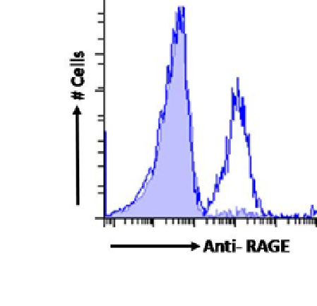
(Flow-cytometry using the Anti-RAGE antibody 2A11 . A549 cells were stained with anti-Fluorescein IgG antibody (4-4-20; isotype control, shaded line) or the rabbit IgG version of 2A11 at a dilution of 1:100 for 1h at RT. After washing, bound antibody was detected using a goat anti-rabbit IgG AlexaFluor 488 antibody at a dilution of 1:1000 and cells analyzed using a FACSCanto flow-cytometer.)
FCM/FACS (Flow Cytometry)
(Flow-cytometry using the Anti-RAGE antibody 2A11 . A549 cells were stained with anti-Fluorescein IgG antibody (4-4-20; isotype control, shaded line) or the rabbit IgG version of 2A11 at a dilution of 1:100 for 1h at RT. After washing, bound antibody was detected using a goat anti-rabbit IgG AlexaFluor 488 antibody at a dilution of 1:1000 and cells analyzed using a FACSCanto flow-cytometer.)
RAGE, Monoclonal Antibody (Cat# AAA72113)


 FCM/FACS (Flow Cytometry)
(Flow-cytometry using the Anti-RAGE antibody 2A11 (AAA72114). A549 cells were stained with anti-Fluorescein IgG antibody (4-4-20; isotype control, shaded line) or the rabbit IgG version of 2A11 (, blue line) at a dilution of 1:100 for 1h at RT. After washing, bound antibody was detected using a goat anti-rabbit IgG AlexaFluor 488 antibody at a dilution of 1:1000 and cells analyzed using a FACSCanto flow-cytometer.)
FCM/FACS (Flow Cytometry)
(Flow-cytometry using the Anti-RAGE antibody 2A11 (AAA72114). A549 cells were stained with anti-Fluorescein IgG antibody (4-4-20; isotype control, shaded line) or the rabbit IgG version of 2A11 (, blue line) at a dilution of 1:100 for 1h at RT. After washing, bound antibody was detected using a goat anti-rabbit IgG AlexaFluor 488 antibody at a dilution of 1:1000 and cells analyzed using a FACSCanto flow-cytometer.)
RAGE, Monoclonal Antibody (Cat# AAA72114)
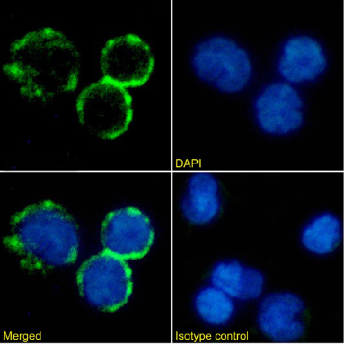
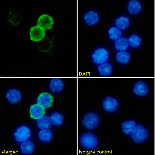
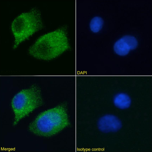
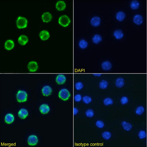 IF (Immunofluorescence)
(Immunofluorescence staining of mouse splenocytes using anti-CD98 antibody RL388. Immunofluorescence analysis of paraformaldehyde fixed mouse splenocytes immobilized on Shi-fix cover-slips and stained with the chimeric rabbit IgG version of RL388 at 10ug/ml followed by Alexa Fluor 488 secondary antibody (2ug/ml), showing membrane staining. The nuclear stain is DAPI (blue). Panels show from left-right, top-bottom DAPI, merged channels and an isotype control. The isotype control was stained with anti-Fluorescein antibody followed by Alexa Fluor 488 secondary antibody.)
IF (Immunofluorescence)
(Immunofluorescence staining of mouse splenocytes using anti-CD98 antibody RL388. Immunofluorescence analysis of paraformaldehyde fixed mouse splenocytes immobilized on Shi-fix cover-slips and stained with the chimeric rabbit IgG version of RL388 at 10ug/ml followed by Alexa Fluor 488 secondary antibody (2ug/ml), showing membrane staining. The nuclear stain is DAPI (blue). Panels show from left-right, top-bottom DAPI, merged channels and an isotype control. The isotype control was stained with anti-Fluorescein antibody followed by Alexa Fluor 488 secondary antibody.)
CD98, Monoclonal Antibody (Cat# AAA72122)
 FCM/FACS (Flow Cytometry)
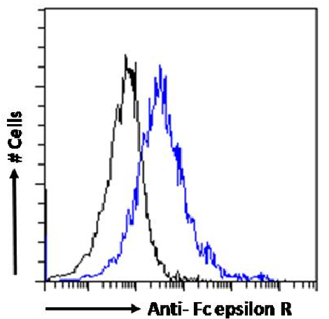
(Flow-cytometry using the Anti-Fc epsilon R antibody IE7 . U937 cells were stained with anti-Fluorescein IgG antibody (4-4-20; isotype control, black line) or the rabbit IgG version of IE7 at a dilution of 1:100 for 1h at RT. After washing, bound antibody was detected using a goat anti-rabbit IgG AlexaFluor 488 antibody at a dilution of 1:1000 and cells analyzed using a FACSCanto flow-cytometer.)
FCM/FACS (Flow Cytometry)
(Flow-cytometry using the Anti-Fc epsilon R antibody IE7 . U937 cells were stained with anti-Fluorescein IgG antibody (4-4-20; isotype control, black line) or the rabbit IgG version of IE7 at a dilution of 1:100 for 1h at RT. After washing, bound antibody was detected using a goat anti-rabbit IgG AlexaFluor 488 antibody at a dilution of 1:1000 and cells analyzed using a FACSCanto flow-cytometer.)
Fc epsilon R, Monoclonal Antibody (Cat# AAA72130)

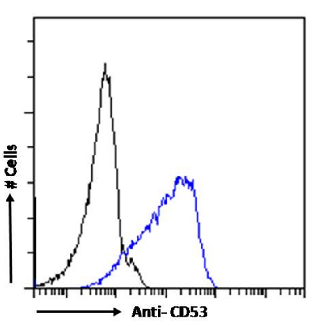 FCM/FACS (Flow Cytometry)
(Flow cytometry using the Anti-CD53 antibody HD77 (AAA72141). Paraformaldehyde fixed Daudi cells were stained with anti-unknown specificity antibody or the rabbit IgG version of HD77 (, blue line) at a dilution of 1:100 for 1h at RT. After washing, the bound antibody was detected using a goat anti-rabbit IgG AlexaFluor 488 antibody at a dilution of 1:1000 and cells analyzed using a FACSCanto flow-cytometer.)
FCM/FACS (Flow Cytometry)
(Flow cytometry using the Anti-CD53 antibody HD77 (AAA72141). Paraformaldehyde fixed Daudi cells were stained with anti-unknown specificity antibody or the rabbit IgG version of HD77 (, blue line) at a dilution of 1:100 for 1h at RT. After washing, the bound antibody was detected using a goat anti-rabbit IgG AlexaFluor 488 antibody at a dilution of 1:1000 and cells analyzed using a FACSCanto flow-cytometer.)
CD53, Monoclonal Antibody (Cat# AAA72141)
 IF (Immunofluorescence)
(Immunofluorescence staining of fixed Daudi cells with anti-CD10 antibody FR4D11 (AAA72152) Immunofluorescence analysis of paraformaldehyde fixed Daudi cells on Shi-fix coverslips stained with the chimeric rabbit IgG version of FR4D11 () at 10ug/ml for 1h followed by Alexa Fluor 488 secondary antibody (2ug/ml), showing membrane staining. The nuclear stain is DAPI (blue). Panels show from left-right, top-bottom , DAPI, merged channels and an isotype control. The isotype control was an unknown specificity antibody followed by staining with Alexa Fluor 488 secondary antibody.)
IF (Immunofluorescence)
(Immunofluorescence staining of fixed Daudi cells with anti-CD10 antibody FR4D11 (AAA72152) Immunofluorescence analysis of paraformaldehyde fixed Daudi cells on Shi-fix coverslips stained with the chimeric rabbit IgG version of FR4D11 () at 10ug/ml for 1h followed by Alexa Fluor 488 secondary antibody (2ug/ml), showing membrane staining. The nuclear stain is DAPI (blue). Panels show from left-right, top-bottom , DAPI, merged channels and an isotype control. The isotype control was an unknown specificity antibody followed by staining with Alexa Fluor 488 secondary antibody.)
CD10, Monoclonal Antibody (Cat# AAA72152)
CD74, Monoclonal Recombinant Antibody (Cat# AAA72010)
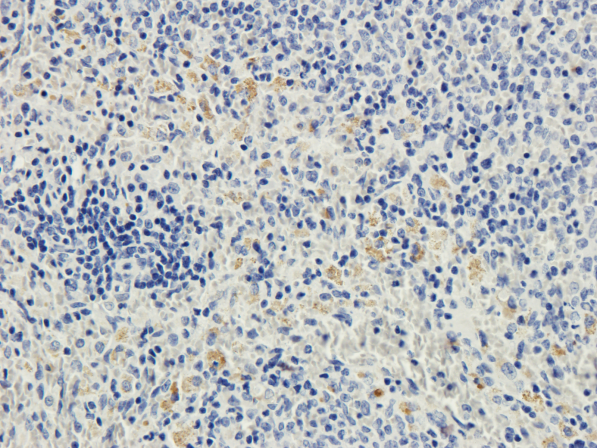 IHC (Immunohiostchemistry)
(Immunohistochemical staining of rat spleen using anti-CD28 antibody Formalin fixed rat spleen slices were were stained at 3 ug/ml.)
IHC (Immunohiostchemistry)
(Immunohistochemical staining of rat spleen using anti-CD28 antibody Formalin fixed rat spleen slices were were stained at 3 ug/ml.)
CD28, Monoclonal Recombinant Antibody (Cat# AAA72022)
 FCM/FACS (Flow Cytometry)
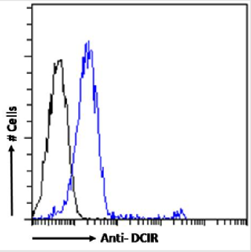
(Flow-cytometry using the anti-DCIR antibody 9E8 or the rabbit IgG1 version of 9E8 at a dilution of 1:100 for 1h at RT. After washing, bound antibody was detected using a goat anti-mouse IgG AlexaFluor 488 antibody at a dilution of 1:1000 and cells analyzed using a FACSCanto flow-cytometer.)
FCM/FACS (Flow Cytometry)
(Flow-cytometry using the anti-DCIR antibody 9E8 or the rabbit IgG1 version of 9E8 at a dilution of 1:100 for 1h at RT. After washing, bound antibody was detected using a goat anti-mouse IgG AlexaFluor 488 antibody at a dilution of 1:1000 and cells analyzed using a FACSCanto flow-cytometer.)
DCIR, Monoclonal Antibody (Cat# AAA72051)
 FCM/FACS (Flow Cytometry)
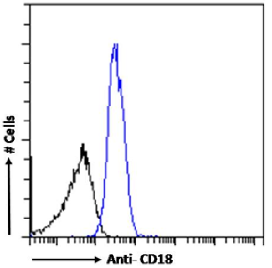
(Flow-cytometry using the anti-CD18 antibody 1B4. Human peripheral blood leukocytes were stained with anti-Fluorescein IgG antibody (4-4-20; isotype control, black line) or the rabbit IgG1 version of 1B4 (blue line) at a dilution of 1:100 for 1h at RT. After washing, bound antibody was detected using a goat anti-mouse IgG AlexaFluor 488 antibody at a dilution of 1:1000 and cells analyzed using a FACSCanto flow-cytometer.)
FCM/FACS (Flow Cytometry)
(Flow-cytometry using the anti-CD18 antibody 1B4. Human peripheral blood leukocytes were stained with anti-Fluorescein IgG antibody (4-4-20; isotype control, black line) or the rabbit IgG1 version of 1B4 (blue line) at a dilution of 1:100 for 1h at RT. After washing, bound antibody was detected using a goat anti-mouse IgG AlexaFluor 488 antibody at a dilution of 1:1000 and cells analyzed using a FACSCanto flow-cytometer.)
CD18, Monoclonal Antibody (Cat# AAA72059)

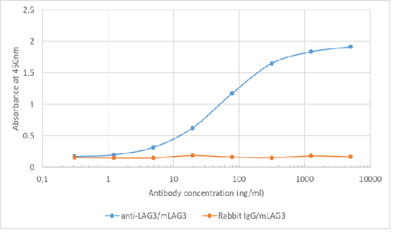 ELISA
(Binding curve of anti-Lag3 antibody C9B7W to recombinant mouse Lag3 Fc-Fusion Protein. ELISA Plate coated with recombinant mouse Lag3 Fc-Fusion Protein at a concentration of 2ug/ml. A 4-fold serial dilution from 5,000ng/ml was performed. For detection, a 1:4000 dilution of HRP-labelled anti-rabbit antibody was used.)
ELISA
(Binding curve of anti-Lag3 antibody C9B7W to recombinant mouse Lag3 Fc-Fusion Protein. ELISA Plate coated with recombinant mouse Lag3 Fc-Fusion Protein at a concentration of 2ug/ml. A 4-fold serial dilution from 5,000ng/ml was performed. For detection, a 1:4000 dilution of HRP-labelled anti-rabbit antibody was used.)
Lag3, Monoclonal Antibody (Cat# AAA72063)
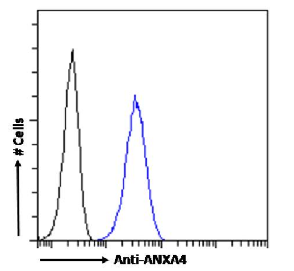
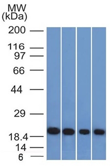
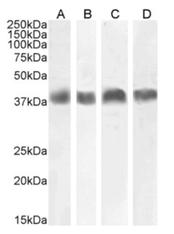
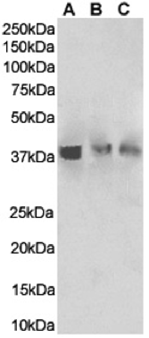
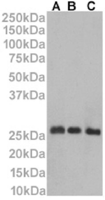
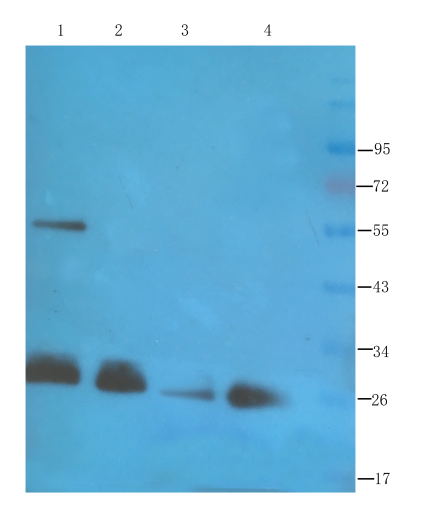
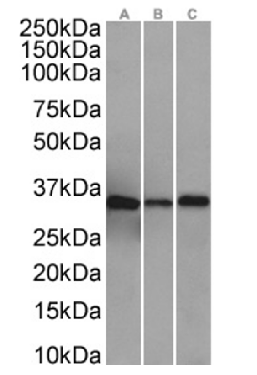 WB (Western Blot)
(Western Blot using anti-ANXA4 antibody SAIC-14C-10F12 (AAA72070). HepG2(A) (0.001ug/ml), A431(B) (0.001ug/ml), and HeLa(C) (0.01ug/ml) cell lysates (35ug protein in RIPA buffer) were resolved on a SDS PAGE gel and blots were probed with the chimeric rabbit version of SAIC-14C-10F12 (), before detection using an anti-rabbit secondary antibody. A primary incubation of 1h was used and protein was detected by chemiluminescence.)
WB (Western Blot)
(Western Blot using anti-ANXA4 antibody SAIC-14C-10F12 (AAA72070). HepG2(A) (0.001ug/ml), A431(B) (0.001ug/ml), and HeLa(C) (0.01ug/ml) cell lysates (35ug protein in RIPA buffer) were resolved on a SDS PAGE gel and blots were probed with the chimeric rabbit version of SAIC-14C-10F12 (), before detection using an anti-rabbit secondary antibody. A primary incubation of 1h was used and protein was detected by chemiluminescence.)
ANXA4, Monoclonal Antibody (Cat# AAA72070)
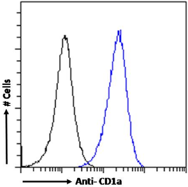 FCM/FACS (Flow Cytometry)
(Flow cytometry using the anti-CD1a antibody CBT6 . Molt4 cells were fixed using 2% PFA and stained with anti-unknown specificity antibody or the rabbit IgG1 version of CBT6 at a dilution of 1:100 for 1h at RT. After washing, the bound antibody was detected using a goat anti-rabbit IgG AlexaFluor 488 antibody at a dilution of 1:1000 and cells analyzed using a FACSCanto flow-cytometer.)
FCM/FACS (Flow Cytometry)
(Flow cytometry using the anti-CD1a antibody CBT6 . Molt4 cells were fixed using 2% PFA and stained with anti-unknown specificity antibody or the rabbit IgG1 version of CBT6 at a dilution of 1:100 for 1h at RT. After washing, the bound antibody was detected using a goat anti-rabbit IgG AlexaFluor 488 antibody at a dilution of 1:1000 and cells analyzed using a FACSCanto flow-cytometer.)
CD1a, Monoclonal Antibody (Cat# AAA72153)

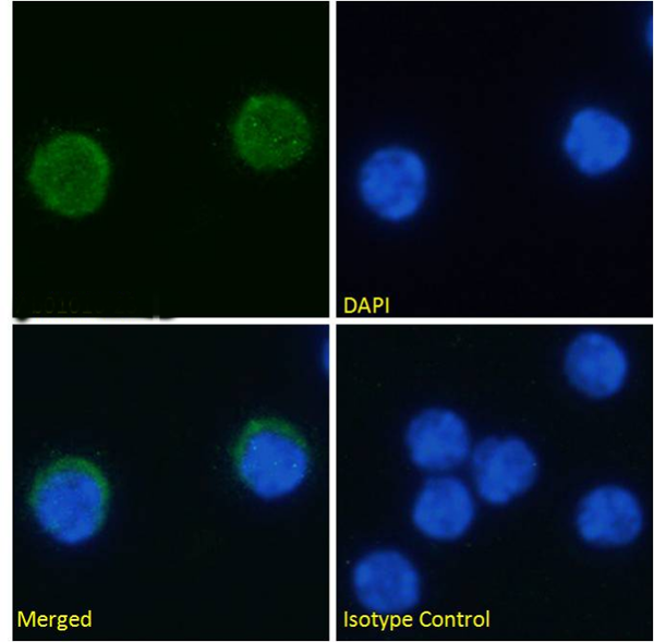
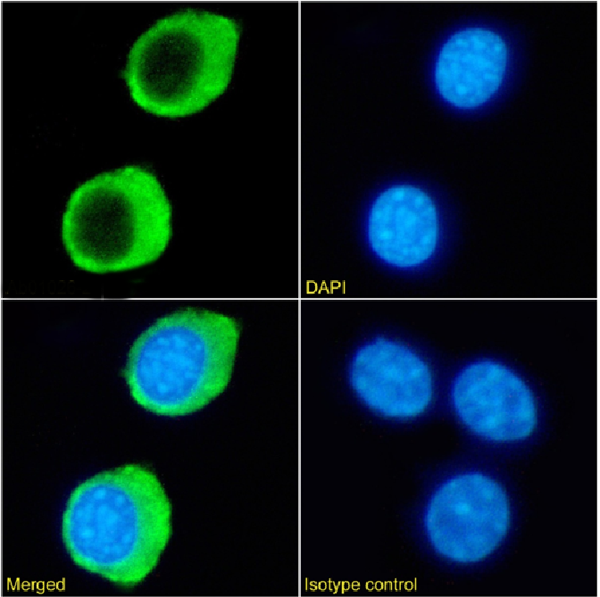
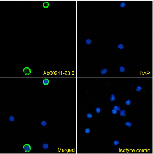
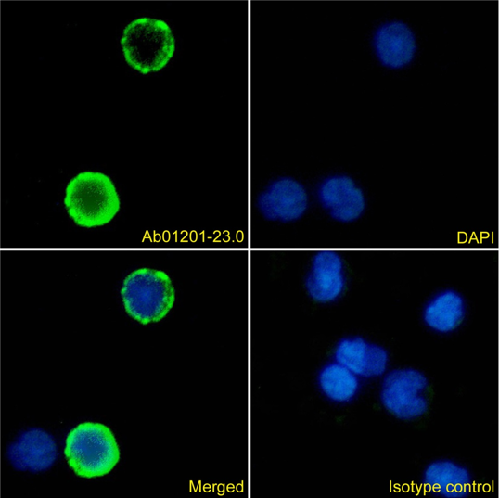
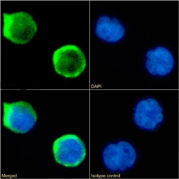
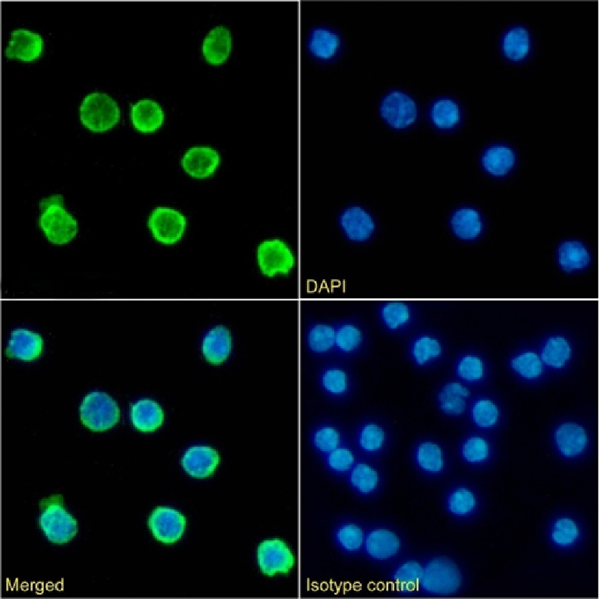 IF (Immunofluorescence)
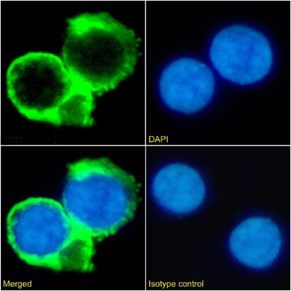
(Immunofluorescence staining of fixed mouse splenocytes with anti-CD79b antibody HM79-16 Immunofluorescence analysis of paraformaldehyde fixed mouse splenocytes on Shi-fix coverslips stained with the chimeric rabbit IgG version HM79-16 () at 10ug/ml for 1h followed by Alexa Fluor 488 secondary antibody (2ug/ml), showing membrane staining. The nuclear stain is DAPI (blue). Panels show from left-right, top-bottom , DAPI, merged channels and an isotype control. The isotype control was an unknown specificity antibody followed by staining with Alexa Fluor 488 secondary antibody.)
IF (Immunofluorescence)
(Immunofluorescence staining of fixed mouse splenocytes with anti-CD79b antibody HM79-16 Immunofluorescence analysis of paraformaldehyde fixed mouse splenocytes on Shi-fix coverslips stained with the chimeric rabbit IgG version HM79-16 () at 10ug/ml for 1h followed by Alexa Fluor 488 secondary antibody (2ug/ml), showing membrane staining. The nuclear stain is DAPI (blue). Panels show from left-right, top-bottom , DAPI, merged channels and an isotype control. The isotype control was an unknown specificity antibody followed by staining with Alexa Fluor 488 secondary antibody.)
CD79b, Monoclonal Antibody (Cat# AAA72170)
What are Monoclonal Antibodies?
Monoclonal antibodies are specialized laboratory-produced proteins developed for binding to specific biological antigens or other molecular targets. Since they come from a single cell (or clone), they are especially consistent and accurate in the data they are involved in producing.
This type of antibody material has been shown to be a powerful tool in finding and subsequently destroying harmful cells in an organism, such as those found in cancers or various autoimmune diseases. This makes them excellent aids in medical testing and research, which is why they are so widely used.
AAA Biotech offers a comprehensive range of high-quality monoclonal antibodies that perform effectively in various laboratory tests, including (amongst others) ELISA, western blotting, immunohistochemistry, and flow cytometry. All of the products in our catalog are thoroughly quality tested to make sure that they are reliable and will consistently perform well in your research.
What Are The Uses of Monoclonal Antibodies
Monoclonal antibodies are used in many lab tests, including (amongst others) ELISA, western blotting, immunohistochemistry, and flow cytometry.
ELISA is a test that helps detect a specific substance/analyte in a sample. It uses antibodies (often monoclonal) bound to a solid surface (such as the well of a microplate) to “capture” the substance/analyte in the sample and immobilize it so that the detection antibody component can then bind to it and produce a signal, which can then be measured.
Western blotting identifies specific proteins in a sample. The sample is first separated on a gel, and then antibodies are applied that will typically bind to the target, which will all be localized to a single band in a lane.
Immunohistochemistry helps locate specific proteins in cells or tissue samples using antibodies.
Flow cytometry looks at and sorts cells. It uses antibodies that are conjugated to reporter molecules called “fluorophores”, which, under special lights, emit light themselves, which can then be measured by a detector instrument.
How Monoclonal Antibodies Are Used as Medicine?
Please note that all of the products listed in AAA Biotech’s also known as AAA Bio or AAABio catalog are strictly for research-use only (RUO).
Monoclonal antibodies can also be used as therapeutic/medical treatments, particularly in the context of cancers. They are designed to find and bind to specific cells or proteins, helping the immune system recognize and attack the cancer. These treatments work in different ways, such as:
- Radioimmunotherapy attaches a small amount of radioactive molecule to the antibody, so it delivers the radiation directly to the cancer cells that the antibody is specifically binding to.
- Antibody-directed enzyme prodrug therapy uses antibodies that are specifically bound to special enzymes. These enzymes activate a harmless drug in the body and turn it into a cancer-killing drug only near the cancer cells—this helps avoid harming healthy cells.
- Immunoliposomes are tiny “bubbles” filled with medicine/drug and coated with antibodies. They carry the drug straight to the cancer cells.
Why Buy Monoclonal Antibodies From Us?
At AAA Biotech, we provide high-performance monoclonal antibodies designed to support a wide range of research needs.
1. Validated for Versatile Applications
The antibodies in our catalog are extensively validated and compatible with multiple techniques, including (but not limited to) ELISA, flow cytometry (FC), immunocytochemistry (ICC), immunofluorescence (IF), immunohistochemistry (IHC), immunoprecipitation (IP), and western blotting (WB).
2. Wide Selection & Specialized Options
We offer antibodies for common and rare species, that are available in various conjugated forms, and also in recombinant formats. Essentially, there is almost anything one might need to meet their experimental model’s requirements.
3. High-Quality Proteins
Our proteins meet high purity standards—90% or more as confirmed by SDS-PAGE. Many are available with tags like His, Flag, GST, or MBP, and we also supply native and biologically active proteins for functional studies.
Frequently Asked Questions
1. Are your monoclonal antibodies validated for specific applications?
Yes, our antibodies are tested and validated for use in methods such as ELISA, western blot, IHC, flow cytometry, and more. Refer to specific product pages or datasheets for individual product information.
2. How do I choose the right monoclonal antibody for my application?
Review the product details directly for application validation, species reactivity, and target information. You may also contact our support team at any time for help.
3. How quickly can I receive my order?
Most orders are processed and shipped within 1–3 business days, depending on product availability and your shipping location.