Filters
▼Clonality
▼Type
▼Reactivity
▼Gene Name
▼Isotype
▼Host
▼Application
▼Clone
▼Monoclonal Antibodies
Get accurate results in your research with our Monoclonal Antibodies, which are specially made to target exactly what you require for your research, and will produce consistent, reliable performance in lab tests.
Viewing 8550-8600 of 27597 product results
Myeloperoxidase, Monoclonal Antibody (Cat# AAA128891)
CD326, Monoclonal Antibody (Cat# AAA129043)
CD133, Monoclonal Antibody (Cat# AAA129053)
CD49d, Monoclonal Antibody (Cat# AAA128396)
CD42b, Monoclonal Antibody (Cat# AAA128413)
HLA-B7, Monoclonal Antibody (Cat# AAA128433)
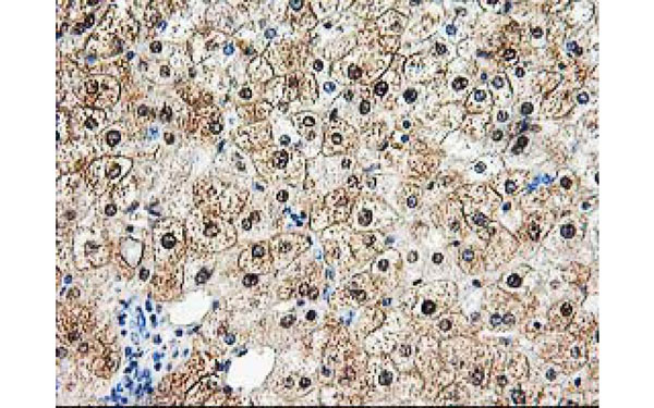
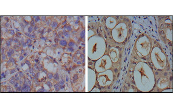
 IHC (Immunohiostchemistry)
(CD14 Antibody-Human Liver: Formalin-Fixed, Paraffin-Embedded (FFPE))
IHC (Immunohiostchemistry)
(CD14 Antibody-Human Liver: Formalin-Fixed, Paraffin-Embedded (FFPE))
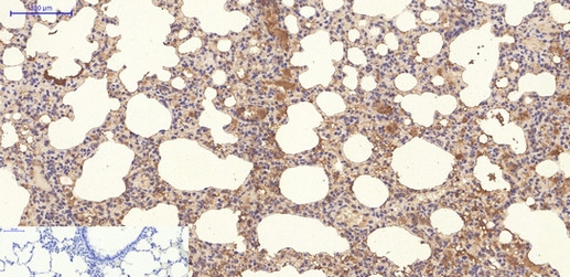
CD14, Monoclonal Antibody (Cat# AAA163089)
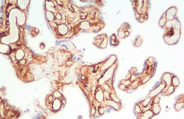
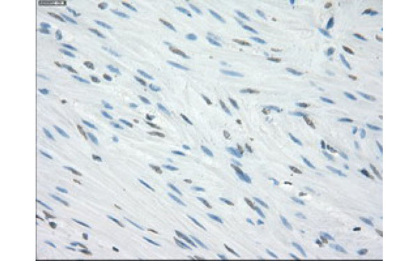
 IHC (Immunohistochemisry)
(Collagen IV Antibody-Human Liver: Formalin-Fixed, Paraffin-Embedded (FFPE))
IHC (Immunohistochemisry)
(Collagen IV Antibody-Human Liver: Formalin-Fixed, Paraffin-Embedded (FFPE))
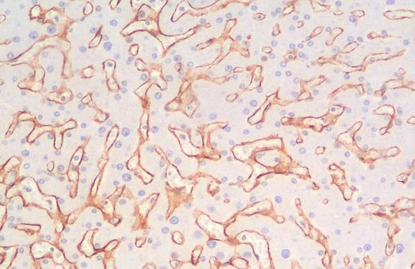
Collagen IV, Monoclonal Antibody (Cat# AAA163091)
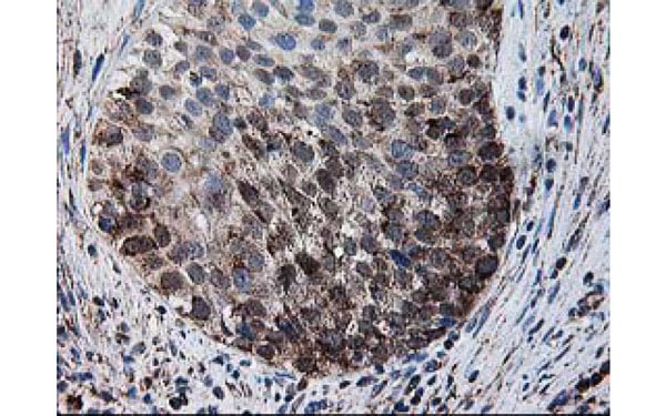
 IHC (Immunohiostchemistry)
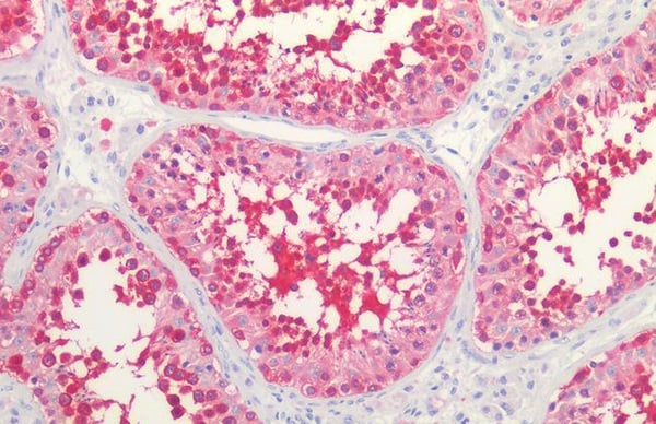
(HSP90/Heat Shock Protein 90 Antibody-Human Testis: Formalin-Fixed, Paraffin-Embedded (FFPE))
IHC (Immunohiostchemistry)
(HSP90/Heat Shock Protein 90 Antibody-Human Testis: Formalin-Fixed, Paraffin-Embedded (FFPE))
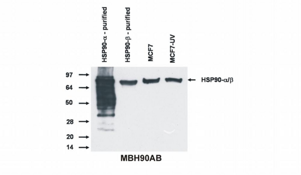
HSP90/Heat Shock Protein 90, Monoclonal Antibody (Cat# AAA163336)
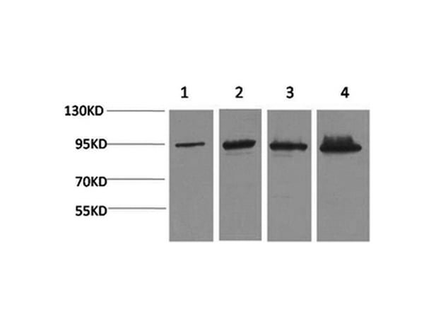
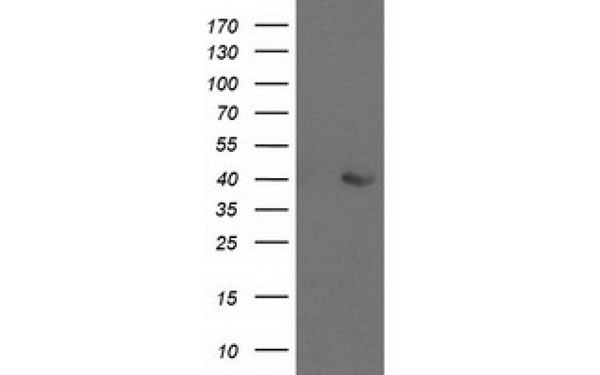
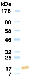
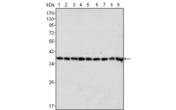
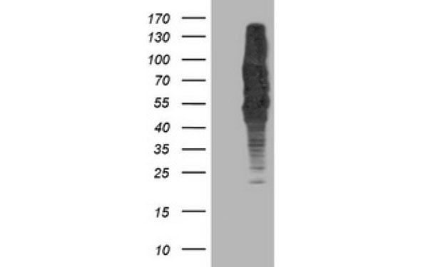
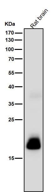 WB (Western Blot)
(All lanes use the Antibody at 1:1K dilution for 1 hour at room temperature.)
WB (Western Blot)
(All lanes use the Antibody at 1:1K dilution for 1 hour at room temperature.)
C Reactive Protein, Monoclonal Antibody (Cat# AAA128113)
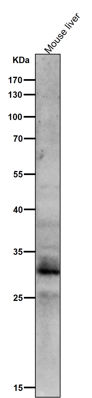
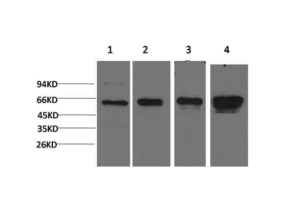
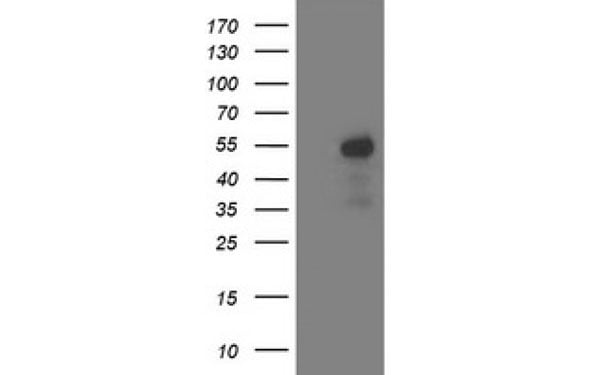
 WB (Western Blot)
(All lanes use the Antibody at 1:1K dilution for 1 hour at room temperature.)
WB (Western Blot)
(All lanes use the Antibody at 1:1K dilution for 1 hour at room temperature.)
MST1/MST2, Monoclonal Antibody (Cat# AAA128143)
HLA-F, Monoclonal Antibody (Cat# AAA128331)
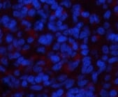
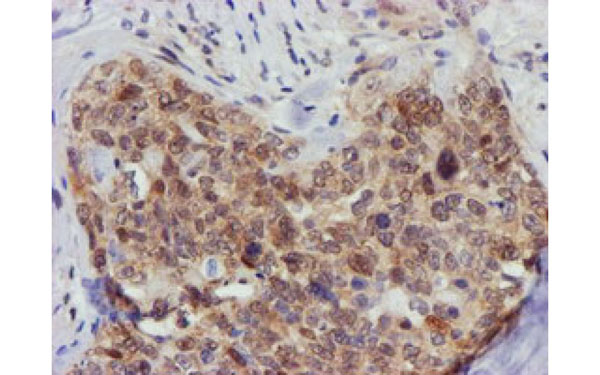
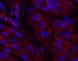 IF (Immunofluorescence)
(Immunofluorescence analysis of Human stomach cancer tissue using Catenin-beta Monoclonal Antibody at dilution of 1:200.)
IF (Immunofluorescence)
(Immunofluorescence analysis of Human stomach cancer tissue using Catenin-beta Monoclonal Antibody at dilution of 1:200.)
Catenin-beta, Monoclonal Antibody (Cat# AAA173651)
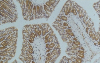 IHC (Immunohiostchemistry)
(Immunohistochemistry of paraffin-embedded Mouse colon tissue using AMPK alpha1 Monoclonal Antibody at dilution of 1:200.)
IHC (Immunohiostchemistry)
(Immunohistochemistry of paraffin-embedded Mouse colon tissue using AMPK alpha1 Monoclonal Antibody at dilution of 1:200.)
AMPK alpha1, Monoclonal Antibody (Cat# AAA173663)
 IF (Immunofluorescence)
(Immunofluorescence analysis of Rat spleen tissue using NBR1 Monoclonal Antibody at dilution of 1:200.)
IF (Immunofluorescence)
(Immunofluorescence analysis of Rat spleen tissue using NBR1 Monoclonal Antibody at dilution of 1:200.)
NBR1, Monoclonal Antibody (Cat# AAA173680)

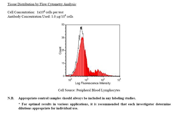
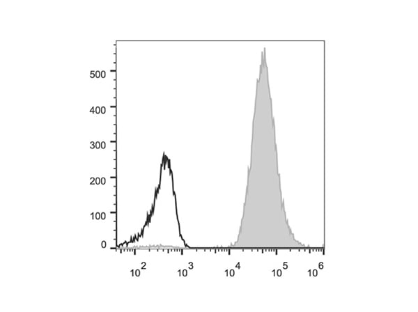 FCM/FACS (Flow Cytometry)
(Human peripheral blood lymphocytes are stained with Anti-Human HLA-A,B,C Monoclonal Antibody(PE Conjugated)(filled gray histogram). Unstained lymphocytes (empty black histogram) are used as control.)
FCM/FACS (Flow Cytometry)
(Human peripheral blood lymphocytes are stained with Anti-Human HLA-A,B,C Monoclonal Antibody(PE Conjugated)(filled gray histogram). Unstained lymphocytes (empty black histogram) are used as control.)
HLA-A,B,C, Monoclonal Antibody (Cat# AAA174661)
Leptin, Monoclonal Antibody (Cat# AAA74613)
IL2Ra, Monoclonal Antibody (Cat# AAA74626)
CD4, Monoclonal Antibody (Cat# AAA74630)
 IHC (Immunohiostchemistry)
(Immunohistochemical analysis of FCGR2A protein in paraffin embedded Human colon tissue using FCGR2A antibody)
IHC (Immunohiostchemistry)
(Immunohistochemical analysis of FCGR2A protein in paraffin embedded Human colon tissue using FCGR2A antibody)
FCGR2A, Monoclonal Antibody (Cat# AAA74687)
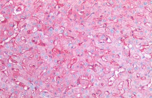
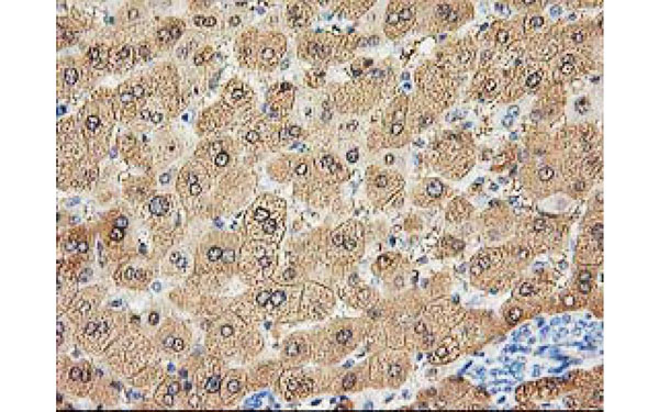 IHC (Immunohiostchemistry)
(Immunohistochemical analysis of ALDOB protein in paraffin embedded Human liver tissue using ALDOB antibody)
IHC (Immunohiostchemistry)
(Immunohistochemical analysis of ALDOB protein in paraffin embedded Human liver tissue using ALDOB antibody)
ALDOB, Monoclonal Antibody (Cat# AAA74724)
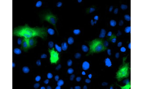
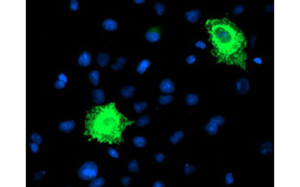
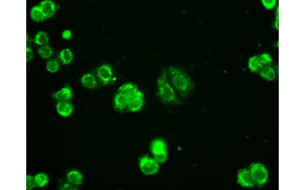 IF (Immunofluorescence)
(Immunofluorescent staining of endogenous AK1 protein in HT29 cells using AK1 antibody)
IF (Immunofluorescence)
(Immunofluorescent staining of endogenous AK1 protein in HT29 cells using AK1 antibody)
AK1, Monoclonal Antibody (Cat# AAA74731)
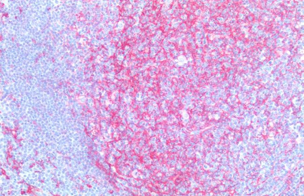
 IHC (Immunohiostchemistry)
(Immunohistochemical analysis of DPP9 protein in paraffin embedded Human lymphoma tissue using DPP9 antibody)
IHC (Immunohiostchemistry)
(Immunohistochemical analysis of DPP9 protein in paraffin embedded Human lymphoma tissue using DPP9 antibody)
DPP9, Monoclonal Antibody (Cat# AAA74746)
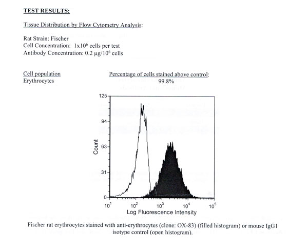
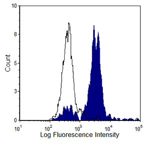 FCM/FACS (Flow Cytometry)
(Human peripheral blood monocytes were stained with anti-CD163 (clone: GHI/61)(filled histogram) or mouse IgG1 isotype control (open histogram).)
FCM/FACS (Flow Cytometry)
(Human peripheral blood monocytes were stained with anti-CD163 (clone: GHI/61)(filled histogram) or mouse IgG1 isotype control (open histogram).)
HLA-ABC, Monoclonal Antibody (Cat# AAA74300)
ApoA-I, Monoclonal Antibody (Cat# AAA74326)
Apo A-II: 0%
Apo B: 0%
ApoA-I antibody was purified by Protein A chromatography.
HBcAg, Monoclonal Antibody (Cat# AAA74529)
HSV1 + HSV2 gB, Monoclonal Antibody (Cat# AAA74559)
EPO, Monoclonal Antibody (Cat# AAA74421)
CD16, Monoclonal Antibody (Cat# AAA74490)
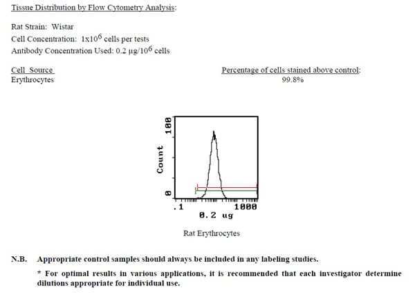
 Application Data
Erythrocytes, Monoclonal Antibody (Cat# AAA74173)
H-2Kd (private; H-2.m31), Monoclonal Antibody (Cat# AAA74180)
 Application Data
HLA-DR, Monoclonal Antibody (Cat# AAA74185)
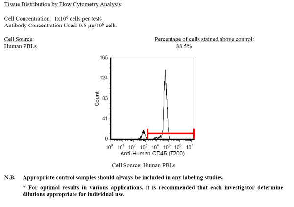 Application Data
CD45 (T200), Monoclonal Antibody (Cat# AAA74197)
 Application Data
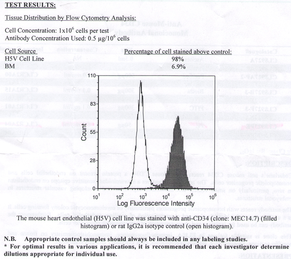
CD34, Monoclonal Antibody (Cat# AAA74209)
 Application Data
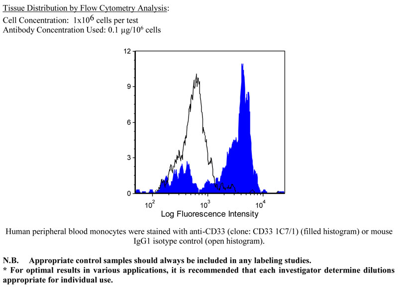
CD33, Monoclonal Antibody (Cat# AAA74217)
HLA-DOB, Monoclonal Antibody (Cat# AAA74218)
NUCLEAR ANTIGEN, Monoclonal Antibody (Cat# AAA49137)
N.B. Antibody reactivity and working conditions may vary between species.
IgE, Monoclonal Antibody (Cat# AAA49156)
FOLATE BINDING PROTEIN, Monoclonal Antibody (Cat# AAA49159)
CORTISOL, Monoclonal Antibody (Cat# AAA49174)
RESPIRATORY SYNCYTIAL VIRUS MAJOR SURFACE GLYCOPROTEIN G, Monoclonal Antibody (Cat# AAA49208)
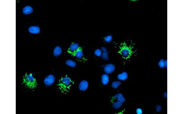
 IF (Immunofluorescence)
(Immunofluorescent staining of COS7 cells transiently transfected with recombinant CAPN9 protein using CAPN9 antibody)
IF (Immunofluorescence)
(Immunofluorescent staining of COS7 cells transiently transfected with recombinant CAPN9 protein using CAPN9 antibody)
CAPN9, Monoclonal Antibody (Cat# AAA74758)
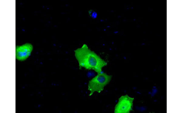 IF (Immunofluorescence)
(Immunofluorescent staining of COS7 cells transiently transfected with recombinant BTK protein using BTK antibody)
IF (Immunofluorescence)
(Immunofluorescent staining of COS7 cells transiently transfected with recombinant BTK protein using BTK antibody)
BTK, Monoclonal Antibody (Cat# AAA74767)
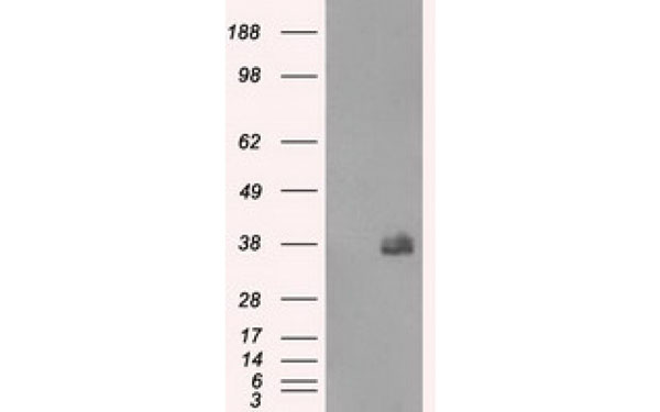
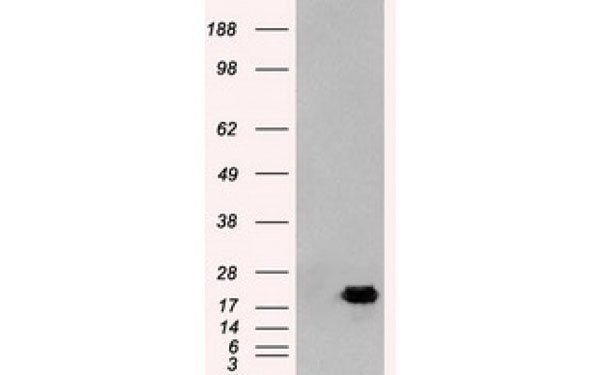
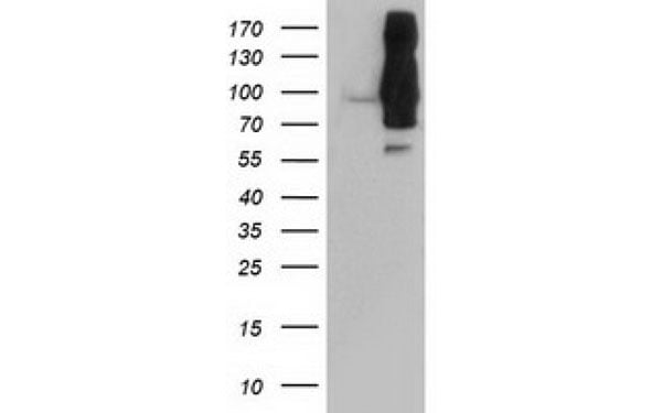
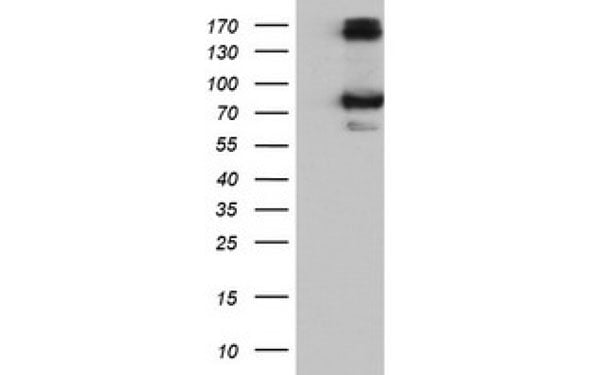
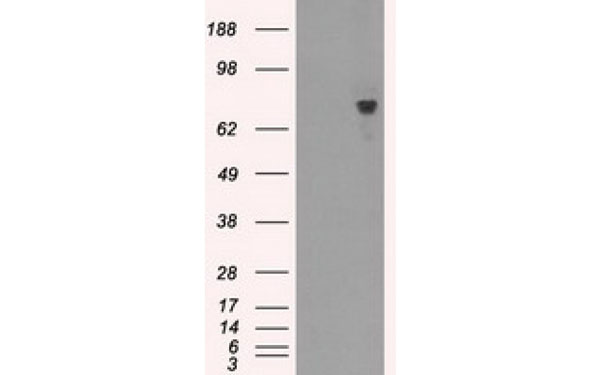
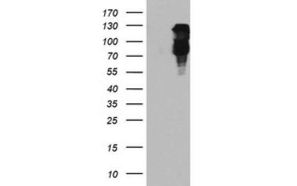
 WB (Western Blot)
(Western Blot analysis using Endostatin antibody)
WB (Western Blot)
(Western Blot analysis using Endostatin antibody)
Endostatin, Monoclonal Antibody (Cat# AAA74771)
 IHC (Immunohiostchemistry)
(Immunohistochemical analysis of ACY1 protein in paraffin embedded Human liver tissue using ACY1 antibody)
IHC (Immunohiostchemistry)
(Immunohistochemical analysis of ACY1 protein in paraffin embedded Human liver tissue using ACY1 antibody)
ACY1, Monoclonal Antibody (Cat# AAA74773)
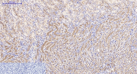
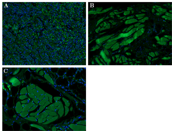
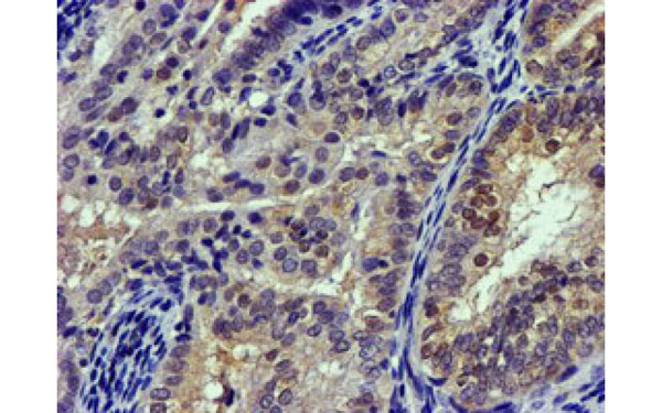
 IHC (Immunohiostchemistry)
(Immunohistochemical analysis of paraffin-embedded human breast carcinoma (left) and kidney carcinoma (right), showing cytoplasmic localization using GAPDH antibody with DAB staining.)
IHC (Immunohiostchemistry)
(Immunohistochemical analysis of paraffin-embedded human breast carcinoma (left) and kidney carcinoma (right), showing cytoplasmic localization using GAPDH antibody with DAB staining.)
GAPDH, Monoclonal Antibody (Cat# AAA74792)
 IF (Immunofluorescence)
(Immunofluorescent staining of COS7 cells transiently transfected with recombinant DLD protein using DLD antibody)
IF (Immunofluorescence)
(Immunofluorescent staining of COS7 cells transiently transfected with recombinant DLD protein using DLD antibody)
DLD, Monoclonal Antibody (Cat# AAA74821)
 IHC (Immunohiostchemistry)
(Immunohistochemical analysis of EPM2AIP1 protein in paraffin embedded Adenocarcinoma of Human breast tissue using EPM2AIP1 antibody)
IHC (Immunohiostchemistry)
(Immunohistochemical analysis of EPM2AIP1 protein in paraffin embedded Adenocarcinoma of Human breast tissue using EPM2AIP1 antibody)
EPM2AIP1, Monoclonal Antibody (Cat# AAA74860)
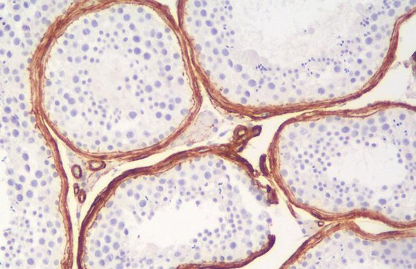
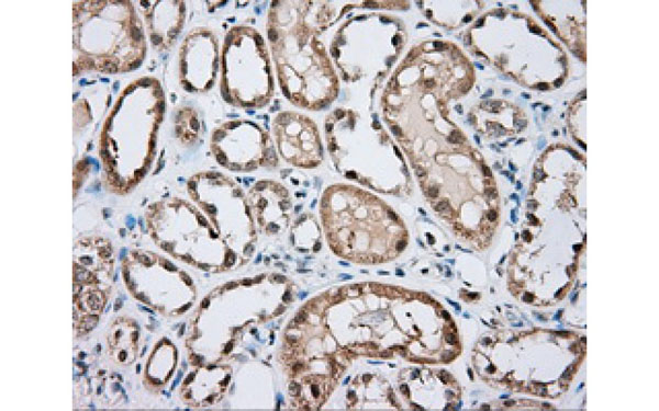
 IHC (Immunohiostchemistry)
(Immunohistochemical analysis of paraffin-embedded human salivary gland tissues (left) and kidney tissues (right) using HK1 antibody with DAB staining.)
IHC (Immunohiostchemistry)
(Immunohistochemical analysis of paraffin-embedded human salivary gland tissues (left) and kidney tissues (right) using HK1 antibody with DAB staining.)
HK1, Monoclonal Antibody (Cat# AAA74870)
 IF (Immunofluorescence)
(Immunofluorescent staining of COS7 cells transiently transfected with recombinant FBXO21 protein using FBXO21 antibody)
IF (Immunofluorescence)
(Immunofluorescent staining of COS7 cells transiently transfected with recombinant FBXO21 protein using FBXO21 antibody)
FBXO21, Monoclonal Antibody (Cat# AAA74877)
What are Monoclonal Antibodies?
Monoclonal antibodies are specialized laboratory-produced proteins developed for binding to specific biological antigens or other molecular targets. Since they come from a single cell (or clone), they are especially consistent and accurate in the data they are involved in producing.
This type of antibody material has been shown to be a powerful tool in finding and subsequently destroying harmful cells in an organism, such as those found in cancers or various autoimmune diseases. This makes them excellent aids in medical testing and research, which is why they are so widely used.
AAA Biotech offers a comprehensive range of high-quality monoclonal antibodies that perform effectively in various laboratory tests, including (amongst others) ELISA, western blotting, immunohistochemistry, and flow cytometry. All of the products in our catalog are thoroughly quality tested to make sure that they are reliable and will consistently perform well in your research.
What Are The Uses of Monoclonal Antibodies
Monoclonal antibodies are used in many lab tests, including (amongst others) ELISA, western blotting, immunohistochemistry, and flow cytometry.
ELISA is a test that helps detect a specific substance/analyte in a sample. It uses antibodies (often monoclonal) bound to a solid surface (such as the well of a microplate) to “capture” the substance/analyte in the sample and immobilize it so that the detection antibody component can then bind to it and produce a signal, which can then be measured.
Western blotting identifies specific proteins in a sample. The sample is first separated on a gel, and then antibodies are applied that will typically bind to the target, which will all be localized to a single band in a lane.
Immunohistochemistry helps locate specific proteins in cells or tissue samples using antibodies.
Flow cytometry looks at and sorts cells. It uses antibodies that are conjugated to reporter molecules called “fluorophores”, which, under special lights, emit light themselves, which can then be measured by a detector instrument.
How Monoclonal Antibodies Are Used as Medicine?
Please note that all of the products listed in AAA Biotech’s also known as AAA Bio or AAABio catalog are strictly for research-use only (RUO).
Monoclonal antibodies can also be used as therapeutic/medical treatments, particularly in the context of cancers. They are designed to find and bind to specific cells or proteins, helping the immune system recognize and attack the cancer. These treatments work in different ways, such as:
- Radioimmunotherapy attaches a small amount of radioactive molecule to the antibody, so it delivers the radiation directly to the cancer cells that the antibody is specifically binding to.
- Antibody-directed enzyme prodrug therapy uses antibodies that are specifically bound to special enzymes. These enzymes activate a harmless drug in the body and turn it into a cancer-killing drug only near the cancer cells—this helps avoid harming healthy cells.
- Immunoliposomes are tiny “bubbles” filled with medicine/drug and coated with antibodies. They carry the drug straight to the cancer cells.
Why Buy Monoclonal Antibodies From Us?
At AAA Biotech, we provide high-performance monoclonal antibodies designed to support a wide range of research needs.
1. Validated for Versatile Applications
The antibodies in our catalog are extensively validated and compatible with multiple techniques, including (but not limited to) ELISA, flow cytometry (FC), immunocytochemistry (ICC), immunofluorescence (IF), immunohistochemistry (IHC), immunoprecipitation (IP), and western blotting (WB).
2. Wide Selection & Specialized Options
We offer antibodies for common and rare species, that are available in various conjugated forms, and also in recombinant formats. Essentially, there is almost anything one might need to meet their experimental model’s requirements.
3. High-Quality Proteins
Our proteins meet high purity standards—90% or more as confirmed by SDS-PAGE. Many are available with tags like His, Flag, GST, or MBP, and we also supply native and biologically active proteins for functional studies.
Frequently Asked Questions
1. Are your monoclonal antibodies validated for specific applications?
Yes, our antibodies are tested and validated for use in methods such as ELISA, western blot, IHC, flow cytometry, and more. Refer to specific product pages or datasheets for individual product information.
2. How do I choose the right monoclonal antibody for my application?
Review the product details directly for application validation, species reactivity, and target information. You may also contact our support team at any time for help.
3. How quickly can I receive my order?
Most orders are processed and shipped within 1–3 business days, depending on product availability and your shipping location.