Filters
▼Clonality
▼Type
▼Reactivity
▼Gene Name
▼Isotype
▼Host
▼Application
▼Clone
▼Monoclonal Antibodies
Get accurate results in your research with our Monoclonal Antibodies, which are specially made to target exactly what you require for your research, and will produce consistent, reliable performance in lab tests.
Viewing 8350-8400 of 27597 product results
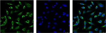 IF (Immunofluorescence)
(IF analysis of Hela with (Left) and DAPI (Right) diluted at 1:100.)
IF (Immunofluorescence)
(IF analysis of Hela with (Left) and DAPI (Right) diluted at 1:100.)
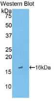
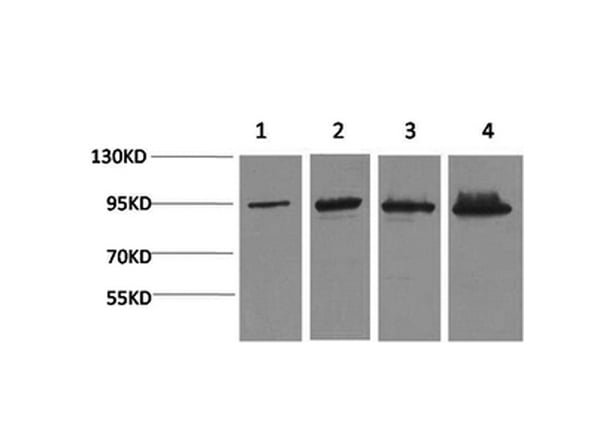
Peroxiredoxin 1, Monoclonal Antibody (Cat# AAA300869)
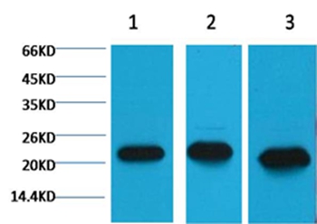
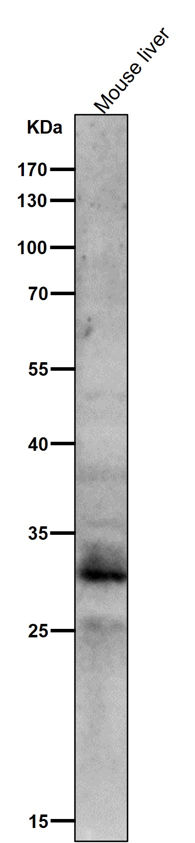
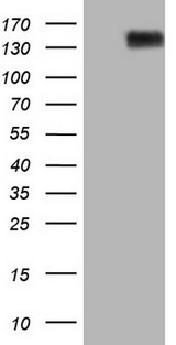
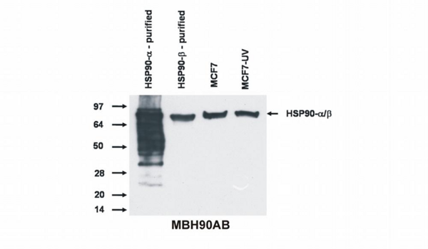
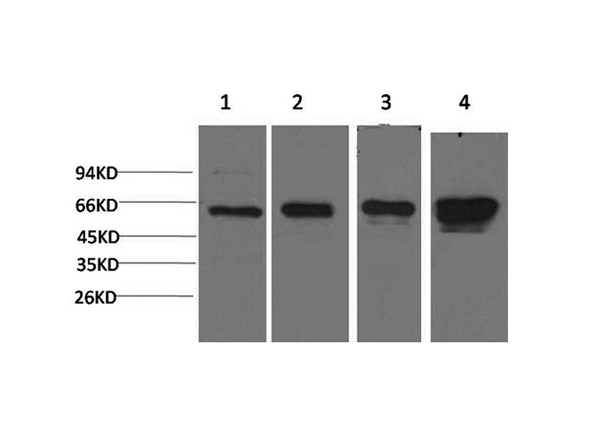
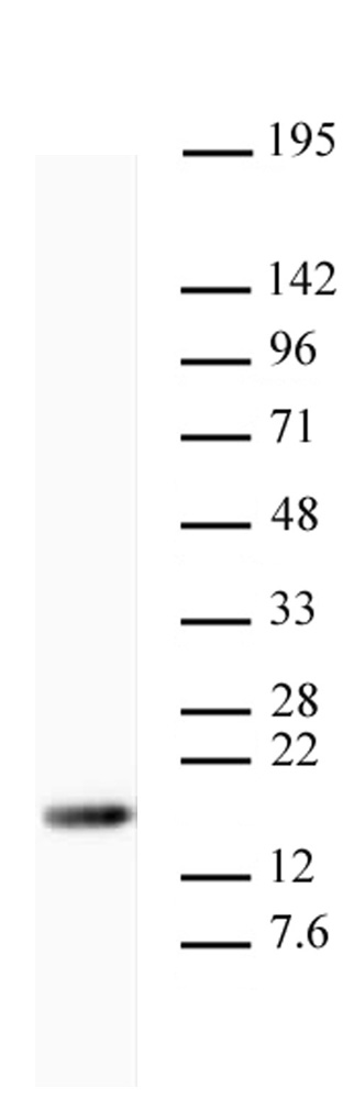
 WB (Western Blot)
(All lanes use the Antibody at 1:1K dilution for 1 hour at room temperature.)
WB (Western Blot)
(All lanes use the Antibody at 1:1K dilution for 1 hour at room temperature.)
C Reactive Protein, Monoclonal Antibody (Cat# AAA128113)
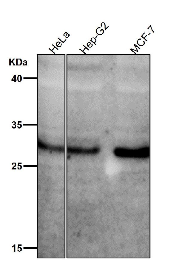 WB (Western Blot)
(All lanes use the Antibody at 1:1K dilution for 1 hour at room temperature.)
WB (Western Blot)
(All lanes use the Antibody at 1:1K dilution for 1 hour at room temperature.)
MST1/MST2, Monoclonal Antibody (Cat# AAA128143)
HLA-F, Monoclonal Antibody (Cat# AAA128331)
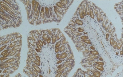
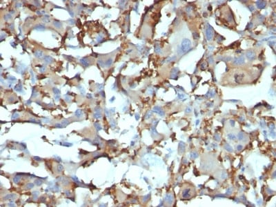
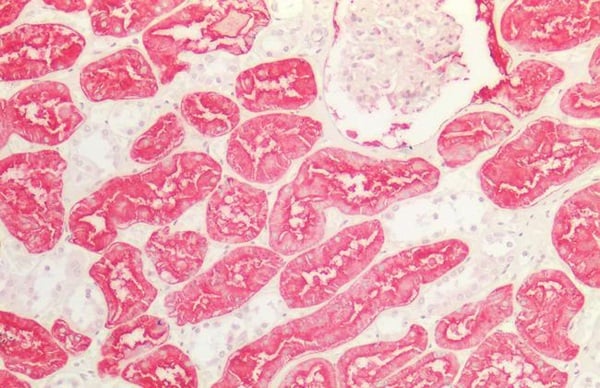 IHC (Immunohistochemistry)
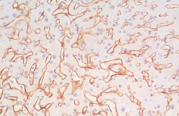
(ANPEP/CD13 Antibody-Human Kidney: Formalin-Fixed, Paraffin-Embedded (FFPE))
IHC (Immunohistochemistry)
(ANPEP/CD13 Antibody-Human Kidney: Formalin-Fixed, Paraffin-Embedded (FFPE))
ANPEP/CD13, Monoclonal Antibody (Cat# AAA162712)
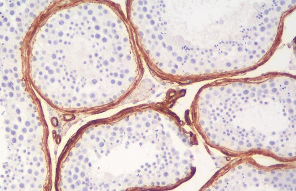
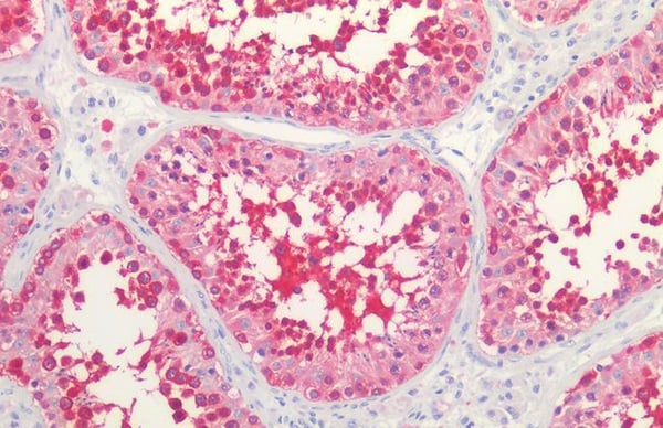 IHC (Immunohiostchemistry)
(HSP90/Heat Shock Protein 90 Antibody-Human Testis: Formalin-Fixed, Paraffin-Embedded (FFPE))
IHC (Immunohiostchemistry)
(HSP90/Heat Shock Protein 90 Antibody-Human Testis: Formalin-Fixed, Paraffin-Embedded (FFPE))
HSP90/Heat Shock Protein 90, Monoclonal Antibody (Cat# AAA163336)
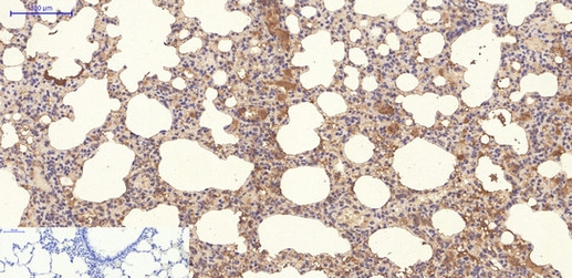
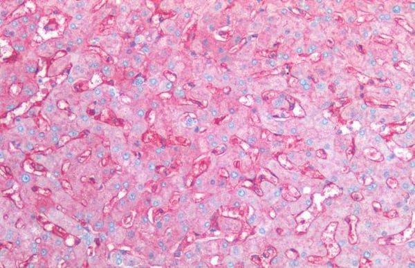 IHC (Immunohiostchemistry)
(CD14 Antibody-Human Liver: Formalin-Fixed, Paraffin-Embedded (FFPE))
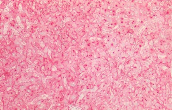
IHC (Immunohiostchemistry)
(CD14 Antibody-Human Liver: Formalin-Fixed, Paraffin-Embedded (FFPE))
CD14, Monoclonal Antibody (Cat# AAA163089)
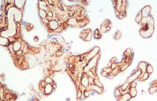
 IHC (Immunohistochemisry)
(Collagen IV Antibody-Human Liver: Formalin-Fixed, Paraffin-Embedded (FFPE))
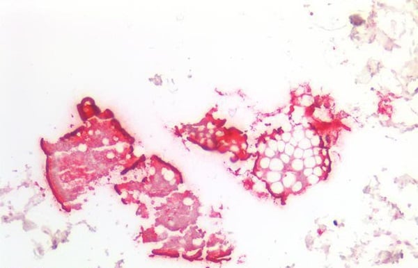
IHC (Immunohistochemisry)
(Collagen IV Antibody-Human Liver: Formalin-Fixed, Paraffin-Embedded (FFPE))
Collagen IV, Monoclonal Antibody (Cat# AAA163091)
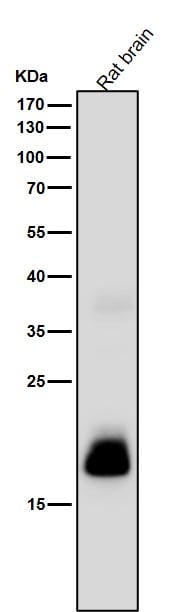
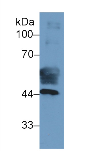
 WB (Western Blot)
(Western Blot; Sample: Recombinant COL4a3, Human.)
WB (Western Blot)
(Western Blot; Sample: Recombinant COL4a3, Human.)
Collagen Type IV Alpha 3 (COL4a3), Monoclonal Antibody (Cat# AAA162094)
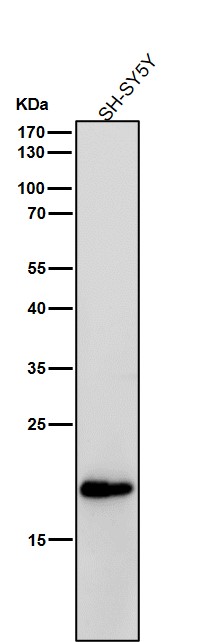
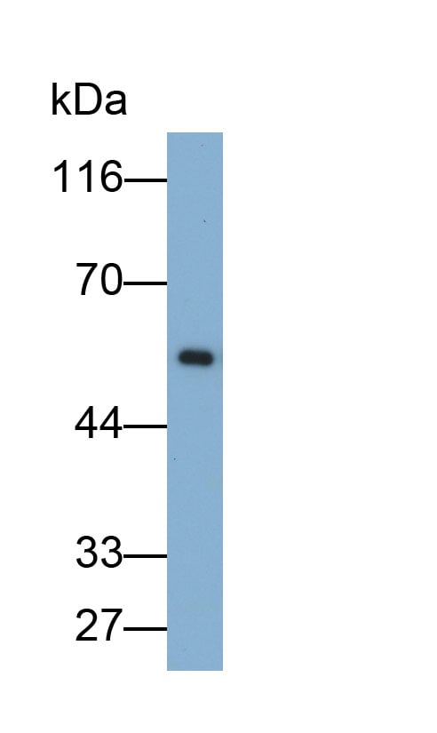
 WB (Western Blot)
(Western Blot: Sample: RecombinantFABP3,Human.)
WB (Western Blot)
(Western Blot: Sample: RecombinantFABP3,Human.)
Fatty Acid Binding Protein 3, Muscle And Heart (FABP3), Monoclonal Antibody (Cat# AAA130606)
GFAP, Monoclonal Antibody (Cat# AAA128545)
CD4, Monoclonal Antibody (Cat# AAA128551)
CD326, Monoclonal Antibody (Cat# AAA129043)
CD15, Monoclonal Antibody (Cat# AAA128880)
Myeloperoxidase, Monoclonal Antibody (Cat# AAA128891)
CD56, Monoclonal Antibody (Cat# AAA128681)
CD120a, Monoclonal Antibody (Cat# AAA128564)
CD229, Monoclonal Antibody (Cat# AAA128595)
CD42b, Monoclonal Antibody (Cat# AAA128413)
HLA-B7, Monoclonal Antibody (Cat# AAA128433)
CD49d, Monoclonal Antibody (Cat# AAA128396)
TCR Vgamma4, Monoclonal Antibody (Cat# AAA129163)
CD133, Monoclonal Antibody (Cat# AAA129053)

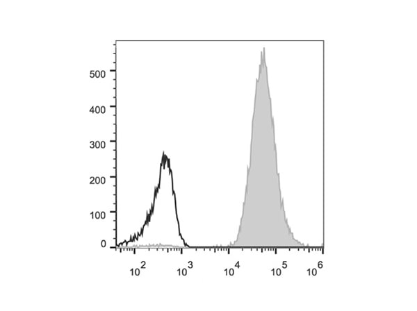 FCM/FACS (Flow Cytometry)
(Human peripheral blood lymphocytes are stained with Anti-Human HLA-A,B,C Monoclonal Antibody(PE Conjugated)(filled gray histogram). Unstained lymphocytes (empty black histogram) are used as control.)
FCM/FACS (Flow Cytometry)
(Human peripheral blood lymphocytes are stained with Anti-Human HLA-A,B,C Monoclonal Antibody(PE Conjugated)(filled gray histogram). Unstained lymphocytes (empty black histogram) are used as control.)
HLA-A,B,C, Monoclonal Antibody (Cat# AAA174661)
 IF (Immunofluorescence)
(Immunofluorescence analysis of C6 cells using Fluor? 594-conjugated GAPDH Monoclonal Antibody at dilution of 1:100)
IF (Immunofluorescence)
(Immunofluorescence analysis of C6 cells using Fluor? 594-conjugated GAPDH Monoclonal Antibody at dilution of 1:100)
GAPDH, Monoclonal Antibody (Cat# AAA179731)
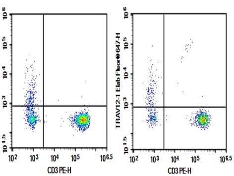 Application Data
(Human peripheral blood lymphocytes were stained with 0.2ug Purified Anti-Human TRAV12-1 Antibody[S511] (Right) and 0.2?g mouse IgG2b,? lsotype Control (Left), followed by Fluor® 647-conjugated goat Anti-mouse IgG Secondary Antibody, then anti-human CD3 PE-conjugated Monoclonal Antibody.)
Application Data
(Human peripheral blood lymphocytes were stained with 0.2ug Purified Anti-Human TRAV12-1 Antibody[S511] (Right) and 0.2?g mouse IgG2b,? lsotype Control (Left), followed by Fluor® 647-conjugated goat Anti-mouse IgG Secondary Antibody, then anti-human CD3 PE-conjugated Monoclonal Antibody.)
TRAV12-1, Monoclonal Antibody (Cat# AAA179732)
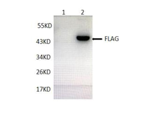
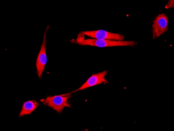
 IF (Immunofluorescence)
(Immunofluorescent analysis of 293F cells transfected with the DYKDDDDK-GFP, using anti-DYKDDDDK-Tag Monoclonal Antibody at dilution 1:2000.)
IF (Immunofluorescence)
(Immunofluorescent analysis of 293F cells transfected with the DYKDDDDK-GFP, using anti-DYKDDDDK-Tag Monoclonal Antibody at dilution 1:2000.)
DYKDDDDK-Tag, Monoclonal Antibody (Cat# AAA178017)
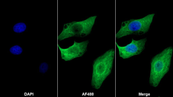
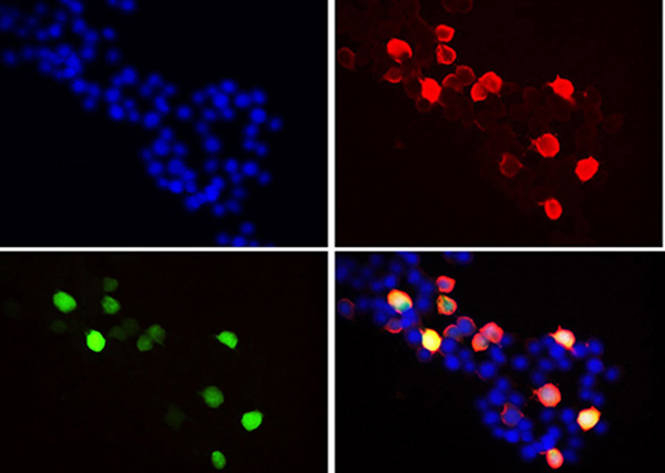
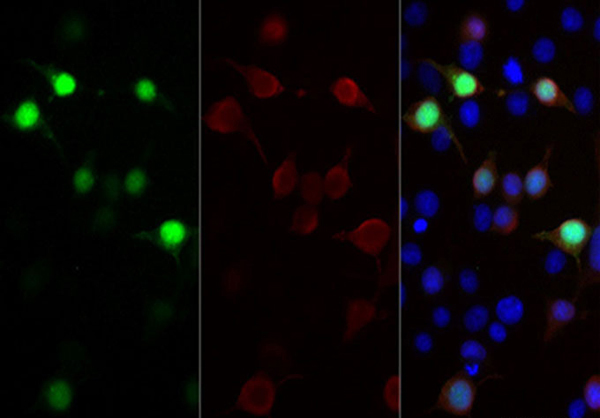
 IF (Immunofluorescence)
(Immunofluorescence of ACOX1 (green) in hela using ACOX1 Rabbit mAb at dilution 1/50, and DAPI(blue))
IF (Immunofluorescence)
(Immunofluorescence of ACOX1 (green) in hela using ACOX1 Rabbit mAb at dilution 1/50, and DAPI(blue))
ACOX1, Monoclonal Antibody (Cat# AAA178830)
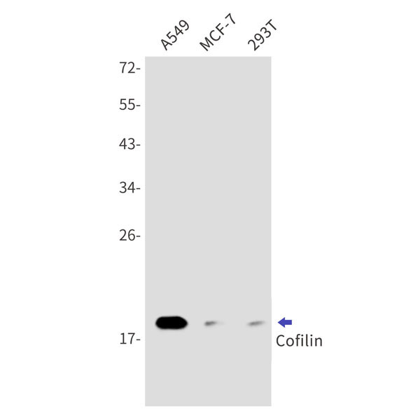
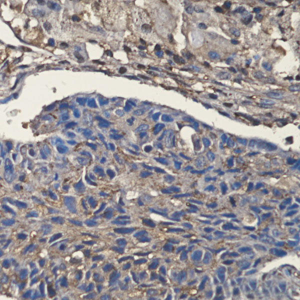 IHC (Immunohiostchemistry)
(Immunohistochemical of Cofilin in Human lung cancer tissue using Cofilin antibody at dilution 1:200)
IHC (Immunohiostchemistry)
(Immunohistochemical of Cofilin in Human lung cancer tissue using Cofilin antibody at dilution 1:200)
Cofilin, Monoclonal Antibody (Cat# AAA178841)
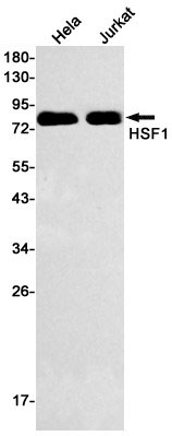
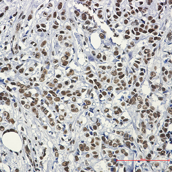 IHC (Immunohiostchemistry)
(Immunohistochemistry of HSF1 in paraffin-embedded Human breast cancer tissue using HSF1 Rabbit mAb at dilution 1:50)
IHC (Immunohiostchemistry)
(Immunohistochemistry of HSF1 in paraffin-embedded Human breast cancer tissue using HSF1 Rabbit mAb at dilution 1:50)
Hsf1, Monoclonal Antibody (Cat# AAA178853)
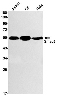
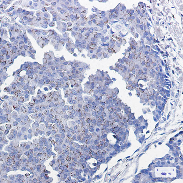 IHC (Immunohiostchemistry)
(Immunohistochemistry of Smad3 in paraffin-embedded Human breast cancer tissue using Smad3 Rabbit mAb at dilution 1:100)
IHC (Immunohiostchemistry)
(Immunohistochemistry of Smad3 in paraffin-embedded Human breast cancer tissue using Smad3 Rabbit mAb at dilution 1:100)
Smad3, Monoclonal Antibody (Cat# AAA178773)
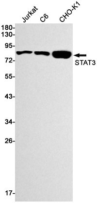
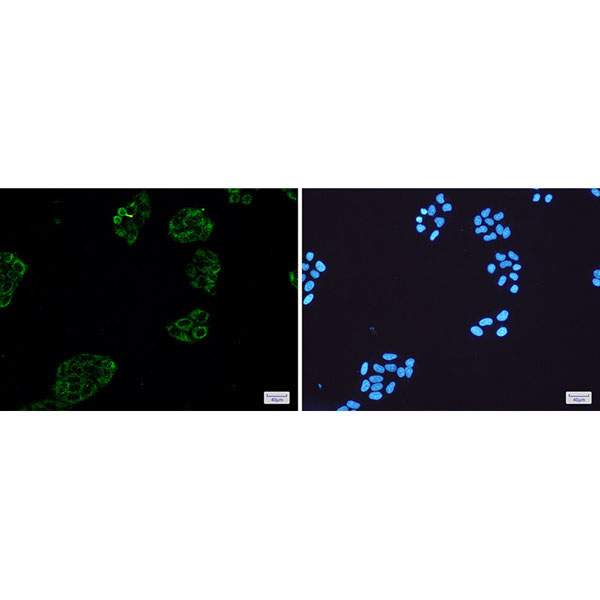
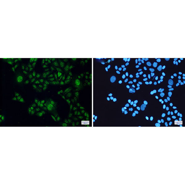 IF (Immunofluorescence)
(Immunofluorescence of STAT3(green) in Hela cells using STAT3 Rabbit mAb at dilution 1:200, and DAPI(blue))
IF (Immunofluorescence)
(Immunofluorescence of STAT3(green) in Hela cells using STAT3 Rabbit mAb at dilution 1:200, and DAPI(blue))
STAT3, Monoclonal Antibody (Cat# AAA178776)
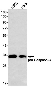
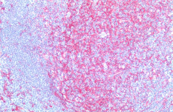
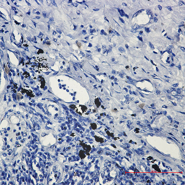
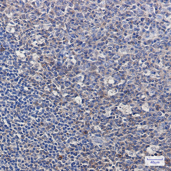 IHC (Immunohiostchemistry)
(Immunohistochemistry of pro Caspase 3 in paraffin-embedded Human tonsil using pro Caspase 3 Rabbit mAb at dilution 1:100)
IHC (Immunohiostchemistry)
(Immunohistochemistry of pro Caspase 3 in paraffin-embedded Human tonsil using pro Caspase 3 Rabbit mAb at dilution 1:100)
Caspase 3, Monoclonal Antibody (Cat# AAA178777)
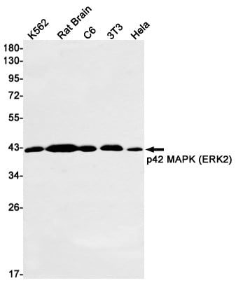
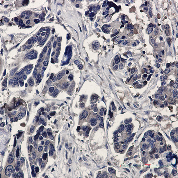 IHC (Immunohiostchemistry)
(Immunohistochemistry of ERK2 in paraffin-embedded Human Cholangiocarcinoma using ERK2 Rabbit mAb at dilution 1:20)
IHC (Immunohiostchemistry)
(Immunohistochemistry of ERK2 in paraffin-embedded Human Cholangiocarcinoma using ERK2 Rabbit mAb at dilution 1:20)
ERK2, Monoclonal Antibody (Cat# AAA178779)
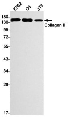
 ICC (Immunocytochemistry)
(Immunocytochemistry of Collagen III(green) in Hela cells using Collagen III Rabbit mAb at dilution 1:50, and DAPI(blue))
ICC (Immunocytochemistry)
(Immunocytochemistry of Collagen III(green) in Hela cells using Collagen III Rabbit mAb at dilution 1:50, and DAPI(blue))
Collagen III alpha 1, Monoclonal Antibody (Cat# AAA178791)
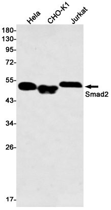
 IHC (Immunohiostchemistry)
(Immunohistochemistry of Smad2 in paraffin-embedded Human lung cancer tissue using Smad2 Rabbit mAb at dilution 1:50)
IHC (Immunohiostchemistry)
(Immunohistochemistry of Smad2 in paraffin-embedded Human lung cancer tissue using Smad2 Rabbit mAb at dilution 1:50)
Smad2, Monoclonal Antibody (Cat# AAA178806)
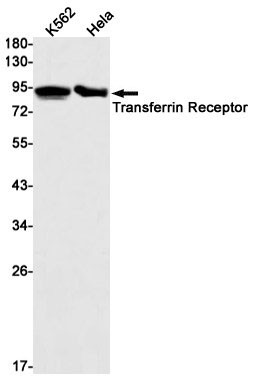
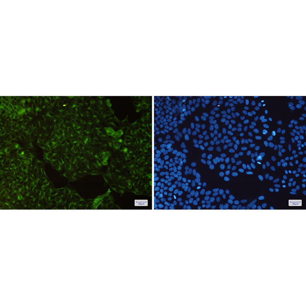 IF (Immunofluorescence)
(Immunofluorescence of Transferrin Receptor(green) in Hela cells using Transferrin Receptor Rabbit mAb at dilution 1:200, and DAPI(blue))
IF (Immunofluorescence)
(Immunofluorescence of Transferrin Receptor(green) in Hela cells using Transferrin Receptor Rabbit mAb at dilution 1:200, and DAPI(blue))
Transferrin Receptor 1, Monoclonal Antibody (Cat# AAA178810)
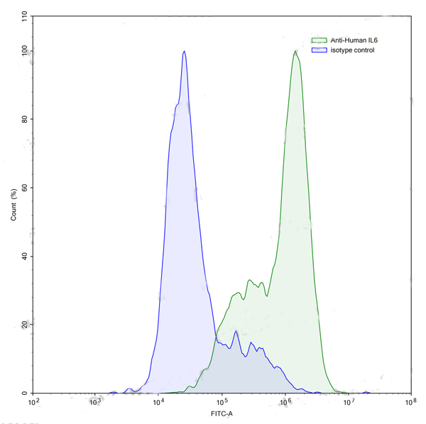
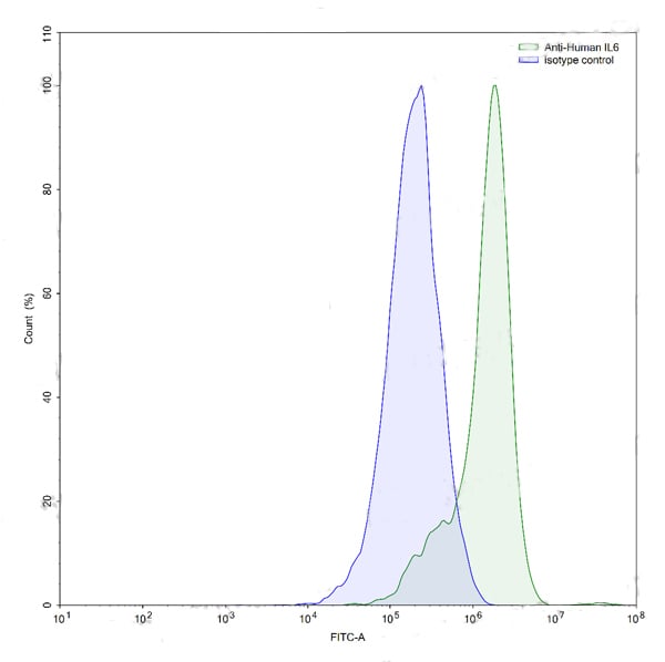 FCM/FACS (Flow Cytometry)
(Flow-cytometry using anti-human IL6 antibody.LPS treated THP-1 cells were stained with an irrelevant antibody (Blue Histogram) or an anti-human IL6 antibody monoclonal antibody (Catalog # RHC15803 ,Green Histogram) at a concentration of 5 ?ug/ml for 30 mins at RT. After washing, bound antibody was detected using a FITC conjugated goat anti-human antibody (Catalog # PHB96441) and cells analysed on a NovoCyte Flow Cytometer.)
FCM/FACS (Flow Cytometry)
(Flow-cytometry using anti-human IL6 antibody.LPS treated THP-1 cells were stained with an irrelevant antibody (Blue Histogram) or an anti-human IL6 antibody monoclonal antibody (Catalog # RHC15803 ,Green Histogram) at a concentration of 5 ?ug/ml for 30 mins at RT. After washing, bound antibody was detected using a FITC conjugated goat anti-human antibody (Catalog # PHB96441) and cells analysed on a NovoCyte Flow Cytometer.)
IL6, Monoclonal Recombinant Antibody (Cat# AAA120379)
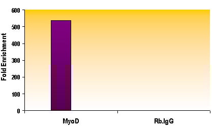
Protein A or G purified from cell culture supernatant.
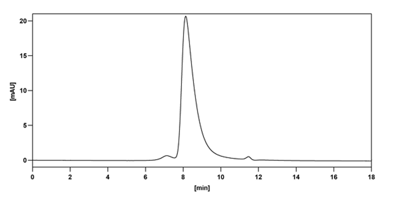 SEC-HPLC
(The purity of this product is >95% as determined by SEC-HPLC.)
SEC-HPLC
(The purity of this product is >95% as determined by SEC-HPLC.)
CD87/PLAUR/suPAR, Monoclonal Recombinant Antibody (Cat# AAA120383)
Protein A or G purified from cell culture supernatant.
Vibrio cholerae LPS/Lipopolysaccharide, Monoclonal Recombinant Antibody (Cat# AAA120264)
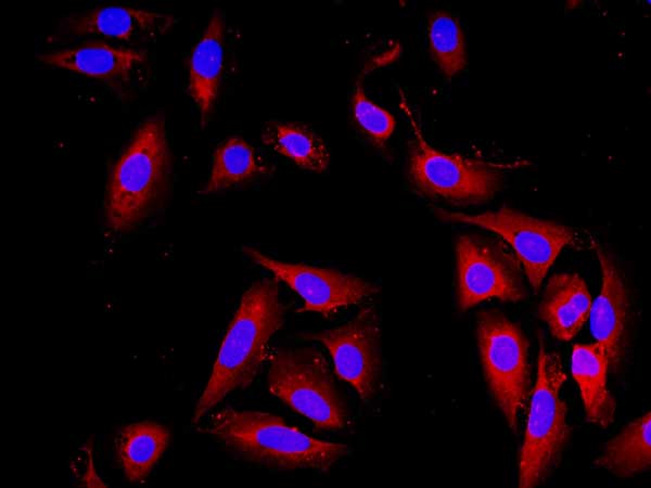
 IF (Immunofluorescence)
(Immunofluorescence analysis of Human stomach cancer tissue using Catenin-beta Monoclonal Antibody at dilution of 1:200.)
IF (Immunofluorescence)
(Immunofluorescence analysis of Human stomach cancer tissue using Catenin-beta Monoclonal Antibody at dilution of 1:200.)
Catenin-beta, Monoclonal Antibody (Cat# AAA173651)
 IHC (Immunohiostchemistry)
(Immunohistochemistry of paraffin-embedded Mouse colon tissue using AMPK alpha1 Monoclonal Antibody at dilution of 1:200.)
IHC (Immunohiostchemistry)
(Immunohistochemistry of paraffin-embedded Mouse colon tissue using AMPK alpha1 Monoclonal Antibody at dilution of 1:200.)
AMPK alpha1, Monoclonal Antibody (Cat# AAA173663)
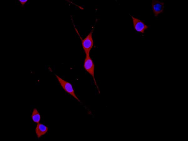
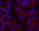
 IF (Immunofluorescence)
(Immunofluorescence analysis of Rat spleen tissue using NBR1 Monoclonal Antibody at dilution of 1:200.)
IF (Immunofluorescence)
(Immunofluorescence analysis of Rat spleen tissue using NBR1 Monoclonal Antibody at dilution of 1:200.)
NBR1, Monoclonal Antibody (Cat# AAA173680)
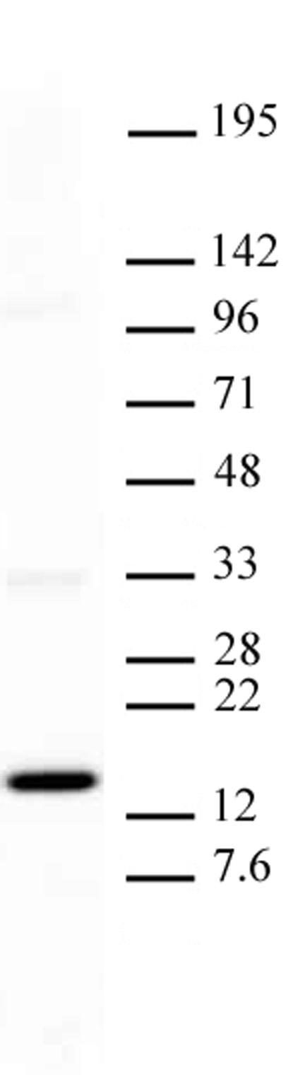
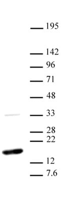
HSV-1 envelope glycoprotein D, Monoclonal Antibody (Cat# AAA59524)
 IHC (Immunohiostchemistry)
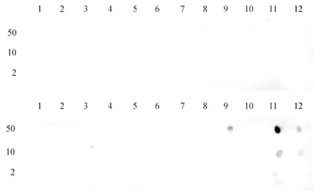
 DB (Dot Blot)
(Histone H3 trimethyl Lys9 antibody tested by dot blot analysis. Dot blot analysis was used to confirm the specificity of Histone H3 trimethyl Lys9 antibody for trimethyl Lys9 of histone H3. Recombinant methylated histone proteins corresponding to the immunogen and related sequences were spotted onto PVDF and probed with Histone H3 trimethyl Lys9 at 2 ug/ml. The amount of protein (picomoles) spotted is indicated next to each row. Lane 1: unmodified H3 protein. Lane 2: monomethyl Lys4 protein. Lane 3: dimethyl Lys4 protein. Lane 4: trimethyl Lys4 protein. Lane 5: monomethyl Lys9 protein. Lane 6: dimethyl Lys9 protein. Lane 7: trimethyl Lys9 protein. Lane 8: monomethyl Lys27 protein. Lane 9: dimethyl Lys27 protein. Lane 10: trimethyl Lys27 protein.)
DB (Dot Blot)
(Histone H3 trimethyl Lys9 antibody tested by dot blot analysis. Dot blot analysis was used to confirm the specificity of Histone H3 trimethyl Lys9 antibody for trimethyl Lys9 of histone H3. Recombinant methylated histone proteins corresponding to the immunogen and related sequences were spotted onto PVDF and probed with Histone H3 trimethyl Lys9 at 2 ug/ml. The amount of protein (picomoles) spotted is indicated next to each row. Lane 1: unmodified H3 protein. Lane 2: monomethyl Lys4 protein. Lane 3: dimethyl Lys4 protein. Lane 4: trimethyl Lys4 protein. Lane 5: monomethyl Lys9 protein. Lane 6: dimethyl Lys9 protein. Lane 7: trimethyl Lys9 protein. Lane 8: monomethyl Lys27 protein. Lane 9: dimethyl Lys27 protein. Lane 10: trimethyl Lys27 protein.)
Histone H3K9me3, Monoclonal Antibody (Cat# AAA59964)

 DB (Dot Blot)
(Histone H3K27me1 antibody (mAb) tested by dot blot analysis. Dot blot analysis was used to confirm the specificity of Histone H3 monomethyl Lys27 antibody for monomethyl Lys27 of histone H3. Recombinant methylated histone proteins corresponding to the immunogen and related sequences were spotted onto PVDF and probed with Histone H3 monomethyl Lys27 at 2 ug/ml. The amount of protein (picomoles) spotted is indicated next to each row. Lane 1: unmodified H3 protein. Lane 2: monomethyl Lys4 protein. Lane 3: dimethyl Lys4 protein. Lane 4: trimethyl Lys4 protein. Lane 5: monomethyl Lys9 protein. Lane 6: dimethyl Lys9 protein. Lane 7: trimethyl Lys9 protein. Lane 8: monomethyl Lys27 protein. Lane 9: dimethyl Lys27 protein. Lane 10: trimethyl Lys27 protein.)
DB (Dot Blot)
(Histone H3K27me1 antibody (mAb) tested by dot blot analysis. Dot blot analysis was used to confirm the specificity of Histone H3 monomethyl Lys27 antibody for monomethyl Lys27 of histone H3. Recombinant methylated histone proteins corresponding to the immunogen and related sequences were spotted onto PVDF and probed with Histone H3 monomethyl Lys27 at 2 ug/ml. The amount of protein (picomoles) spotted is indicated next to each row. Lane 1: unmodified H3 protein. Lane 2: monomethyl Lys4 protein. Lane 3: dimethyl Lys4 protein. Lane 4: trimethyl Lys4 protein. Lane 5: monomethyl Lys9 protein. Lane 6: dimethyl Lys9 protein. Lane 7: trimethyl Lys9 protein. Lane 8: monomethyl Lys27 protein. Lane 9: dimethyl Lys27 protein. Lane 10: trimethyl Lys27 protein.)
Histone H3K27me1, Monoclonal Antibody (Cat# AAA59965)
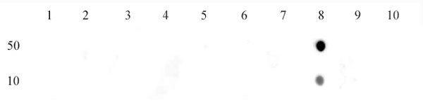
 DB (Dot Blot)
(Dot blot of Histone Histone H3 dimethyl Lys36 antibody. Dot blot analysis was used to confirm the specificity of H3K36me2 antibody for dimethyl Lys36 of histone H3. Recombinant methylated peptides corresponding to the immunogen and related sequences were spotted onto PVDF and probed with H3K36me2 at 2 ug/ml. The amount of protein (picomoles) spotted is indicated next to each row. Top panel - Lane 1: unmodified H3K4. Lane 2: H3K4me1. Lane 3: H3K4me2. Lane 4: H3K4me3. Lane 5: unmodified H3K9. Lane 6: H3K9me1. Lane 7: H3K9me2. Lane 8: H3K9me3. Lane 9: unmodified H3K79. Lane 10: H3K79me1. Lane 11: H3K79me2. Lane 12: H3K79me3. Bottom panel - Lane 1: unmodified H3K23. Lane 2: H3K23me1. Lane 3: H3K23me3. Lane 4: H3K23me3. Lane 5: unmodified H3K27. Lane 6: H3K27me1. Lane 7: H3K27me2. Lane 8: H3K27me3. Lane 9: unmodified H3K36. Lane 10: H3K36me1. Lane 11: H3K36me2. Lane 12: H3K36me3.)
DB (Dot Blot)
(Dot blot of Histone Histone H3 dimethyl Lys36 antibody. Dot blot analysis was used to confirm the specificity of H3K36me2 antibody for dimethyl Lys36 of histone H3. Recombinant methylated peptides corresponding to the immunogen and related sequences were spotted onto PVDF and probed with H3K36me2 at 2 ug/ml. The amount of protein (picomoles) spotted is indicated next to each row. Top panel - Lane 1: unmodified H3K4. Lane 2: H3K4me1. Lane 3: H3K4me2. Lane 4: H3K4me3. Lane 5: unmodified H3K9. Lane 6: H3K9me1. Lane 7: H3K9me2. Lane 8: H3K9me3. Lane 9: unmodified H3K79. Lane 10: H3K79me1. Lane 11: H3K79me2. Lane 12: H3K79me3. Bottom panel - Lane 1: unmodified H3K23. Lane 2: H3K23me1. Lane 3: H3K23me3. Lane 4: H3K23me3. Lane 5: unmodified H3K27. Lane 6: H3K27me1. Lane 7: H3K27me2. Lane 8: H3K27me3. Lane 9: unmodified H3K36. Lane 10: H3K36me1. Lane 11: H3K36me2. Lane 12: H3K36me3.)
Histone H3K36me2, Monoclonal Antibody (Cat# AAA59967)
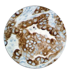
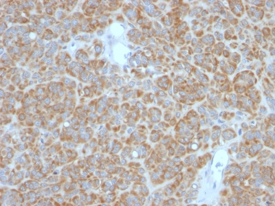 IHC (Immunohiostchemistry)
(Formalin-fixed, paraffin-embedded human Melanoma stained with Bcl-2 Monoclonal Antibody (BCL2/782 + BCL2/796).)
IHC (Immunohiostchemistry)
(Formalin-fixed, paraffin-embedded human Melanoma stained with Bcl-2 Monoclonal Antibody (BCL2/782 + BCL2/796).)
Bcl-2, Monoclonal Antibody (Cat# AAA62864)
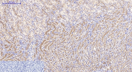
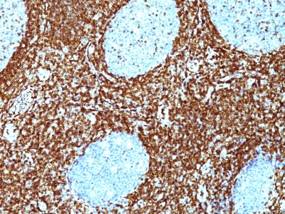
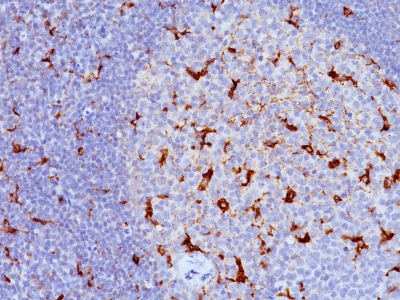 IHC (Immunohiostchemistry)
(Formalin-fixed, paraffin-embedded human Tonsil stained with CD68 Monoclonal Antibody (KP1 + C68/684).)
IHC (Immunohiostchemistry)
(Formalin-fixed, paraffin-embedded human Tonsil stained with CD68 Monoclonal Antibody (KP1 + C68/684).)
CD68, Monoclonal Antibody (Cat# AAA62661)
What are Monoclonal Antibodies?
Monoclonal antibodies are specialized laboratory-produced proteins developed for binding to specific biological antigens or other molecular targets. Since they come from a single cell (or clone), they are especially consistent and accurate in the data they are involved in producing.
This type of antibody material has been shown to be a powerful tool in finding and subsequently destroying harmful cells in an organism, such as those found in cancers or various autoimmune diseases. This makes them excellent aids in medical testing and research, which is why they are so widely used.
AAA Biotech offers a comprehensive range of high-quality monoclonal antibodies that perform effectively in various laboratory tests, including (amongst others) ELISA, western blotting, immunohistochemistry, and flow cytometry. All of the products in our catalog are thoroughly quality tested to make sure that they are reliable and will consistently perform well in your research.
What Are The Uses of Monoclonal Antibodies
Monoclonal antibodies are used in many lab tests, including (amongst others) ELISA, western blotting, immunohistochemistry, and flow cytometry.
ELISA is a test that helps detect a specific substance/analyte in a sample. It uses antibodies (often monoclonal) bound to a solid surface (such as the well of a microplate) to “capture” the substance/analyte in the sample and immobilize it so that the detection antibody component can then bind to it and produce a signal, which can then be measured.
Western blotting identifies specific proteins in a sample. The sample is first separated on a gel, and then antibodies are applied that will typically bind to the target, which will all be localized to a single band in a lane.
Immunohistochemistry helps locate specific proteins in cells or tissue samples using antibodies.
Flow cytometry looks at and sorts cells. It uses antibodies that are conjugated to reporter molecules called “fluorophores”, which, under special lights, emit light themselves, which can then be measured by a detector instrument.
How Monoclonal Antibodies Are Used as Medicine?
Please note that all of the products listed in AAA Biotech’s also known as AAA Bio or AAABio catalog are strictly for research-use only (RUO).
Monoclonal antibodies can also be used as therapeutic/medical treatments, particularly in the context of cancers. They are designed to find and bind to specific cells or proteins, helping the immune system recognize and attack the cancer. These treatments work in different ways, such as:
- Radioimmunotherapy attaches a small amount of radioactive molecule to the antibody, so it delivers the radiation directly to the cancer cells that the antibody is specifically binding to.
- Antibody-directed enzyme prodrug therapy uses antibodies that are specifically bound to special enzymes. These enzymes activate a harmless drug in the body and turn it into a cancer-killing drug only near the cancer cells—this helps avoid harming healthy cells.
- Immunoliposomes are tiny “bubbles” filled with medicine/drug and coated with antibodies. They carry the drug straight to the cancer cells.
Why Buy Monoclonal Antibodies From Us?
At AAA Biotech, we provide high-performance monoclonal antibodies designed to support a wide range of research needs.
1. Validated for Versatile Applications
The antibodies in our catalog are extensively validated and compatible with multiple techniques, including (but not limited to) ELISA, flow cytometry (FC), immunocytochemistry (ICC), immunofluorescence (IF), immunohistochemistry (IHC), immunoprecipitation (IP), and western blotting (WB).
2. Wide Selection & Specialized Options
We offer antibodies for common and rare species, that are available in various conjugated forms, and also in recombinant formats. Essentially, there is almost anything one might need to meet their experimental model’s requirements.
3. High-Quality Proteins
Our proteins meet high purity standards—90% or more as confirmed by SDS-PAGE. Many are available with tags like His, Flag, GST, or MBP, and we also supply native and biologically active proteins for functional studies.
Frequently Asked Questions
1. Are your monoclonal antibodies validated for specific applications?
Yes, our antibodies are tested and validated for use in methods such as ELISA, western blot, IHC, flow cytometry, and more. Refer to specific product pages or datasheets for individual product information.
2. How do I choose the right monoclonal antibody for my application?
Review the product details directly for application validation, species reactivity, and target information. You may also contact our support team at any time for help.
3. How quickly can I receive my order?
Most orders are processed and shipped within 1–3 business days, depending on product availability and your shipping location.