Filters
▼Clonality
▼Type
▼Reactivity
▼Gene Name
▼Isotype
▼Host
▼Application
▼Clone
▼Monoclonal Antibodies
Get accurate results in your research with our Monoclonal Antibodies, which are specially made to target exactly what you require for your research, and will produce consistent, reliable performance in lab tests.
Viewing 8150-8200 of 27597 product results
 IF (Immunofluorescence)
(Immunofluorescent staining of COS7 cells transiently transfected with recombinant DLD protein using DLD antibody)
IF (Immunofluorescence)
(Immunofluorescent staining of COS7 cells transiently transfected with recombinant DLD protein using DLD antibody)
DLD, Monoclonal Antibody (Cat# AAA74821)
 IHC (Immunohiostchemistry)
(Immunohistochemical analysis of FCGR2A protein in paraffin embedded Human colon tissue using FCGR2A antibody)
IHC (Immunohiostchemistry)
(Immunohistochemical analysis of FCGR2A protein in paraffin embedded Human colon tissue using FCGR2A antibody)
FCGR2A, Monoclonal Antibody (Cat# AAA74687)
 IHC (Immunohiostchemistry)
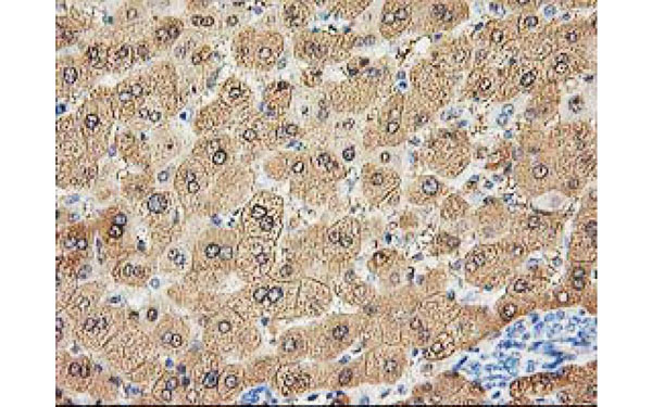
(Immunohistochemical analysis of ALDOB protein in paraffin embedded Human liver tissue using ALDOB antibody)
IHC (Immunohiostchemistry)
(Immunohistochemical analysis of ALDOB protein in paraffin embedded Human liver tissue using ALDOB antibody)
ALDOB, Monoclonal Antibody (Cat# AAA74724)
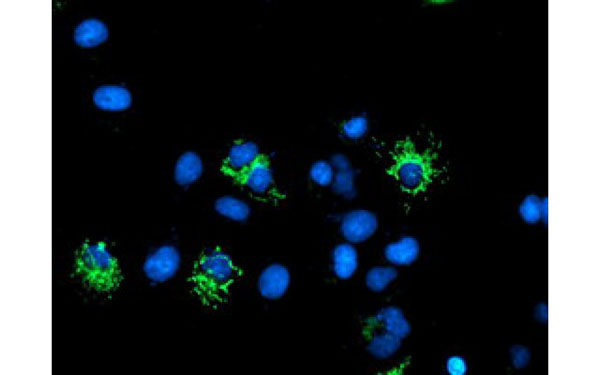
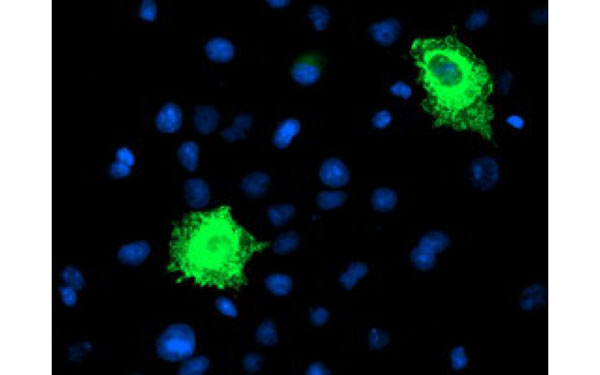
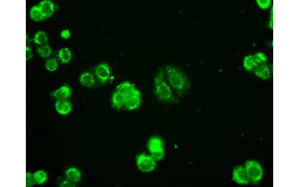 IF (Immunofluorescence)
(Immunofluorescent staining of endogenous AK1 protein in HT29 cells using AK1 antibody)
IF (Immunofluorescence)
(Immunofluorescent staining of endogenous AK1 protein in HT29 cells using AK1 antibody)
AK1, Monoclonal Antibody (Cat# AAA74731)
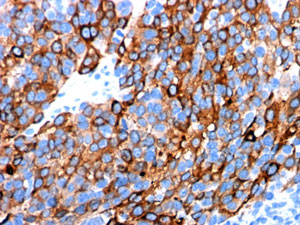
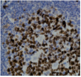
 IHC (Immunohiostchemistry)
(Immunohistochemical analysis of DPP9 protein in paraffin embedded Human lymphoma tissue using DPP9 antibody)
IHC (Immunohiostchemistry)
(Immunohistochemical analysis of DPP9 protein in paraffin embedded Human lymphoma tissue using DPP9 antibody)
DPP9, Monoclonal Antibody (Cat# AAA74746)
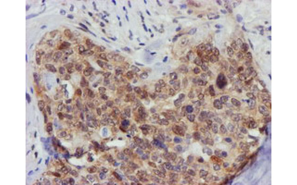
 IHC (Immunohiostchemistry)
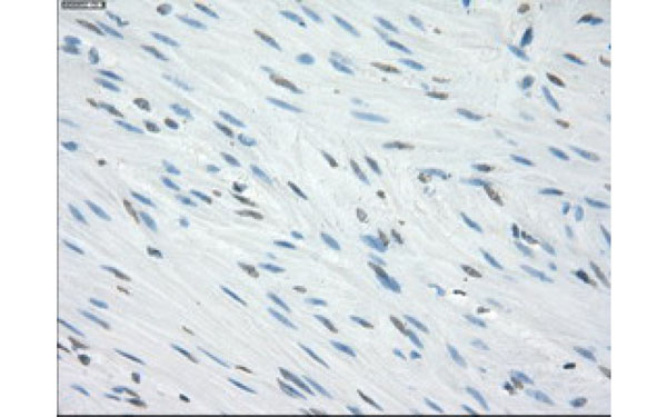
(Immunohistochemical analysis of EPM2AIP1 protein in paraffin embedded Adenocarcinoma of Human breast tissue using EPM2AIP1 antibody)
IHC (Immunohiostchemistry)
(Immunohistochemical analysis of EPM2AIP1 protein in paraffin embedded Adenocarcinoma of Human breast tissue using EPM2AIP1 antibody)
EPM2AIP1, Monoclonal Antibody (Cat# AAA74860)
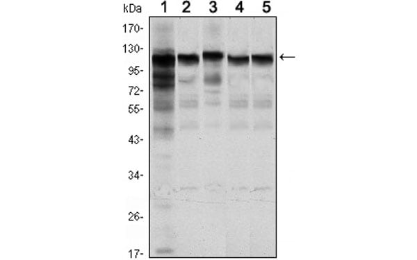
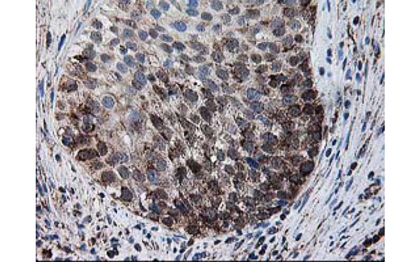
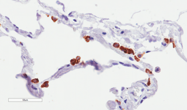
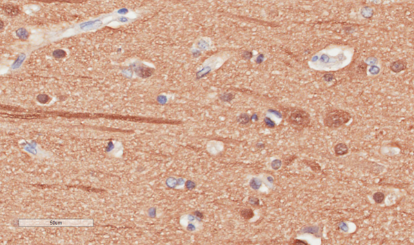
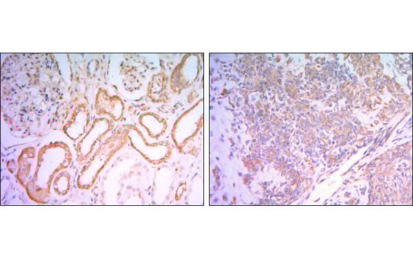 IHC (Immunohiostchemistry)
(Immunohistochemical analysis of paraffin-embedded human salivary gland tissues (left) and kidney tissues (right) using HK1 antibody with DAB staining.)
IHC (Immunohiostchemistry)
(Immunohistochemical analysis of paraffin-embedded human salivary gland tissues (left) and kidney tissues (right) using HK1 antibody with DAB staining.)
HK1, Monoclonal Antibody (Cat# AAA74870)
 IF (Immunofluorescence)
(Immunofluorescent staining of COS7 cells transiently transfected with recombinant FBXO21 protein using FBXO21 antibody)
IF (Immunofluorescence)
(Immunofluorescent staining of COS7 cells transiently transfected with recombinant FBXO21 protein using FBXO21 antibody)
FBXO21, Monoclonal Antibody (Cat# AAA74877)
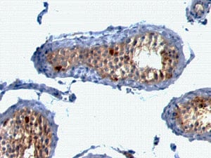 IHC (Immunohiostchemistry)
(Formalin-fixed, paraffin-embedded human Testis stained with MART-1 / Melan-A Monoclonal Antibody (A103 + M2-7C10 + M2-9E3).)
IHC (Immunohiostchemistry)
(Formalin-fixed, paraffin-embedded human Testis stained with MART-1 / Melan-A Monoclonal Antibody (A103 + M2-7C10 + M2-9E3).)
MART-1 / Melan-A / MLANA, Monoclonal Antibody (Cat# AAA62561)
Others not tested
Chondroitin Sulphate Neoepitope, Monoclonal Antibody (Cat# AAA63031)
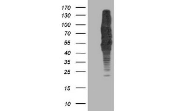
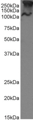 WB (Western Blot)
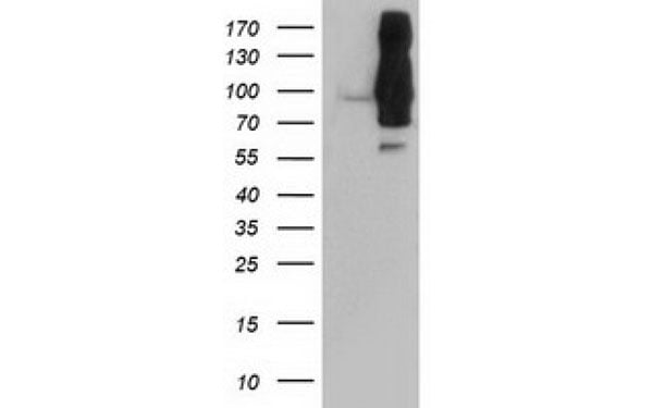
( Western Blot using anti-MUC1 antibody SM3 MCF-7 cell lysate (35ug protein in RIPA buffer) were resolved on a 10% SDS PAGE gel and blots probed with the chimeric rabbit version of SM3 at 1 ug/ml before detection using an anti-rabbit secondary antibody. A primary incubation of 1h was used and protein was detected by chemiluminescence. The predicted band size for unmodified MUC1 is 122.1kDa, though in breast cancer cell lines like MCF-7 MUC1 can be up to 90% glycosylated (c.f. Mueller et al. PMID: 10373415; T47D cells) and expected band sizes are ~250-300kDa. Thus the two bands likely represent processed (>250kDa) and unprocessed (~121kDa) populations of the protein. successfully detected human MUC1 in MCF-7 breast cancer cells.)
WB (Western Blot)
( Western Blot using anti-MUC1 antibody SM3 MCF-7 cell lysate (35ug protein in RIPA buffer) were resolved on a 10% SDS PAGE gel and blots probed with the chimeric rabbit version of SM3 at 1 ug/ml before detection using an anti-rabbit secondary antibody. A primary incubation of 1h was used and protein was detected by chemiluminescence. The predicted band size for unmodified MUC1 is 122.1kDa, though in breast cancer cell lines like MCF-7 MUC1 can be up to 90% glycosylated (c.f. Mueller et al. PMID: 10373415; T47D cells) and expected band sizes are ~250-300kDa. Thus the two bands likely represent processed (>250kDa) and unprocessed (~121kDa) populations of the protein. successfully detected human MUC1 in MCF-7 breast cancer cells.)
MUC1, Monoclonal Recombinant Antibody (Cat# AAA71969)
Protein A Affinity Purified
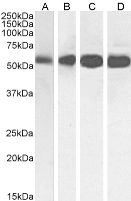 WB (Western Blot)
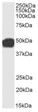
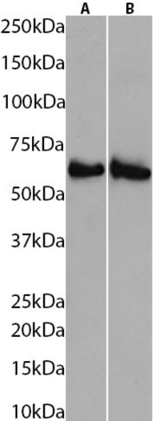
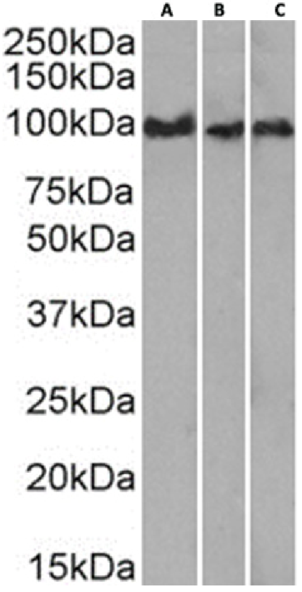
( Western Blot using anti-Beta-Tubulin antibody S11B HeLa (A), A431 (B), HEK293 (C) and MCF-7 (D) cell lysate samples (35ug protein in RIPA buffer) were resolved on a 10% SDS PAGE gel and blots probed with the chimeric rabbit version of S11B at 0.01 ug/ml before detection using an anti-rabbit secondary antibody. A primary incubation of 1h was used and protein was detected by chemiluminescence. The expected band size for Beta-Tubulin is ~54kDa. A431, HEK293 and MCF-7 cell lysate samples.)
WB (Western Blot)
( Western Blot using anti-Beta-Tubulin antibody S11B HeLa (A), A431 (B), HEK293 (C) and MCF-7 (D) cell lysate samples (35ug protein in RIPA buffer) were resolved on a 10% SDS PAGE gel and blots probed with the chimeric rabbit version of S11B at 0.01 ug/ml before detection using an anti-rabbit secondary antibody. A primary incubation of 1h was used and protein was detected by chemiluminescence. The expected band size for Beta-Tubulin is ~54kDa. A431, HEK293 and MCF-7 cell lysate samples.)
Beta-Tubulin, Monoclonal Recombinant Antibody (Cat# AAA71972)
Protein A Affinity Purified
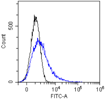
 FCM/FACS (Flow Cytometry)
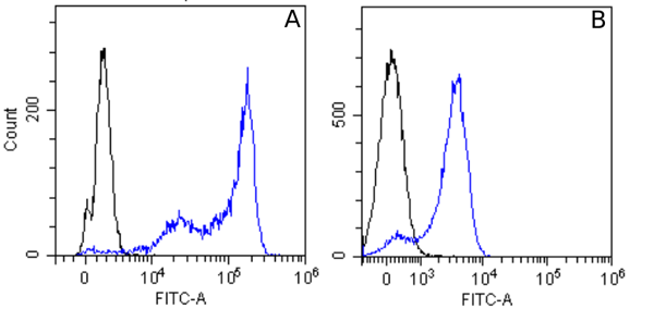
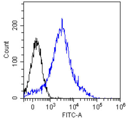
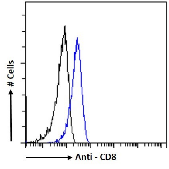
( Flow-cytometry using the anti-CD8 beta antibody YTS 156.7.7 Mouse thymocytes were stained with unimmunized rabbit IgG antibody (black line) or the rabbit-chimeric version of YTS 156.7.7 at a concentration of 10 ug/ml for 30 mins at RT. After washing, bound antibody was detected using an anti-rabbit IgG JK (FITC-conjugate) antibody (129936) at 2 ug/ml and cells analyzed on a FACSCanto flow-cytometer.)
FCM/FACS (Flow Cytometry)
( Flow-cytometry using the anti-CD8 beta antibody YTS 156.7.7 Mouse thymocytes were stained with unimmunized rabbit IgG antibody (black line) or the rabbit-chimeric version of YTS 156.7.7 at a concentration of 10 ug/ml for 30 mins at RT. After washing, bound antibody was detected using an anti-rabbit IgG JK (FITC-conjugate) antibody (129936) at 2 ug/ml and cells analyzed on a FACSCanto flow-cytometer.)
CD8 beta, Monoclonal Recombinant Antibody (Cat# AAA71974)
Protein A Affinity Purified
 IF (Immunofluorescence)
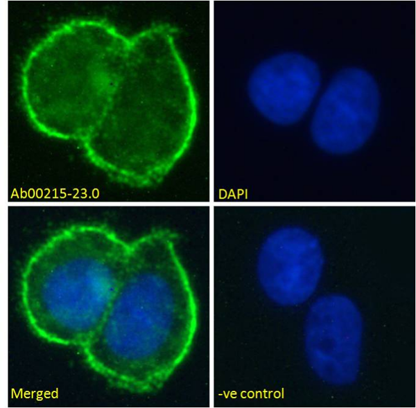
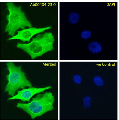
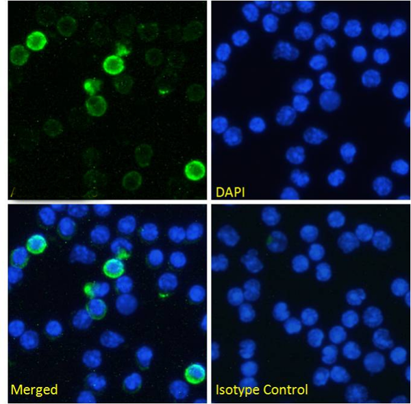
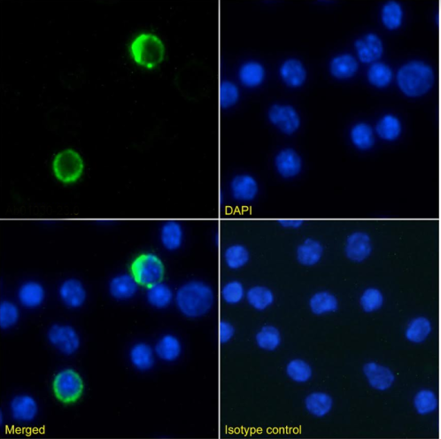
(Immunofluorescencestainingoffixedmousesplenocyteswithanti-CD200antibodyOX-90(AAA72437) ImmunofluorescenceanalysisofparaformaldehydefixedmousesplenocytesonShi-fixcoverslips,permeabilizedwith0.15%TritonandstainedwiththechimericrabbitIgGversionofOX-90(AAA72437)at10ug/mlfor1hfollowedbyAlexaFluor 488secondaryantibody(1ug/ml),showingmembranestaininginasubsetofcells.ThenuclearstainisDAPI(blue).Panelsshowfromleft-right,top-bottomAAA72437,DAPI,mergedchannelsandanisotypecontrol.Theisotypecontrolwasstainedwithananti-FluoresceinantibodyfollowedbyAlexaFluor 488secondaryantibody.)
IF (Immunofluorescence)
(Immunofluorescencestainingoffixedmousesplenocyteswithanti-CD200antibodyOX-90(AAA72437) ImmunofluorescenceanalysisofparaformaldehydefixedmousesplenocytesonShi-fixcoverslips,permeabilizedwith0.15%TritonandstainedwiththechimericrabbitIgGversionofOX-90(AAA72437)at10ug/mlfor1hfollowedbyAlexaFluor 488secondaryantibody(1ug/ml),showingmembranestaininginasubsetofcells.ThenuclearstainisDAPI(blue).Panelsshowfromleft-right,top-bottomAAA72437,DAPI,mergedchannelsandanisotypecontrol.Theisotypecontrolwasstainedwithananti-FluoresceinantibodyfollowedbyAlexaFluor 488secondaryantibody.)
CD200, Monoclonal Recombinant Antibody (Cat# AAA72437)
 IF (Immunofluorescence)
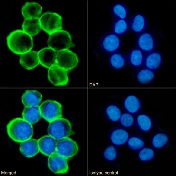
(Immunofluorescencestainingoffixedratsplenocyteswithanti-CD44antibodyOX-49(AAA72448) ImmunofluorescenceanalysisofparaformaldehydefixedratsplenocytesonShi-fixcoverslips,permeabilizedwith0.15%TritonstainedwiththechimericrabbitversionofOX-49(AAA72448)at10ug/mlfor1hfollowedbyAlexaFluor 488secondaryantibody(1ug/ml),showingmembranestaining.ThenuclearstainisDAPI(blue).Panelsshowfromleft-right,top-bottomAAA72448,DAPI,mergedchannelsandanisotypecontrol.Theisotypecontrolwasstainedwithananti-FluoresceinantibodyfollowedbyAlexaFluor 488secondaryantibody.)
IF (Immunofluorescence)
(Immunofluorescencestainingoffixedratsplenocyteswithanti-CD44antibodyOX-49(AAA72448) ImmunofluorescenceanalysisofparaformaldehydefixedratsplenocytesonShi-fixcoverslips,permeabilizedwith0.15%TritonstainedwiththechimericrabbitversionofOX-49(AAA72448)at10ug/mlfor1hfollowedbyAlexaFluor 488secondaryantibody(1ug/ml),showingmembranestaining.ThenuclearstainisDAPI(blue).Panelsshowfromleft-right,top-bottomAAA72448,DAPI,mergedchannelsandanisotypecontrol.Theisotypecontrolwasstainedwithananti-FluoresceinantibodyfollowedbyAlexaFluor 488secondaryantibody.)
CD44, Monoclonal Recombinant Antibody (Cat# AAA72448)
 IF (Immunofluorescence)
(Immunofluorescencestainingoffixedratsplenocyteswithanti-CD25(Interleukin-2receptorsubunitalpha)antibodyOX-39(AAA72451) Immunofluorescenceanalysisofparaformaldehydefixedrat(Rattusnorvegicus)splenocytesonShi-fixcoverslips,permeabilizedwith0.15%TritonstainedwiththechimericrabbitversionofOX-39(AAA72451)at10ug/mlfor1hfollowedbyAlexaFluor 488secondaryantibody(1ug/ml),showingmembranestainingofasubsetofcells.ThenuclearstainisDAPI(blue).Panelsshowfromleft-right,top-bottomAAA72451,DAPI,mergedchannelsandanisotypecontrol.Theisotypecontrolwasstainedwithananti-FluoresceinantibodyfollowedbyAlexaFluor 488secondaryantibody.)
IF (Immunofluorescence)
(Immunofluorescencestainingoffixedratsplenocyteswithanti-CD25(Interleukin-2receptorsubunitalpha)antibodyOX-39(AAA72451) Immunofluorescenceanalysisofparaformaldehydefixedrat(Rattusnorvegicus)splenocytesonShi-fixcoverslips,permeabilizedwith0.15%TritonstainedwiththechimericrabbitversionofOX-39(AAA72451)at10ug/mlfor1hfollowedbyAlexaFluor 488secondaryantibody(1ug/ml),showingmembranestainingofasubsetofcells.ThenuclearstainisDAPI(blue).Panelsshowfromleft-right,top-bottomAAA72451,DAPI,mergedchannelsandanisotypecontrol.Theisotypecontrolwasstainedwithananti-FluoresceinantibodyfollowedbyAlexaFluor 488secondaryantibody.)
CD25, Monoclonal Recombinant Antibody (Cat# AAA72451)
 IF (Immunofluorescence)
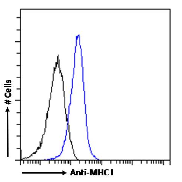
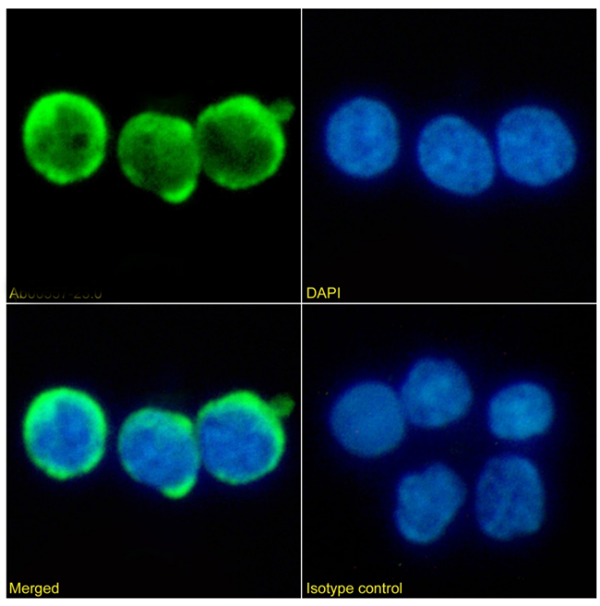
(Immunofluorescencestainingoffixedratsplenocyteswithanti-MHCI(RT1A)antibodyOX-18(AAA72457). ImmunofluorescenceanalysisofparaformaldehydefixedratsplenocytesonShi-fixcover-slipsstainedwiththechimericrabbitIgGversionofOX-18(AAA72457)(1:100dilution)for1hfollowedbyAlexaFluor488secondaryantibody(1:1000dilution),showingmembraneandcytoplasmicstaining.ThenuclearstainisDAPI(blue).Panelsshowfromleft-right,top-bottomAAA72457,DAPI,mergedchannelsandanisotypecontrol.TheisotypecontrolwasanunknownspecificityantibodyfollowedbystainingwithAlexaFluor488secondaryantibody.)
IF (Immunofluorescence)
(Immunofluorescencestainingoffixedratsplenocyteswithanti-MHCI(RT1A)antibodyOX-18(AAA72457). ImmunofluorescenceanalysisofparaformaldehydefixedratsplenocytesonShi-fixcover-slipsstainedwiththechimericrabbitIgGversionofOX-18(AAA72457)(1:100dilution)for1hfollowedbyAlexaFluor488secondaryantibody(1:1000dilution),showingmembraneandcytoplasmicstaining.ThenuclearstainisDAPI(blue).Panelsshowfromleft-right,top-bottomAAA72457,DAPI,mergedchannelsandanisotypecontrol.TheisotypecontrolwasanunknownspecificityantibodyfollowedbystainingwithAlexaFluor488secondaryantibody.)
MHC I (RT1A), Monoclonal Recombinant Antibody (Cat# AAA72457)
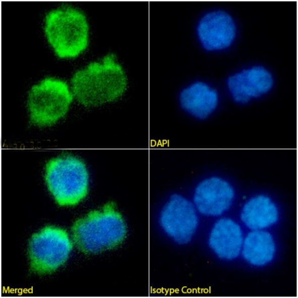 IF (Immunofluorescence)
(Immunofluorescencestainingofratsplenocyteswithanti-CD4antibodyOX-35(AAA72462). ImmunofluorescenceanalysisofparaformaldehydefixedratsplenocytesonShi-fixcoverslipsstainedwiththechimericrabbitIgGversionofOX-35(AAA72462)(1:100dilution)for1hfollowedbyAlexaFluor488secondaryantibody(1:1000dilution),showingmembraneandcytoplasmicstaining.ThenuclearstainisDAPI(blue).Panelsshow,fromleft-right,top-bottom,AAA72462,DAPI,mergedchannelsandanisotypecontrol.TheisotypecontrolwasanunknownspecificityantibodyfollowedbystainingwithAlexaFluor488secondaryantibody.)
IF (Immunofluorescence)
(Immunofluorescencestainingofratsplenocyteswithanti-CD4antibodyOX-35(AAA72462). ImmunofluorescenceanalysisofparaformaldehydefixedratsplenocytesonShi-fixcoverslipsstainedwiththechimericrabbitIgGversionofOX-35(AAA72462)(1:100dilution)for1hfollowedbyAlexaFluor488secondaryantibody(1:1000dilution),showingmembraneandcytoplasmicstaining.ThenuclearstainisDAPI(blue).Panelsshow,fromleft-right,top-bottom,AAA72462,DAPI,mergedchannelsandanisotypecontrol.TheisotypecontrolwasanunknownspecificityantibodyfollowedbystainingwithAlexaFluor488secondaryantibody.)
CD4, Monoclonal Recombinant Antibody (Cat# AAA72462)
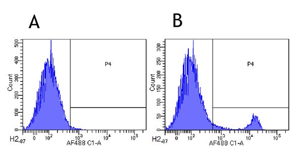
 FCM/FACS (Flow Cytometry)
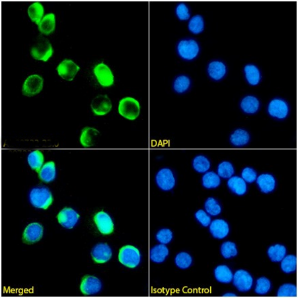
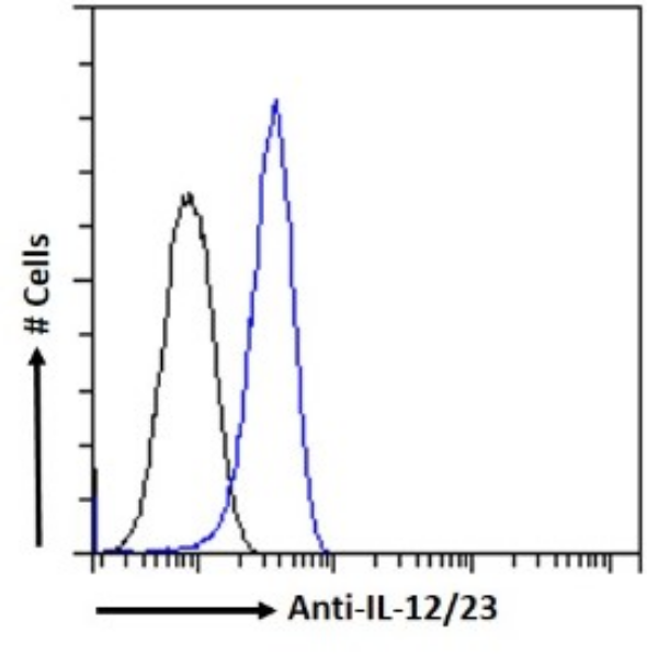
(Flowcytometryusinganti-IL-12/2antibodyABT-874(Briakinumab)(AAA72491). Paraformaldehyde-fixedJurkatcellspermeabilizedwith0.5%Tritonwerestainedwiththeanti-unknownspecificityantibodyortherabbitIgGversionofABT-874(AAA72491,blueline)atadilutionof1:100for1hatRT.Afterwashing,theboundantibodywasdetectedusingagoatanti-rabbitIgGAlexaFluor488antibodyatadilutionof1:1000,andthecellswereanalyzedusingaFACSCantoflow-cytometer.)
FCM/FACS (Flow Cytometry)
(Flowcytometryusinganti-IL-12/2antibodyABT-874(Briakinumab)(AAA72491). Paraformaldehyde-fixedJurkatcellspermeabilizedwith0.5%Tritonwerestainedwiththeanti-unknownspecificityantibodyortherabbitIgGversionofABT-874(AAA72491,blueline)atadilutionof1:100for1hatRT.Afterwashing,theboundantibodywasdetectedusingagoatanti-rabbitIgGAlexaFluor488antibodyatadilutionof1:1000,andthecellswereanalyzedusingaFACSCantoflow-cytometer.)
IL-12/23, Monoclonal Recombinant Antibody (Cat# AAA72491)
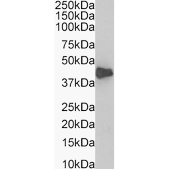
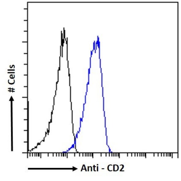
 IF (Immunofluorescence)
(ImmunofluorescencestainingofJurkatcellswithanti-CD2antibodyVIPVIIIC8(AAA72508). ImmunofluorescenceanalysisofparaformaldehydefixedJurkatcellsonShi-fixcoverslipsstainedwiththechimericrabbitIgGversionofVIPVIIIC8(AAA72508)(1:100dilution)for1hfollowedbyAlexaFluor488secondaryantibody(1:1000dilution),showingmembranestaining.ThenuclearstainisDAPI(blue).Panelsshow,fromleft-right,top-bottom,AAA72508,DAPI,mergedchannelsandanisotypecontrol.TheisotypecontrolwasanunknownspecificityantibodyfollowedbystainingwithAlexaFluor488secondaryantibody.)
IF (Immunofluorescence)
(ImmunofluorescencestainingofJurkatcellswithanti-CD2antibodyVIPVIIIC8(AAA72508). ImmunofluorescenceanalysisofparaformaldehydefixedJurkatcellsonShi-fixcoverslipsstainedwiththechimericrabbitIgGversionofVIPVIIIC8(AAA72508)(1:100dilution)for1hfollowedbyAlexaFluor488secondaryantibody(1:1000dilution),showingmembranestaining.ThenuclearstainisDAPI(blue).Panelsshow,fromleft-right,top-bottom,AAA72508,DAPI,mergedchannelsandanisotypecontrol.TheisotypecontrolwasanunknownspecificityantibodyfollowedbystainingwithAlexaFluor488secondaryantibody.)
CD2, Monoclonal Recombinant Antibody (Cat# AAA72508)
 IF (Immunofluorescence)
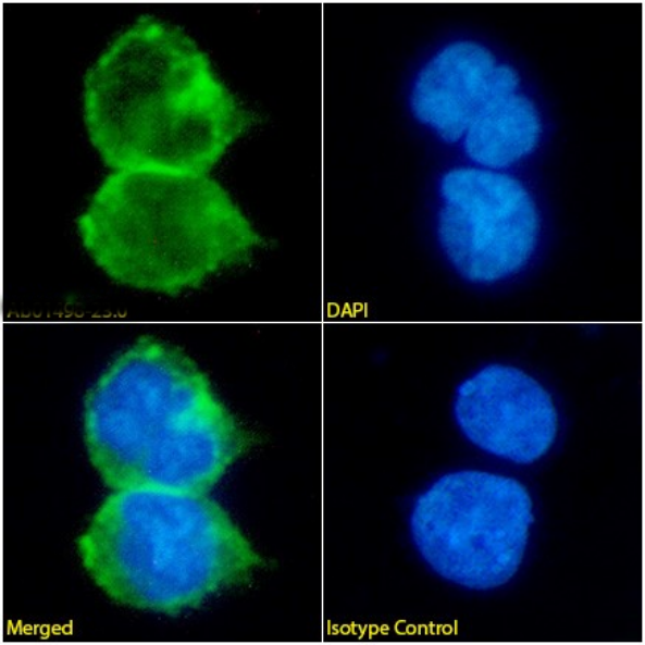
(ImmunofluorescencestainingofJurkatcellswithanti-CD8antibodyBAG45G1(AAA72517). ImmunofluorescenceanalysisofparaformaldehydefixedJurkatcellsonShi-fixcoverslipsstainedwiththechimericrabbitIgGversionofBAG45G1(AAA72517)(1:100dilution)for1hfollowedbyAlexaFluor488secondaryantibody(1:1000dilution),showingmembranestaining.ThenuclearstainisDAPI(blue).Panelsshow,fromleft-right,top-bottom,AAA72517,DAPI,mergedchannelsandanisotypecontrol.TheisotypecontrolwasanunknownspecificityantibodyfollowedbystainingwithAlexaFluor488secondaryantibody.)
IF (Immunofluorescence)
(ImmunofluorescencestainingofJurkatcellswithanti-CD8antibodyBAG45G1(AAA72517). ImmunofluorescenceanalysisofparaformaldehydefixedJurkatcellsonShi-fixcoverslipsstainedwiththechimericrabbitIgGversionofBAG45G1(AAA72517)(1:100dilution)for1hfollowedbyAlexaFluor488secondaryantibody(1:1000dilution),showingmembranestaining.ThenuclearstainisDAPI(blue).Panelsshow,fromleft-right,top-bottom,AAA72517,DAPI,mergedchannelsandanisotypecontrol.TheisotypecontrolwasanunknownspecificityantibodyfollowedbystainingwithAlexaFluor488secondaryantibody.)
CD8, Monoclonal Recombinant Antibody (Cat# AAA72517)
 IF (Immunofluorescence)
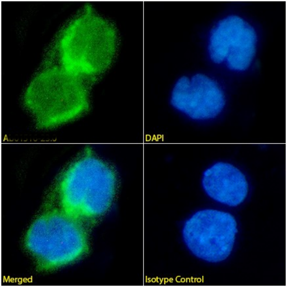
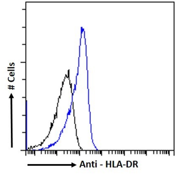
(ImmunofluorescencestainingofDaudicellswithanti-HLA-DRantibodyBAG14D6(AAA72519). ImmunofluorescenceanalysisofparaformaldehydefixedDaudicellsonShi-fixcoverslipsstainedwiththechimericrabbitIgGversionofBAG14D6(AAA72519)(1:100dilution)for1hfollowedbyAlexaFluor488secondaryantibody(1:1000dilution),showingmembranestaining.ThenuclearstainisDAPI(blue).Panelsshow,fromleft-right,top-bottom,AAA72519,DAPI,mergedchannelsandanisotypecontrol.TheisotypecontrolwasanunknownspecificityantibodyfollowedbystainingwithAlexaFluor488secondaryantibody.)
IF (Immunofluorescence)
(ImmunofluorescencestainingofDaudicellswithanti-HLA-DRantibodyBAG14D6(AAA72519). ImmunofluorescenceanalysisofparaformaldehydefixedDaudicellsonShi-fixcoverslipsstainedwiththechimericrabbitIgGversionofBAG14D6(AAA72519)(1:100dilution)for1hfollowedbyAlexaFluor488secondaryantibody(1:1000dilution),showingmembranestaining.ThenuclearstainisDAPI(blue).Panelsshow,fromleft-right,top-bottom,AAA72519,DAPI,mergedchannelsandanisotypecontrol.TheisotypecontrolwasanunknownspecificityantibodyfollowedbystainingwithAlexaFluor488secondaryantibody.)
HLA-DR, Monoclonal Recombinant Antibody (Cat# AAA72519)
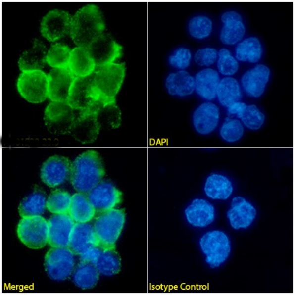
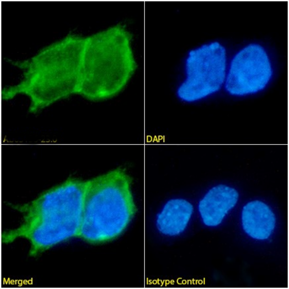
 IF (Immunofluorescence)
(ImmunofluorescencestainingoffixedHeLacellswithanti-desmoglein-3antibody5G11(AAA72527) ImmunofluorescenceanalysisofparaformaldehydefixedA431cellsonShi-fixcoverslipsstainedwiththechimericrabbitIgGversionofHeLacells(AAA72527)at10ug/mlfor1hfollowedbyAlexaFluor488secondaryantibody(2ug/ml),showingmembranestaining.ThenuclearstainisDAPI(blue).Panelsshowfromleft-right,top-bottomAAA72527,DAPI,mergedchannelsandanisotypecontrol.TheisotypecontrolwasanunknownspecificityantibodyfollowedbystainingwithAlexaFluor488secondaryantibody.)
IF (Immunofluorescence)
(ImmunofluorescencestainingoffixedHeLacellswithanti-desmoglein-3antibody5G11(AAA72527) ImmunofluorescenceanalysisofparaformaldehydefixedA431cellsonShi-fixcoverslipsstainedwiththechimericrabbitIgGversionofHeLacells(AAA72527)at10ug/mlfor1hfollowedbyAlexaFluor488secondaryantibody(2ug/ml),showingmembranestaining.ThenuclearstainisDAPI(blue).Panelsshowfromleft-right,top-bottomAAA72527,DAPI,mergedchannelsandanisotypecontrol.TheisotypecontrolwasanunknownspecificityantibodyfollowedbystainingwithAlexaFluor488secondaryantibody.)
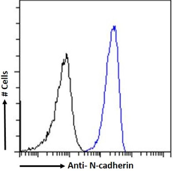
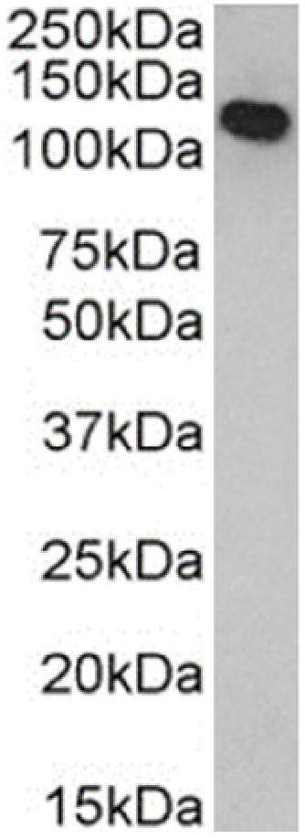
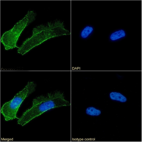
N-cadherin, Monoclonal Recombinant Antibody (Cat# AAA72527)
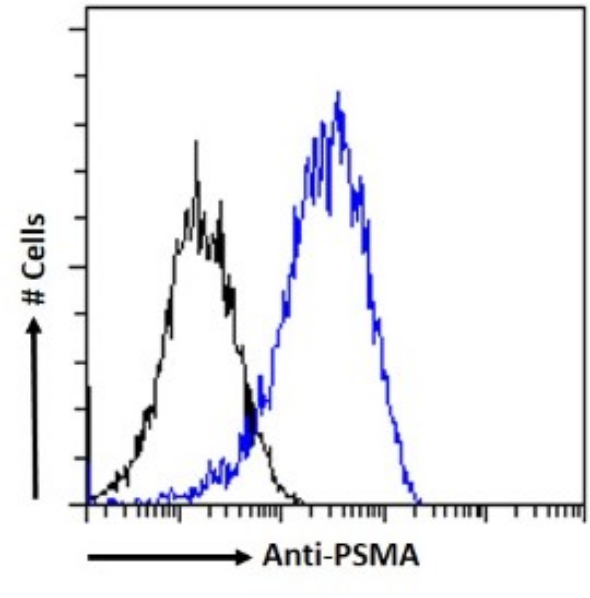 FCM/FACS (Flow Cytometry)
(Flowcytometryusinganti-PSMAantibodyJ591(AAA72547). LNCaPcellswerestainedwiththeanti-unknownspecificityantibodyortherabbitIgGversionofJ591(AAA72547,blueline)atadilutionof1:100for1hatRT.Afterwashing,theboundantibodywasdetectedusingagoatanti-rabbitIgGAlexaFluor488antibodyatadilutionof1:1000,andthecellswereanalyzedusingaFACSCantoflow-cytometer.)
FCM/FACS (Flow Cytometry)
(Flowcytometryusinganti-PSMAantibodyJ591(AAA72547). LNCaPcellswerestainedwiththeanti-unknownspecificityantibodyortherabbitIgGversionofJ591(AAA72547,blueline)atadilutionof1:100for1hatRT.Afterwashing,theboundantibodywasdetectedusingagoatanti-rabbitIgGAlexaFluor488antibodyatadilutionof1:1000,andthecellswereanalyzedusingaFACSCantoflow-cytometer.)
PSMA, Monoclonal Recombinant Antibody (Cat# AAA72547)
Fibronectin, Monoclonal Recombinant Antibody (Cat# AAA72553)

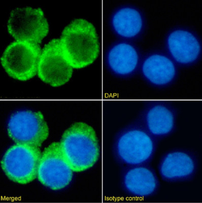
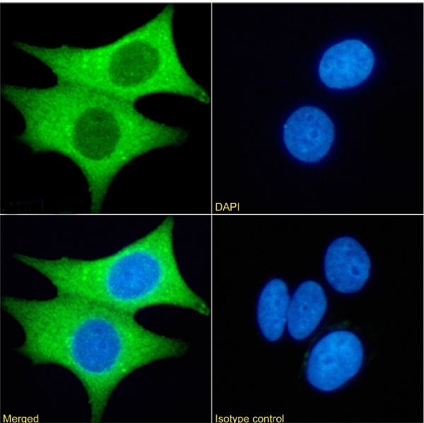
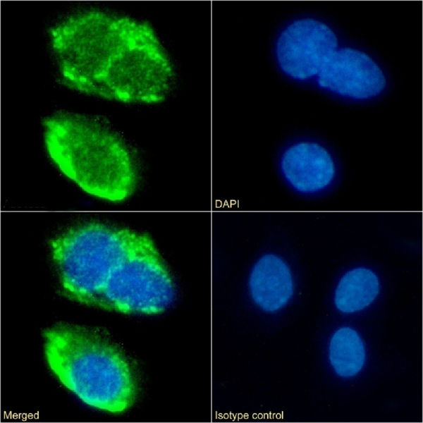 IF (Immunofluorescence)
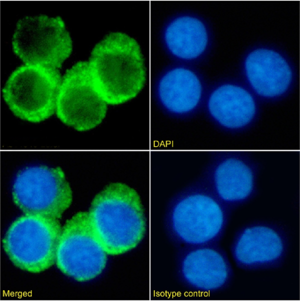
(Immunofluorescence staining of fixed HepG2 cells with anti-CD36 antibody 185-1G2 (B467) (AAA72155) Immunofluorescence analysis of paraformaldehyde fixed HepG2 cells on Shi-fix coverslips stained with the chimeric rabbit IgG version of 185-1G2 (B467) at 10ug/ml for 1h followed by Alexa Fluor 488 secondary antibody (2ug/ml), showing membrane staining. The nuclear stain is DAPI (blue). Panels show from left-right, top-bottom DAPI, merged channels and an isotype control. The isotype control was an unknown specificity antibody followed by staining with Alexa Fluor 488 secondary antibody.)
IF (Immunofluorescence)
(Immunofluorescence staining of fixed HepG2 cells with anti-CD36 antibody 185-1G2 (B467) (AAA72155) Immunofluorescence analysis of paraformaldehyde fixed HepG2 cells on Shi-fix coverslips stained with the chimeric rabbit IgG version of 185-1G2 (B467) at 10ug/ml for 1h followed by Alexa Fluor 488 secondary antibody (2ug/ml), showing membrane staining. The nuclear stain is DAPI (blue). Panels show from left-right, top-bottom DAPI, merged channels and an isotype control. The isotype control was an unknown specificity antibody followed by staining with Alexa Fluor 488 secondary antibody.)
CD36, Monoclonal Antibody (Cat# AAA72155)
 IF (Immunofluorescence)
(Immunofluorescence staining of fixed HepG2 cells with anti-CD36 antibody 185-1G2 (B467) Immunofluorescence analysis of paraformaldehyde fixed HepG2 cells on Shi-fix coverslips stained with the chimeric rabbit IgG version of 185-1G2 (B467) () at 10ug/ml for 1h followed by Alexa Fluor 488 secondary antibody (2ug/ml), showing membrane staining. The nuclear stain is DAPI (blue). Panels show from left-right, top-bottom , DAPI, merged channels and an isotype control. The isotype control was an unknown specificity antibody followed by staining with Alexa Fluor 488 secondary antibody.)
IF (Immunofluorescence)
(Immunofluorescence staining of fixed HepG2 cells with anti-CD36 antibody 185-1G2 (B467) Immunofluorescence analysis of paraformaldehyde fixed HepG2 cells on Shi-fix coverslips stained with the chimeric rabbit IgG version of 185-1G2 (B467) () at 10ug/ml for 1h followed by Alexa Fluor 488 secondary antibody (2ug/ml), showing membrane staining. The nuclear stain is DAPI (blue). Panels show from left-right, top-bottom , DAPI, merged channels and an isotype control. The isotype control was an unknown specificity antibody followed by staining with Alexa Fluor 488 secondary antibody.)
CD36, Monoclonal Antibody (Cat# AAA72156)
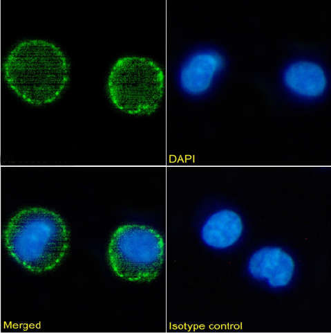
 FCM/FACS (Flow Cytometry)
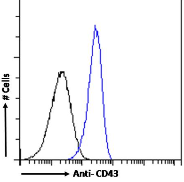
(Flow cytometry using the anti-CD43 antibody 84-3C1 . K562 cells were fixed using 2% PFA and stained with anti-unknown specificity antibody or the rabbit IgG1 version of 84-3C1 at a dilution of 1:100 for 1h at RT. After washing, the bound antibody was detected using a goat anti-rabbit IgG AlexaFluor 488 antibody at a dilution of 1:1000 and cells analyzed using a FACSCanto flow-cytometer.)
FCM/FACS (Flow Cytometry)
(Flow cytometry using the anti-CD43 antibody 84-3C1 . K562 cells were fixed using 2% PFA and stained with anti-unknown specificity antibody or the rabbit IgG1 version of 84-3C1 at a dilution of 1:100 for 1h at RT. After washing, the bound antibody was detected using a goat anti-rabbit IgG AlexaFluor 488 antibody at a dilution of 1:1000 and cells analyzed using a FACSCanto flow-cytometer.)
CD43, Monoclonal Antibody (Cat# AAA72160)

 FCM/FACS (Flow Cytometry)
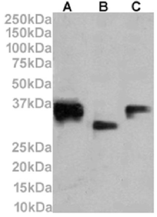
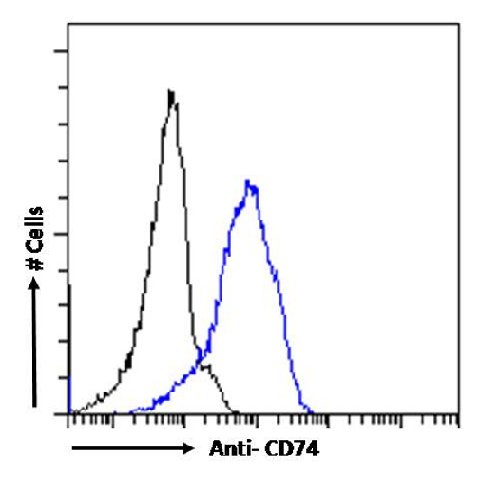
(Flow cytometry using the Anti-CD74 antibody LN-2 . Paraformaldehyde fixed Daudi cells were stained with anti-unknown specificity antibody or the rabbit IgG version of LN-2 at a dilution of 1:100 for 1h at RT. After washing, the bound antibody was detected using a goat anti-rabbit IgG AlexaFluor 488 antibody at a dilution of 1:1000 and cells analyzed using a FACSCanto flow-cytometer.)
FCM/FACS (Flow Cytometry)
(Flow cytometry using the Anti-CD74 antibody LN-2 . Paraformaldehyde fixed Daudi cells were stained with anti-unknown specificity antibody or the rabbit IgG version of LN-2 at a dilution of 1:100 for 1h at RT. After washing, the bound antibody was detected using a goat anti-rabbit IgG AlexaFluor 488 antibody at a dilution of 1:1000 and cells analyzed using a FACSCanto flow-cytometer.)
CD74, Monoclonal Antibody (Cat# AAA72165)
 FCM/FACS (Flow Cytometry)
(Flow cytometry using the Anti-CD74 antibody LN-2 (AAA72166). Paraformaldehyde fixed Daudi cells were stained with anti-unknown specificity antibody followed by staining with Alexa Fluor 488 secondary antibody.)
FCM/FACS (Flow Cytometry)
(Flow cytometry using the Anti-CD74 antibody LN-2 (AAA72166). Paraformaldehyde fixed Daudi cells were stained with anti-unknown specificity antibody followed by staining with Alexa Fluor 488 secondary antibody.)
CD74, Monoclonal Antibody (Cat# AAA72166)
 IF (Immunofluorescence)
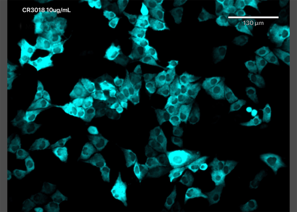
(Immunofluorescence staining of MDCK-SIAT1 cells transfected with SARS-CoV-2 NP with anti-Covid-19 & SARS-CoV Nucleoprotein antibody CR3018 (03-018) (AAA72196) Immunofluorescence analysis of MDCK-SIAT1 cells stably transfected with SARS-CoV-2 NP. The cells were seeded in a flat bottomed 96 well plate overnight, fixed in 10% formalin at 4C for 30min, permeabilised for 20min at RT and then stained with the human IgG1 version of CR3018 (03-018) in PBS/0.1% BSA at 10ug/ml for 1 hour followed by a goat anti-human Alexa Fluor 647 (Invitrogen) secondary antibody. The image is courtesy of Jack Tan, Radcliffe Department of Medicine, University of Oxford.)
IF (Immunofluorescence)
(Immunofluorescence staining of MDCK-SIAT1 cells transfected with SARS-CoV-2 NP with anti-Covid-19 & SARS-CoV Nucleoprotein antibody CR3018 (03-018) (AAA72196) Immunofluorescence analysis of MDCK-SIAT1 cells stably transfected with SARS-CoV-2 NP. The cells were seeded in a flat bottomed 96 well plate overnight, fixed in 10% formalin at 4C for 30min, permeabilised for 20min at RT and then stained with the human IgG1 version of CR3018 (03-018) in PBS/0.1% BSA at 10ug/ml for 1 hour followed by a goat anti-human Alexa Fluor 647 (Invitrogen) secondary antibody. The image is courtesy of Jack Tan, Radcliffe Department of Medicine, University of Oxford.)
COVID 19 Nucleocapsid (NP) Coronavirus, Monoclonal Antibody (Cat# AAA72196)
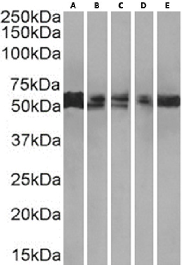 WB (Western Blot)
(Western Blot using anti-PRMT3 antibody RAB-C372 (AAA72212) HEK293(A), HeLa(B), HepG2(C), MCF7(D) and A549(E) cell lysate (35ug protein in RIPA buffer) was resolved on a SDS PAGE gel and blots were probed with the chimeric rabbit version of RAB-C372 at 0.001ug/ml before detection using an anti-rabbit secondary antibody. A primary incubation of 1h was used and protein was detected by chemiluminescence.)
WB (Western Blot)
(Western Blot using anti-PRMT3 antibody RAB-C372 (AAA72212) HEK293(A), HeLa(B), HepG2(C), MCF7(D) and A549(E) cell lysate (35ug protein in RIPA buffer) was resolved on a SDS PAGE gel and blots were probed with the chimeric rabbit version of RAB-C372 at 0.001ug/ml before detection using an anti-rabbit secondary antibody. A primary incubation of 1h was used and protein was detected by chemiluminescence.)
PRMT3, Monoclonal Antibody (Cat# AAA72212)
 FCM/FACS (Flow Cytometry)
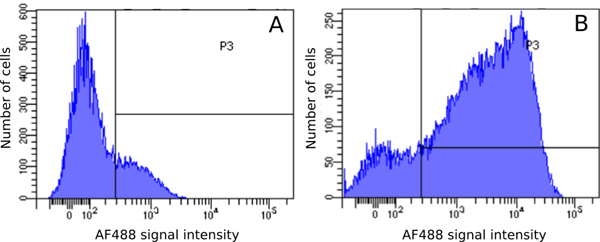
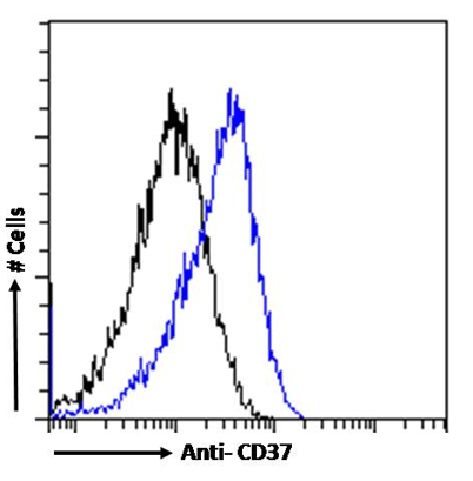
(Flow cytometry using the Anti-CD37 antibody MB-1 . Paraformaldehyde fixed Daudi cells were stained with anti-unknown specificity antibody or the mouse IgG1 version of MB-1 (, blue line) at a dilution of 1:100 for 1h at RT. After washing, the bound antibody was detected using a goat anti-mouse IgG AlexaFluor 488 antibody at a dilution of 1:1000 and cells analyzed using a FACSCanto flow-cytometer.)
FCM/FACS (Flow Cytometry)
(Flow cytometry using the Anti-CD37 antibody MB-1 . Paraformaldehyde fixed Daudi cells were stained with anti-unknown specificity antibody or the mouse IgG1 version of MB-1 (, blue line) at a dilution of 1:100 for 1h at RT. After washing, the bound antibody was detected using a goat anti-mouse IgG AlexaFluor 488 antibody at a dilution of 1:1000 and cells analyzed using a FACSCanto flow-cytometer.)
CD37, Monoclonal Antibody (Cat# AAA72086)
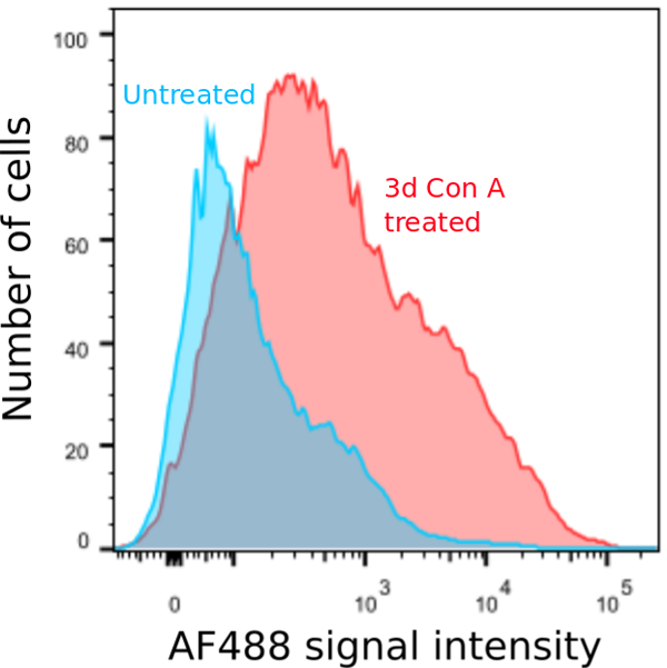
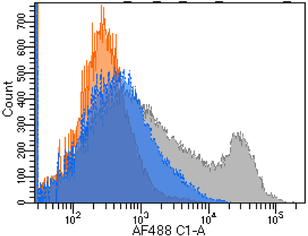
 FCM/FACS (Flow Cytometry)
(Flow-cytometry using the Anti-Ly6G/Ly6C antibody RB6-8C5 (AAA72103). Mouse splenocytes were stained with anti-Fluorescein IgG antibody (4-4-20; isotype control, black line) or the rabbit IgG version of RB6-8C5 (, blue line) at a dilution of 1:100 for 1h at RT. After washing, bound antibody was detected using a goat anti-rabbit IgG AlexaFluor 488 antibody at a dilution of 1:1000 and cells analyzed using a FACSCanto flow-cytometer.)
FCM/FACS (Flow Cytometry)
(Flow-cytometry using the Anti-Ly6G/Ly6C antibody RB6-8C5 (AAA72103). Mouse splenocytes were stained with anti-Fluorescein IgG antibody (4-4-20; isotype control, black line) or the rabbit IgG version of RB6-8C5 (, blue line) at a dilution of 1:100 for 1h at RT. After washing, bound antibody was detected using a goat anti-rabbit IgG AlexaFluor 488 antibody at a dilution of 1:1000 and cells analyzed using a FACSCanto flow-cytometer.)
Ly6G/Ly6C, Monoclonal Antibody (Cat# AAA72103)
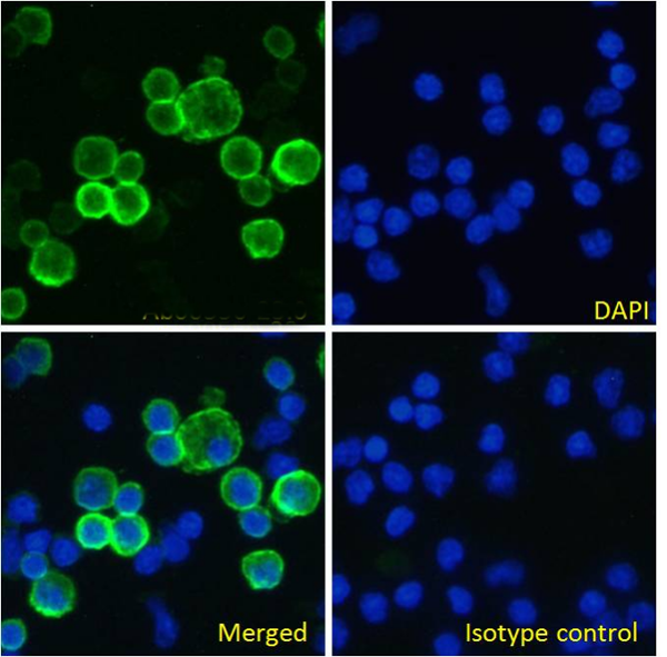
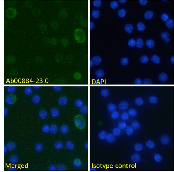
 IF (Immunofluorescence)
( Immunofluorescence staining of fixed mouse splenocytes with anti-GITR (Tumor necrosis factor ligand superfamily member 18) antibody YGITR 860.103.5 Immunofluorescence analysis of paraformaldehyde fixed mouse (Mus musculus) splenocytes on Shi-fix coverslips, permeabilized with 0.15% Triton stained with the chimeric rabbit version of YGITR 860.103.5 at 10 ug/ml for 1h followed by Alexa Fluor 488 secondary antibody (1 ug/ml), showing membrane and some cytoplasmic staining in a subset of cells. The nuclear stain is DAPI (blue). Panels show from left-right, top-bottom DAPI, merged channels and an isotype control. The isotype control was stained with an anti-Fluorescein antibody followed by Alexa Fluor 488 secondary antibody.)
IF (Immunofluorescence)
( Immunofluorescence staining of fixed mouse splenocytes with anti-GITR (Tumor necrosis factor ligand superfamily member 18) antibody YGITR 860.103.5 Immunofluorescence analysis of paraformaldehyde fixed mouse (Mus musculus) splenocytes on Shi-fix coverslips, permeabilized with 0.15% Triton stained with the chimeric rabbit version of YGITR 860.103.5 at 10 ug/ml for 1h followed by Alexa Fluor 488 secondary antibody (1 ug/ml), showing membrane and some cytoplasmic staining in a subset of cells. The nuclear stain is DAPI (blue). Panels show from left-right, top-bottom DAPI, merged channels and an isotype control. The isotype control was stained with an anti-Fluorescein antibody followed by Alexa Fluor 488 secondary antibody.)
GITR, Monoclonal Recombinant Antibody (Cat# AAA71991)
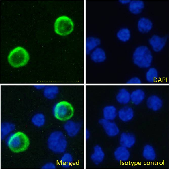
 IF (Immunofluorescence)
( Immunofluorescence staining of fixed mouse splenocytes with anti-B7-H3 (CD276) antibody MJ18 Immunofluorescence analysis of paraformaldehyde fixed mouse (Mus musculus) splenocytes on Shi-fix coverslips, permeabilized with 0.15% Triton stained with the chimeric rabbit version of MJ18 at 10 ug/ml for 1h followed by Alexa Fluor 488 secondary antibody (1 ug/ml), showing weak membrane staining of a subset of cells. The nuclear stain is DAPI (blue). Panels show from left-right, top-bottom DAPI, merged channels and an isotype control. The isotype control was stained with an anti-Fluorescein antibody followed by Alexa Fluor 488 secondary antibody)
IF (Immunofluorescence)
( Immunofluorescence staining of fixed mouse splenocytes with anti-B7-H3 (CD276) antibody MJ18 Immunofluorescence analysis of paraformaldehyde fixed mouse (Mus musculus) splenocytes on Shi-fix coverslips, permeabilized with 0.15% Triton stained with the chimeric rabbit version of MJ18 at 10 ug/ml for 1h followed by Alexa Fluor 488 secondary antibody (1 ug/ml), showing weak membrane staining of a subset of cells. The nuclear stain is DAPI (blue). Panels show from left-right, top-bottom DAPI, merged channels and an isotype control. The isotype control was stained with an anti-Fluorescein antibody followed by Alexa Fluor 488 secondary antibody)
B7-H3, Monoclonal Recombinant Antibody (Cat# AAA72012)
 WB (Western Blot)
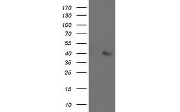
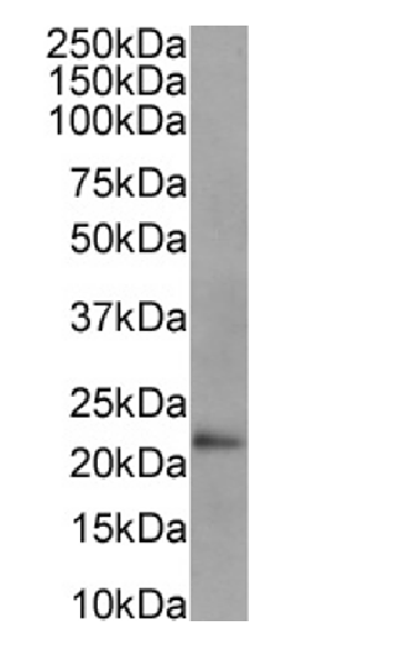
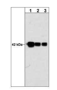
(Western Blot using anti-Cardiac Troponin I antibody scFv 180 Human heart lysate (35ug protein in RIPA buffer) was resolved on a 10% SDS PAGE gel and blots probed with the chimeric mouse IgG1 version of scFv 180 (AAA72032) at 0.001 ug/ml before detection using an anti-mouse secondary antibody. A primary incubation of 1h was used and protein was detected by chemiluminescence. The predicted running size for Cardiac Troponin I is 24.0 kDa. AAA72032 successfully detected Cardiac Troponin I in human heart lysate.)
WB (Western Blot)
(Western Blot using anti-Cardiac Troponin I antibody scFv 180 Human heart lysate (35ug protein in RIPA buffer) was resolved on a 10% SDS PAGE gel and blots probed with the chimeric mouse IgG1 version of scFv 180 (AAA72032) at 0.001 ug/ml before detection using an anti-mouse secondary antibody. A primary incubation of 1h was used and protein was detected by chemiluminescence. The predicted running size for Cardiac Troponin I is 24.0 kDa. AAA72032 successfully detected Cardiac Troponin I in human heart lysate.)
Cardiac Troponin I, Monoclonal Recombinant Antibody (Cat# AAA72032)
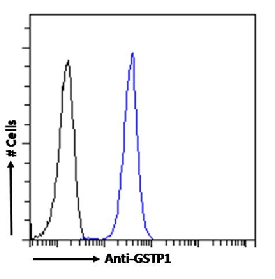
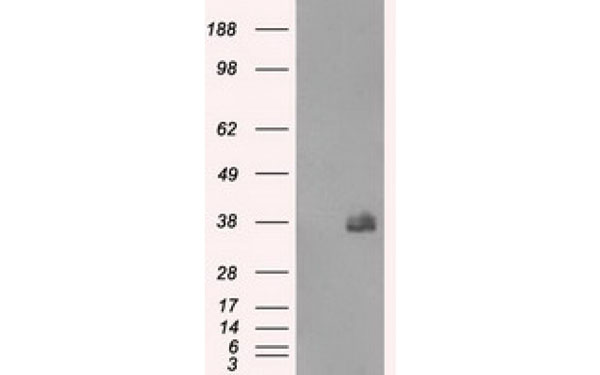
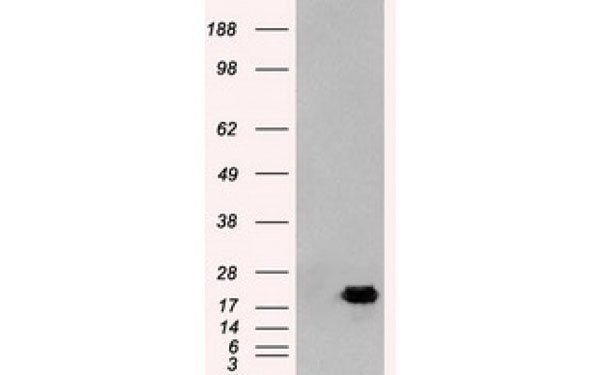
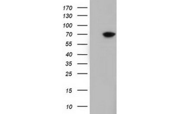
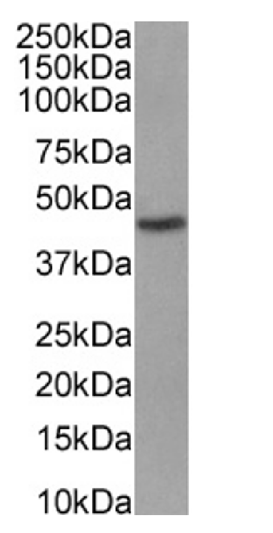
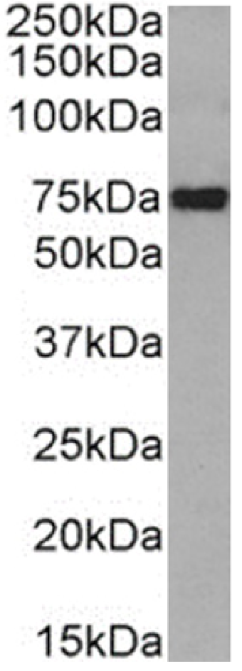
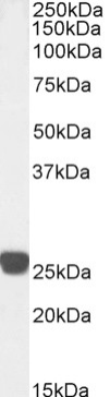
 WB (Western Blot)
(Western Blot using anti-GSTP1 antibody SAIC-22D-22 (AAA72072). Human liver tissue lysates (35ug protein in RIPA buffer) were resolved on a SDS PAGE gel and blots were probed with the chimeric rabbit version of SAIC-22D-22 () at 1ug/ml before detection using an anti-rabbit secondary antibody. A primary incubation of 1h was used and protein was detected by chemiluminescence.)
WB (Western Blot)
(Western Blot using anti-GSTP1 antibody SAIC-22D-22 (AAA72072). Human liver tissue lysates (35ug protein in RIPA buffer) were resolved on a SDS PAGE gel and blots were probed with the chimeric rabbit version of SAIC-22D-22 () at 1ug/ml before detection using an anti-rabbit secondary antibody. A primary incubation of 1h was used and protein was detected by chemiluminescence.)
GSTP1, Monoclonal Antibody (Cat# AAA72072)
HBcAg core IgG2a, Monoclonal Antibody (Cat# AAA71872)
 IHC (Immunohistochemistry)
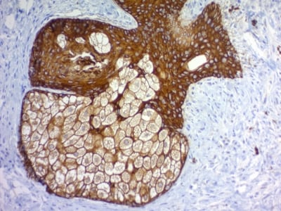
(Formalin-fixed, paraffin-embedded human Skin stained with Cytokeratin, pan Monoclonal Antibody cocktail (PAN-CK).)
IHC (Immunohistochemistry)
(Formalin-fixed, paraffin-embedded human Skin stained with Cytokeratin, pan Monoclonal Antibody cocktail (PAN-CK).)
Cytokeratin, pan, Monoclonal Antibody (Cat# AAA62906)
 FCM/FACS (Flow Cytometry)
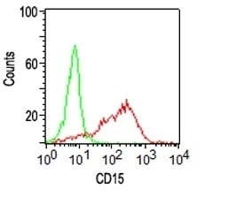
(FACS analysis of human Monocytes using CD15 Monoclonal Antibody (Leu-M1).)
FCM/FACS (Flow Cytometry)
(FACS analysis of human Monocytes using CD15 Monoclonal Antibody (Leu-M1).)
CD15 / FUT4, Monoclonal Antibody (Cat# AAA62941)
HBeAg, Monoclonal Antibody (Cat# AAA72737)
Purification Method: DEAE Purified
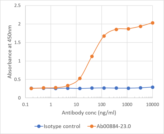
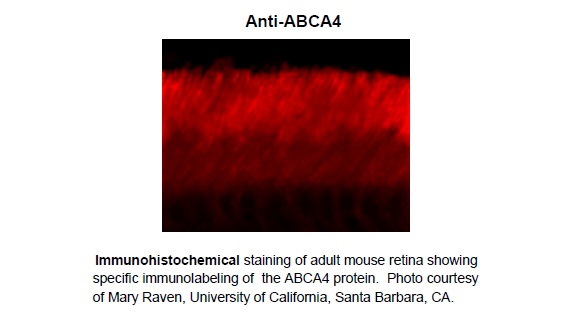 Application Data
ABCA4 (Rim Protein), Monoclonal Antibody (Cat# AAA72744)
 FCM/FACS (Flow Cytometry)
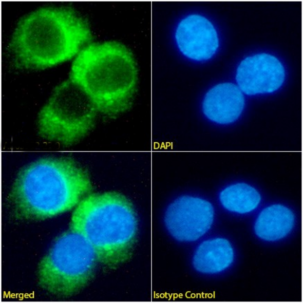
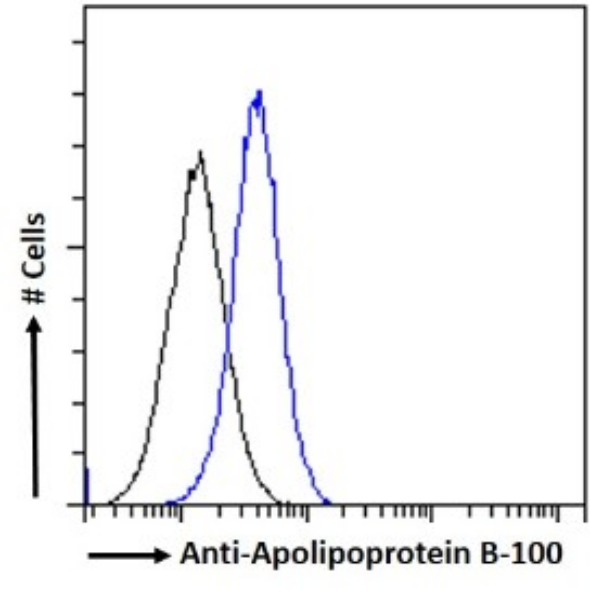
(Flowcytometryusinganti-ApolipoproteinB-100antibodyMabB23(AAA72564). ParaformaldehydefixedHepG2cellspermeabilizedwith0.5%Tritonwerestainedwiththeanti-unknownspecificityantibodyortherabbitIgGversionofMabB23(AAA72564,blueline)atadilutionof1:100for1hatRT.Afterwashing,theboundantibodywasdetectedusingagoatanti-rabbitIgGAlexaFluor488antibodyatadilutionof1:1000,andthecellswereanalyzedusingaFACSCantoflow-cytometer.)
FCM/FACS (Flow Cytometry)
(Flowcytometryusinganti-ApolipoproteinB-100antibodyMabB23(AAA72564). ParaformaldehydefixedHepG2cellspermeabilizedwith0.5%Tritonwerestainedwiththeanti-unknownspecificityantibodyortherabbitIgGversionofMabB23(AAA72564,blueline)atadilutionof1:100for1hatRT.Afterwashing,theboundantibodywasdetectedusingagoatanti-rabbitIgGAlexaFluor488antibodyatadilutionof1:1000,andthecellswereanalyzedusingaFACSCantoflow-cytometer.)
Apolipoprotein B-100, Monoclonal Recombinant Antibody (Cat# AAA72564)
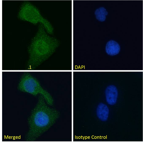
 ICC (Immunocytochemistry)
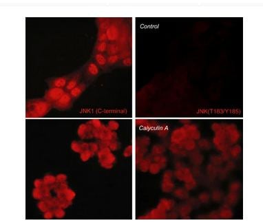
(Immunocytochemical labeling of JNK in control (Top row) or calyculin A-treated A431 cells (Bottom row). The cells were labeled with mouse monoclonal JNK (C-terminal region) (Left) or mouse monoclonal JNK (Thr-183/Tyr-185) (Right). The antibodies were detected using goat anti-mouse DyLight 594.)
ICC (Immunocytochemistry)
(Immunocytochemical labeling of JNK in control (Top row) or calyculin A-treated A431 cells (Bottom row). The cells were labeled with mouse monoclonal JNK (C-terminal region) (Left) or mouse monoclonal JNK (Thr-183/Tyr-185) (Right). The antibodies were detected using goat anti-mouse DyLight 594.)
JNK1, Monoclonal Antibody (Cat# AAA71652)
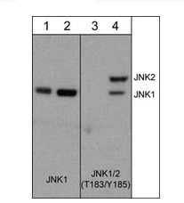
 WB (Western Blot)
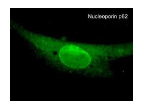
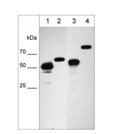
(Western blot image of cell structure markers in NCI-H1915 lung carcinoma cells. The blot was probed with anti-Vimentin intermediate filament protein VM4341 (lane 1), anti-Nucleoporin p62nM4361 (lane 2), anti-Hsp60 mitochondrial protein HM4381 (lane 3), and anti-Calnexin endoplasmic reticulum protein CM4371 (lane 4).)
WB (Western Blot)
(Western blot image of cell structure markers in NCI-H1915 lung carcinoma cells. The blot was probed with anti-Vimentin intermediate filament protein VM4341 (lane 1), anti-Nucleoporin p62nM4361 (lane 2), anti-Hsp60 mitochondrial protein HM4381 (lane 3), and anti-Calnexin endoplasmic reticulum protein CM4371 (lane 4).)
Nucleoporin p62, Monoclonal Antibody (Cat# AAA71674)
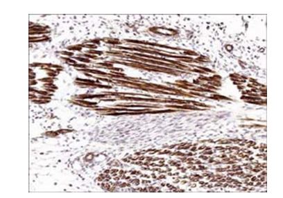 IHC (Immunohiostchemistry)
(Formalin fixed, citric acid treated parafin sections of E18 mouse skeletal muscle. Sections were probed with anti-Actin (AM2021) then anti-Mouse:HRP before detection using DAB. (Images provided by Carl Hobbs and Dr. Pat Doherty at Wolfson Centre for Age-Related Diseases, King's College London).)
IHC (Immunohiostchemistry)
(Formalin fixed, citric acid treated parafin sections of E18 mouse skeletal muscle. Sections were probed with anti-Actin (AM2021) then anti-Mouse:HRP before detection using DAB. (Images provided by Carl Hobbs and Dr. Pat Doherty at Wolfson Centre for Age-Related Diseases, King's College London).)
Actin, Monoclonal Antibody (Cat# AAA71559)
 IHC (Immunohistochemistry)
(Human HPV infected cervical tissue statined with Mouse anti HPV 16 monoclonal antibody (AAA71316) at 1:100 for 30 min. at RT. Staining of formalin-fixed tissue requires boiling tissue sections in 10 mM citrate buffer, pH 6.0 for 10 min followed by cooling at RT for 20 min.)
IHC (Immunohistochemistry)
(Human HPV infected cervical tissue statined with Mouse anti HPV 16 monoclonal antibody (AAA71316) at 1:100 for 30 min. at RT. Staining of formalin-fixed tissue requires boiling tissue sections in 10 mM citrate buffer, pH 6.0 for 10 min followed by cooling at RT for 20 min.)
HPV 16, Monoclonal Antibody (Cat# AAA71316)
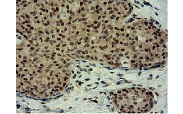
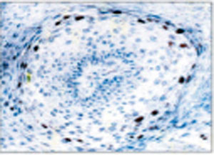
 IHC (Immunohistochemistry)
(IHC: The Tonsil tissue was stained with mouse anti BRCA1 antibody (Cat# AAA71341) at 1:200. (Note: formalin-fixed, paraffinembedded sections need 15 minutes heat-induced epitope retrieval in 10 mM citrate buffer, pH 6.0, and 30 minutes incubation at room temperature with the primary antibody)
IHC (Immunohistochemistry)
(IHC: The Tonsil tissue was stained with mouse anti BRCA1 antibody (Cat# AAA71341) at 1:200. (Note: formalin-fixed, paraffinembedded sections need 15 minutes heat-induced epitope retrieval in 10 mM citrate buffer, pH 6.0, and 30 minutes incubation at room temperature with the primary antibody)
BRCA1, Monoclonal Antibody (Cat# AAA71341)
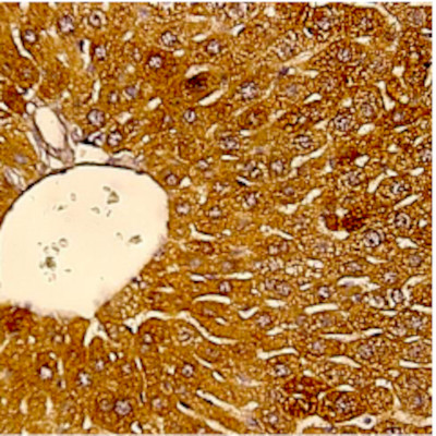 IHC (Immunohistochemistry)
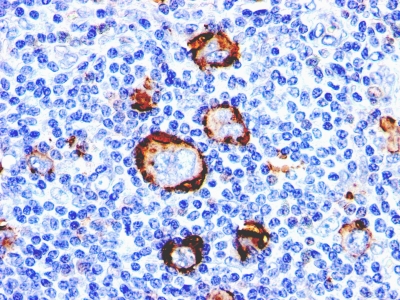
(Immunohistochemistry: Human liver carcinoma (FFPE) stained with Mouse anti-PETH (Clone ZY434) (Cat#AAA71370) at 1:200 for 10 min @ RT. Staining of formalin-fixed tissue requires boiling tissue sections in 10 mM Citrate Buffer, pH 6.0 for 10 min followed by cooling at RT for 20 min.)
IHC (Immunohistochemistry)
(Immunohistochemistry: Human liver carcinoma (FFPE) stained with Mouse anti-PETH (Clone ZY434) (Cat#AAA71370) at 1:200 for 10 min @ RT. Staining of formalin-fixed tissue requires boiling tissue sections in 10 mM Citrate Buffer, pH 6.0 for 10 min followed by cooling at RT for 20 min.)
PETH, Monoclonal Antibody (Cat# AAA71370)
What are Monoclonal Antibodies?
Monoclonal antibodies are specialized laboratory-produced proteins developed for binding to specific biological antigens or other molecular targets. Since they come from a single cell (or clone), they are especially consistent and accurate in the data they are involved in producing.
This type of antibody material has been shown to be a powerful tool in finding and subsequently destroying harmful cells in an organism, such as those found in cancers or various autoimmune diseases. This makes them excellent aids in medical testing and research, which is why they are so widely used.
AAA Biotech offers a comprehensive range of high-quality monoclonal antibodies that perform effectively in various laboratory tests, including (amongst others) ELISA, western blotting, immunohistochemistry, and flow cytometry. All of the products in our catalog are thoroughly quality tested to make sure that they are reliable and will consistently perform well in your research.
What Are The Uses of Monoclonal Antibodies
Monoclonal antibodies are used in many lab tests, including (amongst others) ELISA, western blotting, immunohistochemistry, and flow cytometry.
ELISA is a test that helps detect a specific substance/analyte in a sample. It uses antibodies (often monoclonal) bound to a solid surface (such as the well of a microplate) to “capture” the substance/analyte in the sample and immobilize it so that the detection antibody component can then bind to it and produce a signal, which can then be measured.
Western blotting identifies specific proteins in a sample. The sample is first separated on a gel, and then antibodies are applied that will typically bind to the target, which will all be localized to a single band in a lane.
Immunohistochemistry helps locate specific proteins in cells or tissue samples using antibodies.
Flow cytometry looks at and sorts cells. It uses antibodies that are conjugated to reporter molecules called “fluorophores”, which, under special lights, emit light themselves, which can then be measured by a detector instrument.
How Monoclonal Antibodies Are Used as Medicine?
Please note that all of the products listed in AAA Biotech’s also known as AAA Bio or AAABio catalog are strictly for research-use only (RUO).
Monoclonal antibodies can also be used as therapeutic/medical treatments, particularly in the context of cancers. They are designed to find and bind to specific cells or proteins, helping the immune system recognize and attack the cancer. These treatments work in different ways, such as:
- Radioimmunotherapy attaches a small amount of radioactive molecule to the antibody, so it delivers the radiation directly to the cancer cells that the antibody is specifically binding to.
- Antibody-directed enzyme prodrug therapy uses antibodies that are specifically bound to special enzymes. These enzymes activate a harmless drug in the body and turn it into a cancer-killing drug only near the cancer cells—this helps avoid harming healthy cells.
- Immunoliposomes are tiny “bubbles” filled with medicine/drug and coated with antibodies. They carry the drug straight to the cancer cells.
Why Buy Monoclonal Antibodies From Us?
At AAA Biotech, we provide high-performance monoclonal antibodies designed to support a wide range of research needs.
1. Validated for Versatile Applications
The antibodies in our catalog are extensively validated and compatible with multiple techniques, including (but not limited to) ELISA, flow cytometry (FC), immunocytochemistry (ICC), immunofluorescence (IF), immunohistochemistry (IHC), immunoprecipitation (IP), and western blotting (WB).
2. Wide Selection & Specialized Options
We offer antibodies for common and rare species, that are available in various conjugated forms, and also in recombinant formats. Essentially, there is almost anything one might need to meet their experimental model’s requirements.
3. High-Quality Proteins
Our proteins meet high purity standards—90% or more as confirmed by SDS-PAGE. Many are available with tags like His, Flag, GST, or MBP, and we also supply native and biologically active proteins for functional studies.
Frequently Asked Questions
1. Are your monoclonal antibodies validated for specific applications?
Yes, our antibodies are tested and validated for use in methods such as ELISA, western blot, IHC, flow cytometry, and more. Refer to specific product pages or datasheets for individual product information.
2. How do I choose the right monoclonal antibody for my application?
Review the product details directly for application validation, species reactivity, and target information. You may also contact our support team at any time for help.
3. How quickly can I receive my order?
Most orders are processed and shipped within 1–3 business days, depending on product availability and your shipping location.