Filters
▼Clonality
▼Type
▼Reactivity
▼Gene Name
▼Isotype
▼Host
▼Application
▼Clone
▼Monoclonal Antibodies
Get accurate results in your research with our Monoclonal Antibodies, which are specially made to target exactly what you require for your research, and will produce consistent, reliable performance in lab tests.
Viewing 8250-8300 of 27597 product results
 IP (Immunoprecipitation)
(Dilution: WB (1/2000 - 1/5000), IF/IC (1/200 - 1/500), IP (1/100 - 1/200))
IP (Immunoprecipitation)
(Dilution: WB (1/2000 - 1/5000), IF/IC (1/200 - 1/500), IP (1/100 - 1/200))
V5-tag, Monoclonal Antibody (Cat# AAA293369)
 IP (Immunoprecipitation)
(Dilution: WB (1/2000 - 1/5000), IF/IC (1/200 - 1/500), IP (1/100 - 1/200))
IP (Immunoprecipitation)
(Dilution: WB (1/2000 - 1/5000), IF/IC (1/200 - 1/500), IP (1/100 - 1/200))
VSV-G-tag, Monoclonal Antibody (Cat# AAA293370)
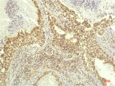
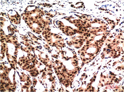
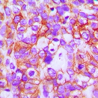 IHC (Immunohiostchemistry)
(Dilution: WB: (1/1000 - 1/2000), IH (1/200 - 1/500)Immunohistochemical analysis of ABCB5 staining in human breast cancer formalin fixed paraffin embedded tissue section. The section was pre-treated using heat mediated antigen retrieval with sodium citrate buffer (pH 6.0). The section was then incubated with the antibody at room temperature and detected using an HRP conjugated compact polymer system. DAB was used as the chromogen. The section was then counterstained with haematoxylin and mounted with DPX.)
IHC (Immunohiostchemistry)
(Dilution: WB: (1/1000 - 1/2000), IH (1/200 - 1/500)Immunohistochemical analysis of ABCB5 staining in human breast cancer formalin fixed paraffin embedded tissue section. The section was pre-treated using heat mediated antigen retrieval with sodium citrate buffer (pH 6.0). The section was then incubated with the antibody at room temperature and detected using an HRP conjugated compact polymer system. DAB was used as the chromogen. The section was then counterstained with haematoxylin and mounted with DPX.)
ABCB5, Monoclonal Antibody (Cat# AAA293371)
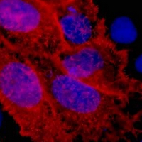
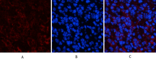
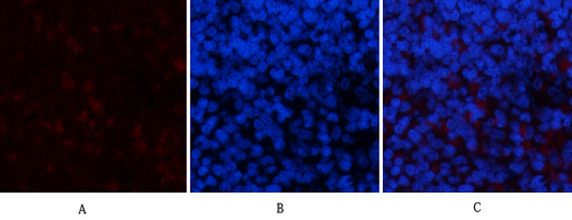
 IF (Immunofluorescence)
(Dilution: WB (1/500 - 1/1000), IH (1/100 - 1/200), IF/IC (1/50 - 1/200))
IF (Immunofluorescence)
(Dilution: WB (1/500 - 1/1000), IH (1/100 - 1/200), IF/IC (1/50 - 1/200))
active Caspase 3, Monoclonal Antibody (Cat# AAA293377)
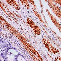
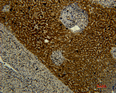
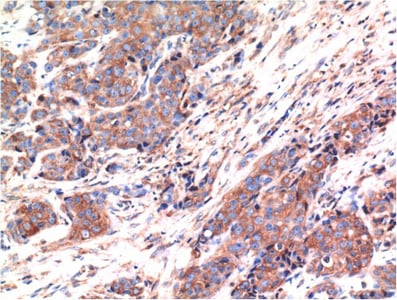
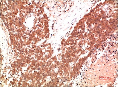
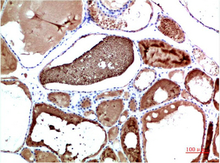
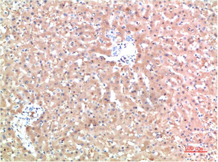
 IHC (Immunohiostchemistry)
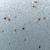
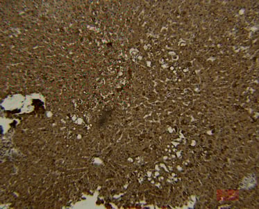
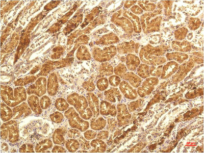
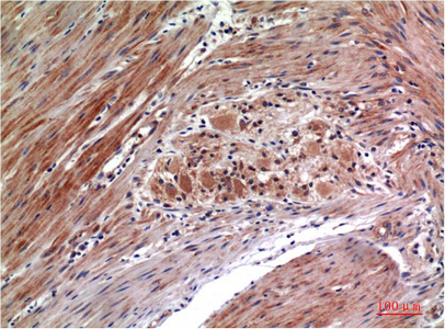
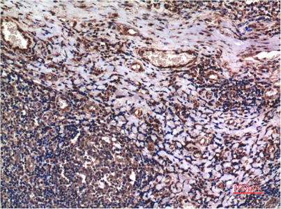
(Dilution: WB: (1/1000 - 1/2000), IH (1/100 - 1/200)Immunohistochemical analysis of Cyclophilin B staining in human heptacancer,human pancreatic cancer formalin fixed paraffin embedded tissue section. The section was pre-treated using heat mediated antigen retrieval with sodium citrate buffer (pH 6.0). The section was then incubated with the antibody at room temperature and detected using an HRP conjugated compact polymer system. DAB was used as the chromogen. The section was then counterstained with haematoxylin and mounted with DPX.)
IHC (Immunohiostchemistry)
(Dilution: WB: (1/1000 - 1/2000), IH (1/100 - 1/200)Immunohistochemical analysis of Cyclophilin B staining in human heptacancer,human pancreatic cancer formalin fixed paraffin embedded tissue section. The section was pre-treated using heat mediated antigen retrieval with sodium citrate buffer (pH 6.0). The section was then incubated with the antibody at room temperature and detected using an HRP conjugated compact polymer system. DAB was used as the chromogen. The section was then counterstained with haematoxylin and mounted with DPX.)
Cyclophilin B, Monoclonal Antibody (Cat# AAA293379)
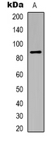
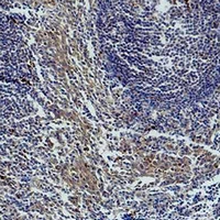
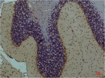
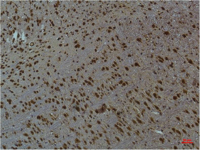
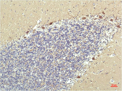
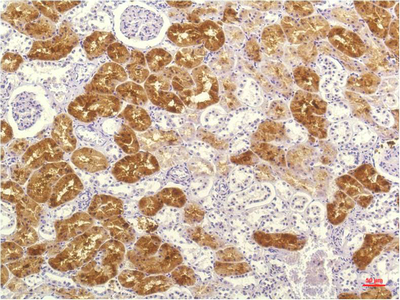
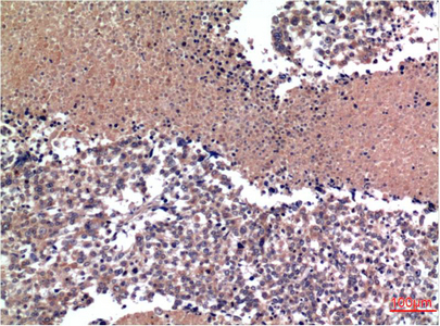
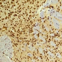 IHC (Immunohiostchemistry)
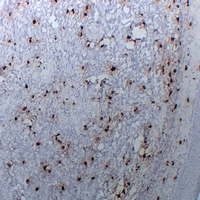
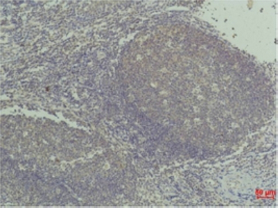
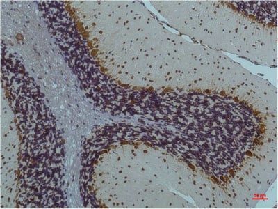
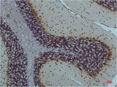
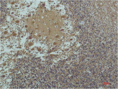
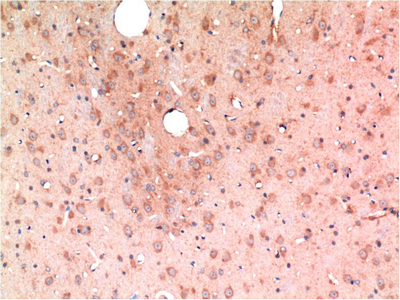
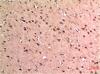
(Dilution: WB: (1/1000 - 1/2000), IH (1/100 - 1/200)Immunohistochemical analysis of HIF1 beta staining in mouse brain formalin fixed paraffin embedded tissue section. The section was pre-treated using heat mediated antigen retrieval with sodium citrate buffer (pH 6.0). The section was then incubated with the antibody at room temperature and detected using an HRP conjugated compact polymer system. DAB was used as the chromogen. The section was then counterstained with haematoxylin and mounted with DPX.)
IHC (Immunohiostchemistry)
(Dilution: WB: (1/1000 - 1/2000), IH (1/100 - 1/200)Immunohistochemical analysis of HIF1 beta staining in mouse brain formalin fixed paraffin embedded tissue section. The section was pre-treated using heat mediated antigen retrieval with sodium citrate buffer (pH 6.0). The section was then incubated with the antibody at room temperature and detected using an HRP conjugated compact polymer system. DAB was used as the chromogen. The section was then counterstained with haematoxylin and mounted with DPX.)
HIF1 beta, Monoclonal Antibody (Cat# AAA293383)
 IP (Immunoprecipitation)
(Dilution: WB (1/1000 - 1/3000), IH (1/200 - 1/500), IP (1/50 - 1/200))
IP (Immunoprecipitation)
(Dilution: WB (1/1000 - 1/3000), IH (1/200 - 1/500), IP (1/50 - 1/200))
Histone H3, Monoclonal Antibody (Cat# AAA293384)
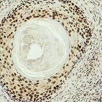
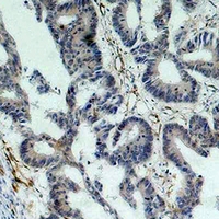
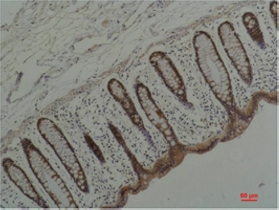
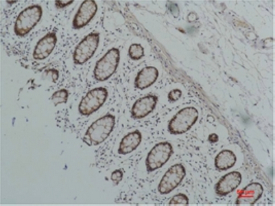
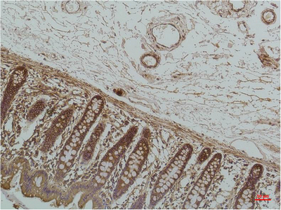
 IHC (Immunohiostchemistry)
(Dilution: WB: (1/1000 - 1/3000), IH (1/200 - 1/500)Immunohistochemical analysis of Histone H3 (DiMethyl K36) staining in human colon formalin fixed paraffin embedded tissue section. The section was pre-treated using heat mediated antigen retrieval with sodium citrate buffer (pH 6.0). The section was then incubated with the antibody at room temperature and detected using an HRP conjugated compact polymer system. DAB was used as the chromogen. The section was then counterstained with haematoxylin and mounted with DPX.)
IHC (Immunohiostchemistry)
(Dilution: WB: (1/1000 - 1/3000), IH (1/200 - 1/500)Immunohistochemical analysis of Histone H3 (DiMethyl K36) staining in human colon formalin fixed paraffin embedded tissue section. The section was pre-treated using heat mediated antigen retrieval with sodium citrate buffer (pH 6.0). The section was then incubated with the antibody at room temperature and detected using an HRP conjugated compact polymer system. DAB was used as the chromogen. The section was then counterstained with haematoxylin and mounted with DPX.)
Histone H3, Monoclonal Antibody (Cat# AAA293385)
 IHC (Immunohiostchemistry)
(Dilution: WB: (1/1000 - 1/3000), IH (1/200 - 1/500)Immunohistochemical analysis of Histone H3 (DiMethyl K9) staining in human skin formalin fixed paraffin embedded tissue section. The section was pre-treated using heat mediated antigen retrieval with sodium citrate buffer (pH 6.0). The section was then incubated with the antibody at room temperature and detected using an HRP conjugated compact polymer system. DAB was used as the chromogen. The section was then counterstained with haematoxylin and mounted with DPX.)
IHC (Immunohiostchemistry)
(Dilution: WB: (1/1000 - 1/3000), IH (1/200 - 1/500)Immunohistochemical analysis of Histone H3 (DiMethyl K9) staining in human skin formalin fixed paraffin embedded tissue section. The section was pre-treated using heat mediated antigen retrieval with sodium citrate buffer (pH 6.0). The section was then incubated with the antibody at room temperature and detected using an HRP conjugated compact polymer system. DAB was used as the chromogen. The section was then counterstained with haematoxylin and mounted with DPX.)
Histone H3, Monoclonal Antibody (Cat# AAA293386)
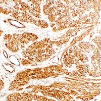
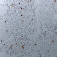
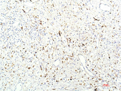
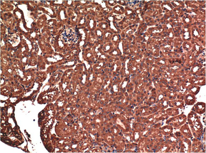
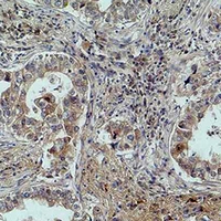 IHC (Immunohiostchemistry)
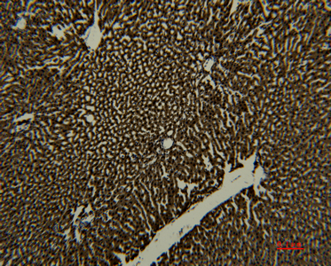
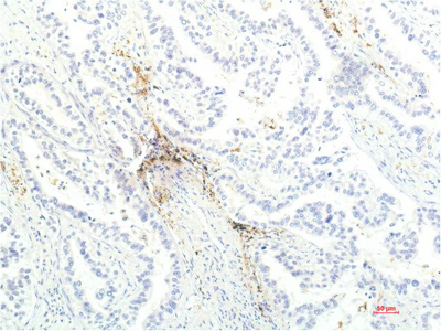
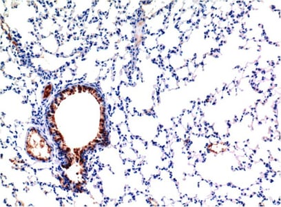
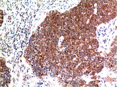
(Dilution: WB: (1/1000 - 1/2000), IH (1/100 - 1/200)Immunohistochemical analysis of MICU1 staining in human lung cancer formalin fixed paraffin embedded tissue section. The section was pre-treated using heat mediated antigen retrieval with sodium citrate buffer (pH 6.0). The section was then incubated with the antibody at room temperature and detected using an HRP conjugated compact polymer system. DAB was used as the chromogen. The section was then counterstained with haematoxylin and mounted with DPX.)
IHC (Immunohiostchemistry)
(Dilution: WB: (1/1000 - 1/2000), IH (1/100 - 1/200)Immunohistochemical analysis of MICU1 staining in human lung cancer formalin fixed paraffin embedded tissue section. The section was pre-treated using heat mediated antigen retrieval with sodium citrate buffer (pH 6.0). The section was then incubated with the antibody at room temperature and detected using an HRP conjugated compact polymer system. DAB was used as the chromogen. The section was then counterstained with haematoxylin and mounted with DPX.)
MICU1, Monoclonal Antibody (Cat# AAA293391)

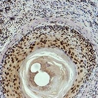
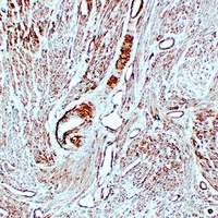
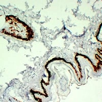
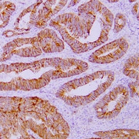
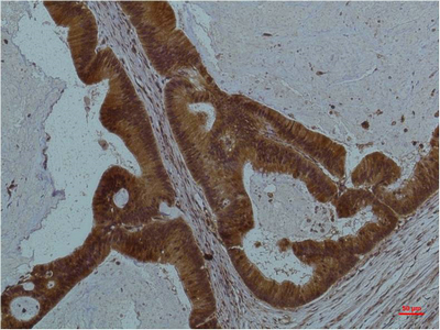
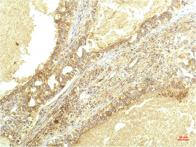
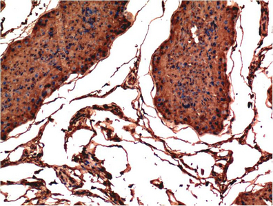
 IHC (Immunohiostchemistry)
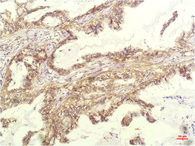
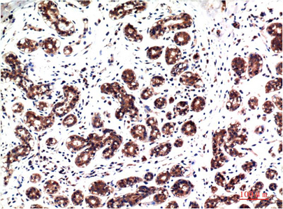
(Dilution: WB: (1/1000 - 1/2000), IH (1/200 - 1/500)Immunohistochemical analysis of Survivin staining in human colon cancer formalin fixed paraffin embedded tissue section. The section was pre-treated using heat mediated antigen retrieval with sodium citrate buffer (pH 6.0). The section was then incubated with the antibody at room temperature and detected using an HRP conjugated compact polymer system. DAB was used as the chromogen. The section was then counterstained with haematoxylin and mounted with DPX.)
IHC (Immunohiostchemistry)
(Dilution: WB: (1/1000 - 1/2000), IH (1/200 - 1/500)Immunohistochemical analysis of Survivin staining in human colon cancer formalin fixed paraffin embedded tissue section. The section was pre-treated using heat mediated antigen retrieval with sodium citrate buffer (pH 6.0). The section was then incubated with the antibody at room temperature and detected using an HRP conjugated compact polymer system. DAB was used as the chromogen. The section was then counterstained with haematoxylin and mounted with DPX.)
Survivin, Monoclonal Antibody (Cat# AAA293395)
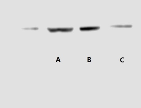
 IP (Immunoprecipitation)
(Dilution: WB (1/500 - 1/1000), IH (1/100 - 1/300))
IP (Immunoprecipitation)
(Dilution: WB (1/500 - 1/1000), IH (1/100 - 1/300))
Caldesmon, Monoclonal Antibody (Cat# AAA293399)
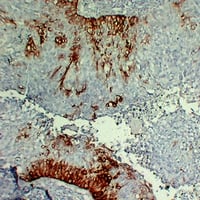
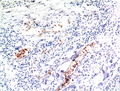
 FCM/FACS (Flow Cytometry)
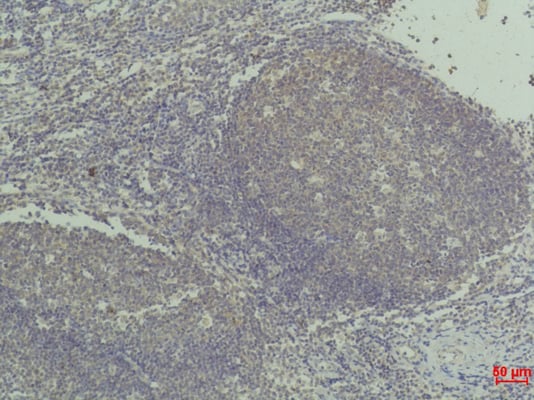
(Dilution: WB (1/500 - 1/1000), IH (1/100 - 1/300)Immunohistochemical analysis of Cytokeratin 15 staining in human tonsil formalin fixed paraffin embedded tissue section. The section was pre-treated using heat mediated antigen retrieval with sodium citrate buffer (pH 6.0). The section was then incubated with the antibody at room temperature and detected using an HRP conjugated compact polymer system. DAB was used as the chromogen. The section was then counterstained with haematoxylin and mounted with DPX.)
FCM/FACS (Flow Cytometry)
(Dilution: WB (1/500 - 1/1000), IH (1/100 - 1/300)Immunohistochemical analysis of Cytokeratin 15 staining in human tonsil formalin fixed paraffin embedded tissue section. The section was pre-treated using heat mediated antigen retrieval with sodium citrate buffer (pH 6.0). The section was then incubated with the antibody at room temperature and detected using an HRP conjugated compact polymer system. DAB was used as the chromogen. The section was then counterstained with haematoxylin and mounted with DPX.)
Cytokeratin 15, Monoclonal Antibody (Cat# AAA293402)


 FCM/FACS (Flow Cytometry)
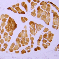
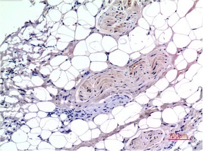
(Dilution: IH (1/100 - 1/300)Immunohistochemical analysis of alpha Smooth Muscle Actin staining in human skeletal muscle formalin fixed paraffin embedded tissue section. The section was pre-treated using heat mediated antigen retrieval with sodium citrate buffer (pH 6.0). The section was then incubated with the antibody at room temperature and detected using an HRP conjugated compact polymer system. DAB was used as the chromogen. The section was then counterstained with haematoxylin and mounted with DPX.)
FCM/FACS (Flow Cytometry)
(Dilution: IH (1/100 - 1/300)Immunohistochemical analysis of alpha Smooth Muscle Actin staining in human skeletal muscle formalin fixed paraffin embedded tissue section. The section was pre-treated using heat mediated antigen retrieval with sodium citrate buffer (pH 6.0). The section was then incubated with the antibody at room temperature and detected using an HRP conjugated compact polymer system. DAB was used as the chromogen. The section was then counterstained with haematoxylin and mounted with DPX.)
alpha Smooth Muscle Actin, Monoclonal Antibody (Cat# AAA293408)
 IP (Immunoprecipitation)
(Dilution: IH (1/100 - 1/300))
IP (Immunoprecipitation)
(Dilution: IH (1/100 - 1/300))
Tryptase alpha/beta, Monoclonal Antibody (Cat# AAA293409)
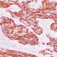
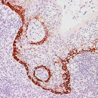
 IF (Immunofluorescence)
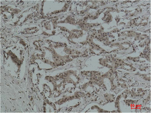
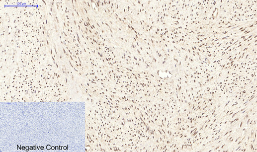
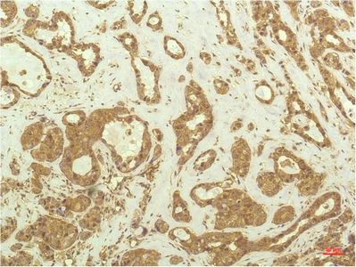
(Dilution: IF WB (1/500 - 1/1000), IH (1/100 - 1/300)Immunohistochemical analysis of Catenin delta 1 staining in human invasive ductal breast carcinoma formalin fixed paraffin embedded tissue section. The section was pre-treated using heat mediated antigen retrieval with sodium citrate buffer (pH 6.0). The section was then incubated with the antibody at room temperature and detected using an HRP conjugated compact polymer system. DAB was used as the chromogen. The section was then counterstained with haematoxylin and mounted with DPX.)
IF (Immunofluorescence)
(Dilution: IF WB (1/500 - 1/1000), IH (1/100 - 1/300)Immunohistochemical analysis of Catenin delta 1 staining in human invasive ductal breast carcinoma formalin fixed paraffin embedded tissue section. The section was pre-treated using heat mediated antigen retrieval with sodium citrate buffer (pH 6.0). The section was then incubated with the antibody at room temperature and detected using an HRP conjugated compact polymer system. DAB was used as the chromogen. The section was then counterstained with haematoxylin and mounted with DPX.)
Catenin delta 1, Monoclonal Antibody (Cat# AAA293411)
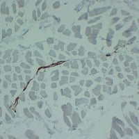
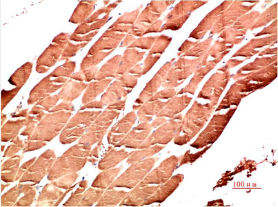
 IF (Immunofluorescence)
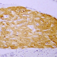
(Dilution: IF IH (1/100 - 1/300)Immunohistochemical analysis of Myoglobin staining in human skeletal muscle formalin fixed paraffin embedded tissue section. The section was pre-treated using heat mediated antigen retrieval with sodium citrate buffer (pH 6.0). The section was then incubated with the antibody at room temperature and detected using an HRP conjugated compact polymer system. DAB was used as the chromogen. The section was then counterstained with haematoxylin and mounted with DPX.)
IF (Immunofluorescence)
(Dilution: IF IH (1/100 - 1/300)Immunohistochemical analysis of Myoglobin staining in human skeletal muscle formalin fixed paraffin embedded tissue section. The section was pre-treated using heat mediated antigen retrieval with sodium citrate buffer (pH 6.0). The section was then incubated with the antibody at room temperature and detected using an HRP conjugated compact polymer system. DAB was used as the chromogen. The section was then counterstained with haematoxylin and mounted with DPX.)
Myoglobin, Monoclonal Antibody (Cat# AAA293412)

 IHC (Immunohiostchemistry)
(Dilution: WB: 1:1000-2000 IHC: 1:200-500)
IHC (Immunohiostchemistry)
(Dilution: WB: 1:1000-2000 IHC: 1:200-500)
Acetyl Lysine, Monoclonal Antibody (Cat# AAA293671)

 Application Data
(Dilution: WB: 1:1000-1:3000 IHC: 1:50-1:200)
Application Data
(Dilution: WB: 1:1000-1:3000 IHC: 1:50-1:200)
TNF alpha, Monoclonal Antibody (Cat# AAA293687)
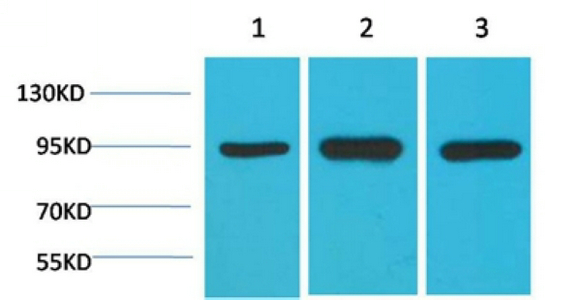
 Application Data
(Dilution: Western Blot: 1/500 - 1/2000. Immunohistochemistry: 1/100 - 1/300. ELISA: 1/40000. Not yet tested in other applications.)
Application Data
(Dilution: Western Blot: 1/500 - 1/2000. Immunohistochemistry: 1/100 - 1/300. ELISA: 1/40000. Not yet tested in other applications.)
HSP90 alpha, Monoclonal Antibody (Cat# AAA293688)
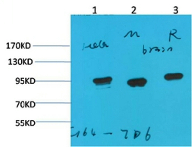
 Application Data
(Dilution: Western Blot: 1/500 - 1/2000. Immunohistochemistry: 1/100 - 1/300. ELISA: 1/40000. Not yet tested in other applications.)
Application Data
(Dilution: Western Blot: 1/500 - 1/2000. Immunohistochemistry: 1/100 - 1/300. ELISA: 1/40000. Not yet tested in other applications.)
HSP90 alpha, Monoclonal Antibody (Cat# AAA293699)
 Application Data
(Dilution: WB 1:1000-2000, IHC 1:100-200)
Application Data
(Dilution: WB 1:1000-2000, IHC 1:100-200)
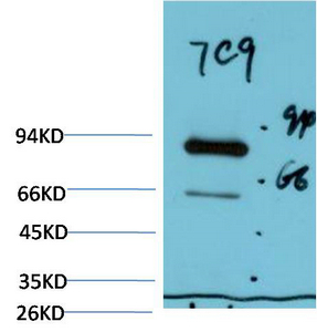
STAT3, Monoclonal Antibody (Cat# AAA293701)
 Application Data
(Dilution: WB 1:1000-2000, IHC 1:100-200)
Application Data
(Dilution: WB 1:1000-2000, IHC 1:100-200)
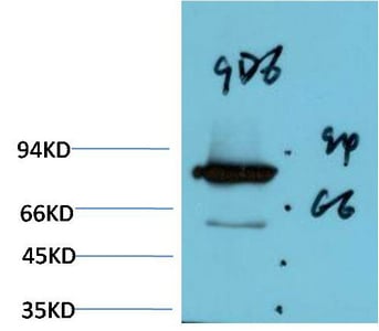
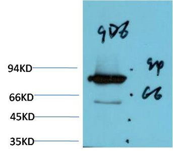
GRP78/Bip, Monoclonal Antibody (Cat# AAA293707)
 Application Data
(Dilution: IHC 1:100-200)
Application Data
(Dilution: IHC 1:100-200)
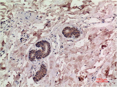
IkappaB beta, Monoclonal Antibody (Cat# AAA293715)

 Application Data
(Dilution: IHC 1:100-200)
Application Data
(Dilution: IHC 1:100-200)
CaMKIIbeta/gamma/delta, Monoclonal Antibody (Cat# AAA293717)
 IHC (Immunohiostchemistry)
(Dilution: IHC 1:100-200)
IHC (Immunohiostchemistry)
(Dilution: IHC 1:100-200)
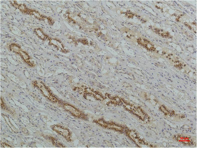
Transferrin, Monoclonal Antibody (Cat# AAA293755)
 Application Data
(Dilution: WB 1:1000-2000, IHC 1:100-200)
Application Data
(Dilution: WB 1:1000-2000, IHC 1:100-200)
alpha-tubulin, Monoclonal Antibody (Cat# AAA293757)
 IHC (Immunohiostchemistry)
(Dilution: IHC 1:100-200)
IHC (Immunohiostchemistry)
(Dilution: IHC 1:100-200)
Transferrin, Monoclonal Antibody (Cat# AAA293758)
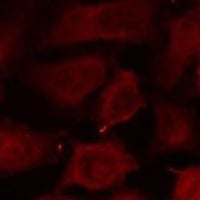
 Application Data
(Dilution: IF: 1:50-200 IHC 1:100-200)
Application Data
(Dilution: IF: 1:50-200 IHC 1:100-200)
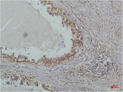
PPAR Delta, Monoclonal Antibody (Cat# AAA293759)
 Application Data
(Dilution: IHC 1:100-200)
Application Data
(Dilution: IHC 1:100-200)
P70 S6 Kinase, Monoclonal Antibody (Cat# AAA293760)
 Application Data
(Dilution: IHC 1:100-200)
Application Data
(Dilution: IHC 1:100-200)
IL-8, Monoclonal Antibody (Cat# AAA293762)
 Application Data
(Dilution: IHC 1:100-200)
Application Data
(Dilution: IHC 1:100-200)
JAK1, Monoclonal Antibody (Cat# AAA293764)
 Application Data
(Dilution: IHC 1:100-200)
Application Data
(Dilution: IHC 1:100-200)
IL-8, Monoclonal Antibody (Cat# AAA293769)
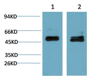
 WB (Western Blot)
(Dilution: WB 1:1000-2000, IHC 1:100-200)
WB (Western Blot)
(Dilution: WB 1:1000-2000, IHC 1:100-200)
Bax, Monoclonal Antibody (Cat# AAA293777)
 Application Data
(Dilution: IHC 1:100-200)
Application Data
(Dilution: IHC 1:100-200)
TGFbeta1, Monoclonal Antibody (Cat# AAA293780)

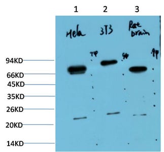
 Application Data
(Dilution: IHC 1:100-200)
Application Data
(Dilution: IHC 1:100-200)
Phospho-ERK 1/2, Monoclonal Antibody (Cat# AAA293783)
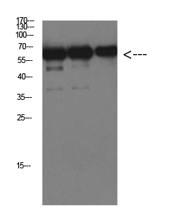
 WB (Western Blot)
(Dilution: IF: 1:50-200 WB 1:1000-2000, IHC 1:100-200 IHC 1:50-300)
WB (Western Blot)
(Dilution: IF: 1:50-200 WB 1:1000-2000, IHC 1:100-200 IHC 1:50-300)
Luciferase, Monoclonal Antibody (Cat# AAA293788)

 Application Data
(Dilution: WB 1:1000-2000, IHC 1:100-200)
Application Data
(Dilution: WB 1:1000-2000, IHC 1:100-200)
Ubiquitin, Monoclonal Antibody (Cat# AAA293789)
 Application Data
(Dilution: IHC 1:100-200)
Application Data
(Dilution: IHC 1:100-200)
Cyclin B1, Monoclonal Antibody (Cat# AAA293790)
 Application Data
(Dilution: WB 1:1000-2000, IHC 1:100-200)
Application Data
(Dilution: WB 1:1000-2000, IHC 1:100-200)
Beclin-1, Monoclonal Antibody (Cat# AAA293791)
 Application Data
(Dilution: WB 1:1000-2000)
Application Data
(Dilution: WB 1:1000-2000)
Luciferase, Monoclonal Antibody (Cat# AAA293793)
 Application Data
(Dilution: WB 1:1000-2000)
Application Data
(Dilution: WB 1:1000-2000)
Luciferase, Monoclonal Antibody (Cat# AAA293794)
 Application Data
(Dilution: WB 1:1000-2000, IHC 1:100-200)
Application Data
(Dilution: WB 1:1000-2000, IHC 1:100-200)
Smad3, Monoclonal Antibody (Cat# AAA293799)
 Application Data
(Dilution: WB 1:1000-2000, IHC 1:100-200)
Application Data
(Dilution: WB 1:1000-2000, IHC 1:100-200)
AMPKalpha1, Monoclonal Antibody (Cat# AAA293809)
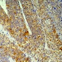
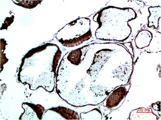
 Application Data
(Dilution: WB 1:500-2000,IHC-p 1:50-300)
Application Data
(Dilution: WB 1:500-2000,IHC-p 1:50-300)
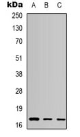
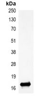
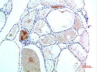
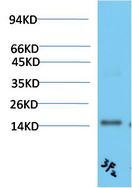
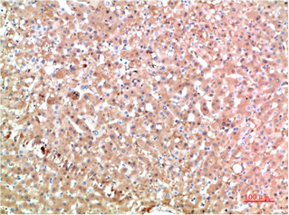
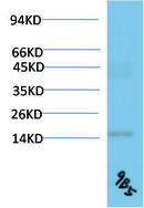
TTR, Monoclonal Antibody (Cat# AAA293833)
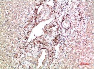
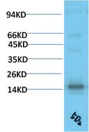 Application Data
(Dilution: WB 1:500-2000,IHC-p 1:50-300)
Application Data
(Dilution: WB 1:500-2000,IHC-p 1:50-300)
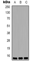
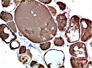
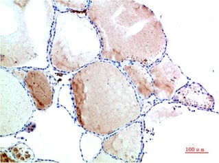
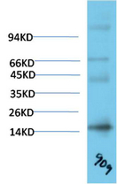
TTR, Monoclonal Antibody (Cat# AAA293838)
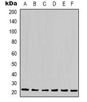
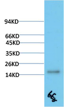 Application Data
(Dilution: WB 1:500-2000,IHC-p 1:50-300)
Application Data
(Dilution: WB 1:500-2000,IHC-p 1:50-300)
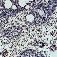
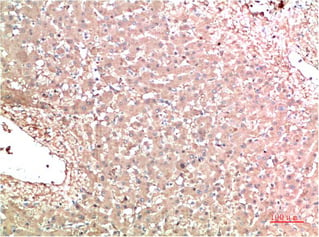
TTR, Monoclonal Antibody (Cat# AAA293841)
 Application Data
(Dilution: WB 1:500-2000,IHC-p 1:50-300)
Application Data
(Dilution: WB 1:500-2000,IHC-p 1:50-300)
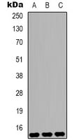
TTR, Monoclonal Antibody (Cat# AAA293842)
 Application Data
(Dilution: WB 1:500-2000,IHC-p 1:50-300)
Application Data
(Dilution: WB 1:500-2000,IHC-p 1:50-300)
TTR, Monoclonal Antibody (Cat# AAA293844)
 Application Data
(Dilution: WB 1:500-2000,IHC-p 1:50-300)
Application Data
(Dilution: WB 1:500-2000,IHC-p 1:50-300)
TTR, Monoclonal Antibody (Cat# AAA293845)
What are Monoclonal Antibodies?
Monoclonal antibodies are specialized laboratory-produced proteins developed for binding to specific biological antigens or other molecular targets. Since they come from a single cell (or clone), they are especially consistent and accurate in the data they are involved in producing.
This type of antibody material has been shown to be a powerful tool in finding and subsequently destroying harmful cells in an organism, such as those found in cancers or various autoimmune diseases. This makes them excellent aids in medical testing and research, which is why they are so widely used.
AAA Biotech offers a comprehensive range of high-quality monoclonal antibodies that perform effectively in various laboratory tests, including (amongst others) ELISA, western blotting, immunohistochemistry, and flow cytometry. All of the products in our catalog are thoroughly quality tested to make sure that they are reliable and will consistently perform well in your research.
What Are The Uses of Monoclonal Antibodies
Monoclonal antibodies are used in many lab tests, including (amongst others) ELISA, western blotting, immunohistochemistry, and flow cytometry.
ELISA is a test that helps detect a specific substance/analyte in a sample. It uses antibodies (often monoclonal) bound to a solid surface (such as the well of a microplate) to “capture” the substance/analyte in the sample and immobilize it so that the detection antibody component can then bind to it and produce a signal, which can then be measured.
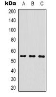
Western blotting identifies specific proteins in a sample. The sample is first separated on a gel, and then antibodies are applied that will typically bind to the target, which will all be localized to a single band in a lane.
Immunohistochemistry helps locate specific proteins in cells or tissue samples using antibodies.
Flow cytometry looks at and sorts cells. It uses antibodies that are conjugated to reporter molecules called “fluorophores”, which, under special lights, emit light themselves, which can then be measured by a detector instrument.
How Monoclonal Antibodies Are Used as Medicine?
Please note that all of the products listed in AAA Biotech’s also known as AAA Bio or AAABio catalog are strictly for research-use only (RUO).
Monoclonal antibodies can also be used as therapeutic/medical treatments, particularly in the context of cancers. They are designed to find and bind to specific cells or proteins, helping the immune system recognize and attack the cancer. These treatments work in different ways, such as:
- Radioimmunotherapy attaches a small amount of radioactive molecule to the antibody, so it delivers the radiation directly to the cancer cells that the antibody is specifically binding to.
- Antibody-directed enzyme prodrug therapy uses antibodies that are specifically bound to special enzymes. These enzymes activate a harmless drug in the body and turn it into a cancer-killing drug only near the cancer cells—this helps avoid harming healthy cells.
- Immunoliposomes are tiny “bubbles” filled with medicine/drug and coated with antibodies. They carry the drug straight to the cancer cells.
Why Buy Monoclonal Antibodies From Us?
At AAA Biotech, we provide high-performance monoclonal antibodies designed to support a wide range of research needs.
1. Validated for Versatile Applications
The antibodies in our catalog are extensively validated and compatible with multiple techniques, including (but not limited to) ELISA, flow cytometry (FC), immunocytochemistry (ICC), immunofluorescence (IF), immunohistochemistry (IHC), immunoprecipitation (IP), and western blotting (WB).
2. Wide Selection & Specialized Options
We offer antibodies for common and rare species, that are available in various conjugated forms, and also in recombinant formats. Essentially, there is almost anything one might need to meet their experimental model’s requirements.
3. High-Quality Proteins
Our proteins meet high purity standards—90% or more as confirmed by SDS-PAGE. Many are available with tags like His, Flag, GST, or MBP, and we also supply native and biologically active proteins for functional studies.
Frequently Asked Questions
1. Are your monoclonal antibodies validated for specific applications?
Yes, our antibodies are tested and validated for use in methods such as ELISA, western blot, IHC, flow cytometry, and more. Refer to specific product pages or datasheets for individual product information.
2. How do I choose the right monoclonal antibody for my application?
Review the product details directly for application validation, species reactivity, and target information. You may also contact our support team at any time for help.
3. How quickly can I receive my order?
Most orders are processed and shipped within 1–3 business days, depending on product availability and your shipping location.