Filters
▼Clonality
▼Type
▼Reactivity
▼Gene Name
▼Isotype
▼Host
▼Application
▼Clone
▼Monoclonal Antibodies
Get accurate results in your research with our Monoclonal Antibodies, which are specially made to target exactly what you require for your research, and will produce consistent, reliable performance in lab tests.
Viewing 8300-8350 of 27597 product results
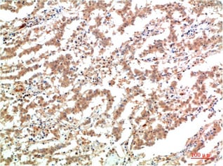
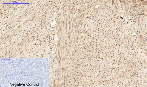
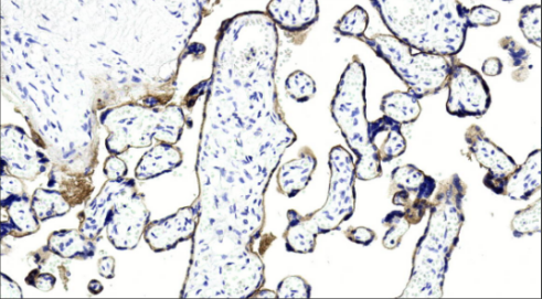
 Application Data
(Dilution: IHC-p 1:50-300)
Application Data
(Dilution: IHC-p 1:50-300)
JAK2, Monoclonal Antibody (Cat# AAA293849)
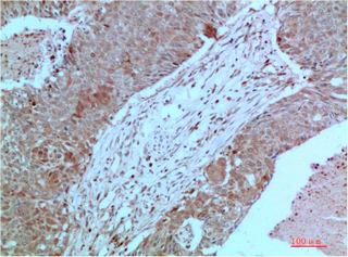
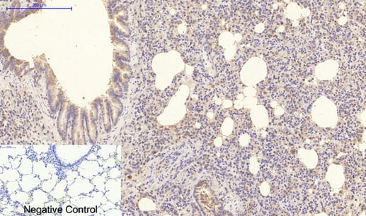
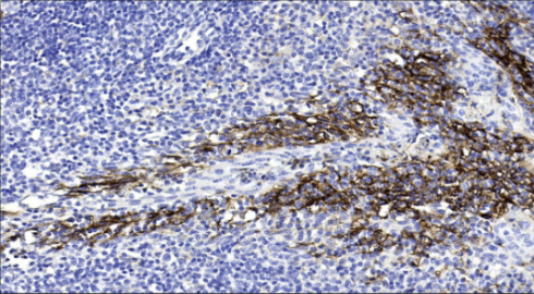
 Application Data
(Dilution: IHC-p 1:50-300)
Application Data
(Dilution: IHC-p 1:50-300)
JAK2, Monoclonal Antibody (Cat# AAA293850)
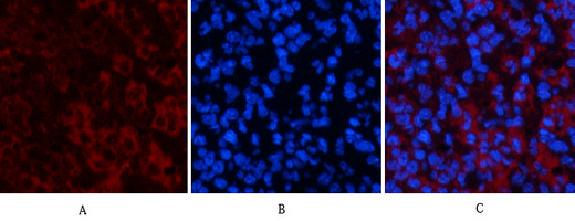
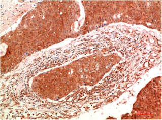
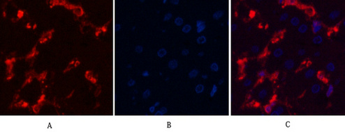
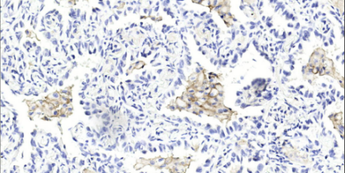
 IHC (Immunohistochemisry)
(Dilution: IF: 1:50-200 IHC-p 1:50-300)
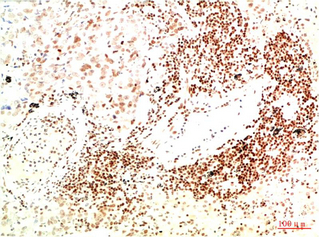
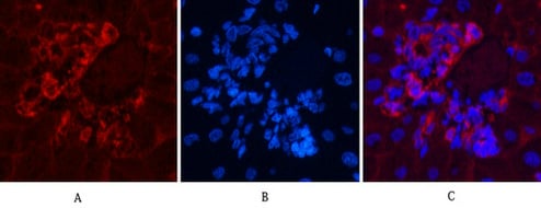
IHC (Immunohistochemisry)
(Dilution: IF: 1:50-200 IHC-p 1:50-300)
ATG7, Monoclonal Antibody (Cat# AAA293853)
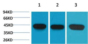 WB (Western Blot)
(Dilution: WB: 1:10000-100000 IF 1:200 IHC:1:1000-2000)
WB (Western Blot)
(Dilution: WB: 1:10000-100000 IF 1:200 IHC:1:1000-2000)
alpha-SMA, Monoclonal Antibody (Cat# AAA293643)
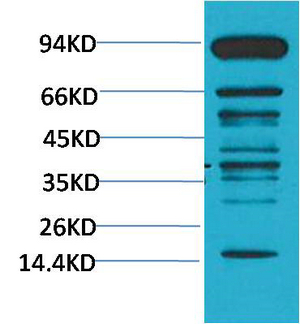
 IHC (Immunohiostchemistry)
(Dilution: WB: 1:1000-2000 IHC: 1:200-500)
IHC (Immunohiostchemistry)
(Dilution: WB: 1:1000-2000 IHC: 1:200-500)
Pan Methylated Lysine, Monoclonal Antibody (Cat# AAA293664)
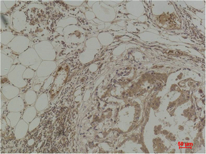
 IHC (Immunohistochemisry)
(Dilution: IHC-P: 1:1000)
IHC (Immunohistochemisry)
(Dilution: IHC-P: 1:1000)
PD-L1 [AK94], Monoclonal Antibody (Cat# AAA296697)
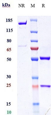
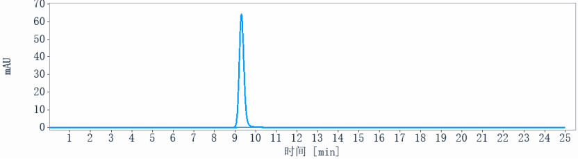 Application Data
(The purity of Anti-ERBB2 / HER2 / CD340 Reference Antibody (Trastuzumab duocarmazine)is more than 95% ,determined by SEC-HPLC.)
Application Data
(The purity of Anti-ERBB2 / HER2 / CD340 Reference Antibody (Trastuzumab duocarmazine)is more than 95% ,determined by SEC-HPLC.)
ERBB2/HER2/CD340, Monoclonal Recombinant Antibody (Cat# AAA297001)
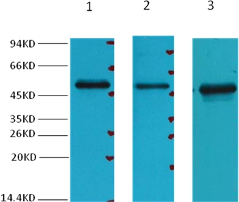
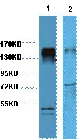
 WB (Western Blot)
(Western blot analysis of 1) Mouse Brain tissue, 2) Rat Brain tissue,, using diluted at 1:3,000.)
WB (Western Blot)
(Western blot analysis of 1) Mouse Brain tissue, 2) Rat Brain tissue,, using diluted at 1:3,000.)
Fumarase, Monoclonal Antibody (Cat# AAA300701)
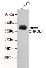
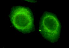
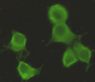 ICC (Immunocytochemistry)
(Immunocytochemistry stain of Hela using CHRDL1 antibody (1:300).)
ICC (Immunocytochemistry)
(Immunocytochemistry stain of Hela using CHRDL1 antibody (1:300).)
CHRDL1, Monoclonal Antibody (Cat# AAA300704)
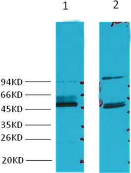
 WB (Western Blot)
(Western blot analysis of recombinant mOrange protein, diluted at 1:10000.)
WB (Western Blot)
(Western blot analysis of recombinant mOrange protein, diluted at 1:10000.)
mOrange, Monoclonal Antibody (Cat# AAA300708)
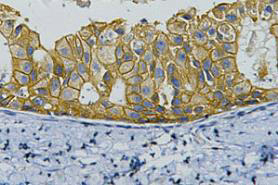 IHC (Immunohiostchemistry)
(IHC staining of human breast cancer tissue, diluted at 1:200.)
IHC (Immunohiostchemistry)
(IHC staining of human breast cancer tissue, diluted at 1:200.)
HER2, Monoclonal Antibody (Cat# AAA300713)
 ICC (Immunocytochemistry)
(Immunocytochemistry of Bim in K562 cells with Bim antibody at 10 ug/mL.)
ICC (Immunocytochemistry)
(Immunocytochemistry of Bim in K562 cells with Bim antibody at 10 ug/mL.)
Bim, Monoclonal Antibody (Cat# AAA300715)
 WB (Western Blot)
(Western blot analysis of Hela, HepG2, using diluted at 1:2,000.)
WB (Western Blot)
(Western blot analysis of Hela, HepG2, using diluted at 1:2,000.)
Insulin Degrading Enzyme, Monoclonal Antibody (Cat# AAA300717)
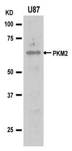
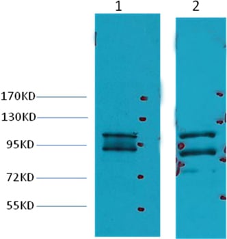
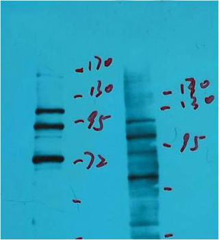
 IP (Immunoprecipitation)
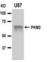
(Immunoprecipitation analysis of U87 cell lysates using PKM2 antibody. IP:PKM2 Monoclonal Antibody. WB:PKM2 Polyclonal Antibody #21578.)
IP (Immunoprecipitation)
(Immunoprecipitation analysis of U87 cell lysates using PKM2 antibody. IP:PKM2 Monoclonal Antibody. WB:PKM2 Polyclonal Antibody #21578.)
PKM2, Monoclonal Antibody (Cat# AAA300720)
 IHC (Immunohiostchemistry)
(Immunohistochemistry of MD-2 in human spleen with MD-2 antibody at 2.5 ug/mL.)
IHC (Immunohiostchemistry)
(Immunohistochemistry of MD-2 in human spleen with MD-2 antibody at 2.5 ug/mL.)
MD-2, Monoclonal Antibody (Cat# AAA300722)
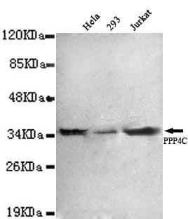
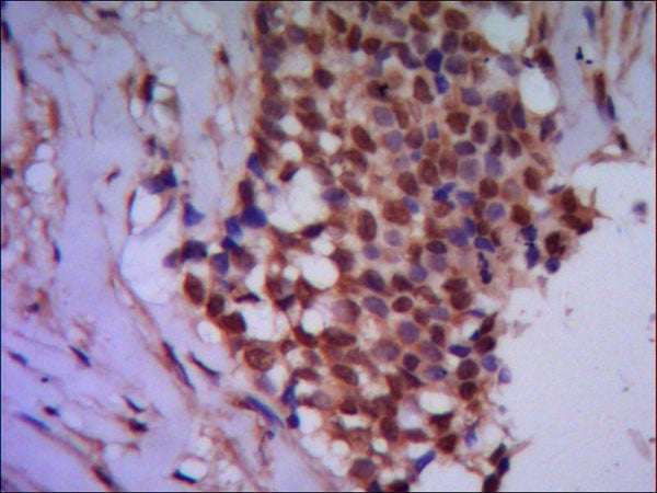 IHC (Immunohistochemisry)
(Paraffin-embedded human breast cancer using anti- PPP4C diluted 1:50-1:100)
IHC (Immunohistochemisry)
(Paraffin-embedded human breast cancer using anti- PPP4C diluted 1:50-1:100)
Protein Phosphatase 4C, Monoclonal Antibody (Cat# AAA300727)
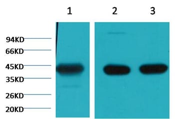
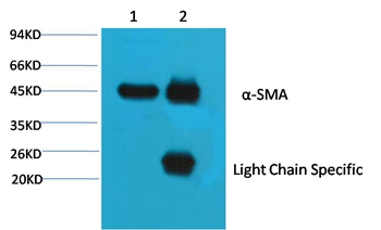 Application Data
(1¡¢Input: Mouse Brain Tissue Lysate 2¡¢IP product: IP dilute 1:200 Western blot analysis: primary antibody: 1:10,000 Secondary antibody: Goat anti-Mouse IgG, Light chain specific, 1:5,000)
Application Data
(1¡¢Input: Mouse Brain Tissue Lysate 2¡¢IP product: IP dilute 1:200 Western blot analysis: primary antibody: 1:10,000 Secondary antibody: Goat anti-Mouse IgG, Light chain specific, 1:5,000)
alpha Skeletal Muslce Actin, Monoclonal Antibody (Cat# AAA300728)
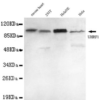
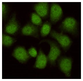
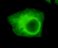 Application Data
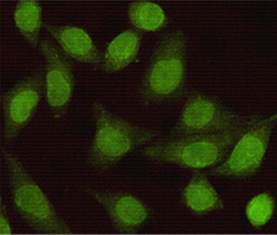
(HeLa cells using anti-UHRF1 (N-terminus)antibody diluted 1:200)
Application Data
(HeLa cells using anti-UHRF1 (N-terminus)antibody diluted 1:200)
UHRF1, Monoclonal Antibody (Cat# AAA300729)
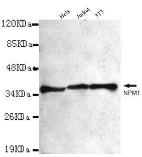
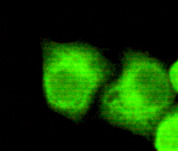 Application Data
(1:200 dilution from a previous lot detected NPM1 in Hela)
Application Data
(1:200 dilution from a previous lot detected NPM1 in Hela)
NPM1, Monoclonal Antibody (Cat# AAA300738)
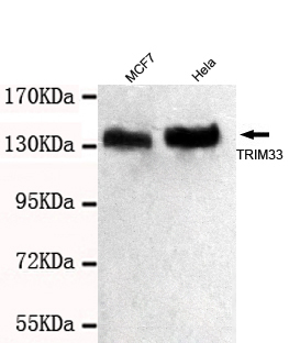
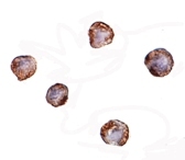
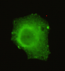 Application Data
(HeLa cells using anti- TRIM33 (C-terminus) antibody diluted 1:50)
Application Data
(HeLa cells using anti- TRIM33 (C-terminus) antibody diluted 1:50)
TRIM33, Monoclonal Antibody (Cat# AAA300741)
 ICC (Immunocytochemistry)
(Immunocytochemistry of XBP-1 in HepG2 cells with XBP-1 antibody at 2 ug/mL.)
ICC (Immunocytochemistry)
(Immunocytochemistry of XBP-1 in HepG2 cells with XBP-1 antibody at 2 ug/mL.)
XBP-1, Monoclonal Antibody (Cat# AAA300751)
 Application Data
(2ug S-Tag fusion protein+ Primary antibody dilution at 1:5,000 2y 1:10,000)
Application Data
(2ug S-Tag fusion protein+ Primary antibody dilution at 1:5,000 2y 1:10,000)
S-Tag, Monoclonal Antibody (Cat# AAA300753)
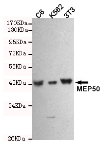 WB (Western Blot)
(Western blot detection of WDR77 antibody in C6,3T3 and K562 cell lysates using WDR77 antibody (1:1000 diluted).Predicted band size:42KDa.Observed band size:42KDa.)
WB (Western Blot)
(Western blot detection of WDR77 antibody in C6,3T3 and K562 cell lysates using WDR77 antibody (1:1000 diluted).Predicted band size:42KDa.Observed band size:42KDa.)
WDR77, Monoclonal Antibody (Cat# AAA300761)
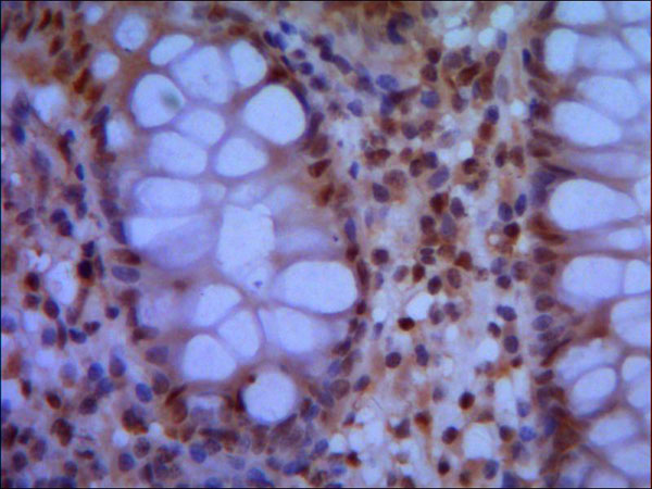
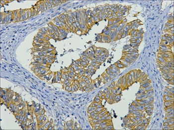 IHC (Immunohistochemistry)
(IHC staining of paraffin-embedded Human colon cancer tissue with CK19 mouse mAb(11F5) diluted at 1:200.)
IHC (Immunohistochemistry)
(IHC staining of paraffin-embedded Human colon cancer tissue with CK19 mouse mAb(11F5) diluted at 1:200.)
CK19, Monoclonal Antibody (Cat# AAA300762)
 IHC (Immunohiostchemistry)
(Immunohistochemistry of MD-2 in human spleen with MD-2 antibody at 2.5 ug/mL.)
IHC (Immunohiostchemistry)
(Immunohistochemistry of MD-2 in human spleen with MD-2 antibody at 2.5 ug/mL.)
MD-2, Monoclonal Antibody (Cat# AAA300770)
 WB (Western Blot)
(Western blot analysis of Hela, diluted at 1:3000.)
WB (Western Blot)
(Western blot analysis of Hela, diluted at 1:3000.)
GAPDH, Monoclonal Antibody (Cat# AAA300777)
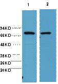
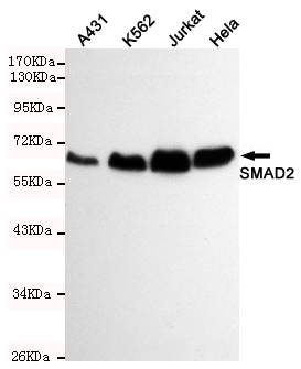 WB (Western Blot)
(Western blot detection of SMAD2 antibody in Hela,A431,Jurkat and K562 cell lysates using SMAD2 antibody (1:500 diluted).Predicted band size:60KDa.Observed band size:60KDa.)
WB (Western Blot)
(Western blot detection of SMAD2 antibody in Hela,A431,Jurkat and K562 cell lysates using SMAD2 antibody (1:500 diluted).Predicted band size:60KDa.Observed band size:60KDa.)
Smad2, Monoclonal Antibody (Cat# AAA300783)
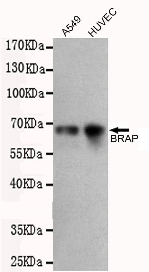
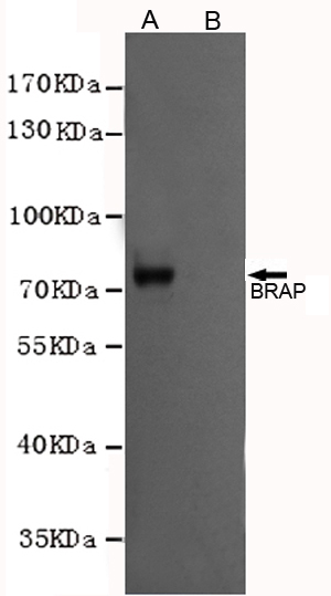 WB (Western Blot)
(Western blot analysis of extracts from CHO-K1 cells, transfected with a human pDYKDDDDK-CMV2-BRAP construct (A) or transfected with a human pDYKDDDDK-CMV2 construct (B), using BRAP antibody (1:1000 diluted).)
WB (Western Blot)
(Western blot analysis of extracts from CHO-K1 cells, transfected with a human pDYKDDDDK-CMV2-BRAP construct (A) or transfected with a human pDYKDDDDK-CMV2 construct (B), using BRAP antibody (1:1000 diluted).)
BRAP, Monoclonal Antibody (Cat# AAA300786)
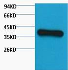
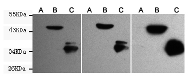
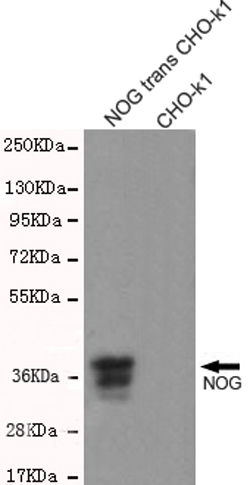 WB (Western Blot)
(Western blot detection of NOG antibody in CHO-K1 cell lysates over-expressing NOG-PDGFR transmembrane domain fused protein using NOG antibody (1:1000 diluted).Predicted band size:26KDa.Observed band size:37KDa.)
WB (Western Blot)
(Western blot detection of NOG antibody in CHO-K1 cell lysates over-expressing NOG-PDGFR transmembrane domain fused protein using NOG antibody (1:1000 diluted).Predicted band size:26KDa.Observed band size:37KDa.)
Noggin, Monoclonal Antibody (Cat# AAA300787)
 WB (Western Blot)
(Western blot analysis of 1) Hela, 2) Mouse Brain, diluted at 1:2000.)
WB (Western Blot)
(Western blot analysis of 1) Hela, 2) Mouse Brain, diluted at 1:2000.)
HSP70, Monoclonal Antibody (Cat# AAA300788)
 WB (Western Blot)
(Western blot analysis of MyD88 in EL4 whole cell lysate with MyD88 antibody at 2 ug/mL.)
WB (Western Blot)
(Western blot analysis of MyD88 in EL4 whole cell lysate with MyD88 antibody at 2 ug/mL.)
MyD88, Monoclonal Antibody (Cat# AAA300792)
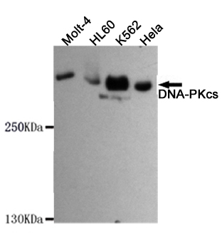
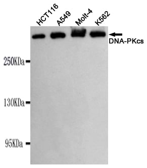 WB (Western Blot)
(Western blot detection of PRKDC in K562,Molt-4,A549 and HCT116 cell lysates using PRKDC antibody(1:1000 diluted).Predicted band size:450KDa.Observed band size:450KDa.)
WB (Western Blot)
(Western blot detection of PRKDC in K562,Molt-4,A549 and HCT116 cell lysates using PRKDC antibody(1:1000 diluted).Predicted band size:450KDa.Observed band size:450KDa.)
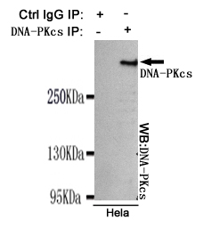
DNA-PKcs, Monoclonal Antibody (Cat# AAA300797)
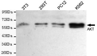
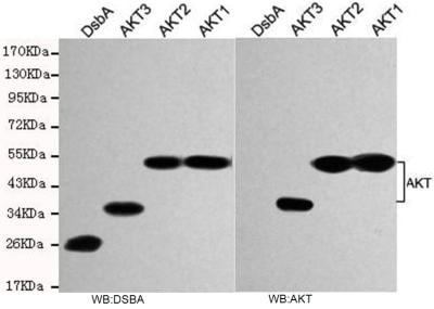
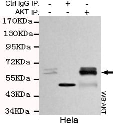 IP (Immunoprecipitation)
(Immunoprecipitation analysis of Hela cell lysates using AKT antibody.)
IP (Immunoprecipitation)
(Immunoprecipitation analysis of Hela cell lysates using AKT antibody.)
total AKT, Monoclonal Antibody (Cat# AAA300801)
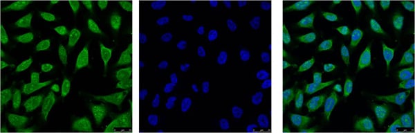
 IHC (Immunohiostchemistry)
(Immunohistochemistry of PD-1 in mouse brain tissue with PD-1 antibody at 2.5 ug/mL.)
IHC (Immunohiostchemistry)
(Immunohistochemistry of PD-1 in mouse brain tissue with PD-1 antibody at 2.5 ug/mL.)
PD-1, Monoclonal Antibody (Cat# AAA300803)
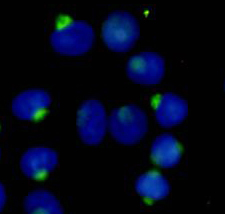
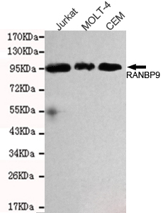
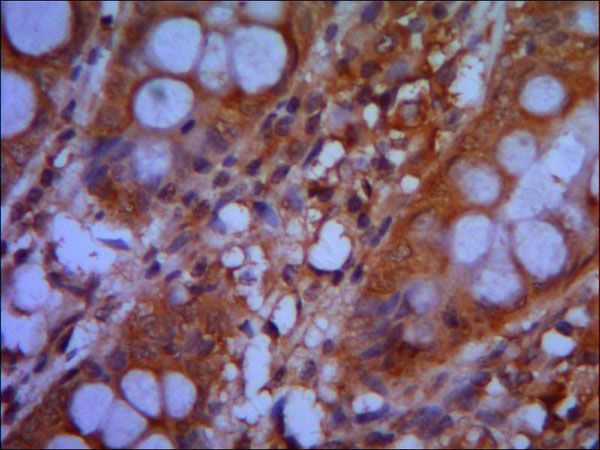
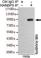 IP (Immunoprecipitation)
(Immunoprecipitation analysis of Hela cell lysates using RANBP9 antibody)
IP (Immunoprecipitation)
(Immunoprecipitation analysis of Hela cell lysates using RANBP9 antibody)
RANBP9, Monoclonal Antibody (Cat# AAA300806)
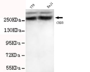
 Application Data
(Hela cells stained with SMC1A (red, 1:100 dilution), followed by FITC-conjugated goat anti-mouse IgG. Black line histogram represents the isotype control, normal mouse IgG)
Application Data
(Hela cells stained with SMC1A (red, 1:100 dilution), followed by FITC-conjugated goat anti-mouse IgG. Black line histogram represents the isotype control, normal mouse IgG)
CHD3, Monoclonal Antibody (Cat# AAA300809)
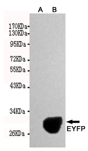
 WB (Western Blot)
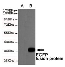
(Western blot detection of EGFP expression in Hela cells non-transfected (A) or transfected (B) with pEGFP C1 using EGFP antibody(1:5000 diluted).Predicted band size: 34KDa Observed band size: 34KDa.)
WB (Western Blot)
(Western blot detection of EGFP expression in Hela cells non-transfected (A) or transfected (B) with pEGFP C1 using EGFP antibody(1:5000 diluted).Predicted band size: 34KDa Observed band size: 34KDa.)
EGFP/EYFP, Monoclonal Antibody (Cat# AAA300812)
 Application Data
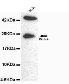
(HeLa cells using anti- RAB2A(C-terminus)antibody diluted 1:200)
Application Data
(HeLa cells using anti- RAB2A(C-terminus)antibody diluted 1:200)
Rab2A, Monoclonal Antibody (Cat# AAA300819)
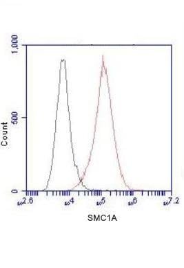 Application Data
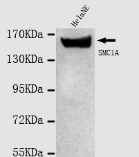
(Hela cells stained with SMC1A (red, 1:100 dilution), followed by FITC-conjugated goat anti-mouse IgG. Black line histogram represents the isotype control, normal mouse IgG)
Application Data
(Hela cells stained with SMC1A (red, 1:100 dilution), followed by FITC-conjugated goat anti-mouse IgG. Black line histogram represents the isotype control, normal mouse IgG)
SMC1A, Monoclonal Antibody (Cat# AAA300820)
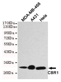 WB (Western Blot)
(Western blot detection of CBR1 in Hela,A431 & MDA-MB-468 cell lysates using CBR1 antibody(1:1000 diluted).Predicted band size:30KDa,Observed band size:30KDa.)
WB (Western Blot)
(Western blot detection of CBR1 in Hela,A431 & MDA-MB-468 cell lysates using CBR1 antibody(1:1000 diluted).Predicted band size:30KDa,Observed band size:30KDa.)
CBR1, Monoclonal Antibody (Cat# AAA300821)
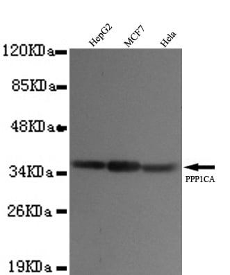
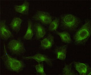
 Application Data
(HeLa cells using anti- PPP1CA(N-terminus)antibody diluted 1:50)
Application Data
(HeLa cells using anti- PPP1CA(N-terminus)antibody diluted 1:50)
PP1A, Monoclonal Antibody (Cat# AAA300831)
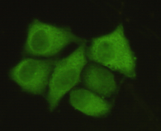
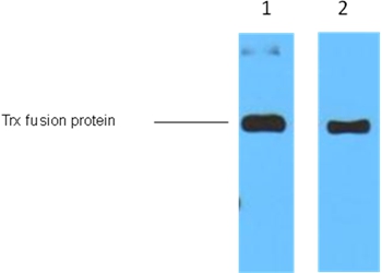 Application Data
(1ug Trx fusion protein+ Primary antibody dilution at 1:5,000 (lane 1) and 1:10,000 (lane 2).)
Application Data
(1ug Trx fusion protein+ Primary antibody dilution at 1:5,000 (lane 1) and 1:10,000 (lane 2).)
Trx-Tag, Monoclonal Antibody (Cat# AAA300837)
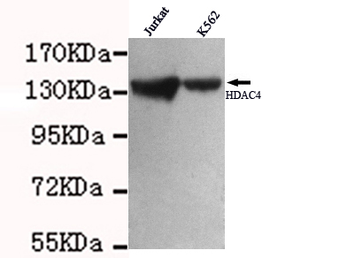
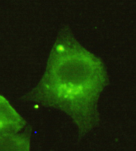 Application Data
(1:150 dilution from a previous lot detected HDAC4 in Hela)
Application Data
(1:150 dilution from a previous lot detected HDAC4 in Hela)
HDAC4, Monoclonal Antibody (Cat# AAA300840)
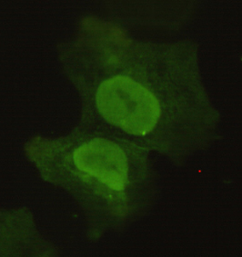
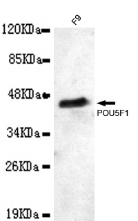
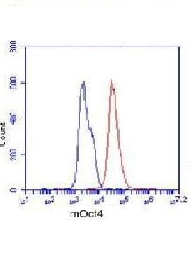 Application Data
(F9 cells stained with POU5F1 (red, 1:100 dilution), followed by FITC-conjugated goat anti-mouse IgG. Blue line histogram represents the isotype control, normal mouse IgG)
Application Data
(F9 cells stained with POU5F1 (red, 1:100 dilution), followed by FITC-conjugated goat anti-mouse IgG. Blue line histogram represents the isotype control, normal mouse IgG)
POU5F1, Monoclonal Antibody (Cat# AAA300842)
 WB (Western Blot)
(Western blot analysis of (A) 5 ng and (B) 25 ng of recombinant HA1 with Hemagglutinin antibody at 1 ug/mL.)
WB (Western Blot)
(Western blot analysis of (A) 5 ng and (B) 25 ng of recombinant HA1 with Hemagglutinin antibody at 1 ug/mL.)
Hemagglutinin, Monoclonal Antibody (Cat# AAA300844)
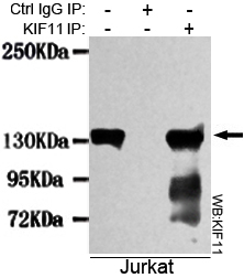 IP (Immunoprecipitation)
(Immunoprecipitation analysis of Jurkat cell lysates using KIF11 antibody)
IP (Immunoprecipitation)
(Immunoprecipitation analysis of Jurkat cell lysates using KIF11 antibody)
KIF11, Monoclonal Antibody (Cat# AAA300851)
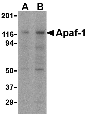
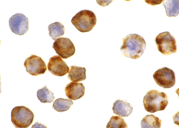 IHC (Immunohiostchemistry)
(Immunohistochemistry of Apaf1 in K562 cells with Apaf1 antibody at 0.5 ug/mL.)
IHC (Immunohiostchemistry)
(Immunohistochemistry of Apaf1 in K562 cells with Apaf1 antibody at 0.5 ug/mL.)
Apaf-1, Monoclonal Antibody (Cat# AAA300855)
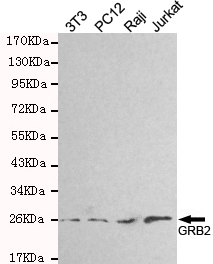
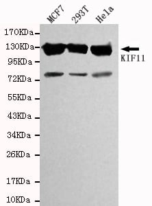
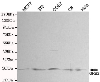 WB (Western Blot)
(Western blot detection of GRB2 in MCF7,3T3,COS7,C6 and Hela cell lysates and using GRB2 antibody(1:300 diluted).Predicted band size: 25KDa.Observed band size: 25KDa.)
WB (Western Blot)
(Western blot detection of GRB2 in MCF7,3T3,COS7,C6 and Hela cell lysates and using GRB2 antibody(1:300 diluted).Predicted band size: 25KDa.Observed band size: 25KDa.)
GRB2, Monoclonal Antibody (Cat# AAA300860)
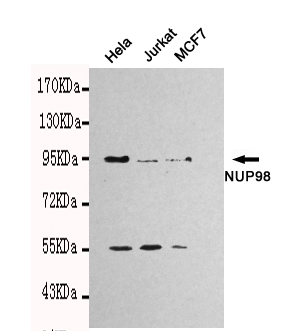
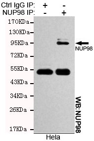 IP (Immunoprecipitation)
(Immunoprecipitation analysis of Hela cell lysates using NUP98 antibody)
IP (Immunoprecipitation)
(Immunoprecipitation analysis of Hela cell lysates using NUP98 antibody)
NUP98, Monoclonal Antibody (Cat# AAA300861)
 Application Data
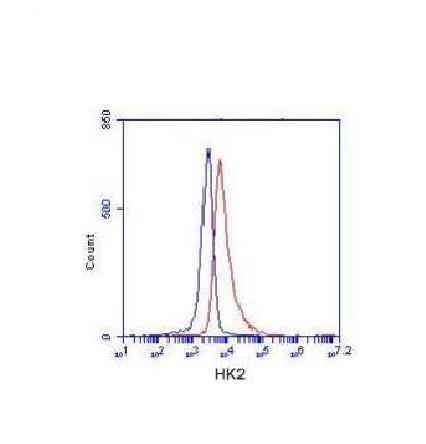
(K562 cells stained with HK2 (red, 1/100 dilution), followed by FITC-conjugated goat anti-mouse IgG. Blue line histogram represents the isotype control, normal mouse IgG)
Application Data
(K562 cells stained with HK2 (red, 1/100 dilution), followed by FITC-conjugated goat anti-mouse IgG. Blue line histogram represents the isotype control, normal mouse IgG)
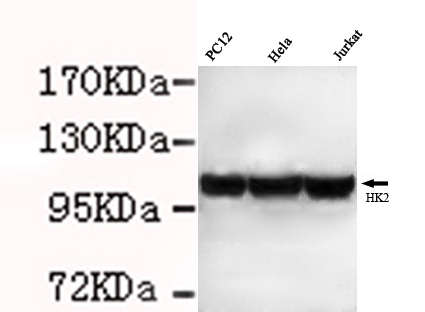
Hexokinase-2, Monoclonal Antibody (Cat# AAA300864)
What are Monoclonal Antibodies?
Monoclonal antibodies are specialized laboratory-produced proteins developed for binding to specific biological antigens or other molecular targets. Since they come from a single cell (or clone), they are especially consistent and accurate in the data they are involved in producing.
This type of antibody material has been shown to be a powerful tool in finding and subsequently destroying harmful cells in an organism, such as those found in cancers or various autoimmune diseases. This makes them excellent aids in medical testing and research, which is why they are so widely used.
AAA Biotech offers a comprehensive range of high-quality monoclonal antibodies that perform effectively in various laboratory tests, including (amongst others) ELISA, western blotting, immunohistochemistry, and flow cytometry. All of the products in our catalog are thoroughly quality tested to make sure that they are reliable and will consistently perform well in your research.
What Are The Uses of Monoclonal Antibodies
Monoclonal antibodies are used in many lab tests, including (amongst others) ELISA, western blotting, immunohistochemistry, and flow cytometry.
ELISA is a test that helps detect a specific substance/analyte in a sample. It uses antibodies (often monoclonal) bound to a solid surface (such as the well of a microplate) to “capture” the substance/analyte in the sample and immobilize it so that the detection antibody component can then bind to it and produce a signal, which can then be measured.
Western blotting identifies specific proteins in a sample. The sample is first separated on a gel, and then antibodies are applied that will typically bind to the target, which will all be localized to a single band in a lane.
Immunohistochemistry helps locate specific proteins in cells or tissue samples using antibodies.
Flow cytometry looks at and sorts cells. It uses antibodies that are conjugated to reporter molecules called “fluorophores”, which, under special lights, emit light themselves, which can then be measured by a detector instrument.
How Monoclonal Antibodies Are Used as Medicine?
Please note that all of the products listed in AAA Biotech’s also known as AAA Bio or AAABio catalog are strictly for research-use only (RUO).
Monoclonal antibodies can also be used as therapeutic/medical treatments, particularly in the context of cancers. They are designed to find and bind to specific cells or proteins, helping the immune system recognize and attack the cancer. These treatments work in different ways, such as:
- Radioimmunotherapy attaches a small amount of radioactive molecule to the antibody, so it delivers the radiation directly to the cancer cells that the antibody is specifically binding to.
- Antibody-directed enzyme prodrug therapy uses antibodies that are specifically bound to special enzymes. These enzymes activate a harmless drug in the body and turn it into a cancer-killing drug only near the cancer cells—this helps avoid harming healthy cells.
- Immunoliposomes are tiny “bubbles” filled with medicine/drug and coated with antibodies. They carry the drug straight to the cancer cells.
Why Buy Monoclonal Antibodies From Us?
At AAA Biotech, we provide high-performance monoclonal antibodies designed to support a wide range of research needs.
1. Validated for Versatile Applications
The antibodies in our catalog are extensively validated and compatible with multiple techniques, including (but not limited to) ELISA, flow cytometry (FC), immunocytochemistry (ICC), immunofluorescence (IF), immunohistochemistry (IHC), immunoprecipitation (IP), and western blotting (WB).
2. Wide Selection & Specialized Options
We offer antibodies for common and rare species, that are available in various conjugated forms, and also in recombinant formats. Essentially, there is almost anything one might need to meet their experimental model’s requirements.
3. High-Quality Proteins
Our proteins meet high purity standards—90% or more as confirmed by SDS-PAGE. Many are available with tags like His, Flag, GST, or MBP, and we also supply native and biologically active proteins for functional studies.
Frequently Asked Questions
1. Are your monoclonal antibodies validated for specific applications?
Yes, our antibodies are tested and validated for use in methods such as ELISA, western blot, IHC, flow cytometry, and more. Refer to specific product pages or datasheets for individual product information.
2. How do I choose the right monoclonal antibody for my application?
Review the product details directly for application validation, species reactivity, and target information. You may also contact our support team at any time for help.
3. How quickly can I receive my order?
Most orders are processed and shipped within 1–3 business days, depending on product availability and your shipping location.