Filters
▼Clonality
▼Type
▼Reactivity
▼Gene Name
▼Isotype
▼Host
▼Application
▼Clone
▼Monoclonal Antibodies
Get accurate results in your research with our Monoclonal Antibodies, which are specially made to target exactly what you require for your research, and will produce consistent, reliable performance in lab tests.
Viewing 8500-8550 of 27597 product results
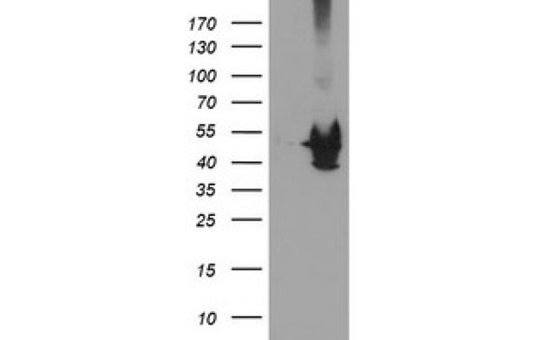
 WB (Western Blot)
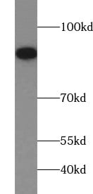
(human heart tissue were subjected to SDS PAGE followed by western blot with AAA102701 (ATP5C1 Antibody) at dilution of 1:1000)
WB (Western Blot)
(human heart tissue were subjected to SDS PAGE followed by western blot with AAA102701 (ATP5C1 Antibody) at dilution of 1:1000)
ATP5C1, Monoclonal Antibody (Cat# AAA102701)
Protein A+G purification
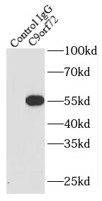
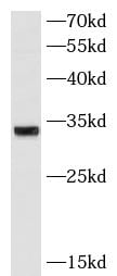
 WB (Western Blot)
(human brain tissue were subjected to SDS PAGE followed by western blot with AAA102718 (C9orf72 antibody) at dilution of 1:1000)
WB (Western Blot)
(human brain tissue were subjected to SDS PAGE followed by western blot with AAA102718 (C9orf72 antibody) at dilution of 1:1000)
C9orf72, Monoclonal Antibody (Cat# AAA102718)
Protein A+G purification
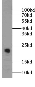
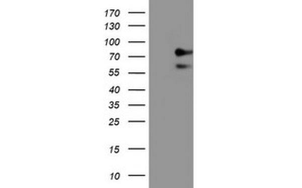
 WB (Western Blot)
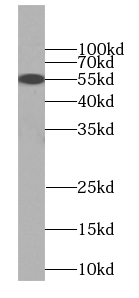
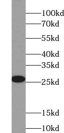
(PC-3 cells were subjected to SDS PAGE followed by western blot with AAA102723 (CAV1 antibody) at dilution of 1:1000)
WB (Western Blot)
(PC-3 cells were subjected to SDS PAGE followed by western blot with AAA102723 (CAV1 antibody) at dilution of 1:1000)
Caveolin-1, Monoclonal Antibody (Cat# AAA102723)
Protein A+G purification
 WB (Western Blot)
(COLO 320 cells were subjected to SDS PAGE followed by western blot with AAA102724 (CCT3 antibody) at dilution of 1:1000)
WB (Western Blot)
(COLO 320 cells were subjected to SDS PAGE followed by western blot with AAA102724 (CCT3 antibody) at dilution of 1:1000)
CCT3, Monoclonal Antibody (Cat# AAA102724)
Protein A+G purification
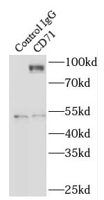
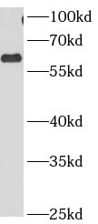
 WB (Western Blot)
(HeLa cells were subjected to SDS PAGE followed by western blot with AAA102740 (CD71 Antibody) at dilution of 1:2000)
WB (Western Blot)
(HeLa cells were subjected to SDS PAGE followed by western blot with AAA102740 (CD71 Antibody) at dilution of 1:2000)
CD71, Monoclonal Antibody (Cat# AAA102740)
Protein A+G purification
 WB (Western Blot)
(Transfected HEK-293 cells were subjected to SDS PAGE followed by western blot with AAA102744 (CGB,hCG Antibody) at dilution of 1:1000)
WB (Western Blot)
(Transfected HEK-293 cells were subjected to SDS PAGE followed by western blot with AAA102744 (CGB,hCG Antibody) at dilution of 1:1000)
CGB, hCG, Monoclonal Antibody (Cat# AAA102744)
Protein A+G purification
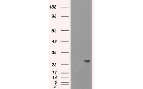
 WB (Western Blot)
(HepG2 cells were subjected to SDS PAGE followed by western blot with AAA102752 (CNPY2, MSAP antibody) at dilution of 1:1000)
WB (Western Blot)
(HepG2 cells were subjected to SDS PAGE followed by western blot with AAA102752 (CNPY2, MSAP antibody) at dilution of 1:1000)
CNPY2, MSAP, Monoclonal Antibody (Cat# AAA102752)
Protein A+G purification
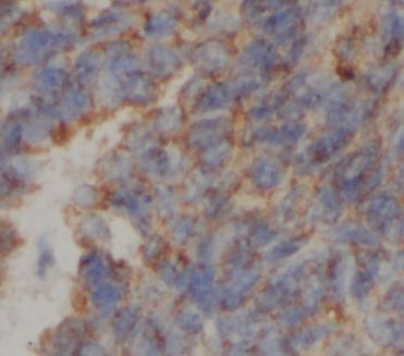
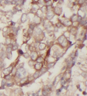
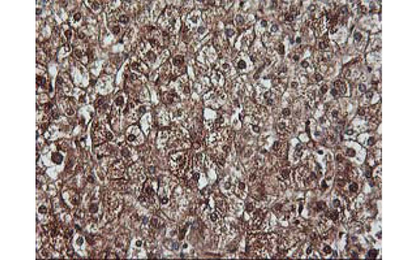
 IHC (Immunohiostchemistry)
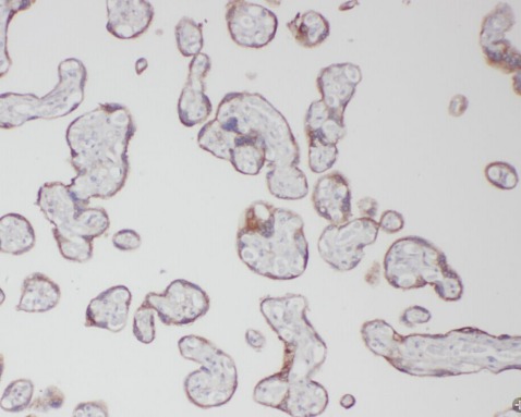
(Immunohistochemical analysis of IFT57 protein in paraffin embedded Adenocarcinoma of Human colon tissue using IFT57 antibody)
IHC (Immunohiostchemistry)
(Immunohistochemical analysis of IFT57 protein in paraffin embedded Adenocarcinoma of Human colon tissue using IFT57 antibody)
IFT57, Monoclonal Antibody (Cat# AAA108057)
Renin, Monoclonal Antibody (Cat# AAA108062)
Mouse anti Human IgG4 antibody, Monoclonal Antibody (Cat# AAA108064)
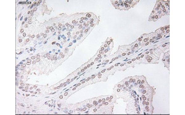
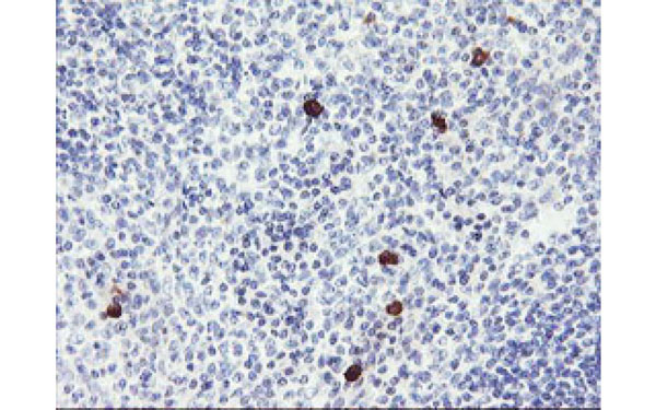 IHC (Immunohistochemisry)
(Immunohistochemical analysis of RPS6KB1 protein in paraffin embedded Human tonsil tissue using RPS6KB1 antibody)
IHC (Immunohistochemisry)
(Immunohistochemical analysis of RPS6KB1 protein in paraffin embedded Human tonsil tissue using RPS6KB1 antibody)
RPS6KB1, Monoclonal Antibody (Cat# AAA108072)
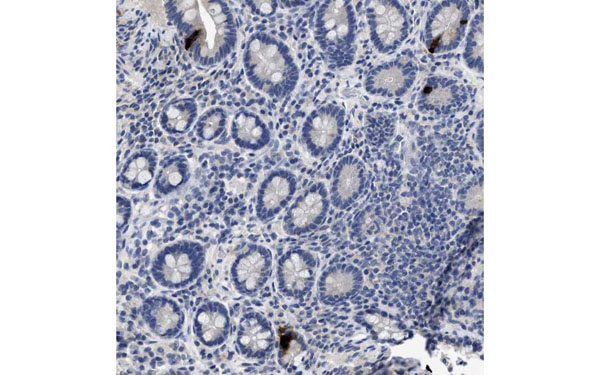
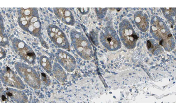
 IHC (Immunohiostchemistry)
(Paraffin-embedded human small intestine tissue immunostained with Peptide YY antibody)
IHC (Immunohiostchemistry)
(Paraffin-embedded human small intestine tissue immunostained with Peptide YY antibody)
Peptide YY, Monoclonal Antibody (Cat# AAA108073)
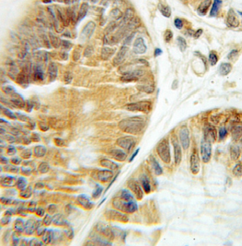
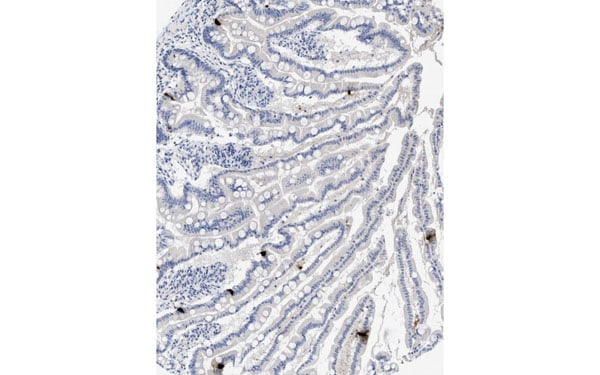
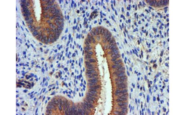
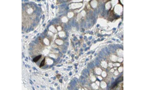 IHC (Immunohiostchemistry)
(Paraffin-embedded human small intestine tissue immunostained with GIP antibody)
IHC (Immunohiostchemistry)
(Paraffin-embedded human small intestine tissue immunostained with GIP antibody)
GIP, Monoclonal Antibody (Cat# AAA108078)
Cholic acid, Monoclonal Antibody (Cat# AAA108091)
 IF (Immunofluorescence)
(Immunofluorescent staining of COS7 cells transiently transfected with recombinant PSMC3 protein using PSMC3 antibody)
IF (Immunofluorescence)
(Immunofluorescent staining of COS7 cells transiently transfected with recombinant PSMC3 protein using PSMC3 antibody)
PSMC3, Monoclonal Antibody (Cat# AAA108111)
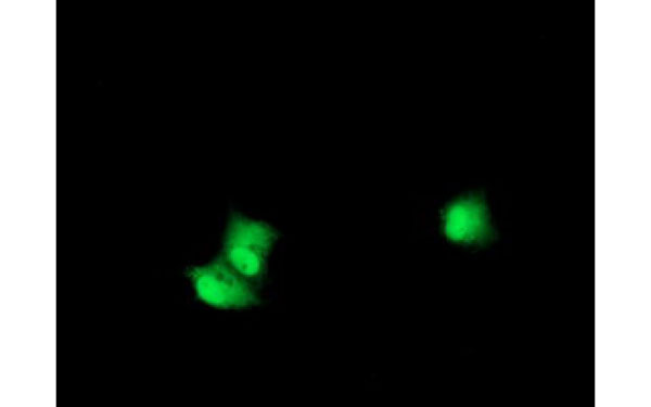
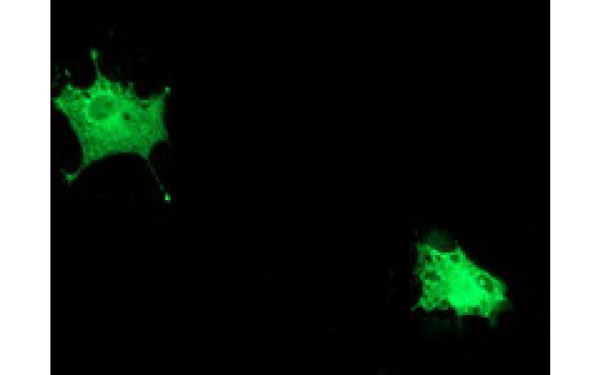
 IF (Immunofluorescence)
(Immunofluorescent staining of COS7 cells transiently transfected with recombinant MAPRE2 protein using MAPRE2 antibody)
IF (Immunofluorescence)
(Immunofluorescent staining of COS7 cells transiently transfected with recombinant MAPRE2 protein using MAPRE2 antibody)
MAPRE2, Monoclonal Antibody (Cat# AAA108132)
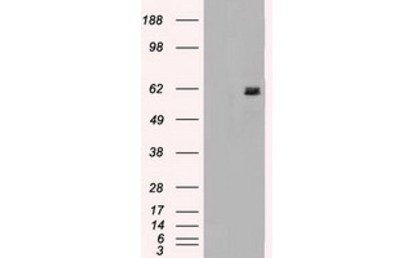
 WB (Western Blot)
Lymphotoxin alpha, Monoclonal Antibody (Cat# AAA108148)
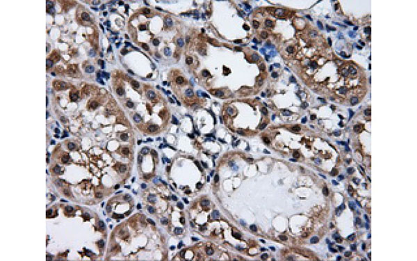
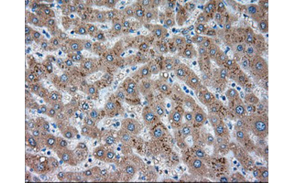
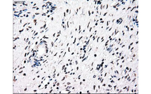 IHC (Immunohistochemisry)
(Immunohistochemical analysis of NRBP1 protein in paraffin embedded Human ovary tissue using NRBP1 antibody)
IHC (Immunohistochemisry)
(Immunohistochemical analysis of NRBP1 protein in paraffin embedded Human ovary tissue using NRBP1 antibody)
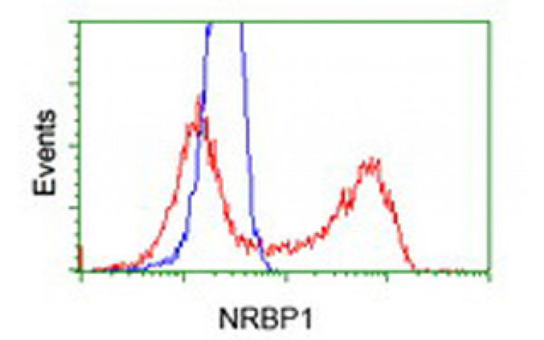
NRBP1, Monoclonal Antibody (Cat# AAA108150)
Fentanyl, Monoclonal Antibody (Cat# AAA108164)
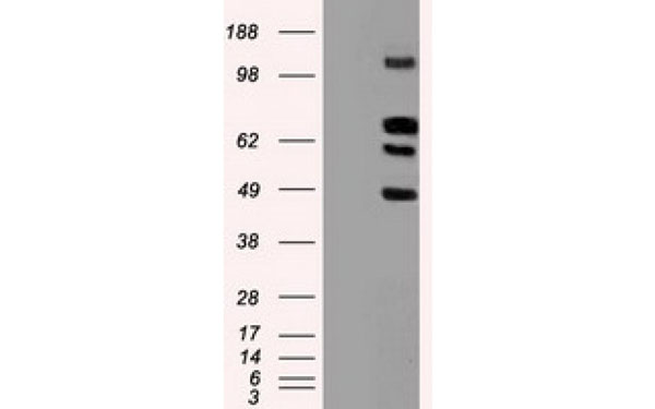
 WB (Western Blot)
(Western Blot analysis of HEK293T cell lysates (5 ug) transfected with either recombinant TNNI3 protein (Right) or empty vector (Left) detected with TNNI3 antibody)
WB (Western Blot)
(Western Blot analysis of HEK293T cell lysates (5 ug) transfected with either recombinant TNNI3 protein (Right) or empty vector (Left) detected with TNNI3 antibody)
TNNI3, Monoclonal Antibody (Cat# AAA108169)
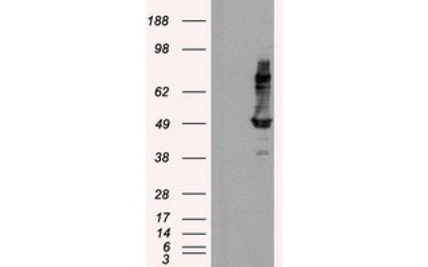
 WB (Western Blot)
(Western Blot analysis of HEK293T cell lysates (5 ug) transfected with either recombinant POLR2E protein (Right) or empty vector (Left) detected with POLR2E antibody)
WB (Western Blot)
(Western Blot analysis of HEK293T cell lysates (5 ug) transfected with either recombinant POLR2E protein (Right) or empty vector (Left) detected with POLR2E antibody)
POLR2E, Monoclonal Antibody (Cat# AAA108181)
Insulin B chain, Monoclonal Antibody (Cat# AAA108198)
THC, Monoclonal Antibody (Cat# AAA108200)
Vitamin D, Monoclonal Antibody (Cat# AAA108203)
HBV Pres 1, Monoclonal Antibody (Cat# AAA108204)
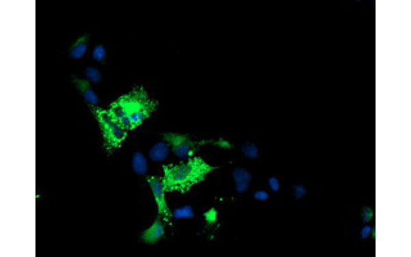
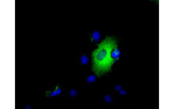
 IF (Immunofluorescence)
(Immunofluorescent staining of COS7 cells transiently transfected with recombinant TACC3 protein using TACC3 antibody)
IF (Immunofluorescence)
(Immunofluorescent staining of COS7 cells transiently transfected with recombinant TACC3 protein using TACC3 antibody)
TACC3, Monoclonal Antibody (Cat# AAA108211)
 IF (Immunofluorescence)
(Immunofluorescent staining of COS7 cells transiently transfected with recombinant MAPK7 protein using MAPK7 antibody)
IF (Immunofluorescence)
(Immunofluorescent staining of COS7 cells transiently transfected with recombinant MAPK7 protein using MAPK7 antibody)
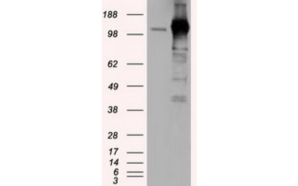
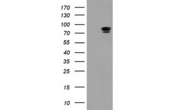
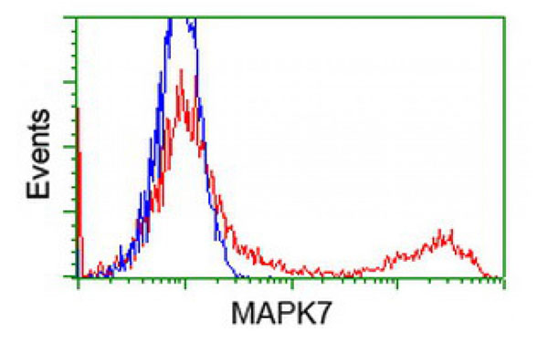
MAPK7, Monoclonal Antibody (Cat# AAA108212)
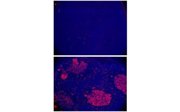
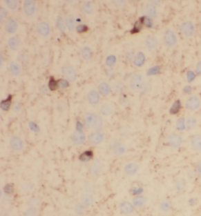
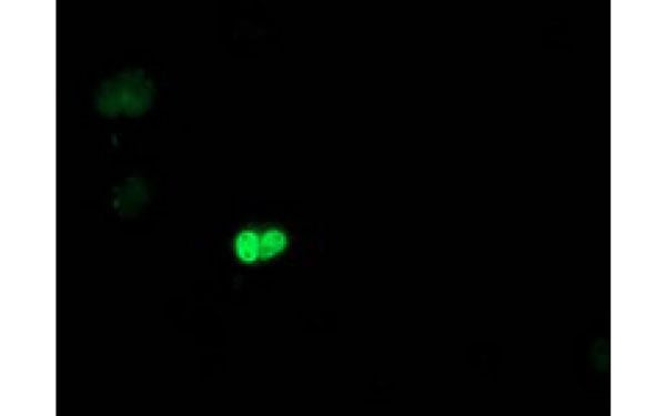
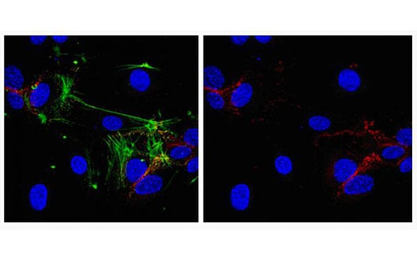
 IF (Immunofluorescence)
(Immunofluorescent staining of AID knock-out (top) or wild type (bottom) mouse intestinal Peyer's patches with AID antibody (AAA107966))
IF (Immunofluorescence)
(Immunofluorescent staining of AID knock-out (top) or wild type (bottom) mouse intestinal Peyer's patches with AID antibody (AAA107966))
AID, Monoclonal Antibody (Cat# AAA107966)
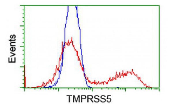
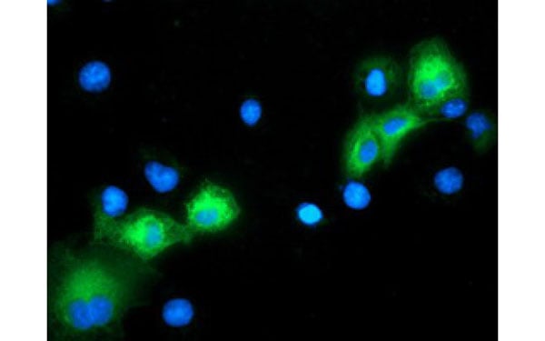
 IF (Immunofluorescence)
(Immunofluorescent staining of COS7 cells transiently transfected with recombinant TMPRSS5 protein using TMPRSS5 antibody)
IF (Immunofluorescence)
(Immunofluorescent staining of COS7 cells transiently transfected with recombinant TMPRSS5 protein using TMPRSS5 antibody)
TMPRSS5, Monoclonal Antibody (Cat# AAA107980)
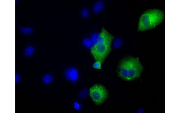
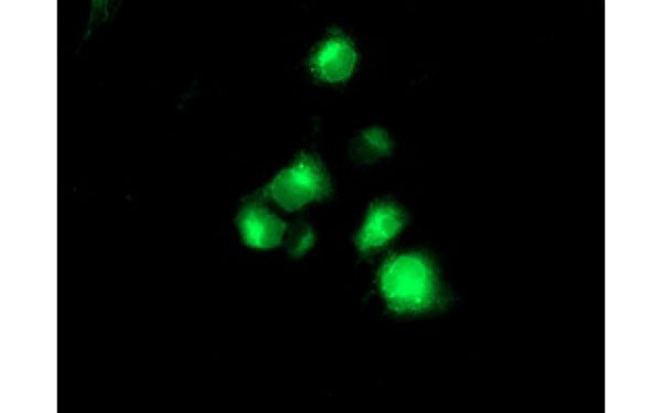
 IF (Immunofluorescence)
(Immunofluorescent staining of COS7 cells transiently transfected with recombinant MAPK7 protein using MAPK7 antibody)
IF (Immunofluorescence)
(Immunofluorescent staining of COS7 cells transiently transfected with recombinant MAPK7 protein using MAPK7 antibody)
MAPK7, Monoclonal Antibody (Cat# AAA108220)
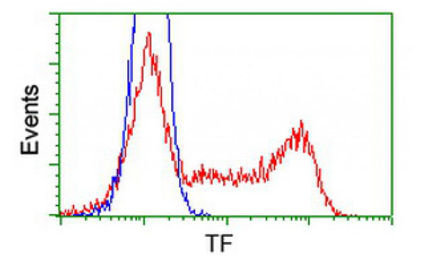
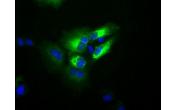 IF (Immunofluorescence)
(Immunofluorescent staining of COS7 cells transiently transfected with recombinant TF protein using TF antibody)
IF (Immunofluorescence)
(Immunofluorescent staining of COS7 cells transiently transfected with recombinant TF protein using TF antibody)
TF, Monoclonal Antibody (Cat# AAA108223)
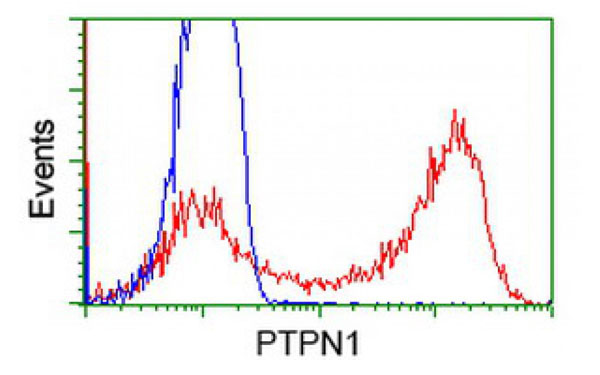
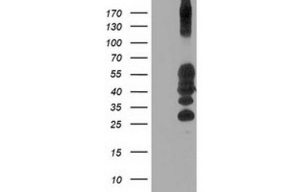 WB (Western Blot)
(Western Blot analysis of HEK293T cell lysates (5 ug) transfected with either recombinant PTPN1 protein (Right) or empty vector (Left) detected with PTPN1 antibody)
WB (Western Blot)
(Western Blot analysis of HEK293T cell lysates (5 ug) transfected with either recombinant PTPN1 protein (Right) or empty vector (Left) detected with PTPN1 antibody)
PTPN1, Monoclonal Antibody (Cat# AAA108232)

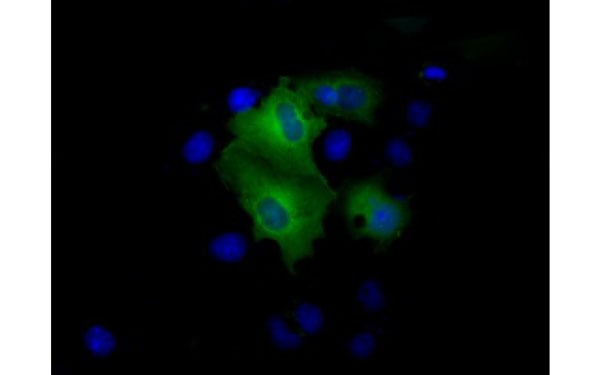
 IF (Immunofluorescence)
(Immunofluorescent staining of COS7 cells transiently transfected with recombinant ALOX15 protein using ALOX15 antibody)
IF (Immunofluorescence)
(Immunofluorescent staining of COS7 cells transiently transfected with recombinant ALOX15 protein using ALOX15 antibody)
ALOX15, Monoclonal Antibody (Cat# AAA108263)
 WB (Western Blot)
(Western Blot analysis of HEK293T cell lysates (5 ug) transfected with either recombinant POLR2E protein (Right) or empty vector (Left) detected with POLR2E antibody)
WB (Western Blot)
(Western Blot analysis of HEK293T cell lysates (5 ug) transfected with either recombinant POLR2E protein (Right) or empty vector (Left) detected with POLR2E antibody)
POLR2E, Monoclonal Antibody (Cat# AAA108267)
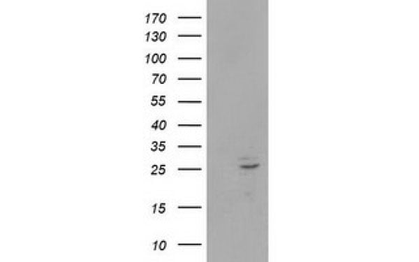
 WB (Western Blot)
(Western Blot analysis of HEK293T cell lysates (5 ug) transfected with either recombinant KCNAB1 protein (Right) or empty vector (Left) detected with KCNAB1 antibody)
WB (Western Blot)
(Western Blot analysis of HEK293T cell lysates (5 ug) transfected with either recombinant KCNAB1 protein (Right) or empty vector (Left) detected with KCNAB1 antibody)
KCNAB1, Monoclonal Antibody (Cat# AAA108011)
 WB (Western Blot)
(Western Blot analysis of HEK293T cell lysates (5 ug) transfected with either recombinant SIGLEC9 protein (Right) or empty vector (Left) detected with SIGLEC9 antibody)
WB (Western Blot)
(Western Blot analysis of HEK293T cell lysates (5 ug) transfected with either recombinant SIGLEC9 protein (Right) or empty vector (Left) detected with SIGLEC9 antibody)
SIGLEC9, Monoclonal Antibody (Cat# AAA108037)
IGFBP4, Monoclonal Antibody (Cat# AAA108051)
 IF (Immunofluorescence)
(Immunofluorescent staining of COS7 cells transiently transfected with recombinant LGALS3BP protein using LGALS3BP antibody)
IF (Immunofluorescence)
(Immunofluorescent staining of COS7 cells transiently transfected with recombinant LGALS3BP protein using LGALS3BP antibody)
LGALS3BP, Monoclonal Antibody (Cat# AAA108052)
Renin, Monoclonal Antibody (Cat# AAA108056)
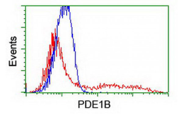
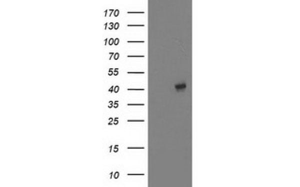
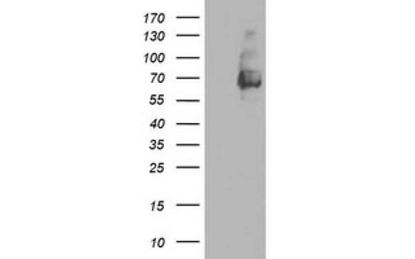
 WB (Western Blot)
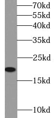
(Western Blot analysis of HEK293T cell lysates (5 ug) transfected with either recombinant PDE1B protein (Right) or empty vector (Left) detected with PDE1B antibody)
WB (Western Blot)
(Western Blot analysis of HEK293T cell lysates (5 ug) transfected with either recombinant PDE1B protein (Right) or empty vector (Left) detected with PDE1B antibody)
PDE1B, Monoclonal Antibody (Cat# AAA107132)
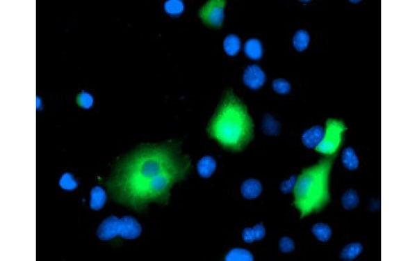
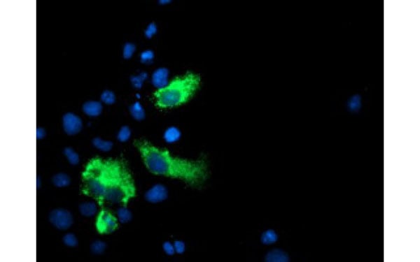
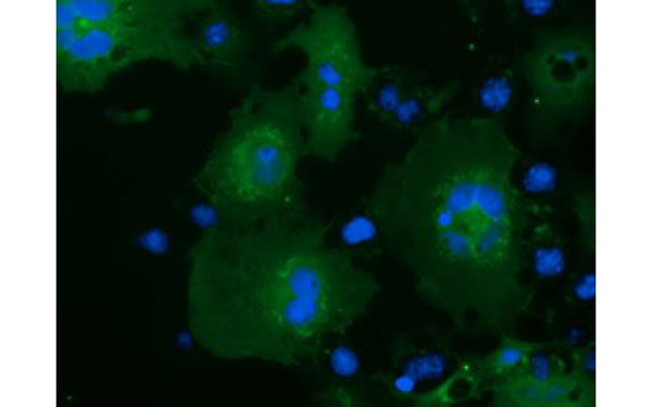 IF (Immunofluorescence)
(Immunofluorescent staining of COS7 cells transiently transfected with recombinant MOBKL2B protein using MOBKL2B antibody)
IF (Immunofluorescence)
(Immunofluorescent staining of COS7 cells transiently transfected with recombinant MOBKL2B protein using MOBKL2B antibody)
MOBKL2B, Monoclonal Antibody (Cat# AAA107171)
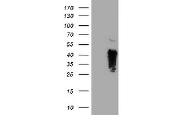
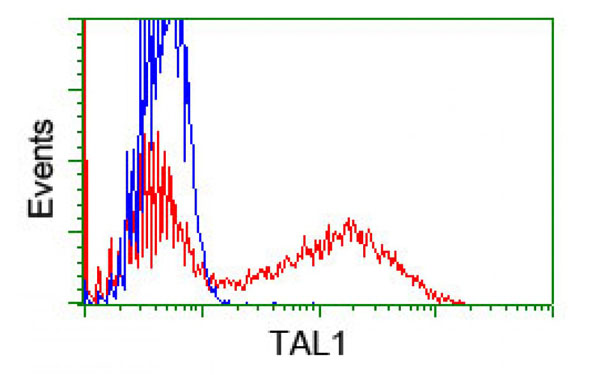
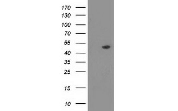
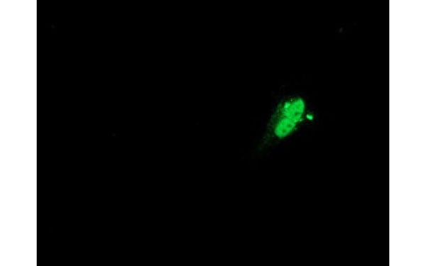
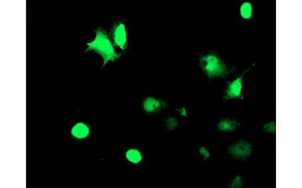 IF (Immunofluorescence)
(Immunofluorescent staining of COS7 cells transiently transfected with recombinant TAL1 protein using TAL1 antibody)
IF (Immunofluorescence)
(Immunofluorescent staining of COS7 cells transiently transfected with recombinant TAL1 protein using TAL1 antibody)
TAL1, Monoclonal Antibody (Cat# AAA107190)
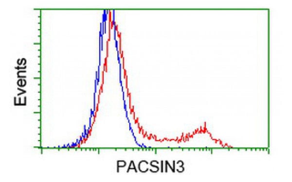
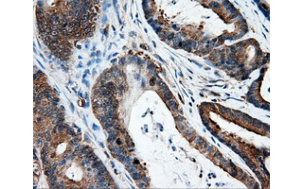
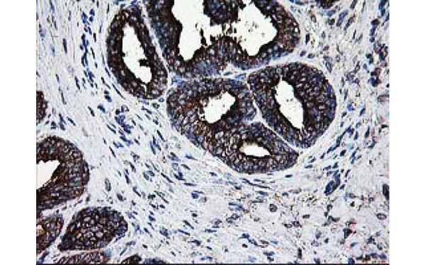
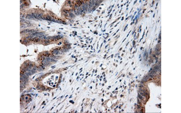
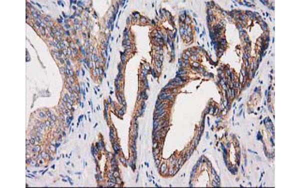 IHC (Immunohistochemisry)
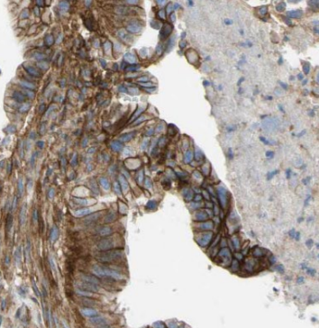
(Immunohistochemical analysis of PACSIN3 protein in paraffin embedded Carcinoma of Human prostate tissue using PACSIN3 antibody)
IHC (Immunohistochemisry)
(Immunohistochemical analysis of PACSIN3 protein in paraffin embedded Carcinoma of Human prostate tissue using PACSIN3 antibody)
PACSIN3, Monoclonal Antibody (Cat# AAA107197)
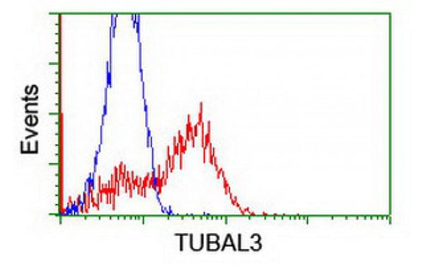
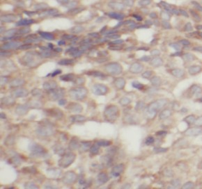
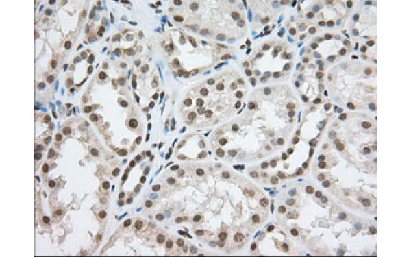
 IHC (Immunohistochemisry)
(Immunohistochemical analysis of TUBAL3 protein in paraffin embedded Human endometrium tissue using TUBAL3 antibody)
IHC (Immunohistochemisry)
(Immunohistochemical analysis of TUBAL3 protein in paraffin embedded Human endometrium tissue using TUBAL3 antibody)
TUBAL3, Monoclonal Antibody (Cat# AAA106690)

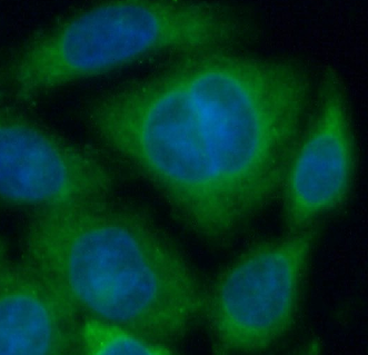
 IF (Immunofluorescence)
(Immunofluorescent staining of COS7 cells transiently transfected with recombinant SAMHD1 protein using SAMHD1 antibody)
IF (Immunofluorescence)
(Immunofluorescent staining of COS7 cells transiently transfected with recombinant SAMHD1 protein using SAMHD1 antibody)
SAMHD1, Monoclonal Antibody (Cat# AAA106713)
 WB (Western Blot)
(Western Blot analysis of HEK293T cell lysates (5 ug) transfected with either recombinant NPTN protein (Right) or empty vector (Left) detected with NPTN antibody)
WB (Western Blot)
(Western Blot analysis of HEK293T cell lysates (5 ug) transfected with either recombinant NPTN protein (Right) or empty vector (Left) detected with NPTN antibody)
NPTN, Monoclonal Antibody (Cat# AAA106726)
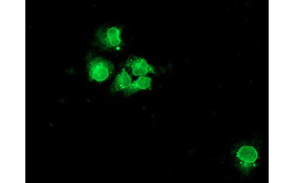
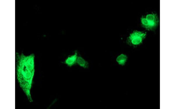 IF (Immunofluorescence)
(Immunofluorescent staining of COS7 cells transiently transfected with recombinant SPINT1 protein using SPINT1 antibody)
IF (Immunofluorescence)
(Immunofluorescent staining of COS7 cells transiently transfected with recombinant SPINT1 protein using SPINT1 antibody)
SPINT1, Monoclonal Antibody (Cat# AAA106728)
Vibrio cholerae O1 Ogawa & Inaba, Monoclonal Antibody (Cat# AAA106741)
 IF (Immunofluorescence)
(Immunofluorescent staining of COS7 cells transiently transfected with recombinant LIPG protein using LIPG antibody)
IF (Immunofluorescence)
(Immunofluorescent staining of COS7 cells transiently transfected with recombinant LIPG protein using LIPG antibody)
LIPG, Monoclonal Antibody (Cat# AAA106744)
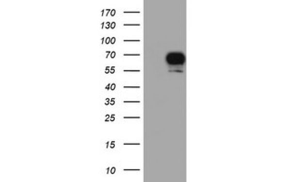
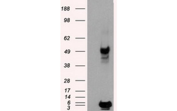
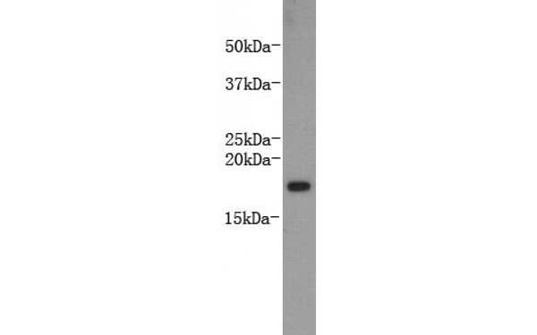
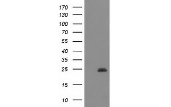
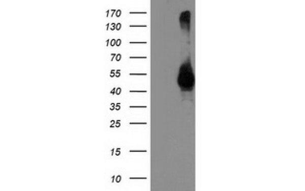
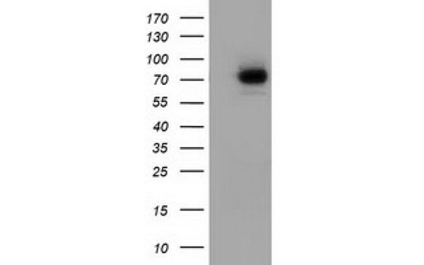
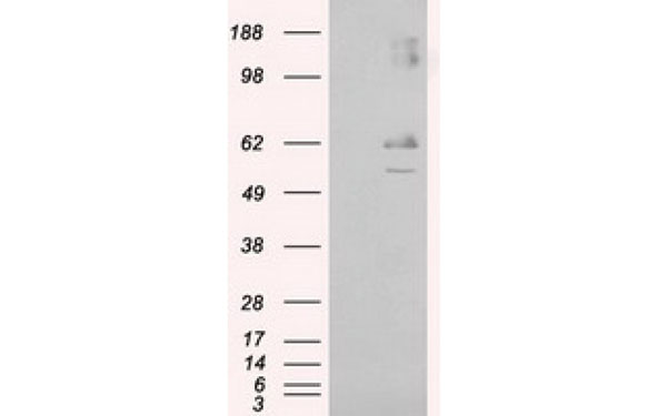
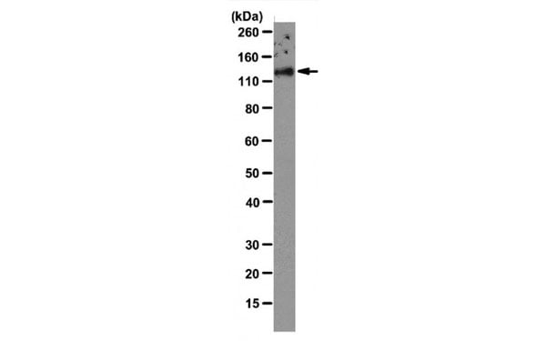 WB (Western Blot)
(HUVEC cell lysate was probed with VE-cadherin antibody (1/2000 dilution).)
WB (Western Blot)
(HUVEC cell lysate was probed with VE-cadherin antibody (1/2000 dilution).)
VE Cadherin, Monoclonal Antibody (Cat# AAA106745)
What are Monoclonal Antibodies?
Monoclonal antibodies are specialized laboratory-produced proteins developed for binding to specific biological antigens or other molecular targets. Since they come from a single cell (or clone), they are especially consistent and accurate in the data they are involved in producing.
This type of antibody material has been shown to be a powerful tool in finding and subsequently destroying harmful cells in an organism, such as those found in cancers or various autoimmune diseases. This makes them excellent aids in medical testing and research, which is why they are so widely used.
AAA Biotech offers a comprehensive range of high-quality monoclonal antibodies that perform effectively in various laboratory tests, including (amongst others) ELISA, western blotting, immunohistochemistry, and flow cytometry. All of the products in our catalog are thoroughly quality tested to make sure that they are reliable and will consistently perform well in your research.
What Are The Uses of Monoclonal Antibodies
Monoclonal antibodies are used in many lab tests, including (amongst others) ELISA, western blotting, immunohistochemistry, and flow cytometry.
ELISA is a test that helps detect a specific substance/analyte in a sample. It uses antibodies (often monoclonal) bound to a solid surface (such as the well of a microplate) to “capture” the substance/analyte in the sample and immobilize it so that the detection antibody component can then bind to it and produce a signal, which can then be measured.
Western blotting identifies specific proteins in a sample. The sample is first separated on a gel, and then antibodies are applied that will typically bind to the target, which will all be localized to a single band in a lane.
Immunohistochemistry helps locate specific proteins in cells or tissue samples using antibodies.
Flow cytometry looks at and sorts cells. It uses antibodies that are conjugated to reporter molecules called “fluorophores”, which, under special lights, emit light themselves, which can then be measured by a detector instrument.
How Monoclonal Antibodies Are Used as Medicine?
Please note that all of the products listed in AAA Biotech’s also known as AAA Bio or AAABio catalog are strictly for research-use only (RUO).
Monoclonal antibodies can also be used as therapeutic/medical treatments, particularly in the context of cancers. They are designed to find and bind to specific cells or proteins, helping the immune system recognize and attack the cancer. These treatments work in different ways, such as:
- Radioimmunotherapy attaches a small amount of radioactive molecule to the antibody, so it delivers the radiation directly to the cancer cells that the antibody is specifically binding to.
- Antibody-directed enzyme prodrug therapy uses antibodies that are specifically bound to special enzymes. These enzymes activate a harmless drug in the body and turn it into a cancer-killing drug only near the cancer cells—this helps avoid harming healthy cells.
- Immunoliposomes are tiny “bubbles” filled with medicine/drug and coated with antibodies. They carry the drug straight to the cancer cells.
Why Buy Monoclonal Antibodies From Us?
At AAA Biotech, we provide high-performance monoclonal antibodies designed to support a wide range of research needs.
1. Validated for Versatile Applications
The antibodies in our catalog are extensively validated and compatible with multiple techniques, including (but not limited to) ELISA, flow cytometry (FC), immunocytochemistry (ICC), immunofluorescence (IF), immunohistochemistry (IHC), immunoprecipitation (IP), and western blotting (WB).
2. Wide Selection & Specialized Options
We offer antibodies for common and rare species, that are available in various conjugated forms, and also in recombinant formats. Essentially, there is almost anything one might need to meet their experimental model’s requirements.
3. High-Quality Proteins
Our proteins meet high purity standards—90% or more as confirmed by SDS-PAGE. Many are available with tags like His, Flag, GST, or MBP, and we also supply native and biologically active proteins for functional studies.
Frequently Asked Questions
1. Are your monoclonal antibodies validated for specific applications?
Yes, our antibodies are tested and validated for use in methods such as ELISA, western blot, IHC, flow cytometry, and more. Refer to specific product pages or datasheets for individual product information.
2. How do I choose the right monoclonal antibody for my application?
Review the product details directly for application validation, species reactivity, and target information. You may also contact our support team at any time for help.
3. How quickly can I receive my order?
Most orders are processed and shipped within 1–3 business days, depending on product availability and your shipping location.