Filters
▼Clonality
▼Type
▼Reactivity
▼Gene Name
▼Isotype
▼Host
▼Application
▼Clone
▼Monoclonal Antibodies
Get accurate results in your research with our Monoclonal Antibodies, which are specially made to target exactly what you require for your research, and will produce consistent, reliable performance in lab tests.
Viewing 6450-6500 of 27597 product results
 IHC (Immunohiostchemistry)
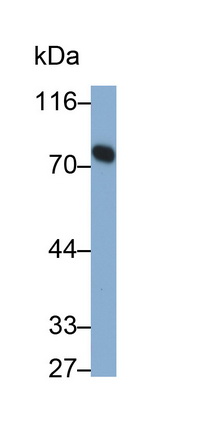
(DAB staining on IHCP;Samples: Human Stomach Tissue; Primary Ab: 10ug/ml Mouse AntiHuman TK1 AntibodySecond Ab: 2ug/mL HRPLinked Caprine AntiMouse IgG Polyclonal Antibody(Catalog: SAA544Mu19))
IHC (Immunohiostchemistry)
(DAB staining on IHCP;Samples: Human Stomach Tissue; Primary Ab: 10ug/ml Mouse AntiHuman TK1 AntibodySecond Ab: 2ug/mL HRPLinked Caprine AntiMouse IgG Polyclonal Antibody(Catalog: SAA544Mu19))
Thymidine Kinase 1, Soluble (TK1), Monoclonal Antibody (Cat# AAA151818)
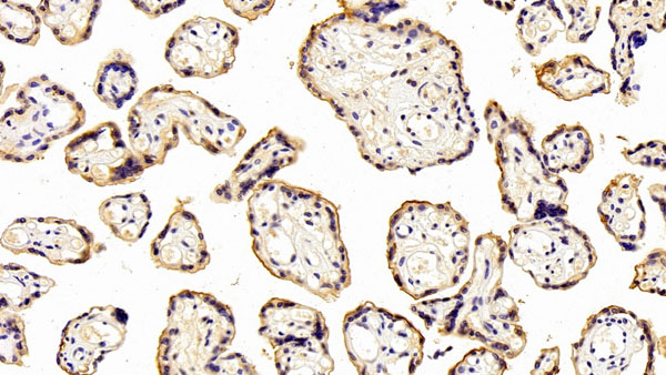
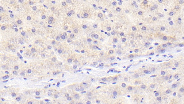
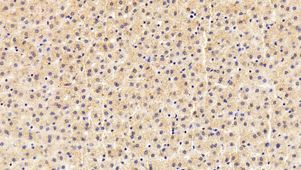
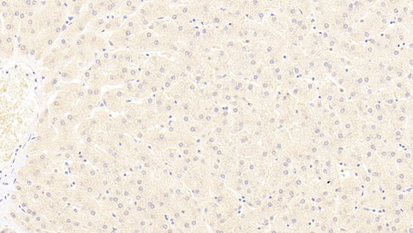 IHC (Immunohiostchemistry)
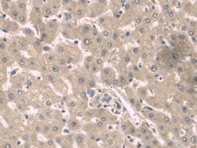
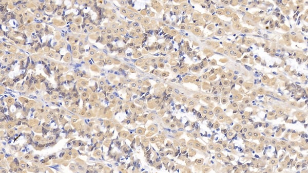
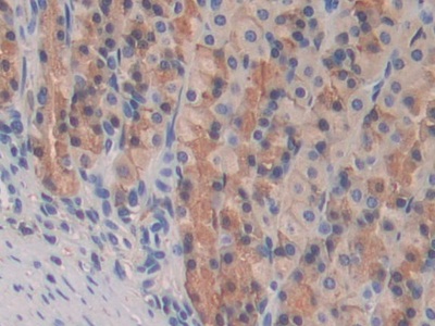
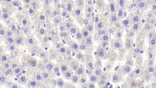
(DAB staining on IHCP;Sample: Human Liver Tissue; Primary Ab: 20ug/ml Mouse AntiHuman LRP6 AntibodySecond Ab: 2ug/mL HRPLinked Caprine AntiMouse IgG Polyclonal Antibody(Catalog: SAA544Mu19))
IHC (Immunohiostchemistry)
(DAB staining on IHCP;Sample: Human Liver Tissue; Primary Ab: 20ug/ml Mouse AntiHuman LRP6 AntibodySecond Ab: 2ug/mL HRPLinked Caprine AntiMouse IgG Polyclonal Antibody(Catalog: SAA544Mu19))
Low Density Lipoprotein Receptor Related Protein 6 (LRP6), Monoclonal Antibody (Cat# AAA151831)

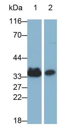
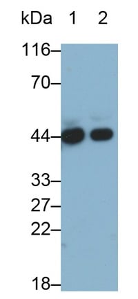
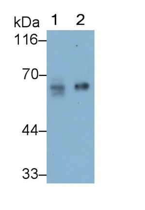
 WB (Western Blot)
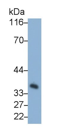
(Western Blot; Sample: Hela cell lysate Primary Ab: 2ug/ml Mouse AntiHuman SQSTM1 Antibody Second Ab: 0.2ug/mL HRPLinked Caprine AntiMouse IgG Polyclonal Antibody (Catalog: SAA544Mu19))
WB (Western Blot)
(Western Blot; Sample: Hela cell lysate Primary Ab: 2ug/ml Mouse AntiHuman SQSTM1 Antibody Second Ab: 0.2ug/mL HRPLinked Caprine AntiMouse IgG Polyclonal Antibody (Catalog: SAA544Mu19))
Sequestosome 1 (SQSTM1), Monoclonal Antibody (Cat# AAA151832)
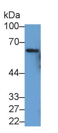
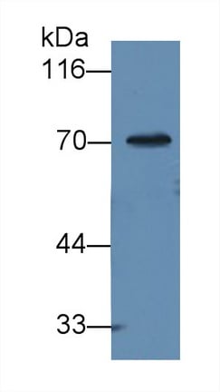
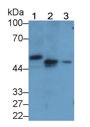
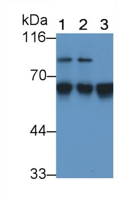
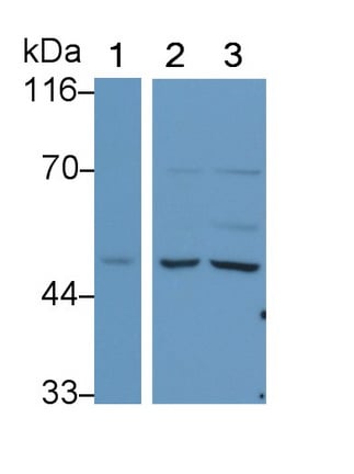
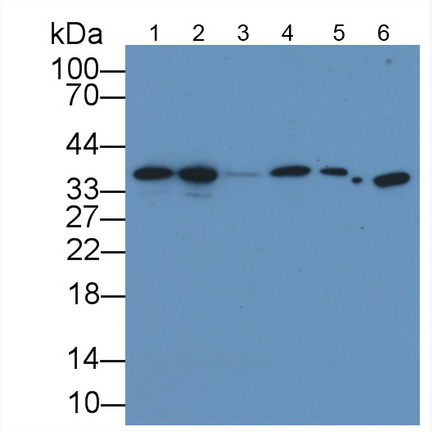
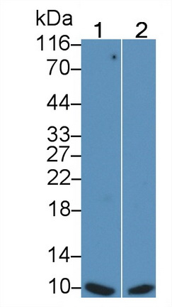
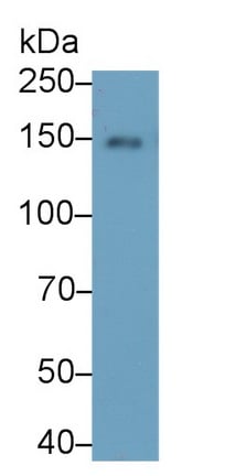
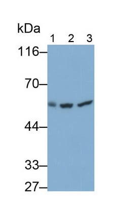
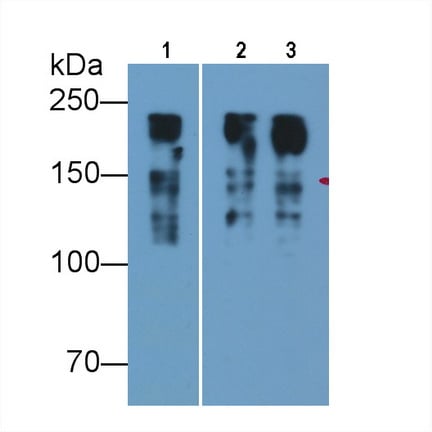
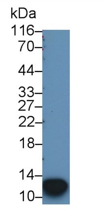
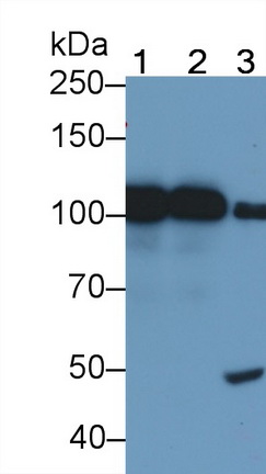
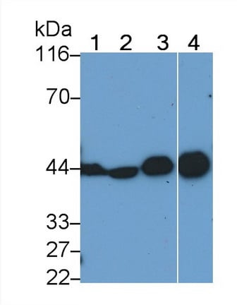
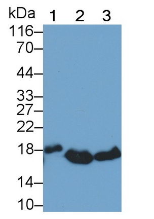
 WB (Western Blot)
(Western Blot; Sample: Lane1: Porcine Heart lysate; Lane2: Rat Heart lysate Primary Ab: 2ug/ml Mouse AntiHuman TNNT2 Antibody Second Ab: 0.2ug/mL HRPLinked Caprine AntiMouse IgG Polyclonal Antibody (Catalog: SAA544Mu19))
WB (Western Blot)
(Western Blot; Sample: Lane1: Porcine Heart lysate; Lane2: Rat Heart lysate Primary Ab: 2ug/ml Mouse AntiHuman TNNT2 Antibody Second Ab: 0.2ug/mL HRPLinked Caprine AntiMouse IgG Polyclonal Antibody (Catalog: SAA544Mu19))
Troponin T Type 2, Cardiac (TNNT2), Monoclonal Antibody (Cat# AAA151836)
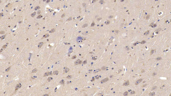
 WB (Western Blot)
(Western Blot; Sample: Lane1: Canine Cerebrum lysate; Lane2: Rabbit Cerebrum lysate; Lane3: Cavia Cerebrum lysate Primary Ab: 1ug/ml Mouse AntiHuman NEFL Antibody Second Ab: 0.2ug/mL HRPLinked Caprine AntiMouse IgG Polyclonal Antibody (Catalog: SAA544Mu19))
WB (Western Blot)
(Western Blot; Sample: Lane1: Canine Cerebrum lysate; Lane2: Rabbit Cerebrum lysate; Lane3: Cavia Cerebrum lysate Primary Ab: 1ug/ml Mouse AntiHuman NEFL Antibody Second Ab: 0.2ug/mL HRPLinked Caprine AntiMouse IgG Polyclonal Antibody (Catalog: SAA544Mu19))
Neurofilament, Light Polypeptide (NEFL), Monoclonal Antibody (Cat# AAA151845)
 WB (Western Blot)
(Western Blot; Sample: Human Serum Primary Ab: 2ug/ml Mouse AntiHuman a1BG Antibody Second Ab: 0.2ug/mL HRPLinked Rabbit AntiMouse IgG Polyclonal Antibody (Catalog: SAA544Mu19))
WB (Western Blot)
(Western Blot; Sample: Human Serum Primary Ab: 2ug/ml Mouse AntiHuman a1BG Antibody Second Ab: 0.2ug/mL HRPLinked Rabbit AntiMouse IgG Polyclonal Antibody (Catalog: SAA544Mu19))
Alpha1BGlycoprotein (a1BG), Monoclonal Antibody (Cat# AAA151852)
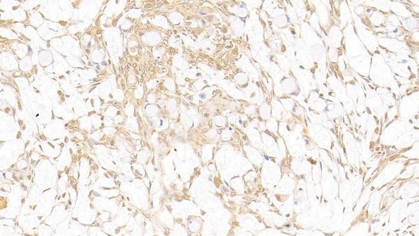
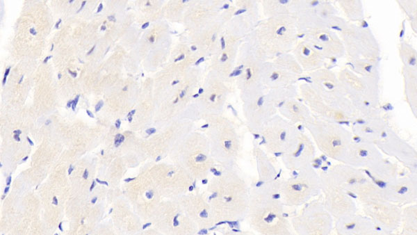
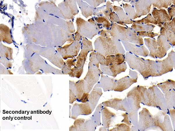
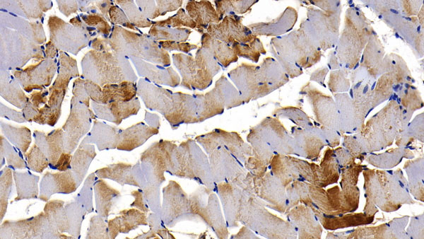
 IHC (Immunohiostchemistry)
(DAB staining on IHCP;Sample: Human Cardiac Muscle Tissue;Primary Ab: 30ug/ml Mouse AntiHuman BAI3 AntibodySecond Ab: 2ug/mL HRPLinked Caprine AntiMouse IgG Polyclonal Antibody(Catalog: SAA544Mu19))
IHC (Immunohiostchemistry)
(DAB staining on IHCP;Sample: Human Cardiac Muscle Tissue;Primary Ab: 30ug/ml Mouse AntiHuman BAI3 AntibodySecond Ab: 2ug/mL HRPLinked Caprine AntiMouse IgG Polyclonal Antibody(Catalog: SAA544Mu19))
Brain Specific Angiogenesis Inhibitor 3 (BAI3), Monoclonal Antibody (Cat# AAA151865)
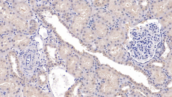
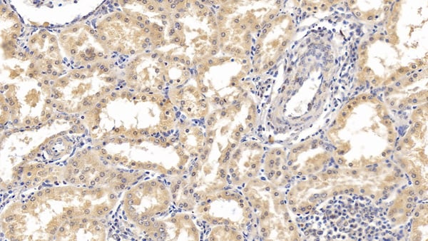
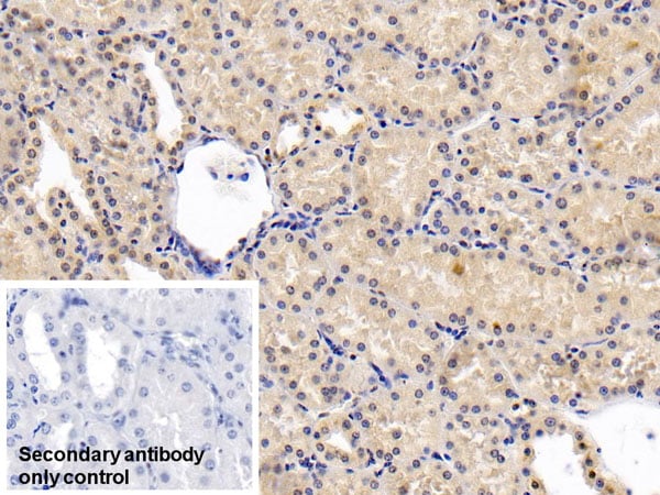
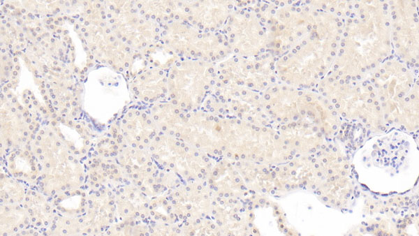
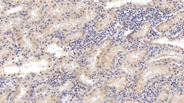
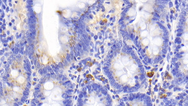
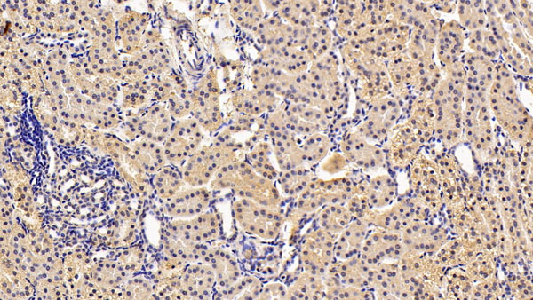
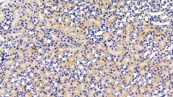
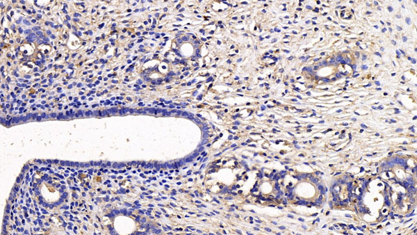
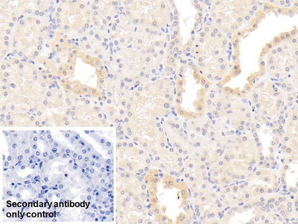
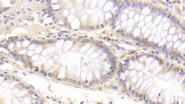
 IHC (Immunohiostchemistry)
(DAB staining on IHCP;Sample: Human Kidney Tissue; Primary Ab: 20ug/ml Mouse AntiHuman SFRP1 AntibodySecond Ab: 2ug/mL HRPLinked Caprine AntiMouse IgG Polyclonal Antibody(Catalog: SAA544Mu19))
IHC (Immunohiostchemistry)
(DAB staining on IHCP;Sample: Human Kidney Tissue; Primary Ab: 20ug/ml Mouse AntiHuman SFRP1 AntibodySecond Ab: 2ug/mL HRPLinked Caprine AntiMouse IgG Polyclonal Antibody(Catalog: SAA544Mu19))
Secreted Frizzled Related Protein 1 (SFRP1), Monoclonal Antibody (Cat# AAA151872)
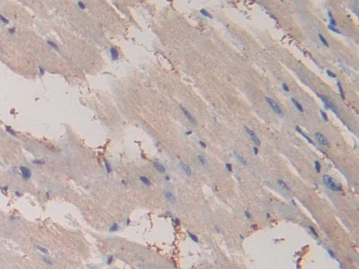
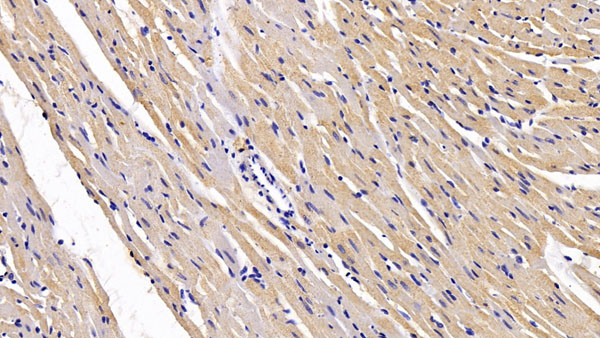
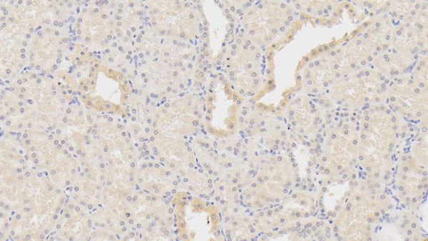
 IHC (Immunohiostchemistry)
(DAB staining on IHCP;Sample: Human Cardiac Muscle Tissue; Primary Ab: 20ug/ml Mouse AntiHuman SFRP1 AntibodySecond Ab: 2ug/mL HRPLinked Caprine AntiMouse IgG Polyclonal Antibody(Catalog: SAA544Mu19))
IHC (Immunohiostchemistry)
(DAB staining on IHCP;Sample: Human Cardiac Muscle Tissue; Primary Ab: 20ug/ml Mouse AntiHuman SFRP1 AntibodySecond Ab: 2ug/mL HRPLinked Caprine AntiMouse IgG Polyclonal Antibody(Catalog: SAA544Mu19))
Secreted Frizzled Related Protein 1 (SFRP1), Monoclonal Antibody (Cat# AAA151873)
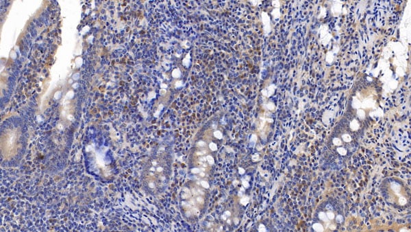
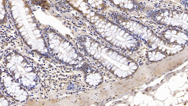
 IHC (Immunohiostchemistry)
(DAB staining on IHCP;Samples: Human Kidney Tissue; Primary Ab: 10ug/ml Mouse AntiHuman IFNa21 AntibodySecond Ab: 2ug/mL HRPLinked Caprine AntiMouse IgG Polyclonal Antibody(Catalog: SAA544Mu19))
IHC (Immunohiostchemistry)
(DAB staining on IHCP;Samples: Human Kidney Tissue; Primary Ab: 10ug/ml Mouse AntiHuman IFNa21 AntibodySecond Ab: 2ug/mL HRPLinked Caprine AntiMouse IgG Polyclonal Antibody(Catalog: SAA544Mu19))
Interferon Alpha 21 (IFNa21), Monoclonal Antibody (Cat# AAA151879)
 IHC (Immunohistochemisry)
(DAB staining on IHCP;Samples: Rat Stomach Tissue; Primary Ab: 30ug/ml Mouse AntiRat PPARg AntibodySecond Ab: 2ug/mL HRPLinked Caprine AntiMouse IgG Polyclonal Antibody(Catalog: SAA544Mu19))
IHC (Immunohistochemisry)
(DAB staining on IHCP;Samples: Rat Stomach Tissue; Primary Ab: 30ug/ml Mouse AntiRat PPARg AntibodySecond Ab: 2ug/mL HRPLinked Caprine AntiMouse IgG Polyclonal Antibody(Catalog: SAA544Mu19))
Peroxisome Proliferator Activated Receptor Gamma (PPARg), Monoclonal Antibody (Cat# AAA151648)
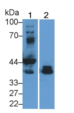
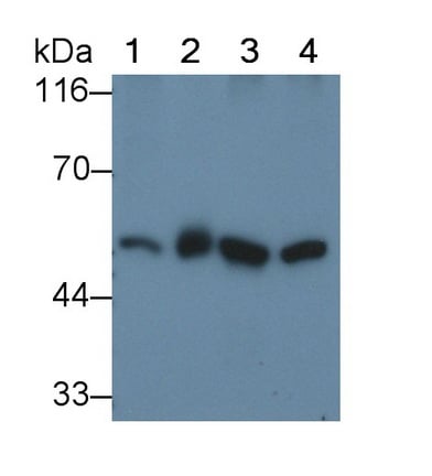
 WB (Western Blot)
(Western Blot; Sample: Lane1: Porcine Kidney lysate; Lane2: Porcine Lung lysate; Lane3: HepG2 cell lysate Primary Ab: 2ug/ml Mouse Anti-human PAI1 Antibody Second Ab: 0.2ug/mL HRP-Linked Caprine Anti-Mouse IgG Polyclonal Antibody)
WB (Western Blot)
(Western Blot; Sample: Lane1: Porcine Kidney lysate; Lane2: Porcine Lung lysate; Lane3: HepG2 cell lysate Primary Ab: 2ug/ml Mouse Anti-human PAI1 Antibody Second Ab: 0.2ug/mL HRP-Linked Caprine Anti-Mouse IgG Polyclonal Antibody)
Plasminogen Activator Inhibitor 1 (PAI1), Monoclonal Antibody (Cat# AAA152625)
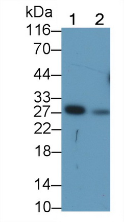
 WB (Western Blot)
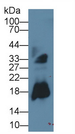
(Western Blot; Sample: Rat Colon lysate; Primary Ab: 2ug/ml Mouse Anti-human GAL3 Antibody Second Ab: 0.2ug/ml HRP-Linked Caprine Anti-Mouse IgG Polyclonal Antibody)
WB (Western Blot)
(Western Blot; Sample: Rat Colon lysate; Primary Ab: 2ug/ml Mouse Anti-human GAL3 Antibody Second Ab: 0.2ug/ml HRP-Linked Caprine Anti-Mouse IgG Polyclonal Antibody)
Galectin 3 (GAL3), Monoclonal Antibody (Cat# AAA152626)
 WB (Western Blot)
(Western Blot; Sample: Lane1: human SeuM; Lane2: human Plasma; Lane3: human Placenta lysate; Lane4: Rat Plasma Primary Ab: 0.5ug/ml Mouse Anti-human AT Antibody Second Ab: 0.2ug/mL HRP-Linked Caprine Anti-Mouse IgG Polyclonal Antibody)
WB (Western Blot)
(Western Blot; Sample: Lane1: human SeuM; Lane2: human Plasma; Lane3: human Placenta lysate; Lane4: Rat Plasma Primary Ab: 0.5ug/ml Mouse Anti-human AT Antibody Second Ab: 0.2ug/mL HRP-Linked Caprine Anti-Mouse IgG Polyclonal Antibody)
Antithrombin (AT), Monoclonal Antibody (Cat# AAA152630)
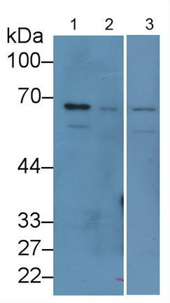
 WB (Western Blot)
(Western Blot;Sample:Lane 1: Rat CerebuM lysate;Lane 2: Mouse CerebuM lysate;Lane 3: Bovine CerebuM lysate;Lane 4: Canine CerebuM lysatePrimary Ab: 0.2ug/ml Mouse Anti-ulti-species H3 AntibodySecond Ab: 0.2ug/mL HRP-Linked Caprine Anti-Mouse IgG Polyclonal Antibody)
WB (Western Blot)
(Western Blot;Sample:Lane 1: Rat CerebuM lysate;Lane 2: Mouse CerebuM lysate;Lane 3: Bovine CerebuM lysate;Lane 4: Canine CerebuM lysatePrimary Ab: 0.2ug/ml Mouse Anti-ulti-species H3 AntibodySecond Ab: 0.2ug/mL HRP-Linked Caprine Anti-Mouse IgG Polyclonal Antibody)
Histone H3 (H3), Monoclonal Antibody (Cat# AAA152635)
 WB (Western Blot)
(Western Blot; Sample: Lane1: human SeuM; Lane2: Porcine Skeletal muscle lysate; Lane3: Hela cell lysate Primary Ab: 2ug/ml Mouse Anti-human ADP Antibody Second Ab: 0.2ug/mL HRP-Linked Caprine Anti-Mouse IgG Polyclonal Antibody)
WB (Western Blot)
(Western Blot; Sample: Lane1: human SeuM; Lane2: Porcine Skeletal muscle lysate; Lane3: Hela cell lysate Primary Ab: 2ug/ml Mouse Anti-human ADP Antibody Second Ab: 0.2ug/mL HRP-Linked Caprine Anti-Mouse IgG Polyclonal Antibody)
Adiponectin (ADPN), Monoclonal Antibody (Cat# AAA152636)
 WB (Western Blot)
(Western Blot; Sample: Jurkat cell lysate Primary Ab: 2ug/ml Mouse Anti-human CD3d Antibody Second Ab: 0.2ug/mL HRP-Linked Caprine Anti-Mouse IgG Polyclonal Antibody)
WB (Western Blot)
(Western Blot; Sample: Jurkat cell lysate Primary Ab: 2ug/ml Mouse Anti-human CD3d Antibody Second Ab: 0.2ug/mL HRP-Linked Caprine Anti-Mouse IgG Polyclonal Antibody)
Cluster Of Differentiation 3d (CD3d), Monoclonal Antibody (Cat# AAA152638)
 WB (Western Blot)
(Western Blot; Sample: Lane1: HepG2 cell lysate; Lane2: U2OS cell lysate; Lane3: Hela cell lysate Primary Ab: 1ug/ml Mouse Anti-human LCAT Antibody Second Ab: 0.2ug/mL HRP-Linked Caprine Anti-Mouse IgG Polyclonal Antibody)
WB (Western Blot)
(Western Blot; Sample: Lane1: HepG2 cell lysate; Lane2: U2OS cell lysate; Lane3: Hela cell lysate Primary Ab: 1ug/ml Mouse Anti-human LCAT Antibody Second Ab: 0.2ug/mL HRP-Linked Caprine Anti-Mouse IgG Polyclonal Antibody)
Lecithin Cholesterol Acyltransferase (LCAT), Monoclonal Antibody (Cat# AAA152648)
 WB (Western Blot)
(Western Blot; Sample: Lane1: Rat Liver lysate; Lane2: Rat Testis lysate; Lane3: A549 cell lysate Primary Ab: 2 ug/ml Mouse Anti-Mouse HSPA5 Antibody Second Ab: 0.2ug/mL HRP-Linked Caprine Anti-Mouse IgG Polyclonal Antibody)
WB (Western Blot)
(Western Blot; Sample: Lane1: Rat Liver lysate; Lane2: Rat Testis lysate; Lane3: A549 cell lysate Primary Ab: 2 ug/ml Mouse Anti-Mouse HSPA5 Antibody Second Ab: 0.2ug/mL HRP-Linked Caprine Anti-Mouse IgG Polyclonal Antibody)
Heat Shock 70kDa Protein 5 (HSPA5), Monoclonal Antibody (Cat# AAA152650)
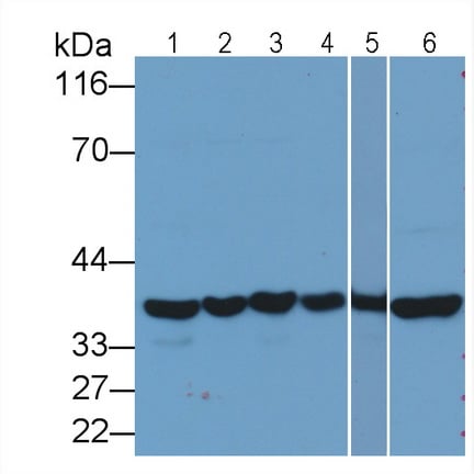

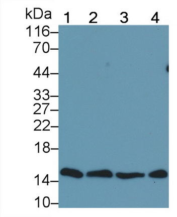
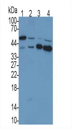
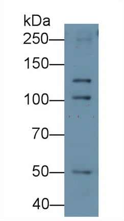
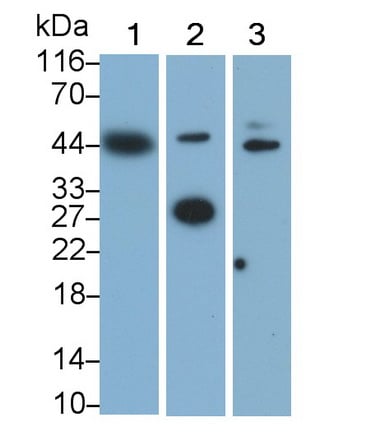
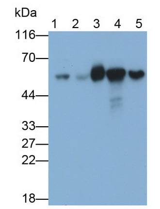
 WB (Western Blot)
(Western Blot; Sample: Lane1: Gallus Kidney lysate; Lane2: Rabbit Liver lysate; Lane3: Rabbit Kidney lysate; Lane4: Caprine Liver lysate; Lane5: Mouse Liver lysate; Lane6: Rat Liver lysate; Lane7: Rat Kidney lysatePrimary Ab: 0.5ug/ml Mouse Anti-human GAPDH AntibodySecond Ab: 0.2ug/mL HRP-Linked Caprine Anti-Mouse IgG Polyclonal Antibody)
WB (Western Blot)
(Western Blot; Sample: Lane1: Gallus Kidney lysate; Lane2: Rabbit Liver lysate; Lane3: Rabbit Kidney lysate; Lane4: Caprine Liver lysate; Lane5: Mouse Liver lysate; Lane6: Rat Liver lysate; Lane7: Rat Kidney lysatePrimary Ab: 0.5ug/ml Mouse Anti-human GAPDH AntibodySecond Ab: 0.2ug/mL HRP-Linked Caprine Anti-Mouse IgG Polyclonal Antibody)
Glyceraldehyde-3-Phosphate Dehydrogenase (GAPDH), Monoclonal Antibody (Cat# AAA152651)
 WB (Western Blot)
(Western Blot; Sample: Lane1: HT1080 cell lysate; Lane2: HepG2 cell lysate Primary Ab: 0.2 ug/ml Mouse Anti-human S100 Antibody Second Ab: 0.2ug/mL HRP-Linked Caprine Anti-Mouse IgG Polyclonal Antibody)
WB (Western Blot)
(Western Blot; Sample: Lane1: HT1080 cell lysate; Lane2: HepG2 cell lysate Primary Ab: 0.2 ug/ml Mouse Anti-human S100 Antibody Second Ab: 0.2ug/mL HRP-Linked Caprine Anti-Mouse IgG Polyclonal Antibody)
S100 CalcuM Binding Protein (S100), Monoclonal Antibody (Cat# AAA152658)
 WB (Western Blot)
(Western Blot; Sample: Rat Spleen lysate Primary Ab: 3ug/ml Mouse Anti-Rat CD163 Antibody Second Ab: 0.2ug/mL HRP-Linked Caprine Anti-Mouse IgG Polyclonal Antibody)
WB (Western Blot)
(Western Blot; Sample: Rat Spleen lysate Primary Ab: 3ug/ml Mouse Anti-Rat CD163 Antibody Second Ab: 0.2ug/mL HRP-Linked Caprine Anti-Mouse IgG Polyclonal Antibody)
Cluster Of Differentiation (CD163), Monoclonal Antibody (Cat# AAA152670)
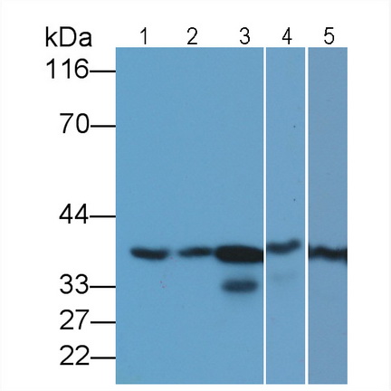
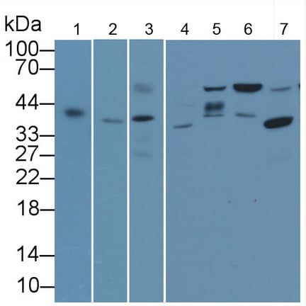 WB (Western Blot)
(Western Blot; Sample: Lane1: 293T cell lysate; Lane2: Cavia Testis lysate; Lane3: Mouse Testis lysate; Lane4: Rabbit Kidney lysate; Lane5: Caprine Testis lysate; Lane6: Porcine Kidney lysate; Lane7: Rat Testis lysatePrimary Ab: 1ug/ml Mouse Anti-human TBP AntibodySecond Ab: 0.2ug/mL HRP-Linked Caprine Anti-Mouse IgG Polyclonal Antibody)
WB (Western Blot)
(Western Blot; Sample: Lane1: 293T cell lysate; Lane2: Cavia Testis lysate; Lane3: Mouse Testis lysate; Lane4: Rabbit Kidney lysate; Lane5: Caprine Testis lysate; Lane6: Porcine Kidney lysate; Lane7: Rat Testis lysatePrimary Ab: 1ug/ml Mouse Anti-human TBP AntibodySecond Ab: 0.2ug/mL HRP-Linked Caprine Anti-Mouse IgG Polyclonal Antibody)
TATA Binding Protein (TBP), Monoclonal Antibody (Cat# AAA152681)
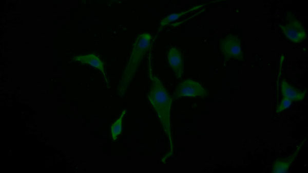
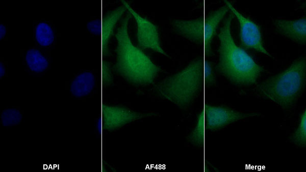
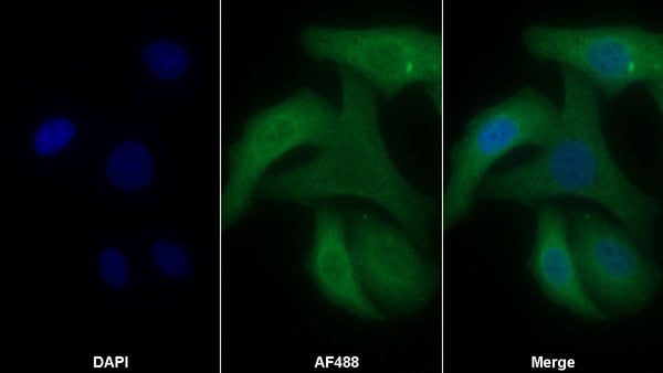
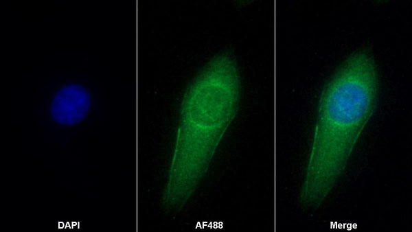
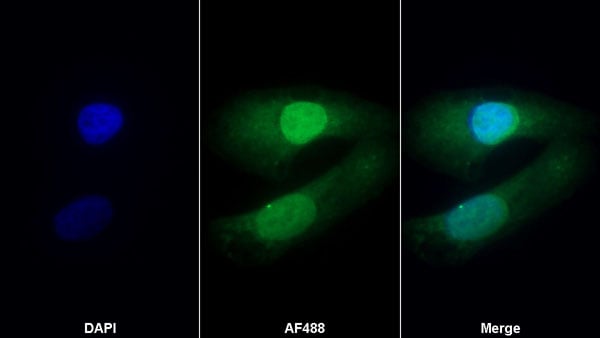
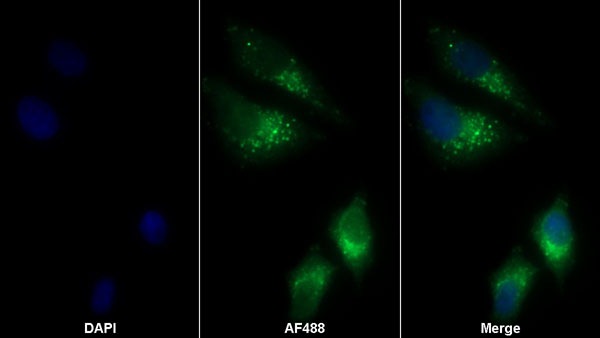
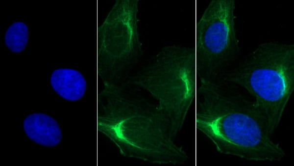
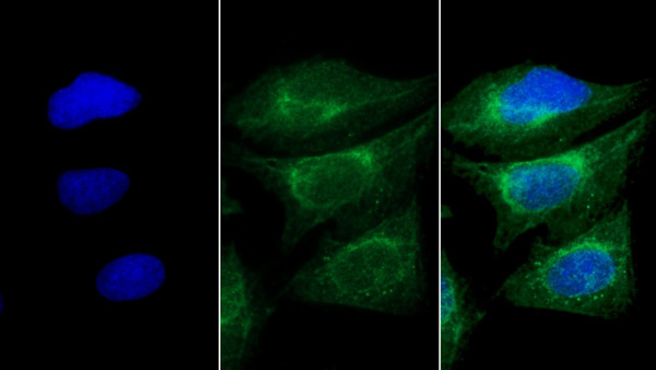
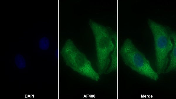
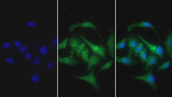
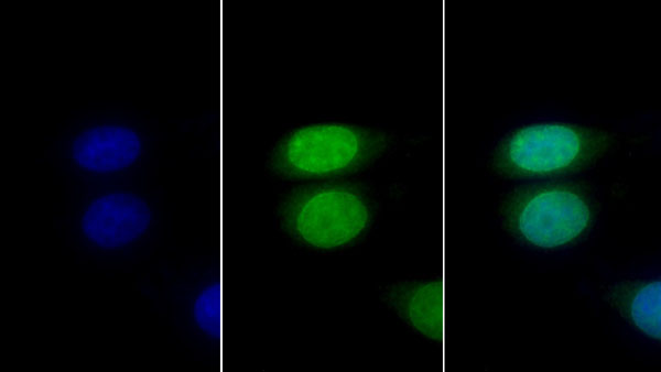
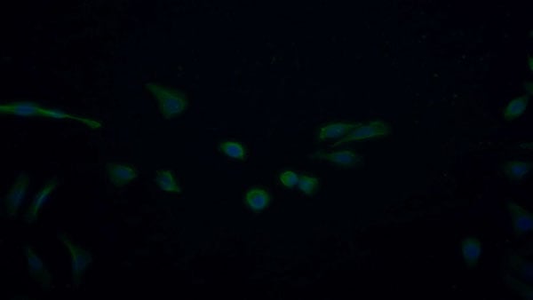 IF (Immunofluorescence)
(FITC staining on IF;Sample: human HepG2 cell; Primary Ab: 40ug/ml Mouse Anti-human NGF AntibodySecond Ab: 5ug/ml FITC-Linked Caprine Anti-Mouse IgG Polyclonal Antibody)
IF (Immunofluorescence)
(FITC staining on IF;Sample: human HepG2 cell; Primary Ab: 40ug/ml Mouse Anti-human NGF AntibodySecond Ab: 5ug/ml FITC-Linked Caprine Anti-Mouse IgG Polyclonal Antibody)
Nerve Growth Factor (NGF), Monoclonal Antibody (Cat# AAA152762)
 WB (Western Blot)
(Western Blot; Sample: Hela cell lysate Primary Ab: 1.5 ug/ml Mouse Anti-human DSG3 Antibody Second Ab: 0.2ug/mL HRP-Linked Caprine Anti-Mouse IgG Polyclonal Antibody)
WB (Western Blot)
(Western Blot; Sample: Hela cell lysate Primary Ab: 1.5 ug/ml Mouse Anti-human DSG3 Antibody Second Ab: 0.2ug/mL HRP-Linked Caprine Anti-Mouse IgG Polyclonal Antibody)
Desmoglein 3 (DSG3), Monoclonal Antibody (Cat# AAA152769)
 WB (Western Blot)
(Western Blot; Sample: Lane1: human Urine; Lane2: human Leukocyte lysatePrimary Ab: 0.3ug/ml Mouse Anti-human RNASE2 AntibodySecond Ab: 0.2ug/mL HRP-Linked Caprine Anti-Mouse IgG Polyclonal Antibody)
WB (Western Blot)
(Western Blot; Sample: Lane1: human Urine; Lane2: human Leukocyte lysatePrimary Ab: 0.3ug/ml Mouse Anti-human RNASE2 AntibodySecond Ab: 0.2ug/mL HRP-Linked Caprine Anti-Mouse IgG Polyclonal Antibody)
Ribonuclease A2 (RNASE2), Monoclonal Antibody (Cat# AAA152777)
 WB (Western Blot)
(Western Blot; Sample: Lane1: Rat Liver lysate; Lane2: Rat Kidney lysate; Lane3: Porcine Liver lysate; Lane4: HepG2 cell lysate Primary Ab: 0.2ug/ml Mouse Anti-human HPD Antibody Second Ab: 0.2ug/mL HRP-Linked Caprine Anti-Mouse IgG Polyclonal Antibody)
WB (Western Blot)
(Western Blot; Sample: Lane1: Rat Liver lysate; Lane2: Rat Kidney lysate; Lane3: Porcine Liver lysate; Lane4: HepG2 cell lysate Primary Ab: 0.2ug/ml Mouse Anti-human HPD Antibody Second Ab: 0.2ug/mL HRP-Linked Caprine Anti-Mouse IgG Polyclonal Antibody)
4-Hydroxyphenylpyruvate Dioxygenase (HPD), Monoclonal Antibody (Cat# AAA152790)
 WB (Western Blot)
(Western Blot; Sample: Lane1: Porcine Testis lysate; Lane2: Hela cell lysate; Lane3: U2OS cell lysate Primary Ab: 0.3ug/ml Mouse Anti-human CCT2 Antibody Second Ab: 0.2ug/mL HRP-Linked Caprine Anti-Mouse IgG Polyclonal Antibody)
WB (Western Blot)
(Western Blot; Sample: Lane1: Porcine Testis lysate; Lane2: Hela cell lysate; Lane3: U2OS cell lysate Primary Ab: 0.3ug/ml Mouse Anti-human CCT2 Antibody Second Ab: 0.2ug/mL HRP-Linked Caprine Anti-Mouse IgG Polyclonal Antibody)
Chaperonin Containing TCP1, Subunit 2 (CCT2), Monoclonal Antibody (Cat# AAA152794)
 WB (Western Blot)
(Western Blot; Sample: Lane1: human Lung lysate; Lane2: Rat Liver lysate Primary Ab: 0.2ug/ml Mouse Anti-human ANXA4 Antibody Second Ab: 0.2ug/mL HRP-Linked Caprine Anti-Mouse IgG Polyclonal Antibody)
WB (Western Blot)
(Western Blot; Sample: Lane1: human Lung lysate; Lane2: Rat Liver lysate Primary Ab: 0.2ug/ml Mouse Anti-human ANXA4 Antibody Second Ab: 0.2ug/mL HRP-Linked Caprine Anti-Mouse IgG Polyclonal Antibody)
Annexin A4 (ANXA4), Monoclonal Antibody (Cat# AAA152797)
 WB (Western Blot)
(Western Blot; Sample: Lane1: human SeuM; Lane2: human Plasma Primary Ab: 0.2ug/ml Mouse Anti-human aZGP1 Antibody Second Ab: 0.2ug/mL HRP-Linked Caprine Anti-Mouse IgG Polyclonal Antibody)
WB (Western Blot)
(Western Blot; Sample: Lane1: human SeuM; Lane2: human Plasma Primary Ab: 0.2ug/ml Mouse Anti-human aZGP1 Antibody Second Ab: 0.2ug/mL HRP-Linked Caprine Anti-Mouse IgG Polyclonal Antibody)
Alpha-2-Glycoprotein 1, Zinc Binding (aZGP1), Monoclonal Antibody (Cat# AAA152798)
 WB (Western Blot)
(Western Blot; Sample: Lane1: human Placenta lysate; Lane2: Rat Liver lysate; Lane3: Rat Thymus lysate Primary Ab: 0.2ug/ml Mouse Anti-human LOX1 Antibody Second Ab: 0.2ug/mL HRP-Linked Caprine Anti-Mouse IgG Polyclonal Antibody)
WB (Western Blot)
(Western Blot; Sample: Lane1: human Placenta lysate; Lane2: Rat Liver lysate; Lane3: Rat Thymus lysate Primary Ab: 0.2ug/ml Mouse Anti-human LOX1 Antibody Second Ab: 0.2ug/mL HRP-Linked Caprine Anti-Mouse IgG Polyclonal Antibody)
Lectin Like Oxidized Low Density Lipoprotein Receptor 1 (LOX1), Monoclonal Antibody (Cat# AAA152799)
 WB (Western Blot)
(Western Blot; Sample: Lane1: 293T cell lysate; Lane2: A431 cell lysate Primary Ab: 0.8ug/ml Mouse Anti-human PSAP Antibody Second Ab: 0.2ug/mL HRP-Linked Caprine Anti-Mouse IgG Polyclonal Antibody)
WB (Western Blot)
(Western Blot; Sample: Lane1: 293T cell lysate; Lane2: A431 cell lysate Primary Ab: 0.8ug/ml Mouse Anti-human PSAP Antibody Second Ab: 0.2ug/mL HRP-Linked Caprine Anti-Mouse IgG Polyclonal Antibody)
Prosaposin (PSAP), Monoclonal Antibody (Cat# AAA152800)
 WB (Western Blot)
(Western Blot; Sample: Porcine Skeletal muscle lysate Primary Ab: 0.01ug/ml Mouse Anti-human MYH8 Antibody Second Ab: 0.2ug/mL HRP-Linked Caprine Anti-Mouse IgG Polyclonal Antibody)
WB (Western Blot)
(Western Blot; Sample: Porcine Skeletal muscle lysate Primary Ab: 0.01ug/ml Mouse Anti-human MYH8 Antibody Second Ab: 0.2ug/mL HRP-Linked Caprine Anti-Mouse IgG Polyclonal Antibody)
Myosin Heavy Chain 8, Skeletal Muscle, Perinatal (MYH8), Monoclonal Antibody (Cat# AAA152807)
 WB (Western Blot)
(Western Blot; Sample: Lane1: Porcine Skeletal muscle lysate; Lane2: Porcine Esophagus lysate; Lane3: Rat Skeletal muscle lysate; Lane4: Mouse Skeletal muscle lysate Primary Ab: 0.01ug/ml Mouse Anti-human MYH8 Antibody Second Ab: 0.2ug/mL HRP-Linked Caprine Anti-Mouse IgG Polyclonal Antibody)
WB (Western Blot)
(Western Blot; Sample: Lane1: Porcine Skeletal muscle lysate; Lane2: Porcine Esophagus lysate; Lane3: Rat Skeletal muscle lysate; Lane4: Mouse Skeletal muscle lysate Primary Ab: 0.01ug/ml Mouse Anti-human MYH8 Antibody Second Ab: 0.2ug/mL HRP-Linked Caprine Anti-Mouse IgG Polyclonal Antibody)
Myosin Heavy Chain 8, Skeletal Muscle, Perinatal (MYH8), Monoclonal Antibody (Cat# AAA152809)
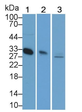
 WB (Western Blot)
(Western Blot; Sample: Lane1: Porcine Heart lysate; Lane2: MCF7 cell lysate; Lane3: Hela cell lysate Primary Ab: 0.2ug/ml Mouse Anti-human PLS3 Antibody Second Ab: 0.2ug/mL HRP-Linked Caprine Anti-Mouse IgG Polyclonal Antibody)
WB (Western Blot)
(Western Blot; Sample: Lane1: Porcine Heart lysate; Lane2: MCF7 cell lysate; Lane3: Hela cell lysate Primary Ab: 0.2ug/ml Mouse Anti-human PLS3 Antibody Second Ab: 0.2ug/mL HRP-Linked Caprine Anti-Mouse IgG Polyclonal Antibody)
Plastin 3 (PLS3), Monoclonal Antibody (Cat# AAA152817)
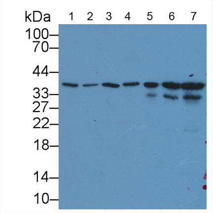
 WB (Western Blot)
(Western Blot; Sample: Lane1: human Lung lysate; Lane2: Rat Testis lysate; Lane3: Porcine Testis lysate; Lane4: Hela cell lysate; Lane5: U2OS cell lysatePrimary Ab: 0.6ug/ml Mouse Anti-human CCT2 AntibodySecond Ab: 0.2ug/mL HRP-Linked Caprine Anti-Mouse IgG Polyclonal Antibody)
WB (Western Blot)
(Western Blot; Sample: Lane1: human Lung lysate; Lane2: Rat Testis lysate; Lane3: Porcine Testis lysate; Lane4: Hela cell lysate; Lane5: U2OS cell lysatePrimary Ab: 0.6ug/ml Mouse Anti-human CCT2 AntibodySecond Ab: 0.2ug/mL HRP-Linked Caprine Anti-Mouse IgG Polyclonal Antibody)
Chaperonin Containing TCP1, Subunit 2 (CCT2), Monoclonal Antibody (Cat# AAA152828)
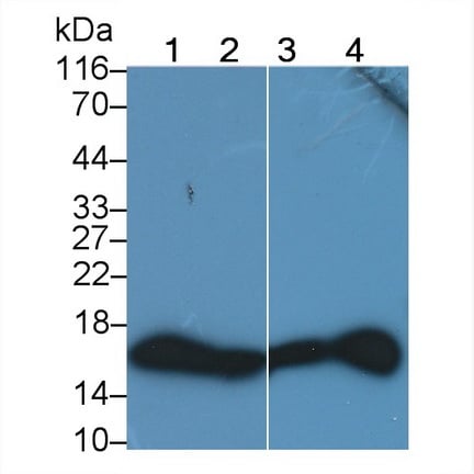
 WB (Western Blot)
(Western Blot; Sample: human Leukocyte lysate Primary Ab: 2ug/ml Mouse Anti-human S100A12 Antibody Second Ab: 0.2ug/mL HRP-Linked Caprine Anti-Mouse IgG Polyclonal Antibody)
WB (Western Blot)
(Western Blot; Sample: human Leukocyte lysate Primary Ab: 2ug/ml Mouse Anti-human S100A12 Antibody Second Ab: 0.2ug/mL HRP-Linked Caprine Anti-Mouse IgG Polyclonal Antibody)
S100 CalcuM Binding Protein A12 (S100A12), Monoclonal Antibody (Cat# AAA152829)
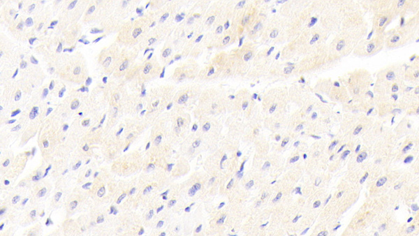
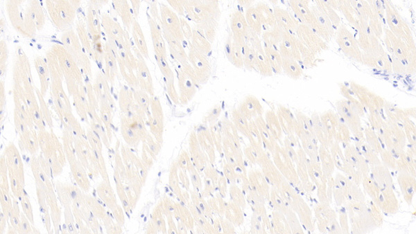
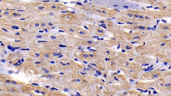
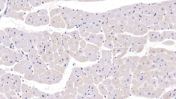
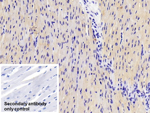
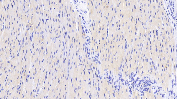
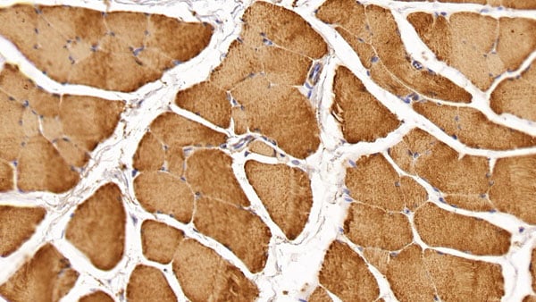
 IHC (Immunohiostchemistry)
(DAB staining on IHC-P;Sample: Rat Cardiac Muscle Tissue; Primary Ab: 30ug/ml Mouse Anti-Rat SFRP1 AntibodySecond Ab: 2ug/mL HRP-Linked Caprine Anti-Mouse IgG Polyclonal Antibody)
IHC (Immunohiostchemistry)
(DAB staining on IHC-P;Sample: Rat Cardiac Muscle Tissue; Primary Ab: 30ug/ml Mouse Anti-Rat SFRP1 AntibodySecond Ab: 2ug/mL HRP-Linked Caprine Anti-Mouse IgG Polyclonal Antibody)
Secreted Frizzled Related Protein 1 (SFRP1), Monoclonal Antibody (Cat# AAA152844)
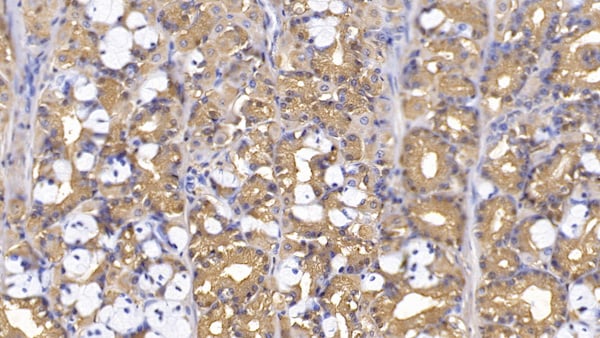
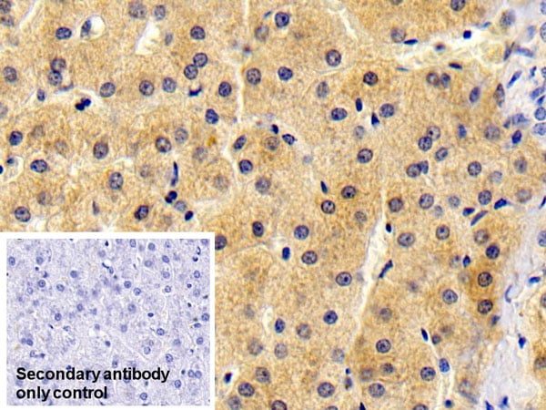
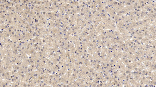
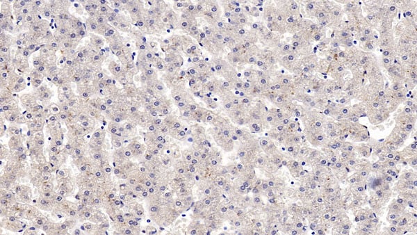
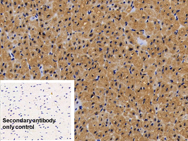
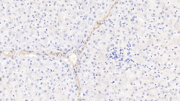 IHC (Immunohiostchemistry)
(DAB staining on IHC-P;Sample: human Liver Tissue; Primary Ab: 30ug/ml Mouse Anti-human PINP AntibodySecond Ab: 2ug/mL HRP-Linked Caprine Anti-Mouse IgG Polyclonal Antibody)
IHC (Immunohiostchemistry)
(DAB staining on IHC-P;Sample: human Liver Tissue; Primary Ab: 30ug/ml Mouse Anti-human PINP AntibodySecond Ab: 2ug/mL HRP-Linked Caprine Anti-Mouse IgG Polyclonal Antibody)
Procollagen I N-Terminal Propeptide (PINP), Monoclonal Antibody (Cat# AAA152696)
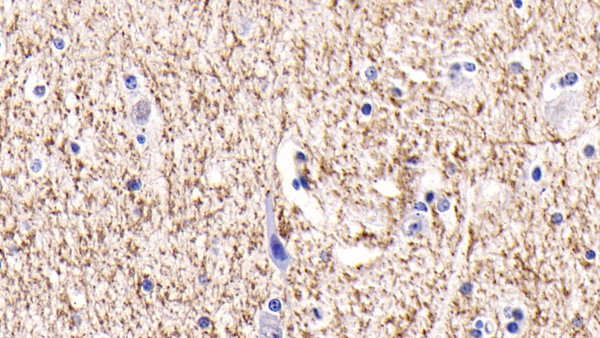
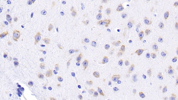
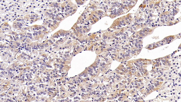
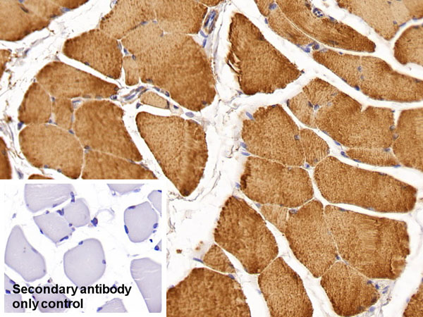
 IHC (Immunohistochemisry)
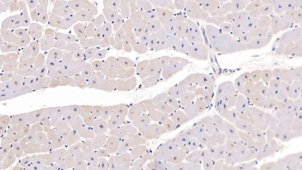
(DAB staining on IHC-P;Sample: human Cardiac Muscle Tissue; Primary Ab: 20ug/ml Mouse Anti-human OPG AntibodySecond Ab: 2ug/mL HRP-Linked Caprine Anti-Mouse IgG Polyclonal Antibody)
IHC (Immunohistochemisry)
(DAB staining on IHC-P;Sample: human Cardiac Muscle Tissue; Primary Ab: 20ug/ml Mouse Anti-human OPG AntibodySecond Ab: 2ug/mL HRP-Linked Caprine Anti-Mouse IgG Polyclonal Antibody)
Osteoprotegerin (OPG), Monoclonal Antibody (Cat# AAA152704)

 WB (Western Blot)
(Western Blot; Sample: Lane1: human SeuM; Lane2: human Plasma; Lane3: human Placenta lysate Primary Ab: 2 ug/ml Mouse Anti-human Plg Antibody Second Ab: 0.2ug/mL HRP-Linked Caprine Anti-Mouse IgG Polyclonal Antibody)
WB (Western Blot)
(Western Blot; Sample: Lane1: human SeuM; Lane2: human Plasma; Lane3: human Placenta lysate Primary Ab: 2 ug/ml Mouse Anti-human Plg Antibody Second Ab: 0.2ug/mL HRP-Linked Caprine Anti-Mouse IgG Polyclonal Antibody)
Plasminogen (Plg), Monoclonal Antibody (Cat# AAA152721)
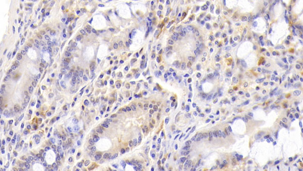
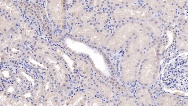
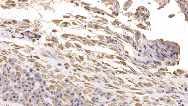
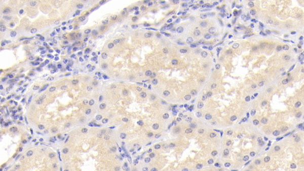
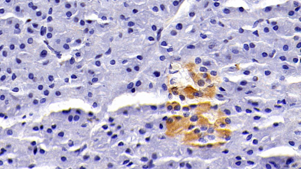
 IHC (Immunohistochemisry)
(DAB staining on IHC-P;Sample: human Kidney Tissue; Primary Ab: 20ug/ml Mouse Anti-human MMP7 AntibodySecond Ab: 2ug/mL HRP-Linked Caprine Anti-Mouse IgG Polyclonal Antibody)
IHC (Immunohistochemisry)
(DAB staining on IHC-P;Sample: human Kidney Tissue; Primary Ab: 20ug/ml Mouse Anti-human MMP7 AntibodySecond Ab: 2ug/mL HRP-Linked Caprine Anti-Mouse IgG Polyclonal Antibody)
Matrix Metalloproteinase 7 (MMP7), Monoclonal Antibody (Cat# AAA152725)
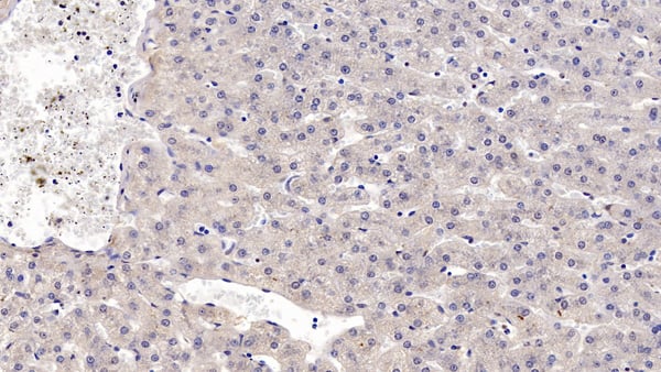
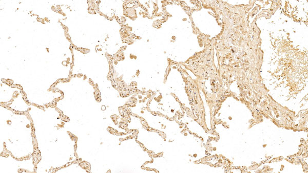
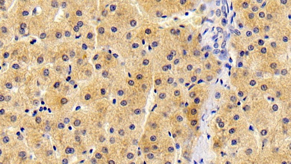
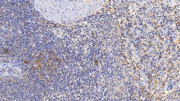
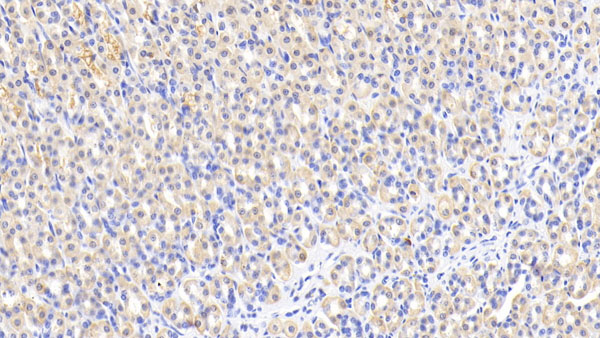
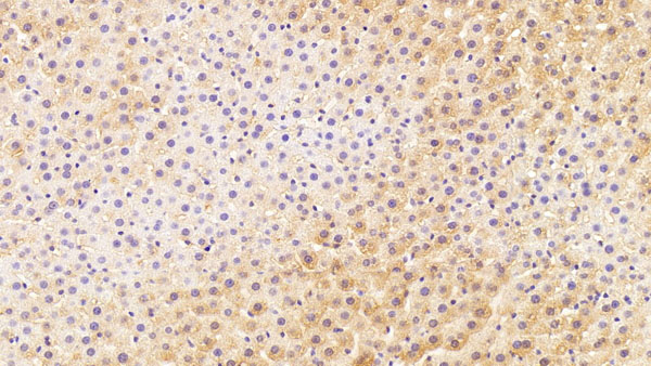
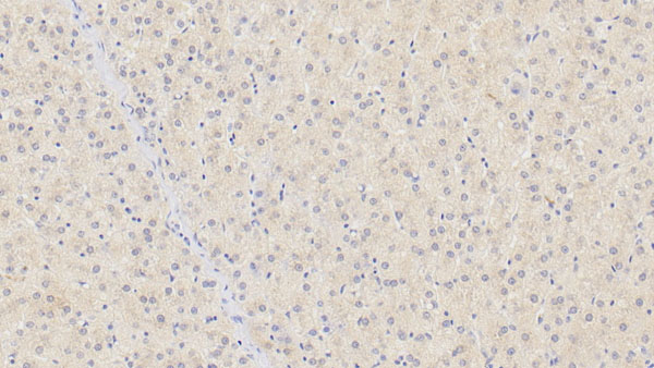
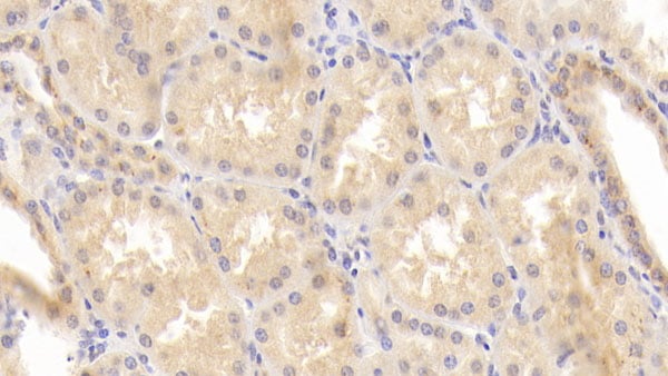
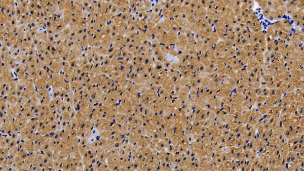
 IHC (Immunohiostchemistry)
(DAB staining on IHC-P;Samples: human Liver Tissue; Primary Ab: 10ug/ml Mouse Anti-human IGFBP4 AntibodySecond Ab: 2ug/mL HRP-Linked Caprine Anti-Mouse IgG Polyclonal Antibody)
IHC (Immunohiostchemistry)
(DAB staining on IHC-P;Samples: human Liver Tissue; Primary Ab: 10ug/ml Mouse Anti-human IGFBP4 AntibodySecond Ab: 2ug/mL HRP-Linked Caprine Anti-Mouse IgG Polyclonal Antibody)
Inulin Like Growth Factor Binding Protein 4 (IGFBP4), Monoclonal Antibody (Cat# AAA152726)
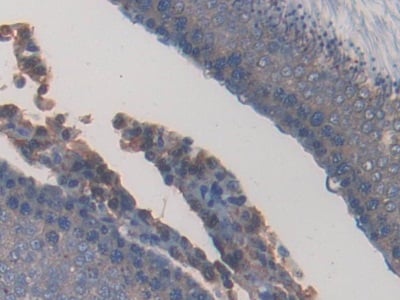
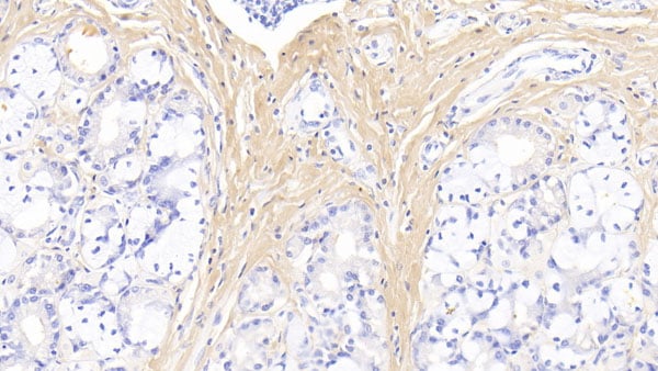
 IHC (Immunohiostchemistry)
(DAB staining on IHC-P;Sample: Rabbit Pancreas Tissue; Primary Ab: 10ug/ml Mouse Anti-Rabbit CLU AntibodySecond Ab: 2ug/mL HRP-Linked Caprine Anti-Mouse IgG Polyclonal Antibody)
IHC (Immunohiostchemistry)
(DAB staining on IHC-P;Sample: Rabbit Pancreas Tissue; Primary Ab: 10ug/ml Mouse Anti-Rabbit CLU AntibodySecond Ab: 2ug/mL HRP-Linked Caprine Anti-Mouse IgG Polyclonal Antibody)
Clusterin (CLU), Monoclonal Antibody (Cat# AAA152729)
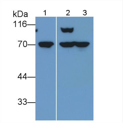
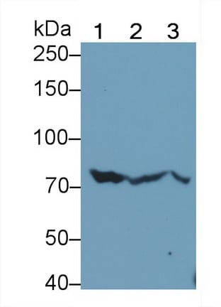
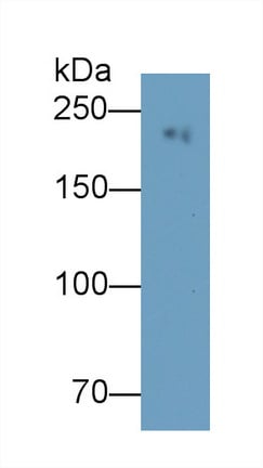
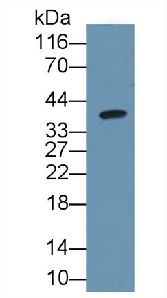
 WB (Western Blot)
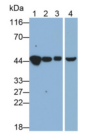
(Western Blot; Sample: Lane1: human Lung lysate; Lane2: 293T cell lysate; Lane3: Rabbit Lung lysate Primary Ab: 3 ug/ml Mouse Anti-human LMNB1 Antibody Second Ab: 0.2ug/mL HRP-Linked Caprine Anti-Mouse IgG Polyclonal Antibody)
WB (Western Blot)
(Western Blot; Sample: Lane1: human Lung lysate; Lane2: 293T cell lysate; Lane3: Rabbit Lung lysate Primary Ab: 3 ug/ml Mouse Anti-human LMNB1 Antibody Second Ab: 0.2ug/mL HRP-Linked Caprine Anti-Mouse IgG Polyclonal Antibody)
Lamin B1 (LMNB1), Monoclonal Antibody (Cat# AAA152732)

 WB (Western Blot)
(Western Blot; Sample: Lane1: human SeuM; Lane2: Hela cell lysate; Lane3: Rat Heart lysate; Lane4: Mouse Heart lysate Primary Ab: 0.1ug/ml Mouse Anti-human CKM Antibody Second Ab: 0.2ug/mL HRP-Linked Caprine Anti-Mouse IgG Polyclonal Antibody)
WB (Western Blot)
(Western Blot; Sample: Lane1: human SeuM; Lane2: Hela cell lysate; Lane3: Rat Heart lysate; Lane4: Mouse Heart lysate Primary Ab: 0.1ug/ml Mouse Anti-human CKM Antibody Second Ab: 0.2ug/mL HRP-Linked Caprine Anti-Mouse IgG Polyclonal Antibody)
Creatine Kinase, Muscle (CKM), Monoclonal Antibody (Cat# AAA152736)
 WB (Western Blot)
(Western Blot; Sample: Lane1: Porcine Heart lysate; Lane2: Rat Heart lysate; Lane3: Mouse Heart lysate Primary Ab: 0.2ug/ml Mouse Anti-Porcine MYO Antibody Second Ab: 0.2ug/mL HRP-Linked Caprine Anti-Mouse IgG Polyclonal Antibody)
WB (Western Blot)
(Western Blot; Sample: Lane1: Porcine Heart lysate; Lane2: Rat Heart lysate; Lane3: Mouse Heart lysate Primary Ab: 0.2ug/ml Mouse Anti-Porcine MYO Antibody Second Ab: 0.2ug/mL HRP-Linked Caprine Anti-Mouse IgG Polyclonal Antibody)
Myoglobin (MYO), Monoclonal Antibody (Cat# AAA152746)
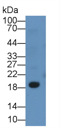
 WB (Western Blot)
(Western Blot;Sample:Lane 1: Rat Spleen lysate;Lane 2: Rat Lung lysatePrimary Ab: 1ug/ml Mouse Anti-Mouse GZMK AntibodySecond Ab: 0.2ug/mL HRP-Linked Caprine Anti-Mouse IgG Polyclonal Antibody)
WB (Western Blot)
(Western Blot;Sample:Lane 1: Rat Spleen lysate;Lane 2: Rat Lung lysatePrimary Ab: 1ug/ml Mouse Anti-Mouse GZMK AntibodySecond Ab: 0.2ug/mL HRP-Linked Caprine Anti-Mouse IgG Polyclonal Antibody)
Granzyme K (GZMK), Monoclonal Antibody (Cat# AAA152751)
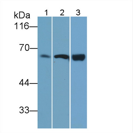
 WB (Western Blot)
(Western Blot; Sample: Rat Lung lysatePrimary Ab: 0.2ug/ml Mouse Anti-Rat CA4 AntibodySecond Ab: 0.2ug/mL HRP-Linked Caprine Anti-Mouse IgG Polyclonal Antibody)
WB (Western Blot)
(Western Blot; Sample: Rat Lung lysatePrimary Ab: 0.2ug/ml Mouse Anti-Rat CA4 AntibodySecond Ab: 0.2ug/mL HRP-Linked Caprine Anti-Mouse IgG Polyclonal Antibody)
Carbonic Anhydrase IV (CA4), Monoclonal Antibody (Cat# AAA152755)
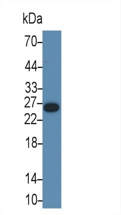
 WB (Western Blot)
(Western Blot; Sample: human SeuM Primary Ab: 0.04ug/ml Mouse Anti-human TF Antibody Second Ab: 0.2ug/mL HRP-Linked Caprine Anti-Mouse IgG Polyclonal Antibody)
WB (Western Blot)
(Western Blot; Sample: human SeuM Primary Ab: 0.04ug/ml Mouse Anti-human TF Antibody Second Ab: 0.2ug/mL HRP-Linked Caprine Anti-Mouse IgG Polyclonal Antibody)
Transferrin (TF), Monoclonal Antibody (Cat# AAA152757)
What are Monoclonal Antibodies?
Monoclonal antibodies are specialized laboratory-produced proteins developed for binding to specific biological antigens or other molecular targets. Since they come from a single cell (or clone), they are especially consistent and accurate in the data they are involved in producing.
This type of antibody material has been shown to be a powerful tool in finding and subsequently destroying harmful cells in an organism, such as those found in cancers or various autoimmune diseases. This makes them excellent aids in medical testing and research, which is why they are so widely used.
AAA Biotech offers a comprehensive range of high-quality monoclonal antibodies that perform effectively in various laboratory tests, including (amongst others) ELISA, western blotting, immunohistochemistry, and flow cytometry. All of the products in our catalog are thoroughly quality tested to make sure that they are reliable and will consistently perform well in your research.
What Are The Uses of Monoclonal Antibodies
Monoclonal antibodies are used in many lab tests, including (amongst others) ELISA, western blotting, immunohistochemistry, and flow cytometry.
ELISA is a test that helps detect a specific substance/analyte in a sample. It uses antibodies (often monoclonal) bound to a solid surface (such as the well of a microplate) to “capture” the substance/analyte in the sample and immobilize it so that the detection antibody component can then bind to it and produce a signal, which can then be measured.
Western blotting identifies specific proteins in a sample. The sample is first separated on a gel, and then antibodies are applied that will typically bind to the target, which will all be localized to a single band in a lane.
Immunohistochemistry helps locate specific proteins in cells or tissue samples using antibodies.
Flow cytometry looks at and sorts cells. It uses antibodies that are conjugated to reporter molecules called “fluorophores”, which, under special lights, emit light themselves, which can then be measured by a detector instrument.
How Monoclonal Antibodies Are Used as Medicine?
Please note that all of the products listed in AAA Biotech’s also known as AAA Bio or AAABio catalog are strictly for research-use only (RUO).
Monoclonal antibodies can also be used as therapeutic/medical treatments, particularly in the context of cancers. They are designed to find and bind to specific cells or proteins, helping the immune system recognize and attack the cancer. These treatments work in different ways, such as:
- Radioimmunotherapy attaches a small amount of radioactive molecule to the antibody, so it delivers the radiation directly to the cancer cells that the antibody is specifically binding to.
- Antibody-directed enzyme prodrug therapy uses antibodies that are specifically bound to special enzymes. These enzymes activate a harmless drug in the body and turn it into a cancer-killing drug only near the cancer cells—this helps avoid harming healthy cells.
- Immunoliposomes are tiny “bubbles” filled with medicine/drug and coated with antibodies. They carry the drug straight to the cancer cells.
Why Buy Monoclonal Antibodies From Us?
At AAA Biotech, we provide high-performance monoclonal antibodies designed to support a wide range of research needs.
1. Validated for Versatile Applications
The antibodies in our catalog are extensively validated and compatible with multiple techniques, including (but not limited to) ELISA, flow cytometry (FC), immunocytochemistry (ICC), immunofluorescence (IF), immunohistochemistry (IHC), immunoprecipitation (IP), and western blotting (WB).
2. Wide Selection & Specialized Options
We offer antibodies for common and rare species, that are available in various conjugated forms, and also in recombinant formats. Essentially, there is almost anything one might need to meet their experimental model’s requirements.
3. High-Quality Proteins
Our proteins meet high purity standards—90% or more as confirmed by SDS-PAGE. Many are available with tags like His, Flag, GST, or MBP, and we also supply native and biologically active proteins for functional studies.
Frequently Asked Questions
1. Are your monoclonal antibodies validated for specific applications?
Yes, our antibodies are tested and validated for use in methods such as ELISA, western blot, IHC, flow cytometry, and more. Refer to specific product pages or datasheets for individual product information.
2. How do I choose the right monoclonal antibody for my application?
Review the product details directly for application validation, species reactivity, and target information. You may also contact our support team at any time for help.
3. How quickly can I receive my order?
Most orders are processed and shipped within 1–3 business days, depending on product availability and your shipping location.