Filters
▼Clonality
▼Type
▼Reactivity
▼Gene Name
▼Isotype
▼Host
▼Application
▼Clone
▼Monoclonal Antibodies
Get accurate results in your research with our Monoclonal Antibodies, which are specially made to target exactly what you require for your research, and will produce consistent, reliable performance in lab tests.
Viewing 6400-6450 of 27597 product results
Corticotropin Releasing Hormone (CRH), Monoclonal Antibody (Cat# AAA148000)
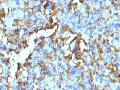
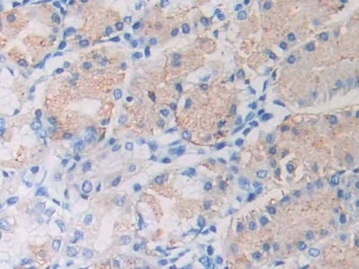
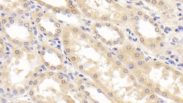
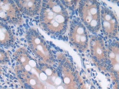
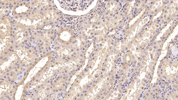 IHC (Immunohiostchemistry)
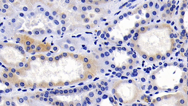
(DAB staining on IHC-P; Samples: Human Kidney Tissue; Primary Ab: 20ug/ml Mouse Anti-Human IFNa21 Antibody Second Ab: 2ug/mL HRP-Linked Caprine Anti-Mouse IgG Polyclonal Antibody)
IHC (Immunohiostchemistry)
(DAB staining on IHC-P; Samples: Human Kidney Tissue; Primary Ab: 20ug/ml Mouse Anti-Human IFNa21 Antibody Second Ab: 2ug/mL HRP-Linked Caprine Anti-Mouse IgG Polyclonal Antibody)
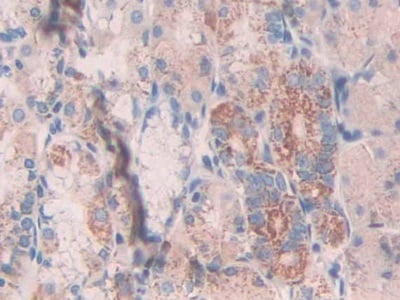
Interferon Alpha 2 (IFNa2), Monoclonal Antibody (Cat# AAA149514)
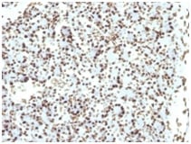
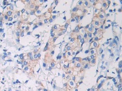
 IHC (Immunohiostchemistry)
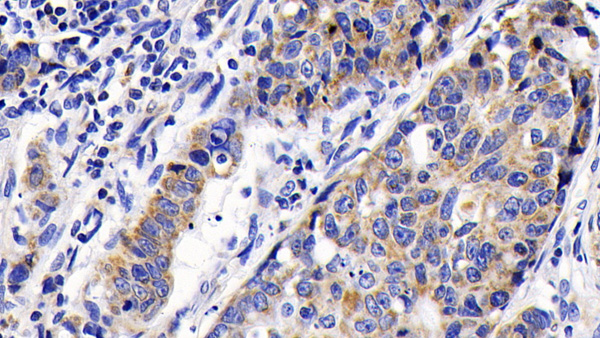
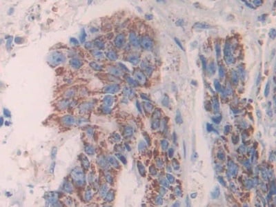
(DAB staining on IHC-P;Samples: Human Ovarian cancer Tissue;Primary Ab: 10ug/ml Mouse Anti-Second Ab: PD1 AntibodySecond Ab: 2ug/mL HRP-Linked Anti-Mouse IgG Polyclonal Antibody (Catalog: ))
IHC (Immunohiostchemistry)
(DAB staining on IHC-P;Samples: Human Ovarian cancer Tissue;Primary Ab: 10ug/ml Mouse Anti-Second Ab: PD1 AntibodySecond Ab: 2ug/mL HRP-Linked Anti-Mouse IgG Polyclonal Antibody (Catalog: ))
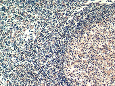
Programmed Cell Death Protein 1 (PD1), Monoclonal Antibody (Cat# AAA149436)
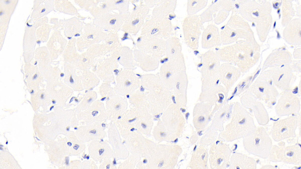
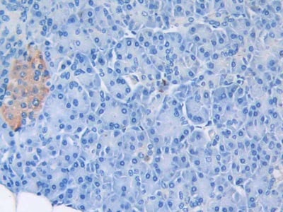
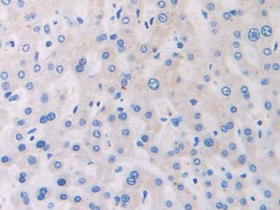
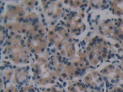
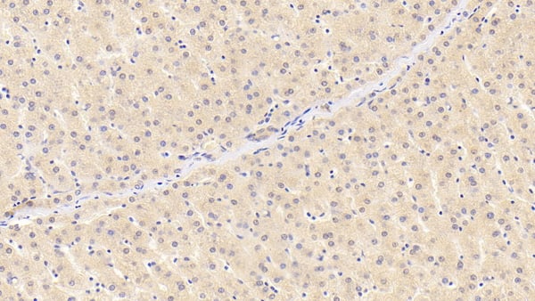
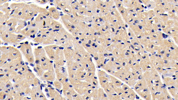
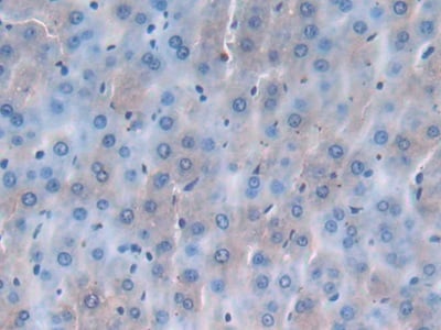
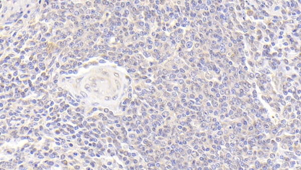
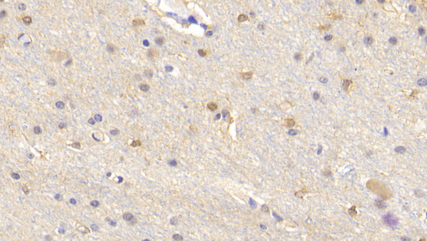
 IHC (Immunohiostchemistry)
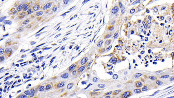
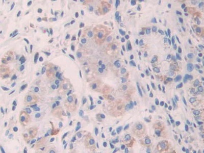
(DAB staining on IHC-P; Samples: Human Liver Tissue))
IHC (Immunohiostchemistry)
(DAB staining on IHC-P; Samples: Human Liver Tissue))
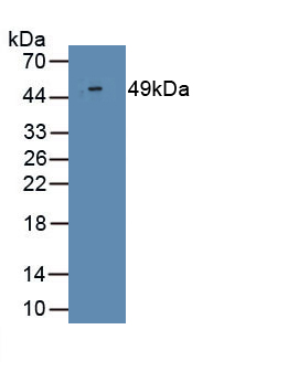
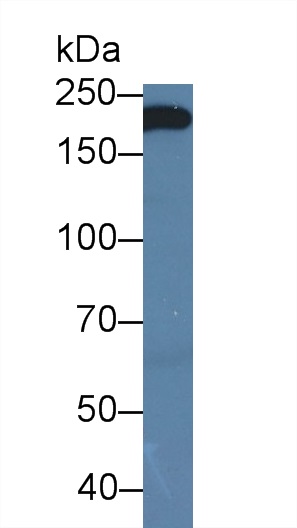
Catenin Beta 1 (CTNNb1), Monoclonal Antibody (Cat# AAA145963)
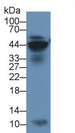
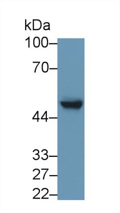
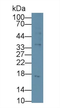
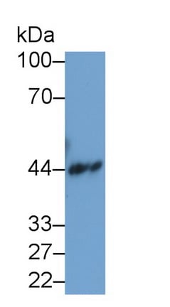
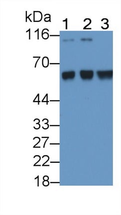
 WB (Western Blot)
(Western Blot: Sample: Recombinant VCL, Human.)
WB (Western Blot)
(Western Blot: Sample: Recombinant VCL, Human.)
Vinculin (VCL), Monoclonal Antibody (Cat# AAA145971)
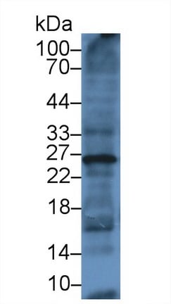
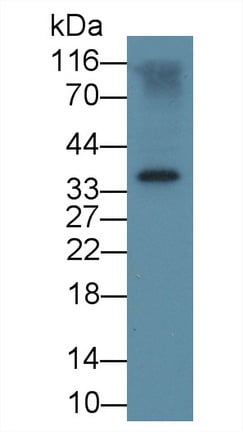
Transforming Growth Factor Beta 1 (TGFb1), Monoclonal Antibody (Cat# AAA146019)
 WB (Western Blot)
(Western Blot; Sample: Human HT1080 cell lysate; Primary Ab: 2ug/mL Mouse Anti-Human S100 Antibody Second Ab: 0.2ug/mL HRP-Linked Caprine Anti-Mouse IgG Polyclonal Antibody)
WB (Western Blot)
(Western Blot; Sample: Human HT1080 cell lysate; Primary Ab: 2ug/mL Mouse Anti-Human S100 Antibody Second Ab: 0.2ug/mL HRP-Linked Caprine Anti-Mouse IgG Polyclonal Antibody)
Calcium Binding Protein (S100), Monoclonal Antibody (Cat# AAA150782)
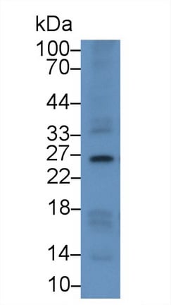
 WB (Western Blot)
(Western Blot; Sample: Rat Liver lysate; Primary Ab: 3ug/mL Mouse Anti-Multi-species Ub AntibodySecond Ab: 0.2ug/mL HRP-Linked Caprine Anti-Mouse IgG Polyclonal Antibody)
WB (Western Blot)
(Western Blot; Sample: Rat Liver lysate; Primary Ab: 3ug/mL Mouse Anti-Multi-species Ub AntibodySecond Ab: 0.2ug/mL HRP-Linked Caprine Anti-Mouse IgG Polyclonal Antibody)
Ubiquitin (Ub), Monoclonal Antibody (Cat# AAA150798)

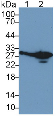
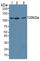
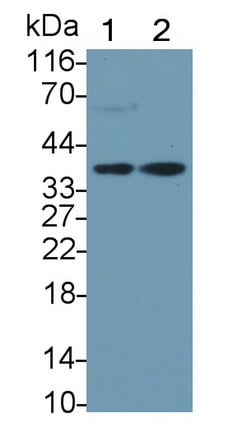
 WB (Western Blot)
(Western Blot; Sample: Lane1: Human Skeletal muscle lysate; Lane2: Human MCF7 cell lysate; Primary Ab)
WB (Western Blot)
(Western Blot; Sample: Lane1: Human Skeletal muscle lysate; Lane2: Human MCF7 cell lysate; Primary Ab)
Insulin Like Growth Factor 1 Receptor (IGF1R), Monoclonal Antibody (Cat# AAA150809)

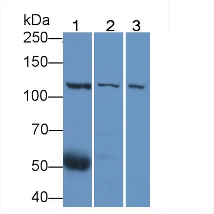
 WB (Western Blot)
(Western Blot; Sample: Lane1: Human Skeletal muscle lysate; Lane2: Human MCF7 cell lysate; Primary Ab)
WB (Western Blot)
(Western Blot; Sample: Lane1: Human Skeletal muscle lysate; Lane2: Human MCF7 cell lysate; Primary Ab)
Insulin Like Growth Factor 1 Receptor (IGF1R), Monoclonal Antibody (Cat# AAA150810)
 WB (Western Blot)
(Western Blot; Sample: Lane1: Human Skeletal muscle lysate; Lane2: Human MCF7 cell lysate; Primary Ab)
WB (Western Blot)
(Western Blot; Sample: Lane1: Human Skeletal muscle lysate; Lane2: Human MCF7 cell lysate; Primary Ab)
Insulin Like Growth Factor 1 Receptor (IGF1R), Monoclonal Antibody (Cat# AAA150813)
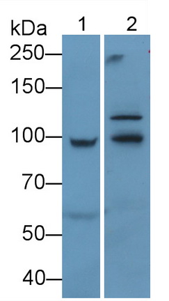
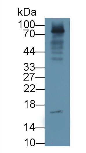
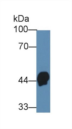
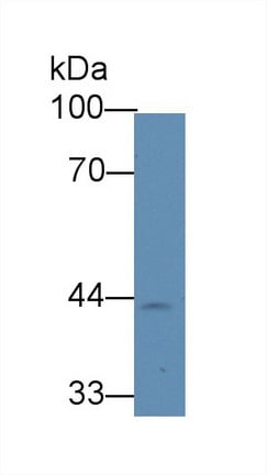
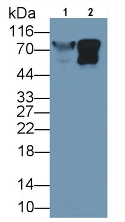
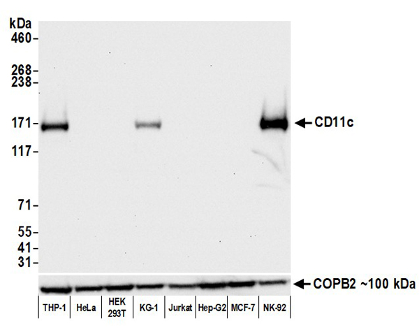 WB (Western Blot)
(Detection of human CD11c by western blot. Samples: Whole cell lysate (50 ug) from THP-1, HeLa, HEK293T, KG-1, Jurkat, Hep-G2, MCF-7, and NK-92 cells prepared using NETN lysis buffer. Antibody: Rabbit anti-CD11c recombinant monoclonal antibody (AAA213599 lot 1) used at 1:1000. Secondary: HRP-conjugated goat anti-rabbit IgG . Detection: Chemiluminescence with an exposure time of 10 seconds. Lower Panel: Rabbit anti-COPB2 antibody .)
WB (Western Blot)
(Detection of human CD11c by western blot. Samples: Whole cell lysate (50 ug) from THP-1, HeLa, HEK293T, KG-1, Jurkat, Hep-G2, MCF-7, and NK-92 cells prepared using NETN lysis buffer. Antibody: Rabbit anti-CD11c recombinant monoclonal antibody (AAA213599 lot 1) used at 1:1000. Secondary: HRP-conjugated goat anti-rabbit IgG . Detection: Chemiluminescence with an exposure time of 10 seconds. Lower Panel: Rabbit anti-COPB2 antibody .)
CD11c, Monoclonal Recombinant Antibody (Cat# AAA213599)
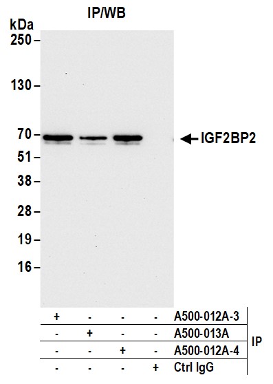
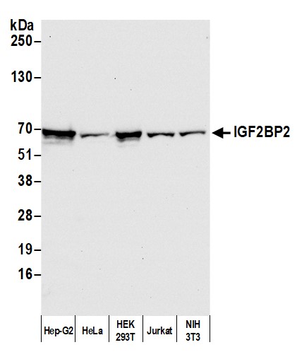 WB (Western Blot)
(Detection of human and mouse IGF2BP2 by western blot. Samples: Whole cell lysate (50 ug) from Hep-G2, HeLa, HEK293T, Jurkat, and NIH 3T3 cells prepared using NETN lysis buffer. Antibody: Mouse anti-IGF2BP2 monoclonal antibody [1E3.01E5] (AAA213498 lot 4) used at 1:1000. Secondary: HRP-conjugated goat anti-mouse IgG . Detection: Chemiluminescence with an exposure time of 10 seconds.)
WB (Western Blot)
(Detection of human and mouse IGF2BP2 by western blot. Samples: Whole cell lysate (50 ug) from Hep-G2, HeLa, HEK293T, Jurkat, and NIH 3T3 cells prepared using NETN lysis buffer. Antibody: Mouse anti-IGF2BP2 monoclonal antibody [1E3.01E5] (AAA213498 lot 4) used at 1:1000. Secondary: HRP-conjugated goat anti-mouse IgG . Detection: Chemiluminescence with an exposure time of 10 seconds.)
IGF2BP2, Monoclonal Antibody (Cat# AAA213498)
B2 Microglobulin, Monoclonal Antibody (Cat# AAA214365)
 Application Data
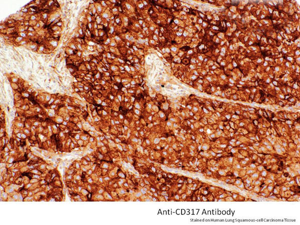
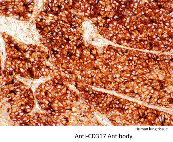
CD317, Monoclonal Antibody (Cat# AAA214370)

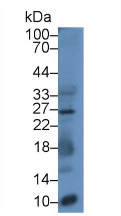
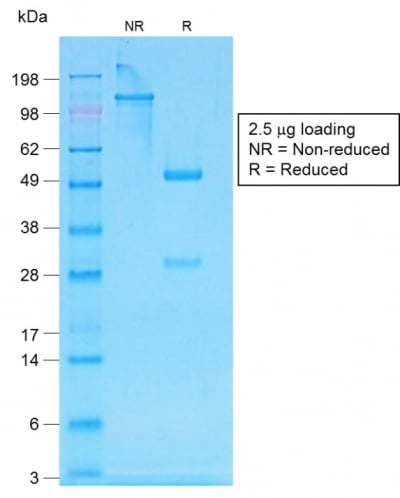
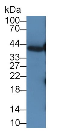
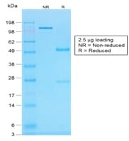 SDS-PAGE
(SDS-PAGE Analysis of Purified Histone H1 Mouse Recombinant Monoclonal Antibody (r1415-1).)
SDS-PAGE
(SDS-PAGE Analysis of Purified Histone H1 Mouse Recombinant Monoclonal Antibody (r1415-1).)
Histone H1, Monoclonal Antibody (Cat# AAA214376)
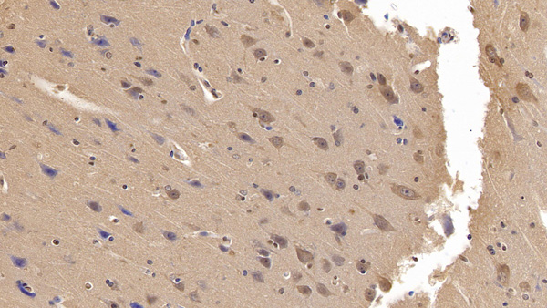
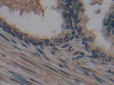
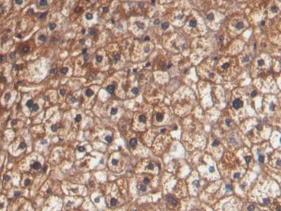
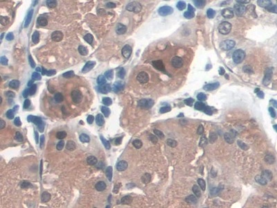
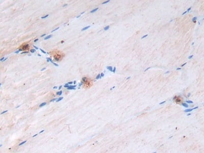
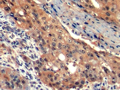
 IHC (Immunohistochemisry)
(Formalin-fixed, paraffin-embedded human Renal Cell Carcinoma stained with Interferon gamma Monoclonal Antibody (IFNG/466))
IHC (Immunohistochemisry)
(Formalin-fixed, paraffin-embedded human Renal Cell Carcinoma stained with Interferon gamma Monoclonal Antibody (IFNG/466))
Interferon gamma (IFNG), Monoclonal Antibody (Cat# AAA214392)
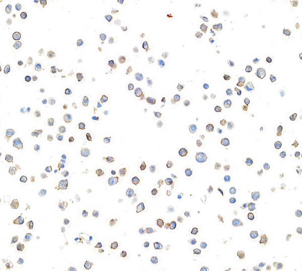
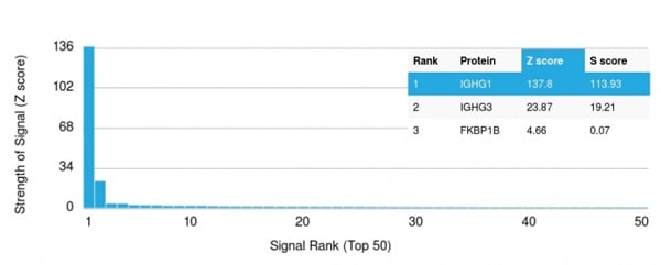 Application Data
(Analysis of Protein Array containing more than 19, 000 full-length human proteins using Anti-human IgG (IGHG1) Rabbit Recombinant Monoclonal Antibody (IG507R). Z- and S- Score: The Z-score represents the strength of a signal that a monoclonal antibody (MAb) (in combination with a fluorescently-tagged anti-IgG secondary antibody) produces when binding to a particular protein on the HuProtTM array. Z-scores are described in units of standard deviations (SD's) above the mean value of all signals generated on that array. If targets on HuProtTM are arranged in descending order of the Z-score, the S-score is the difference (also in units of SD's) between the Z-score. S-score therefore represents the relative target specificity of a MAb to its intended target. A MAb is considered to specific to its intended target, if the MAb has an S-score of at least 2.5. For example, if a MAb binds to protein X with a Z-score of 43 and to protein Y with a Z-score of 14, then the S-score for the binding of that MAb to protein X is equal to 29.)
Application Data
(Analysis of Protein Array containing more than 19, 000 full-length human proteins using Anti-human IgG (IGHG1) Rabbit Recombinant Monoclonal Antibody (IG507R). Z- and S- Score: The Z-score represents the strength of a signal that a monoclonal antibody (MAb) (in combination with a fluorescently-tagged anti-IgG secondary antibody) produces when binding to a particular protein on the HuProtTM array. Z-scores are described in units of standard deviations (SD's) above the mean value of all signals generated on that array. If targets on HuProtTM are arranged in descending order of the Z-score, the S-score is the difference (also in units of SD's) between the Z-score. S-score therefore represents the relative target specificity of a MAb to its intended target. A MAb is considered to specific to its intended target, if the MAb has an S-score of at least 2.5. For example, if a MAb binds to protein X with a Z-score of 43 and to protein Y with a Z-score of 14, then the S-score for the binding of that MAb to protein X is equal to 29.)
IgG (Immunoglobulin Gamma Heavy Chain), Monoclonal Antibody (Cat# AAA214393)
 IHC (Immunohiostchemistry)
(Formalin-fixed, paraffin-embedded human Histiocytoma stained with HLA-DR Monoclonal Antibody (LN-3 + HLA-DRB/1067).)
IHC (Immunohiostchemistry)
(Formalin-fixed, paraffin-embedded human Histiocytoma stained with HLA-DR Monoclonal Antibody (LN-3 + HLA-DRB/1067).)
IgG4 (Ig Heavy Constant Gamma 4), Monoclonal Antibody (Cat# AAA214394)
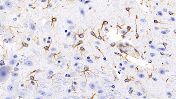
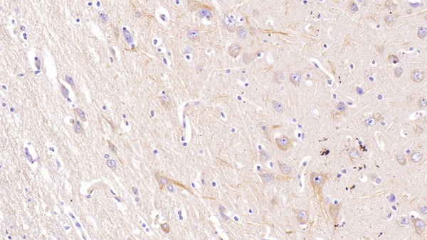
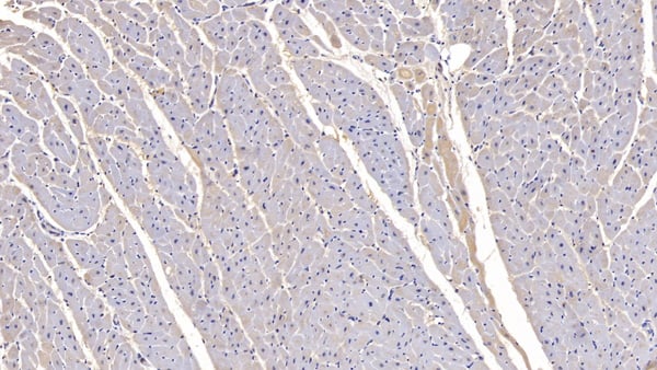
 WB (Western Blot)
(Western Blot; Sample: Rat Cerebrum lysate; Primary Ab: 2ug/ml Mouse AntiMouse GFAP Antibody Second Ab: 0.2ug/mL HRPLinked Caprine AntiMouse IgG Polyclonal Antibody (Catalog: SAA544Mu19))
WB (Western Blot)
(Western Blot; Sample: Rat Cerebrum lysate; Primary Ab: 2ug/ml Mouse AntiMouse GFAP Antibody Second Ab: 0.2ug/mL HRPLinked Caprine AntiMouse IgG Polyclonal Antibody (Catalog: SAA544Mu19))
Glial Fibrillary Acidic Protein (GFAP), Monoclonal Antibody (Cat# AAA151454)
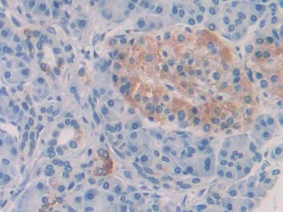
 IHC (Immunohistochemistry)
(DAB staining on IHCP;Sample: Human Spleen Tissue; Primary Ab: 30ug/ml Mouse AntiHuman IL6 AntibodySecond Ab: 2ug/mL HRPLinked Caprine AntiMouse IgG Polyclonal Antibody(Catalog: SAA544Mu19))
IHC (Immunohistochemistry)
(DAB staining on IHCP;Sample: Human Spleen Tissue; Primary Ab: 30ug/ml Mouse AntiHuman IL6 AntibodySecond Ab: 2ug/mL HRPLinked Caprine AntiMouse IgG Polyclonal Antibody(Catalog: SAA544Mu19))
Interleukin 6 (IL6), Monoclonal Antibody (Cat# AAA151462)
 IHC (Immunohiostchemistry)
(DAB staining on IHCP;Sample: Human Spleen Tissue; Primary Ab: 30ug/ml Mouse AntiHuman IL6 AntibodySecond Ab: 2ug/mL HRPLinked Caprine AntiMouse IgG Polyclonal Antibody(Catalog: SAA544Mu19))
IHC (Immunohiostchemistry)
(DAB staining on IHCP;Sample: Human Spleen Tissue; Primary Ab: 30ug/ml Mouse AntiHuman IL6 AntibodySecond Ab: 2ug/mL HRPLinked Caprine AntiMouse IgG Polyclonal Antibody(Catalog: SAA544Mu19))
Interleukin 6 (IL6), Monoclonal Antibody (Cat# AAA151464)
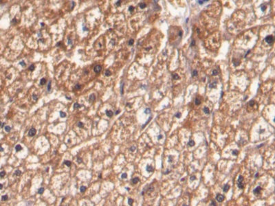
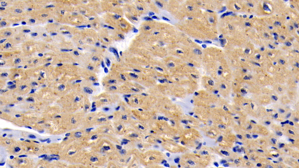
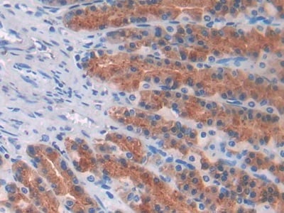
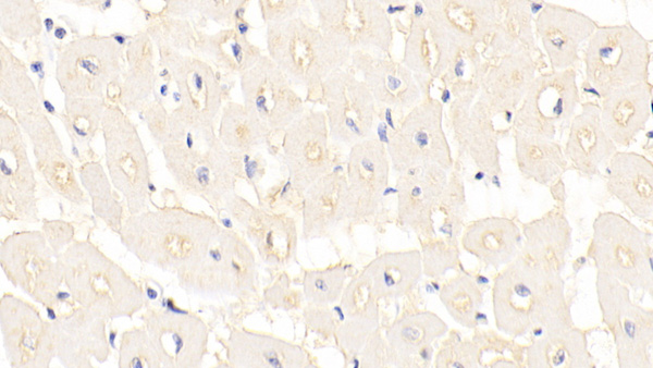
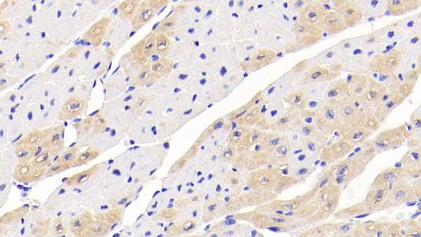
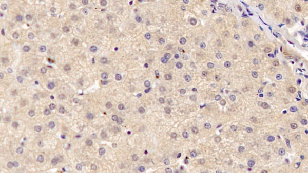
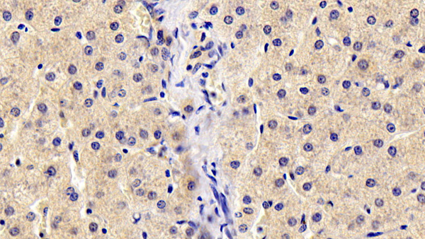
 IHC (Immunohistochemistry)
(DAB staining on IHCP;Sample: Human Liver Tissue; Primary Ab: 30ug/ml Mouse AntiHuman SDF1 AntibodySecond Ab: 2ug/mL HRPLinked Caprine AntiMouse IgG Polyclonal Antibody(Catalog: SAA544Mu19))
IHC (Immunohistochemistry)
(DAB staining on IHCP;Sample: Human Liver Tissue; Primary Ab: 30ug/ml Mouse AntiHuman SDF1 AntibodySecond Ab: 2ug/mL HRPLinked Caprine AntiMouse IgG Polyclonal Antibody(Catalog: SAA544Mu19))
Stromal Cell Derived Factor 1 (SDF1), Monoclonal Antibody (Cat# AAA151486)
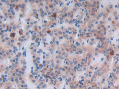
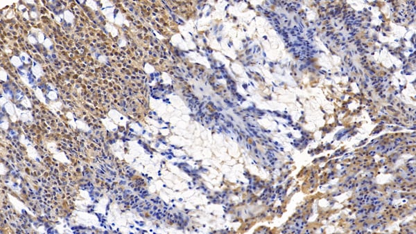
 IHC (Immunohistochemistry)
(DAB staining on IHCP;Sample: Human Lung cancer Tissue; Primary Ab: 20ug/ml Mouse AntiHuman CASP9 AntibodySecond Ab: 2ug/mL HRPLinked Caprine AntiMouse IgG Polyclonal Antibody(Catalog: SAA544Mu19))
IHC (Immunohistochemistry)
(DAB staining on IHCP;Sample: Human Lung cancer Tissue; Primary Ab: 20ug/ml Mouse AntiHuman CASP9 AntibodySecond Ab: 2ug/mL HRPLinked Caprine AntiMouse IgG Polyclonal Antibody(Catalog: SAA544Mu19))
Caspase 9 (CASP9), Monoclonal Antibody (Cat# AAA151588)
 IHC (Immunohiostchemistry)
(DAB staining on IHCP;Sample: Human Prostate Tissue; Primary Ab: 30ug/ml Mouse AntiHuman NTXI AntibodySecond Ab: 2ug/mL HRPLinked Caprine AntiMouse IgG Polyclonal Antibody(Catalog: SAA544Mu19))
IHC (Immunohiostchemistry)
(DAB staining on IHCP;Sample: Human Prostate Tissue; Primary Ab: 30ug/ml Mouse AntiHuman NTXI AntibodySecond Ab: 2ug/mL HRPLinked Caprine AntiMouse IgG Polyclonal Antibody(Catalog: SAA544Mu19))
Cross Linked NTelopeptide Of Type I Collagen (NTXI), Monoclonal Antibody (Cat# AAA151592)
 WB (Western Blot)
(Western Blot; Sample: Porcine Liver lysatePrimary Ab: 5ug/ml Mouse AntiHuman PCT AntibodySecond Ab: 0.2ug/mL HRPLinked Caprine AntiMouse IgG Polyclonal Antibody(Catalog: SAA544Mu19))
WB (Western Blot)
(Western Blot; Sample: Porcine Liver lysatePrimary Ab: 5ug/ml Mouse AntiHuman PCT AntibodySecond Ab: 0.2ug/mL HRPLinked Caprine AntiMouse IgG Polyclonal Antibody(Catalog: SAA544Mu19))
Procalcitonin (PCT), Monoclonal Antibody (Cat# AAA151604)
 WB (Western Blot)
(Western Blot; Sample: Rat Heart lysatePrimary Ab: 3ug/ml Mouse AntiHuman Hpt AntibodySecond Ab: 0.2ug/mL HRPLinked Caprine AntiMouse IgG Polyclonal Antibody(Catalog: SAA544Mu19))
WB (Western Blot)
(Western Blot; Sample: Rat Heart lysatePrimary Ab: 3ug/ml Mouse AntiHuman Hpt AntibodySecond Ab: 0.2ug/mL HRPLinked Caprine AntiMouse IgG Polyclonal Antibody(Catalog: SAA544Mu19))
Haptoglobin (Hpt), Monoclonal Antibody (Cat# AAA151629)
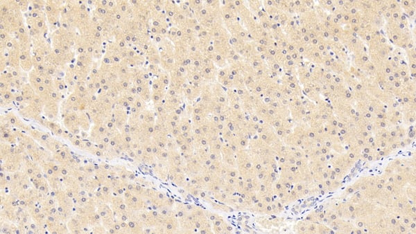
 IHC (Immunohiostchemistry)
(DAB staining on IHC-P;Samples: Human Liver Tissue;Primary Ab: 30ug/ml Mouse Anti-Human RBP4 AntibodySecond Ab: 2ug/mL HRP-Linked Caprine Anti-Mouse IgG Polyclonal Antibody)
IHC (Immunohiostchemistry)
(DAB staining on IHC-P;Samples: Human Liver Tissue;Primary Ab: 30ug/ml Mouse Anti-Human RBP4 AntibodySecond Ab: 2ug/mL HRP-Linked Caprine Anti-Mouse IgG Polyclonal Antibody)
Retinol Binding Protein 4 (RBP4), Monoclonal Antibody (Cat# AAA151123)
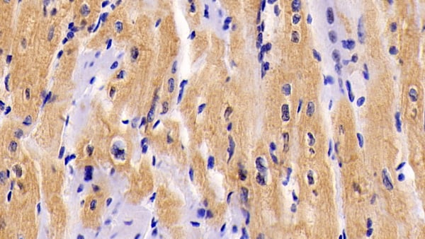
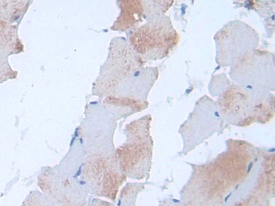
 IHC (Immunohistochemisry)
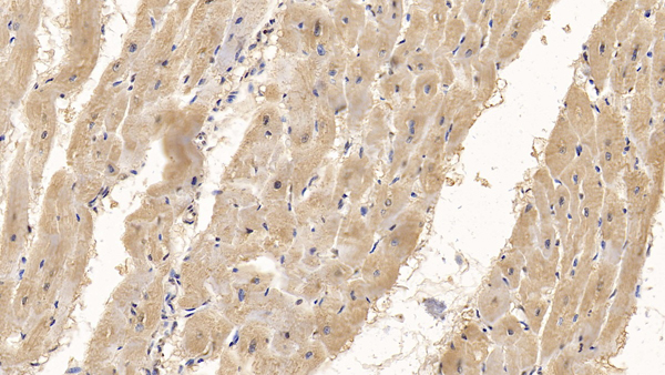
(DAB staining on IHC-P;Sample: Human Cardiac Muscle Tissue;Primary Ab: 20ug/ml Mouse Anti-Human SQSTM1 AntibodySecond Ab: 2ug/mL HRP-Linked Caprine Anti-Mouse IgG Polyclonal Antibody)
IHC (Immunohistochemisry)
(DAB staining on IHC-P;Sample: Human Cardiac Muscle Tissue;Primary Ab: 20ug/ml Mouse Anti-Human SQSTM1 AntibodySecond Ab: 2ug/mL HRP-Linked Caprine Anti-Mouse IgG Polyclonal Antibody)
Sequestosome 1 (SQSTM1), Monoclonal Antibody (Cat# AAA151130)
 WB (Western Blot)
(Western Blot; Sample: Human Urine Primary Ab: 3ug/ml Mouse AntiHuman PGA Antibody Second Ab: 0.2ug/mL HRPLinked Caprine AntiMouse IgG Polyclonal Antibody (Catalog: SAA544Mu19))
WB (Western Blot)
(Western Blot; Sample: Human Urine Primary Ab: 3ug/ml Mouse AntiHuman PGA Antibody Second Ab: 0.2ug/mL HRPLinked Caprine AntiMouse IgG Polyclonal Antibody (Catalog: SAA544Mu19))
Pepsinogen A (PGA), Monoclonal Antibody (Cat# AAA151503)
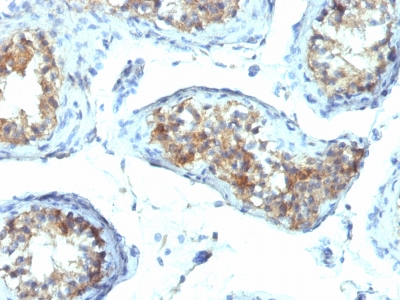
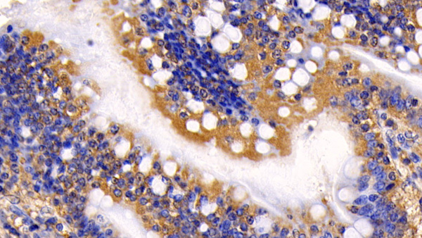
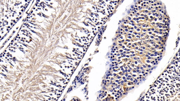 IHC (Immunohiostchemistry)
(DAB staining on IHCP;Sample: Rat Testis Tissue; Primary Ab: 30ug/ml Mouse AntiHuman AMH AntibodySecond Ab: 2ug/mL HRPLinked Caprine AntiMouse IgG Polyclonal Antibody(Catalog: SAA544Mu19))
IHC (Immunohiostchemistry)
(DAB staining on IHCP;Sample: Rat Testis Tissue; Primary Ab: 30ug/ml Mouse AntiHuman AMH AntibodySecond Ab: 2ug/mL HRPLinked Caprine AntiMouse IgG Polyclonal Antibody(Catalog: SAA544Mu19))
AntiMullerian Hormone (AMH), Monoclonal Antibody (Cat# AAA151513)
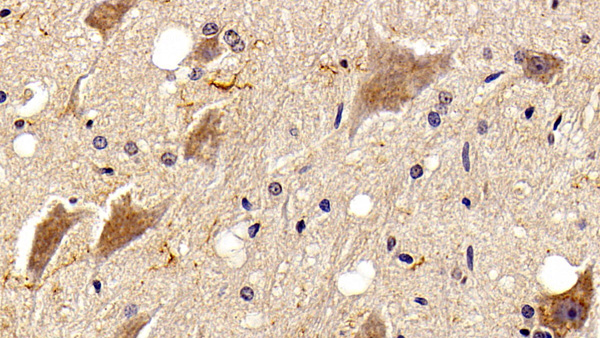
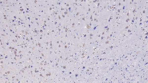
 WB (Western Blot)
(Western Blot; Sample: Lane1: Mouse Cerebrum lysate; Lane2: Rat Cerebrum lysate Primary Ab: 2ug/mL Mouse AntiHuman NRGN Antibody Second Ab: 0.2ug/mL HRPLinked Caprine AntiMouse IgG Polyclonal Antibody (Catalog: SAA544Mu19))
WB (Western Blot)
(Western Blot; Sample: Lane1: Mouse Cerebrum lysate; Lane2: Rat Cerebrum lysate Primary Ab: 2ug/mL Mouse AntiHuman NRGN Antibody Second Ab: 0.2ug/mL HRPLinked Caprine AntiMouse IgG Polyclonal Antibody (Catalog: SAA544Mu19))
Neurogranin (NRGN), Monoclonal Antibody (Cat# AAA151533)
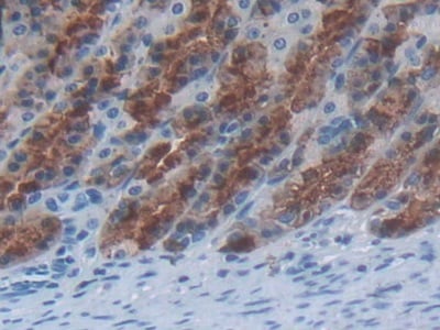
 IHC (Immunohistochemistry)
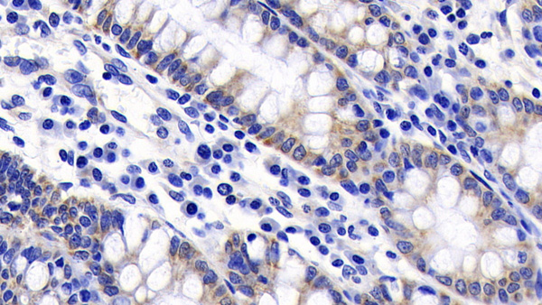
(DAB staining on IHCP;Sample: Rat Small intestine Tissue; Primary Ab: 20ug/ml Mouse AntiRat EGF AntibodySecond Ab: 2ug/mL HRPLinked Caprine AntiMouse IgG Polyclonal Antibody(Catalog: SAA544Mu19))
IHC (Immunohistochemistry)
(DAB staining on IHCP;Sample: Rat Small intestine Tissue; Primary Ab: 20ug/ml Mouse AntiRat EGF AntibodySecond Ab: 2ug/mL HRPLinked Caprine AntiMouse IgG Polyclonal Antibody(Catalog: SAA544Mu19))
Epidermal Growth Factor (EGF), Monoclonal Antibody (Cat# AAA151561)
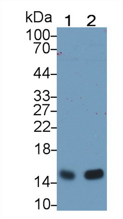
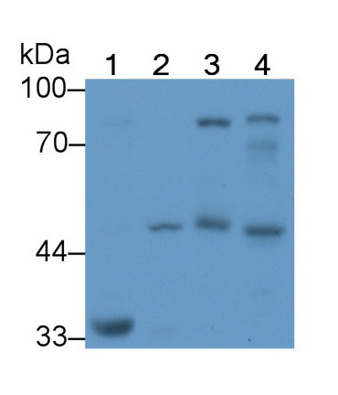
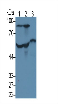
 WB (Western Blot)
(Western Blot; Sample: Lane1: Human Lung lysate; Lane2: Human Serum Primary Ab: 3ug/ml Mouse AntiHuman LUM Antibody Second Ab: 0.2ug/mL HRPLinked Caprine AntiMouse IgG Polyclonal Antibody (Catalog: SAA544Mu19))
WB (Western Blot)
(Western Blot; Sample: Lane1: Human Lung lysate; Lane2: Human Serum Primary Ab: 3ug/ml Mouse AntiHuman LUM Antibody Second Ab: 0.2ug/mL HRPLinked Caprine AntiMouse IgG Polyclonal Antibody (Catalog: SAA544Mu19))
Lumican (LUM), Monoclonal Antibody (Cat# AAA151720)
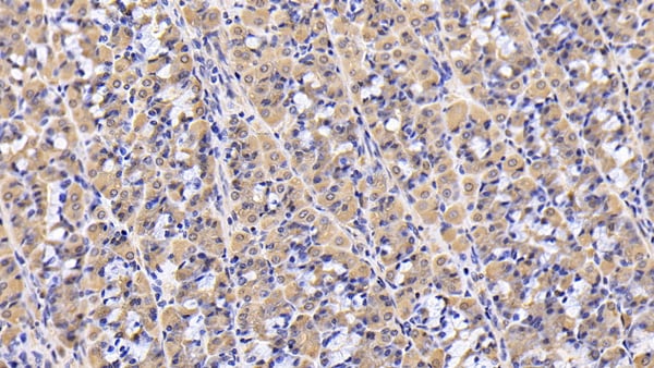
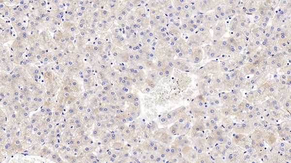
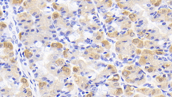
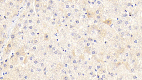
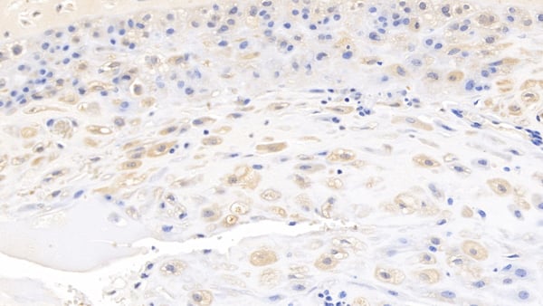
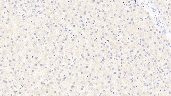 IHC (Immunohistochemisry)
(DAB staining on IHCP;Samples: Human Liver Tissue;Primary Ab: 20ug/ml Mouse AntiHuman a1AT AntibodySecond Ab: 2ug/mL HRPLinked Caprine AntiMouse IgG Polyclonal Antibody(Catalog: SAA544Mu19))
IHC (Immunohistochemisry)
(DAB staining on IHCP;Samples: Human Liver Tissue;Primary Ab: 20ug/ml Mouse AntiHuman a1AT AntibodySecond Ab: 2ug/mL HRPLinked Caprine AntiMouse IgG Polyclonal Antibody(Catalog: SAA544Mu19))
Alpha1Antitrypsin (a1AT), Monoclonal Antibody (Cat# AAA151737)
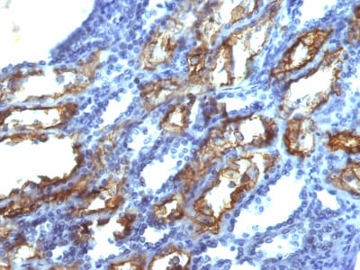
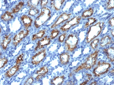
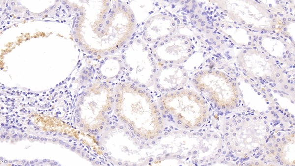
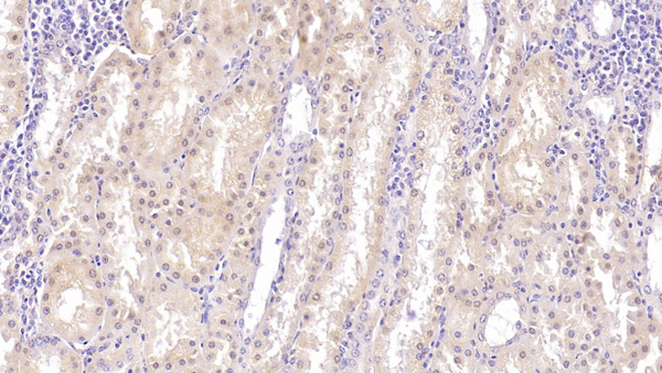
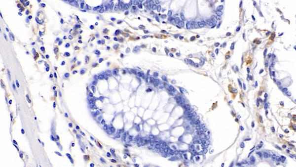
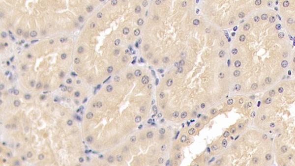
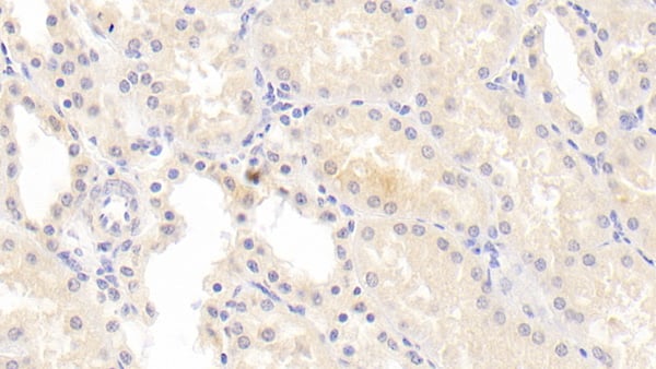
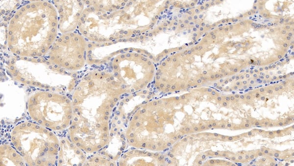
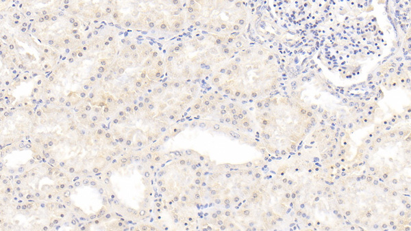 IHC (Immunohistochemisry)
(DAB staining on IHCP;Sample: Human Kidney Tissue; Primary Ab: 20ug/ml Mouse AntiHuman INHbB AntibodySecond Ab: 2ug/mL HRPLinked Caprine AntiMouse IgG Polyclonal Antibody(Catalog: SAA544Mu19))
IHC (Immunohistochemisry)
(DAB staining on IHCP;Sample: Human Kidney Tissue; Primary Ab: 20ug/ml Mouse AntiHuman INHbB AntibodySecond Ab: 2ug/mL HRPLinked Caprine AntiMouse IgG Polyclonal Antibody(Catalog: SAA544Mu19))
Inhibin Beta B (INHbB), Monoclonal Antibody (Cat# AAA151743)

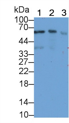
 WB (Western Blot)
(Western Blot; Sample: Lane1: Human Serum; Lane2: Human Lung lysate; Lane3: Human Placenta lysate; Lane4: Human Plasma; Lane5: Rat PlasmaPrimary Ab: 3ug/ml Mouse AntiHuman DBP AntibodySecond Ab: 0.2ug/mL HRPLinked Caprine AntiMouse IgG Polyclonal Antibody(Catalog: SAA544Mu19))
WB (Western Blot)
(Western Blot; Sample: Lane1: Human Serum; Lane2: Human Lung lysate; Lane3: Human Placenta lysate; Lane4: Human Plasma; Lane5: Rat PlasmaPrimary Ab: 3ug/ml Mouse AntiHuman DBP AntibodySecond Ab: 0.2ug/mL HRPLinked Caprine AntiMouse IgG Polyclonal Antibody(Catalog: SAA544Mu19))
Vitamin D Binding Protein (DBP), Monoclonal Antibody (Cat# AAA151750)
 WB (Western Blot)
(Western Blot; Sample: 293T cell lysate Primary Ab: 2ug/ml Mouse AntiHuman CKBB Antibody Second Ab: 0.2ug/mL HRPLinked Caprine AntiMouse IgG Polyclonal Antibody (Catalog: SAA544Mu19))
WB (Western Blot)
(Western Blot; Sample: 293T cell lysate Primary Ab: 2ug/ml Mouse AntiHuman CKBB Antibody Second Ab: 0.2ug/mL HRPLinked Caprine AntiMouse IgG Polyclonal Antibody (Catalog: SAA544Mu19))
Creatine Kinase B (CKBB), Monoclonal Antibody (Cat# AAA151784)
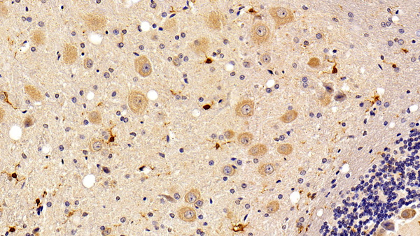
 IHC (Immunohiostchemistry)
(DAB staining on IHCP;Sample: Rat Stomach Tissue; Primary Ab: 30ug/ml Mouse AntiRat IL24 AntibodySecond Ab: 2ug/mL HRPLinked Caprine AntiMouse IgG Polyclonal Antibody(Catalog: SAA544Mu19))
IHC (Immunohiostchemistry)
(DAB staining on IHCP;Sample: Rat Stomach Tissue; Primary Ab: 30ug/ml Mouse AntiRat IL24 AntibodySecond Ab: 2ug/mL HRPLinked Caprine AntiMouse IgG Polyclonal Antibody(Catalog: SAA544Mu19))
Interleukin 24 (IL24), Monoclonal Antibody (Cat# AAA151785)
 IHC (Immunohistochemisry)
(DAB staining on IHCP;Samples: Human Spleen Tissue;Primary Ab: 30ug/ml Mouse AntiHuman MHCDRb1 AntibodySecond Ab: 2ug/mL HRPLinked Caprine AntiMouse IgG Polyclonal Antibody(Catalog: SAA544Mu19))
IHC (Immunohistochemisry)
(DAB staining on IHCP;Samples: Human Spleen Tissue;Primary Ab: 30ug/ml Mouse AntiHuman MHCDRb1 AntibodySecond Ab: 2ug/mL HRPLinked Caprine AntiMouse IgG Polyclonal Antibody(Catalog: SAA544Mu19))
HLA Class II Histocompatibility Antigen, DRB1 Beta Chain (HLADRB1), Monoclonal Antibody (Cat# AAA151796)
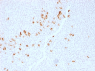
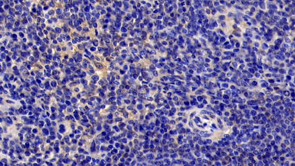
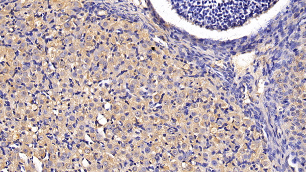
 IHC (Immunohistochemistry)
(DAB staining on IHCP;Sample: Human Pancreas Tissue; Primary Ab: 40ug/ml Mouse AntiRat AGR2 AntibodySecond Ab: 2ug/mL HRPLinked Caprine AntiMouse IgG Polyclonal Antibody(Catalog: SAA544Mu19))
IHC (Immunohistochemistry)
(DAB staining on IHCP;Sample: Human Pancreas Tissue; Primary Ab: 40ug/ml Mouse AntiRat AGR2 AntibodySecond Ab: 2ug/mL HRPLinked Caprine AntiMouse IgG Polyclonal Antibody(Catalog: SAA544Mu19))
Anterior Gradient 2 (AGR2), Monoclonal Antibody (Cat# AAA151798)
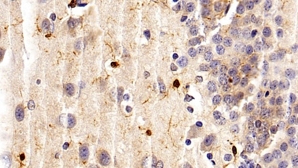
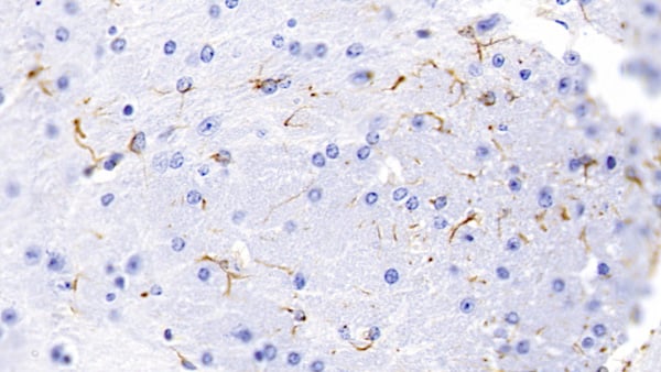
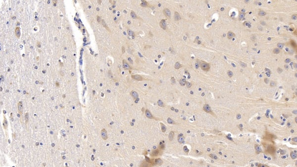
 IHC (Immunohistochemisry)
(DAB staining on IHCP;Sample: Rat Cerebellum Tissue; Primary Ab: 10ug/ml Mouse AntiRat IBA1 AntibodySecond Ab: 2ug/mL HRPLinked Caprine AntiMouse IgG Polyclonal Antibody(Catalog: SAA544Mu19))
IHC (Immunohistochemisry)
(DAB staining on IHCP;Sample: Rat Cerebellum Tissue; Primary Ab: 10ug/ml Mouse AntiRat IBA1 AntibodySecond Ab: 2ug/mL HRPLinked Caprine AntiMouse IgG Polyclonal Antibody(Catalog: SAA544Mu19))
Ionized Calciumbinding Adapter Molecule 1 (IBA1), Monoclonal Antibody (Cat# AAA151799)
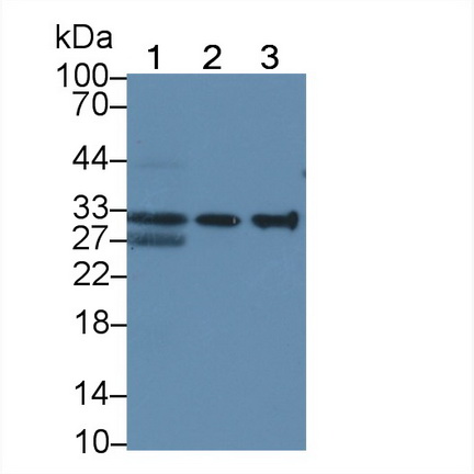
 WB (Western Blot)
(Western Blot; Sample: Lane1: Human Serum; Lane2: Human Plasma; Lane3: Human Placenta lysate Primary Ab: 3ug/ml Mouse AntiHuman Antithrombin Antibody Second Ab: 0.2ug/mL HRPLinked Caprine AntiMouse IgG Polyclonal Antibody (Catalog: SAA544Mu19))
WB (Western Blot)
(Western Blot; Sample: Lane1: Human Serum; Lane2: Human Plasma; Lane3: Human Placenta lysate Primary Ab: 3ug/ml Mouse AntiHuman Antithrombin Antibody Second Ab: 0.2ug/mL HRPLinked Caprine AntiMouse IgG Polyclonal Antibody (Catalog: SAA544Mu19))
Antithrombin (AT), Monoclonal Antibody (Cat# AAA151801)
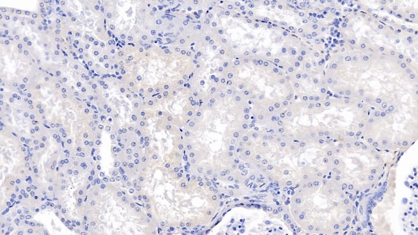
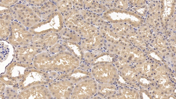
 IHC (Immunohiostchemistry)
(DAB staining on IHCP;Sample: Human Kidney Tissue; Primary Ab: 20ug/ml Mouse AntiHuman AT AntibodySecond Ab: 2ug/mL HRPLinked Caprine AntiMouse IgG Polyclonal Antibody(Catalog: SAA544Mu19))
IHC (Immunohiostchemistry)
(DAB staining on IHCP;Sample: Human Kidney Tissue; Primary Ab: 20ug/ml Mouse AntiHuman AT AntibodySecond Ab: 2ug/mL HRPLinked Caprine AntiMouse IgG Polyclonal Antibody(Catalog: SAA544Mu19))
Antithrombin (AT), Monoclonal Antibody (Cat# AAA151802)
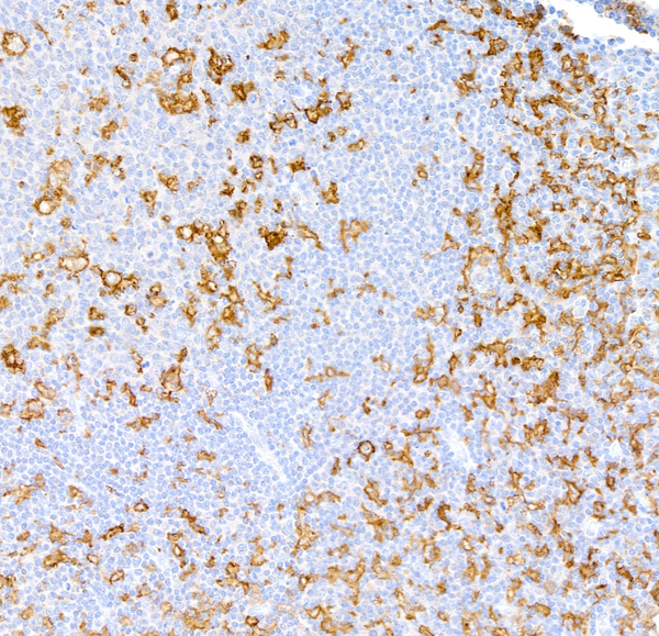
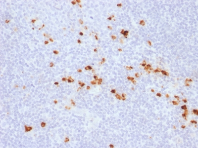
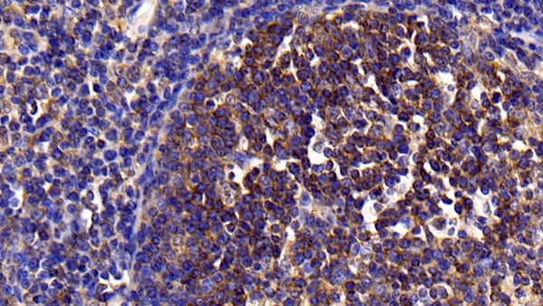
 IHC (Immunohiostchemistry)
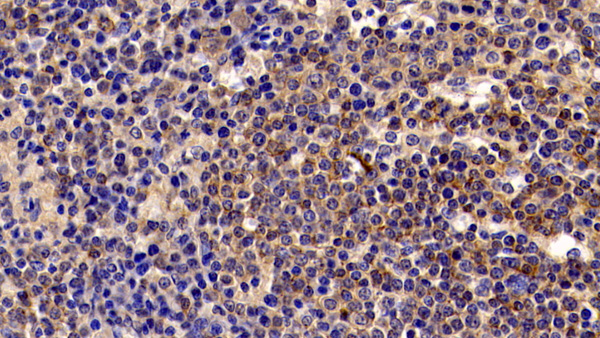
(DAB staining on IHCP;Sample: Human Lymphoma Tissue; Primary Ab: 10ug/ml Mouse AntiHuman HAVCR2 AntibodySecond Ab: 2ug/mL HRPLinked Caprine AntiMouse IgG Polyclonal Antibody(Catalog: SAA544Mu19))
IHC (Immunohiostchemistry)
(DAB staining on IHCP;Sample: Human Lymphoma Tissue; Primary Ab: 10ug/ml Mouse AntiHuman HAVCR2 AntibodySecond Ab: 2ug/mL HRPLinked Caprine AntiMouse IgG Polyclonal Antibody(Catalog: SAA544Mu19))
Hepatitis A Virus Cellular Receptor 2 (HAVCR2), Monoclonal Antibody (Cat# AAA151886)
 WB (Western Blot)
(Western Blot; Sample: Human Placenta lysate; Primary Ab: 3ug/ml Mouse AntiHuman FSTL1 Antibody Second Ab: 0.2ug/mL HRPLinked Caprine AntiMouse IgG Polyclonal Antibody (Catalog: SAA544Mu19))
WB (Western Blot)
(Western Blot; Sample: Human Placenta lysate; Primary Ab: 3ug/ml Mouse AntiHuman FSTL1 Antibody Second Ab: 0.2ug/mL HRPLinked Caprine AntiMouse IgG Polyclonal Antibody (Catalog: SAA544Mu19))
Follistatin Like Protein 1 (FSTL1), Monoclonal Antibody (Cat# AAA151887)
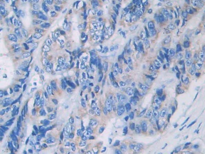
 IHC (Immunohistochemisry)
(DAB staining on IHCP;Sample: Porcine Liver Tissue; Primary Ab: 10ug/ml Mouse AntiHuman LCAT AntibodySecond Ab: 2ug/mL HRPLinked Caprine AntiMouse IgG Polyclonal Antibody(Catalog: SAA544Mu19))
IHC (Immunohistochemisry)
(DAB staining on IHCP;Sample: Porcine Liver Tissue; Primary Ab: 10ug/ml Mouse AntiHuman LCAT AntibodySecond Ab: 2ug/mL HRPLinked Caprine AntiMouse IgG Polyclonal Antibody(Catalog: SAA544Mu19))
Lecithin Cholesterol Acyltransferase (LCAT), Monoclonal Antibody (Cat# AAA151893)

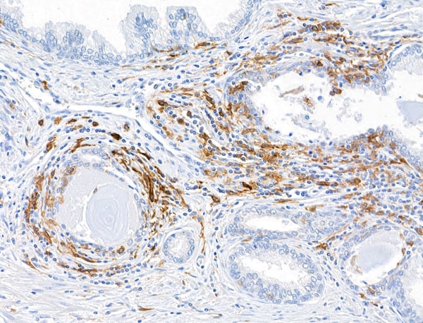
 IHC (Immunohistochemistry)
(DAB staining on IHCP;Sample: Human Spleen Tissue; Primary Ab: 10ug/ml Mouse AntiHuman WNT4 AntibodySecond Ab: 2ug/mL HRPLinked Caprine AntiMouse IgG Polyclonal Antibody(Catalog: SAA544Mu19))
IHC (Immunohistochemistry)
(DAB staining on IHCP;Sample: Human Spleen Tissue; Primary Ab: 10ug/ml Mouse AntiHuman WNT4 AntibodySecond Ab: 2ug/mL HRPLinked Caprine AntiMouse IgG Polyclonal Antibody(Catalog: SAA544Mu19))
Wingless Type MMTV Integration Site Family, Member 4 (WNT4), Monoclonal Antibody (Cat# AAA151902)
 IHC (Immunohiostchemistry)
(DAB staining on IHCP;Sample: Human Small intestine Tissue; Primary Ab: 10ug/ml Mouse AntiHuman WNT3A AntibodySecond Ab: 2ug/mL HRPLinked Caprine AntiMouse IgG Polyclonal Antibody(Catalog: SAA544Mu19))
IHC (Immunohiostchemistry)
(DAB staining on IHCP;Sample: Human Small intestine Tissue; Primary Ab: 10ug/ml Mouse AntiHuman WNT3A AntibodySecond Ab: 2ug/mL HRPLinked Caprine AntiMouse IgG Polyclonal Antibody(Catalog: SAA544Mu19))
Wingless Type MMTV Integration Site Family, Member 3A (WNT3A), Monoclonal Antibody (Cat# AAA151911)
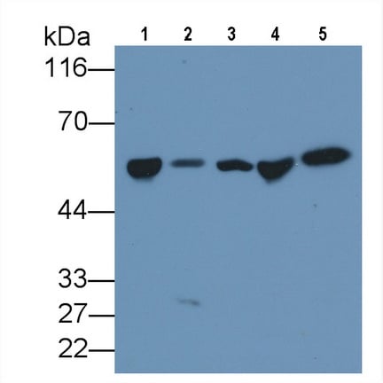
 WB (Western Blot)
(Western Blot; Sample: Lane1: Human Serum; Lane2: Human Plasma; Lane3: Human Placenta lysate Primary Ab: 0.5ug/ml Mouse AntiHuman Antithrombin Antibody Second Ab: 0.2ug/mL HRPLinked Caprine AntiMouse IgG Polyclonal Antibody (Catalog: SAA544Mu19))
WB (Western Blot)
(Western Blot; Sample: Lane1: Human Serum; Lane2: Human Plasma; Lane3: Human Placenta lysate Primary Ab: 0.5ug/ml Mouse AntiHuman Antithrombin Antibody Second Ab: 0.2ug/mL HRPLinked Caprine AntiMouse IgG Polyclonal Antibody (Catalog: SAA544Mu19))
Antithrombin (AT), Monoclonal Antibody (Cat# AAA151803)
What are Monoclonal Antibodies?
Monoclonal antibodies are specialized laboratory-produced proteins developed for binding to specific biological antigens or other molecular targets. Since they come from a single cell (or clone), they are especially consistent and accurate in the data they are involved in producing.
This type of antibody material has been shown to be a powerful tool in finding and subsequently destroying harmful cells in an organism, such as those found in cancers or various autoimmune diseases. This makes them excellent aids in medical testing and research, which is why they are so widely used.
AAA Biotech offers a comprehensive range of high-quality monoclonal antibodies that perform effectively in various laboratory tests, including (amongst others) ELISA, western blotting, immunohistochemistry, and flow cytometry. All of the products in our catalog are thoroughly quality tested to make sure that they are reliable and will consistently perform well in your research.
What Are The Uses of Monoclonal Antibodies
Monoclonal antibodies are used in many lab tests, including (amongst others) ELISA, western blotting, immunohistochemistry, and flow cytometry.
ELISA is a test that helps detect a specific substance/analyte in a sample. It uses antibodies (often monoclonal) bound to a solid surface (such as the well of a microplate) to “capture” the substance/analyte in the sample and immobilize it so that the detection antibody component can then bind to it and produce a signal, which can then be measured.
Western blotting identifies specific proteins in a sample. The sample is first separated on a gel, and then antibodies are applied that will typically bind to the target, which will all be localized to a single band in a lane.
Immunohistochemistry helps locate specific proteins in cells or tissue samples using antibodies.
Flow cytometry looks at and sorts cells. It uses antibodies that are conjugated to reporter molecules called “fluorophores”, which, under special lights, emit light themselves, which can then be measured by a detector instrument.
How Monoclonal Antibodies Are Used as Medicine?
Please note that all of the products listed in AAA Biotech’s also known as AAA Bio or AAABio catalog are strictly for research-use only (RUO).
Monoclonal antibodies can also be used as therapeutic/medical treatments, particularly in the context of cancers. They are designed to find and bind to specific cells or proteins, helping the immune system recognize and attack the cancer. These treatments work in different ways, such as:
- Radioimmunotherapy attaches a small amount of radioactive molecule to the antibody, so it delivers the radiation directly to the cancer cells that the antibody is specifically binding to.
- Antibody-directed enzyme prodrug therapy uses antibodies that are specifically bound to special enzymes. These enzymes activate a harmless drug in the body and turn it into a cancer-killing drug only near the cancer cells—this helps avoid harming healthy cells.
- Immunoliposomes are tiny “bubbles” filled with medicine/drug and coated with antibodies. They carry the drug straight to the cancer cells.
Why Buy Monoclonal Antibodies From Us?
At AAA Biotech, we provide high-performance monoclonal antibodies designed to support a wide range of research needs.
1. Validated for Versatile Applications
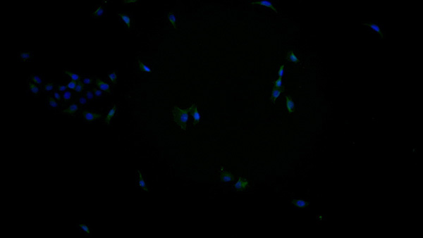
The antibodies in our catalog are extensively validated and compatible with multiple techniques, including (but not limited to) ELISA, flow cytometry (FC), immunocytochemistry (ICC), immunofluorescence (IF), immunohistochemistry (IHC), immunoprecipitation (IP), and western blotting (WB).
2. Wide Selection & Specialized Options
We offer antibodies for common and rare species, that are available in various conjugated forms, and also in recombinant formats. Essentially, there is almost anything one might need to meet their experimental model’s requirements.
3. High-Quality Proteins
Our proteins meet high purity standards—90% or more as confirmed by SDS-PAGE. Many are available with tags like His, Flag, GST, or MBP, and we also supply native and biologically active proteins for functional studies.
Frequently Asked Questions
1. Are your monoclonal antibodies validated for specific applications?
Yes, our antibodies are tested and validated for use in methods such as ELISA, western blot, IHC, flow cytometry, and more. Refer to specific product pages or datasheets for individual product information.
2. How do I choose the right monoclonal antibody for my application?
Review the product details directly for application validation, species reactivity, and target information. You may also contact our support team at any time for help.
3. How quickly can I receive my order?
Most orders are processed and shipped within 1–3 business days, depending on product availability and your shipping location.